Proceeding Paper

Synthesis and Characterization of a Water-Soluble Pentamethine Indocyanine Dye for Peptide Labeling †

by
Cátia D. F. Martins
,
Maria Manuela M. Raposo
and
Susana P. G. Costa
*
Centre of Chemistry, University of Minho, Campus de Gualtar, 4710-057 Braga, Portugal
*
Author to whom correspondence should be addressed.
Presented at the 25th International Electronic Conference on Synthetic Organic Chemistry, 15–30 November 2021; Available online: https://ecsoc-25.sciforum.net/.
Chem. Proc. 2022, 8(1), 91; https://doi.org/10.3390/ecsoc-25-11768
Published: 18 July 2022

Abstract

:
A water-soluble pentamethine cyanine dye was synthesized for further conjugation with peptides. The dye preparation was accomplished by solution-phase chemistry and the synthesized compounds were characterized by the usual techniques (nuclear magnetic resonance, UV-vis absorption and fluorescence spectroscopies and mass spectrometry). The photophysical properties of cyanine 4 were studied in aqueous media, namely in phosphate-buffered saline (PBS) at pH 7.4. Sulfo-Cy5 carboxylic acid 4 exhibited a narrow absorption band and a high molar extinction coefficient (log ε = 5.11) at 646 nm. The wavelength of maximum fluorescence was found in the near-infrared (NIR) region at 661 nm. Therefore, due to its excellent spectral proprieties, good water solubility and emission in the NIR region, dye 4 can be employed in the synthesis of fluorescence resonance energy transfer (FRET) probes for studying enzymatic hydrolysis in vitro.

1. Introduction

Cyanine dyes are widely used as fluorescent labels in many biomedical applications such as bioimaging and diseases diagnosis [1], nucleic acid detection [2] and biomolecular labeling [3]. This is due to their excellent spectral properties, including narrow absorption bands, large molar absorptivities, good stability and high sensitivity [4,5,6].
The basic structure of cyanine dyes includes two heterocyclic rings containing nitrogen centers, which are linked by a conjugated chain of methine groups [6]. Depending on the length of the cyanine methine chain, the fluorescence range extends from the visible to the near-infrared (NIR) region [7,8]. For example, trimethine cyanine (Cy3) dyes with 3 methine groups emit visible light, while pentamethine cyanine (Cy5) dyes with 5 methine groups emit in the NIR region (>650 nm) [1,9,10].
Fluorescence detection in the NIR has many advantages, such as low tissue autofluorescence, small light scattering, background interference and deep tissue penetration, among others [11,12,13]. Therefore, due to the NIR fluorescence characteristics, pentamethine dyes have been employed as donor/acceptor groups in fluorescence resonance energy transfer (FRET) based enzyme probes to study biological processes in vitro and in vivo [14,15].
Among cyanine dyes, water-soluble sulfoindocyanine dye developed by Waggoner’s group is one of the most used type of dyes for biological applications [16]. Sulfoindocyanine dyes have two or more SO3 groups on the ring systems, which enhance water solubility, prevent aggregation, and reduce non-specific binding to biomolecules and cellular constituents [4,17].
Bearing these facts in mind and considering our interest in the synthesis of FRET-labelled peptide substrates for in vitro validation of a nanoconstructed drug-delivery system, we report herein the synthesis of a water-soluble pentamethine cyanine dye. The dye preparation was performed by solution-phase chemistry and its photophysical characterization was investigated in phosphate-buffered saline (PBS) at pH 7.4.

2. Experimental Section

2.1. Instruments and Materials

Nuclear Magnetic Resonance (NMR) spectra were obtained on a Bruker Avance III 400 at an operating frequency of 400 MHz for 1H and 100.6 MHz for 13C using the solvent peak as an internal reference. The solvents are indicated in parentheses before the chemical shift values (δ relative to tetramethylsilane (TMS) and given in ppm). Assignments were supported by two-dimensional heteronuclear correlation techniques. Mass spectrometry analyses were performed at the “C.A.C.T.I. Unidad de Espectrometria de Masas” at the University of Vigo, Spain. Thin-layer chromatography (TLC) analyses were carried out on 0.25 mm-thick silica plates coated with fluorescent indicator F254 (Merck KGaA, Darmstadt, Germany) and spots were visualized in a CN15 viewing cabinet under UV lamp at 365 nm (Vilber Lourmat, Marne-la-Vallée, France). Normal-phase column chromatography was performed on silica gel 0.035–0.070 mm, 60 Å (Acros Organics, Geel, Belgium). UV/Vis absorption spectra were obtained using a Shimadzu UV/2501PC spectrophotometer (Shimadzu Europa GmbH, Duisburg, Germany) and fluorescence spectra were collected using a FluoroMax-4 spectrofluorometer (HORIBA Europe GmbH, Darmstadt, Germany) in standard quartz cuvettes. All reagents were purchased from Acros Organics and Sigma-Aldrich (St. Louis, MO, USA), and used as received.

2.2. Synthesis

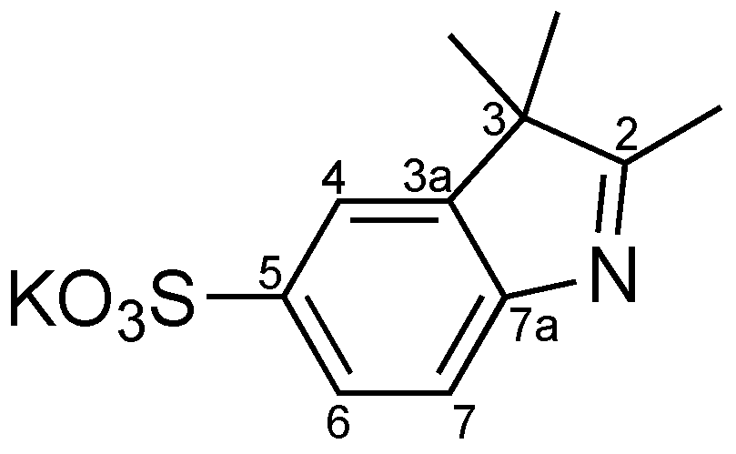
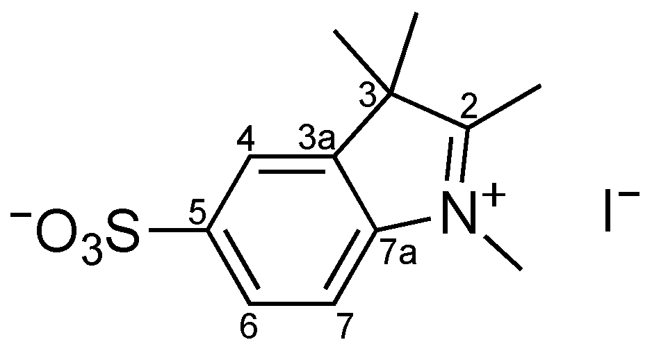
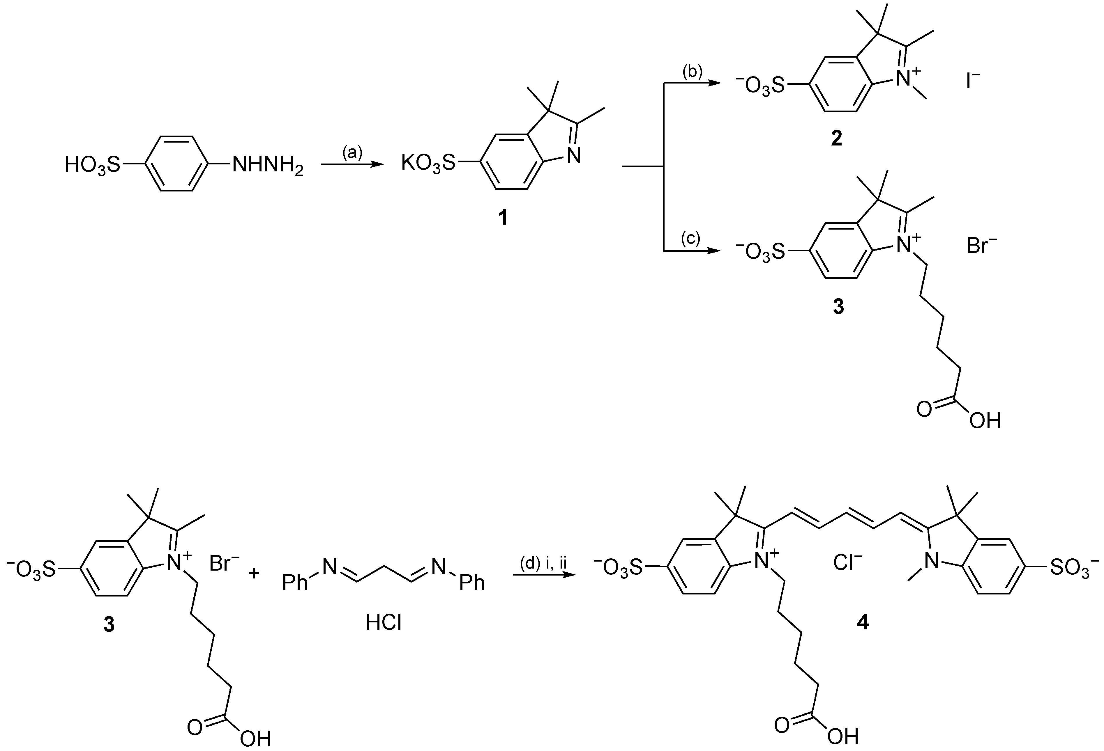
2.2.1. Preparation of Sulfonated Indolium Salts

Potassium 2,3,3-trimethyl-3H-indole-5-sulfonate (1): 4-Hydrazinobenzenesulfonic acid hemihydrate (25 g, 127 mmol) and 3-methyl-2-butanone (40 mL, 371 mmol) were dissolved in acetic acid (75 mL) and the mixture was heated to reflux for 3 h. The solution was cooled to 4 °C, and the precipitate was collected by filtration. The residue was dissolved in methanol and then precipitated by addition of a saturated solution of potassium hydroxide in 2-propanol. The product was collected by filtration, washed with 2-propanol and diethyl ether, and dried in vacuo. Compound 1 (Figure 1) was obtained as purple solid (8.41 g, 24%). The product was pure enough to be used for the next reaction.
1H NMR (400 MHz, D2O): δ = 1.34 (6H, s, 2xC3-CH3), 7.57 (1H, d, J = 8.0 Hz, H-7), 7.81 (1H, dd, J = 2.0 and 8.0 Hz, H-6), 7.86 (1H, d, J = 1.6 Hz, H-4) ppm. The CH3 group at position 2 was not observed in D2O, in agreement with the literature [16].
13C NMR (100.6 MHz, D2O): δ = 21.64 (2xC3-CH3), 54.17 (C3), 118.75 (C7), 119.30 (C4), 125.50 (C6), 139.67 (C5), 146.28 (C3a), 153.84 (C7a), 194.98 (C2) ppm.
1,2,3,3-Tetramethyl-3H-indolium-5-sulfonate iodide (2): The potassium salt of 2,3,3-trimethyl-3H-indole-5-sulfonate 1 (4 g, 14.4 mmol) was suspended in iodomethane (15 mL) and the reaction mixture was stirred at 40 °C for 6 h. The precipitate was collected by filtration, washed with acetone and dried in vacuo. Compound 2 (Figure 2) was obtained as purple solid (3.02 g, 55%). The product was used in the next reaction without additional purification.
1H NMR (400 MHz, D2O): δ = 1.58 (6H, s, 2xC3-CH3), 4.05 (3H, s, NCH3), 7.56 (1H, d, J = 8.0 Hz, H-7), 7.81 (1H, dd, J = 1.6 and 8.0 Hz, H-6), 7.86 (1H, d, J = 1.6 Hz, H-4) ppm. The C2-CH3 group was not observed in D2O, in agreement with the literature [16].
13C NMR (100.6 MHz, D2O): δ = 21.73 (2xC3-CH3), 34.63 (NCH3), 54.27 (C3), 118.73 (C7), 119.46 (C4), 125.64 (C6), 140.02 (C5), 146.21 (C3a), 153.27 (C7a), 198.85 (C2) ppm.
MS m/z (ESI, %): 254 ([M+H]+, 100). HRMS: m/z (ESI) calc. for C12H16NO3S 254.0845; found 254.0848.
1-(5-Carboxypentyl)-2,3,3-trimethyl-3H-indolium-5-sulfonate bromide (3): The potassium salt of 2,3,3-trimethyl-3H-indole-5-sulfonate 1 (4 g, 14.4 mmol) and 6-bromohexanoic acid (3.25 g, 16.7 mmol) were mixed in chlorobenzene (35 mL) and heated at 110 °C for 12 h. The mixture was cooled, chlorobenzene was decanted, and the residue was triturated with 2-propanol, collected by filtration, washed with 2-propanol and dried in vacuo. Compound 3 (Figure 3) was obtained as purple solid (3.86 g, 62%). The product was used in the next reaction without additional purification.
1H NMR (400 MHz, D2O): δ = 1.47 (2H, qui, CH2-c), 1.62 (6H, s, 2xC3-CH3), 1.68 (2H, qui, CH2-d), 2.00 (2H, qui, CH2-b), 2.39 (2H, t, J = 7.6 Hz, CH2-e), 4.54 (2H, t, J = 7.6 Hz, CH2-a), 7.92 (1H, d, J = 8.4 Hz, H-7), 8.04 (1H, dd, J = 1.6 and 8.4 Hz, H-6), 8.14 (1H, d, J = 1.6 Hz, H-4) ppm. The C2-CH3 group was not observed in D2O, in agreement with the literature [16].
13C NMR (100.6 MHz, D2O): δ = 21.54 (2xC3-CH3), 23.74 (CH2-d), 25.22 (CH2-c), 26.76 (CH2-b); 33.37 (CH2-e); 48.16 (CH2-a), 54.88 (C3), 115.91 (C7), 120.88 (C4), 126.88 (C6), 142.57 (C3a), 142.74 (C7a), 144.18 (C5), 178.52 (C=O), 199.01 (C2) ppm.
MS m/z (ESI, %): 354 ([M+H]+, 100). HRMS: m/z (ESI) calc. for C17H24NO5S 354.1370; found 354.1367.

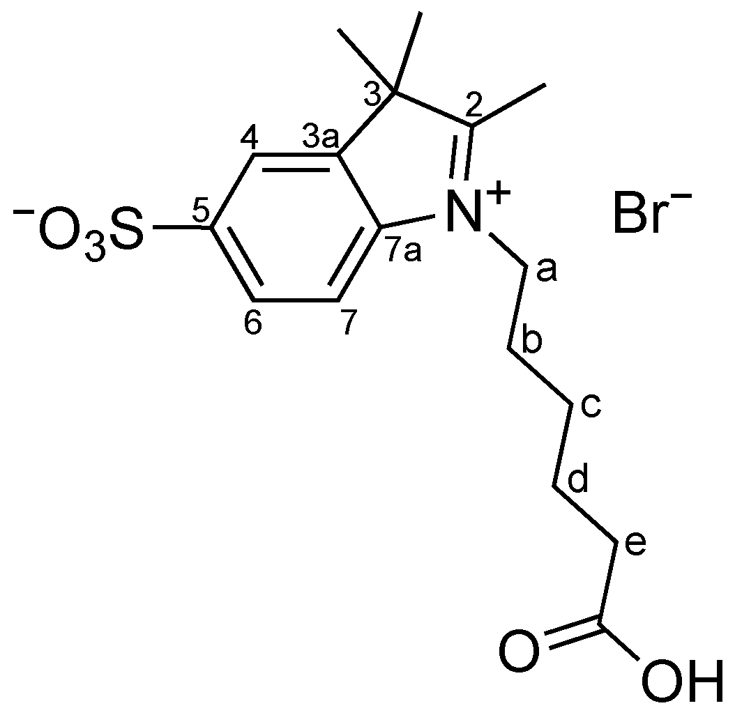
2.2.2. Synthesis of Sulfo-Cy5 Carboxylic Acid 4

1-(5-Carboxypentyl)-3,3-dimethyl-2-[5-(1′,3′,3′-trimethyl-5′-sulfonatoindol-1-ium-2′-yl)penta-2,4-dienylidene]indole-5-sulfonate chloride, sulfo-Cy5 carboxylic acid (4): A solution of indolium salt 3 (200 mg, 0.56 mmol) and malonaldehyde dianilide hydrochloride (176 mg, 0.68 mmol) in acetic anhydride (1.5 mL) was stirred at 120 °C for 30 min. The reaction mixture was cooled to room temperature and a solution of the indolium salt 2 (199 mg, 0.79 mmol) in pyridine (1.5 mL) was added. The reaction mixture was stirred at room temperature in the dark for 24 h. The product was precipitated by addition of ethyl acetate and collected by filtration. The crude residue was purified by a silica gel chromatography column, using as eluent mixtures of ethyl acetate and methanol with increasing order of polarity (ethyl acetate: methanol, 7:3 > 6.5:3.5 > 6:4). The sulfo-Cy5 carboxylic acid 4 (Figure 4) was obtained as dark blue solid (72.8 mg, 19%).
1H NMR (400 MHz, MeOH-d4): δ = 1.49–1.55 (2H, m, CH2-c), 1.67–1.77 (14H, m, CH2-d + 4xCH3), 1.81–1.87 (2H, q, CH2-b), 2.30 (2H, t, J = 7.2 Hz, CH2-e), 3.67 (1H, d, J = 3.6 Hz, NCH3), 4.15 (2H, t, J = 6.8 Hz, CH2-a); 6.36 (2H, dd, J = 5.6 and 13.6 Hz, H-α + H-α′), 6.71 (1H, t, J = 12.4 Hz, H-γ), 7.34–7.37 (2H, m, H-7 + H-7′), 7.89–7.91 (4H, m, H-4 + H-6 + H-4′+ H-6′), 8.32 (2H, t, J = 13.2 Hz, H-β + H-β′) ppm.
13C NMR (100.6 MHz, MeOH-d4): δ = 26.11 (CH2-c), 27.42 (CH2-d), 27.71 (2xCH3), 27.84 (2xCH3), 28.12 (CH2-b), 31.77 (NCH3), 35.83 (CH2-e), 45.06 (CH2-a), 50.50 (C-3 or C-3′), 50.55 (C-3 or C-3′), 105.22 (C-α or C-α′), 105.34 (C-α or C-α′), 111.43 (C-7 or C-7′), 111.64 (C-7 or C-7′), 121.22 (C-6 or C-6′), 121.33 (C-6 or C-6′), 127.75 (C-4 or C-4′ or C-γ), 128.01 (C-4 or C-4′ or C-γ), 128.06 (C-4 or C-4′ or C-γ), 142.48 (C-3a or C-3a′), 142.59 (C-3a or C-3a′), 143.31 (C-5 or C-5′), 143.35 (C-5 or C-5′), 144.93 (C-7a), 145.60 (C-7a′), 156.21 (C-β + C-β′), 175.26 (C-2), 176.00 (C-2′), 178.88 (C=O) ppm.

2.3. Photophysical Characterization

The photophysical characterization of sulfo-Cy5 carboxylic acid 4 was carried out by UV-vis absorption and fluorescence spectroscopy of a 1 × 10⁻⁶ M solution in phosphate-buffered saline (PBS) at pH 7.4. The fluorescence spectrum of dye 4 was obtained by excitation at the wavelength of maximum absorption.
The relative fluorescence quantum yield was calculated relative to a standard solution of Nile Blue in methanol (ΦF = 0.27) [18].

3. Results and Discussion

3.1. Synthesis of Sulfo-Cy5 Carboxylic Acid 4

The preparation of sulfo-Cy5 carboxylic acid 4 was accomplished by solution-phase chemistry described by Waggoner and co-workers [16], according to Scheme 1. The key intermediate, indoleninium-5-sulfonate, was prepared from 4-hydrazinobenzenesulfonic acid hemihydrate and 3-methyl-2-butanone by conventional Fisher indole synthesis [16]. The indolenine thus obtained was converted into a potassium salt before quaternization in 24% yield. This compound was alkylated using iodomethane or 6-bromohexanoic acid to give compound 2 in 55% and compound 3 in 62%, respectively, which were used in the next reaction steps without additional purification.
Stepwise condensation reaction of the indolenines 2 and 3 with a polyene-chain precursor (malonaldehyde dianilide hydrochloride) gave good conversion to the desired sulfoindocyanine 4. This was precipitated from the reaction mixture by addition of ethyl acetate and the precipitate was further purified by column chromatography on silica gel. Isolation of this compound proved to be very difficult, as side products and cleavage product had nearly the same polarity. Nevertheless, sulfo-Cy5 carboxylic acid 4 was obtained in the form of a dark blue solid in 19% yield.
The synthesized compounds were characterized by 1H and 13C NMR spectroscopy and mass spectroscopy, and the obtained data were in agreement with the expected structure.

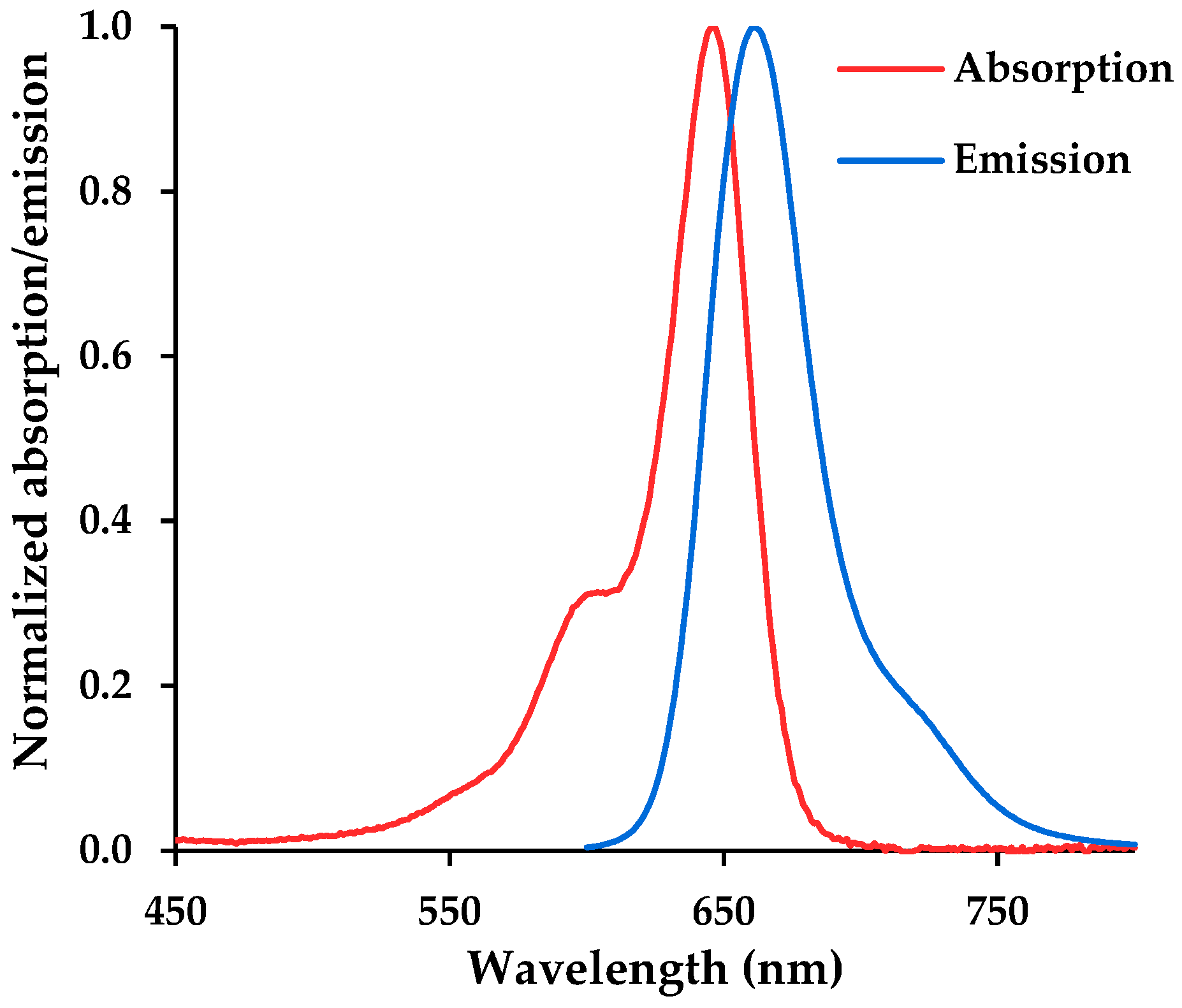
3.2. Photophysical Characterization

A solution of sulfo-Cy5 carboxylic acid 4 (1 × 10⁻⁶ M) at pH 7.4 in phosphate-buffered saline (PBS) was analyzed and the wavelengths of maximum absorption and fluorescence, λabs and λflu, molar absorptivity at the absorption maximum, ε, relative fluorescence quantum yield, ΦF, and Stokes’ shifts are compiled in Table 1.
The synthesized dye 4 exhibited a narrow absorption band (Figure 5), a high molar absorptivity (log ε = 5.11) at 646 nm, and a sharp emission band with a wavelength of maximum fluorescence in the NIR region (λflu = 661 nm). The relative fluorescence quantum yield, determined by using Nile Blue in methanol as standard (ΦF = 0.27), was found to be low (ΦF = 0.27), which is in agreement with the literature [16]. Although the extension of the polymethine bridge presents red shifted emission, lower quantum yields were found for NIR cyanine dyes in water. However, the presence of sulfo groups could significantly enhance water solubility and prevent aggregation and fluorescence self-quenching in aqueous media [4,17]. Taking this into account, sulfo-Cy5 carboxylic acid 4 can be activated to form succinimidyl ester for conjugation with a specific peptide.

4. Conclusions

In summary, a water-soluble pentamethine dye bearing two sulfo groups was synthesized by solution solid phase synthesis in 19% yield. However, future work will be developed to optimize the isolation method to increase the overall synthesis yield.
Photophysical characterization of sulfo-Cy5 carboxylic acid 4 showed an intense absorption band with a high molar absorptivity at 646 nm and the wavelength of maximum fluorescence was found in the NIR region at 661 nm. Therefore, due to its excellent spectral properties, good solubility in water and emission in the NIR region, dye 4 can be used for our intentions for it as a donor group in FRET probes to study enzymatic hydrolysis in vitro.

Author Contributions

Conceptualization, S.P.G.C.; methodology, C.D.F.M. and S.P.G.C.; validation, S.P.G.C. and M.M.M.R.; formal analysis, C.D.F.M., M.M.M.R. and S.P.G.C.; investigation, C.D.F.M.; resources, S.P.G.C. and M.M.M.R.; writing—original draft preparation, C.D.F.M.; writing—review and editing, C.D.F.M., M.M.M.R. and S.P.G.C.; supervision, S.P.G.C. and M.M.M.R.; project administration, S.P.G.C.; funding acquisition, M.M.M.R. and S.P.G.C. All authors have read and agreed to the published version of the manuscript.

Funding

This research was funded by the Foundation for Science and Technology (FCT) for financial support to CQ/UM (UID/QUI/00686/2020) and project PTDC/QUI-COL/28052/2017 and a PhD grant to C. D. F. Martins (SFRH/BD/05277/2020). The NMR spectrometer Bruker Avance III 400 is part of the National NMR Network and was purchased within the framework of the National Program for Scientific Re-equipment, contract REDE/1517/RMN/2005 with funds from POCI 2010 (FEDER) and FCT.

Institutional Review Board Statement

Not applicable.

Informed Consent Statement

Not applicable.

Data Availability Statement

Not applicable.

Conflicts of Interest

The authors declare no conflict of interest.

References

  1. An, H.W.; Wang, M.D. The self-assembly of cyanine dyes for biomedical application in vivo. In In Vivo Self-Assembly Nano-technology for Biomedical Applications; Wang, H., Li, L.L., Eds.; Springer: Singapore, 2018; pp. 31–55. [Google Scholar]
  2. Zhang, X.-H.; Wang, L.-Y.; Nan, Z.-X.; Tan, S.-H.; Zhang, Z.-X. Microwave-assisted solvent-free synthesis and spectral properties of some dimethine cyanine dyes as fluorescent dyes for DNA detection. Dyes Pigm. 2008, 79, 205–209. [Google Scholar] [CrossRef]
  3. Patonay, G.; Salon, J.; Sowell, J.; Strekowski, L. Noncovalent labeling of biomolecules with red and near-infrared dyes. Molecules 2004, 9, 40–49. [Google Scholar] [CrossRef]
  4. Wang, L.; Fan, J.; Qiao, X.; Peng, X.; Dai, B.; Wang, B.; Sun, S.; Zhang, L.; Zhang, Y. Novel asymmetric Cy5 dyes: Synthesis, photostabilities and high sensitivity in protein fluorescence labeling. J. Photochem. Photobiol. A Chem. 2010, 210, 168–172. [Google Scholar] [CrossRef]
  5. Owens, E.A.; Bruschi, N.; Tawney, J.G.; Henary, M. A microwave-assisted and environmentally benign approach to the synthesis of near-infrared fluorescent pentamethine cyanine dyes. Dyes Pigm. 2015, 113, 27–37. [Google Scholar] [CrossRef]
  6. Pham, W.; Medarova, Z.; Moore, A. Synthesis and application of a water-soluble near-infrared dye for cancer detection using optical imaging. Bioconjugate Chem. 2005, 16, 735–740. [Google Scholar] [CrossRef] [PubMed]
  7. Wolf, N.; Kersting, L.; Herok, C.; Mihm, C.; Seibel, J. High-yielding water-soluble asymmetric cyanine dyes for labeling applications. J. Org. Chem. 2020, 85, 9751–9760. [Google Scholar] [CrossRef] [PubMed]
  8. Pisoni, D.S.; Todeschini, L.; Borges, A.C.A.; Petzhold, C.L.; Rodembusch, F.S.; Campo, L.F. Symmetrical and asymmetrical cyanine dyes. Synthesis, spectral properties, and BSA association study. J. Org. Chem. 2014, 79, 5511–5520. [Google Scholar] [CrossRef] [PubMed]
  9. Shindy, H.A. Fundamentals in the chemistry of cyanine dyes: A review. Dyes Pigm. 2017, 145, 505–513. [Google Scholar] [CrossRef]
  10. Bříza, T.; Rimpelová, S.; Králová, J.; Záruba, K.; Kejík, Z.; Ruml, T.; Martásek, P.; Král, V. Pentamethinium fluorescent probes: The impact of molecular structure on photophysical properties and subcellular localization. Dyes Pigm. 2014, 107, 51–59. [Google Scholar] [CrossRef]
  11. Yi, X.; Wang, F.; Qin, W.; Yang, X.; Yuan, J. Near-infrared fluorescent probes in cancer imaging and therapy: An emerging field. Int. J. Nanomed. 2014, 9, 1347–1365. [Google Scholar] [CrossRef] [PubMed] [Green Version]
  12. Luo, S.; Zhang, E.; Su, Y.; Cheng, T.; Shi, C. A review of NIR dyes in cancer targeting and imaging. Biomaterials 2011, 32, 7127–7138. [Google Scholar] [CrossRef] [PubMed]
  13. Feng, L.; Chen, W.; Ma, X.; Liu, S.H.; Yin, J. Near-infrared heptamethine cyanines (Cy7): From structure, property to application. Org. Biomol. Chem. 2020, 18, 9385–9397. [Google Scholar] [CrossRef] [PubMed]
  14. Lee, S.; Park, K.; Lee, S.-Y.; Ryu, J.H.; Park, J.W.; Ahn, H.J.; Kwon, I.C.; Youn, I.-C.; Kim, K.; Choi, K. Dark quenched matrix metalloproteinase fluorogenic probe for imaging osteoarthritis development in vivo. Bioconjugate Chem. 2008, 19, 1743–1747. [Google Scholar] [CrossRef] [PubMed]
  15. Jia, X.; Chen, Q.; Yang, Y.; Tang, Y.; Wang, R.; Xu, Y.; Zhu, W.; Qian, X. FRET-based mito-specific fluorescent probe for ratiometric detection and imaging of endogenous peroxynitrite: Dyad of Cy3 and Cy5. J. Am. Chem. Soc. 2016, 138, 10778–10781. [Google Scholar] [CrossRef] [PubMed]
  16. Mujumdar, R.B.; Ernst, L.A.; Mujumdar, S.R.; Lewis, C.J.; Waggoner, A.S. Cyanine dye labeling reagents: Sulfoindocyanine succinimidyl esters. Bioconjugate Chem. 1993, 4, 105–111. [Google Scholar] [CrossRef] [PubMed]
  17. Lopalco, M.; Koini, E.N.; Cho, J.K.; Bradley, M. Catch and release microwave mediated synthesis of cyanine dyes. Org. Biomol. Chem. 2009, 7, 856–859. [Google Scholar] [CrossRef] [PubMed]
  18. Jose, J.; Ueno, Y.; Burgess, K. Water-soluble Nile Blue derivatives: Syntheses and photophysical properties. Chem. Eur. J. 2008, 15, 418–423. [Google Scholar] [CrossRef]
Figure 1. Structure of indolenine 1.
Chemproc 08 00091 g001
Figure 2. Structure of indolium salt 2.
Chemproc 08 00091 g002
Figure 3. Structure of indolium salt 3.
Chemproc 08 00091 g003
Figure 4. Structure of dye 4.
Chemproc 08 00091 g004
Scheme 1. Synthesis of sulfo-Cy5 carboxylic acid 4: (a) 3-methyl-2-butanone, acetic acid, 3 h, reflux; (b) iodomethane, 6 h, 40 °C; (c) 6-bromohexanoic acid, chlorobenzene, 12 h, 110 °C; (d) (i) acetic anhydride, 30 min, 120 °C; (ii) 2, pyridine, 24, r.t.
Chemproc 08 00091 sch001
Figure 5. Normalized absorption and emission spectra of sulfo-Cy5 carboxylic acid 4 (1 × 10⁻⁶ M) at pH 7.4 in PBS.
Chemproc 08 00091 g005
Table 1. UV-vis absorption and fluorescence data of sulfo-Cy5 carboxylic acid 4 (1 × 10⁻⁶ M) at pH 7.4 in PBS.
CompoundUV-Vis AbsorptionFluorescence
λabs (nm)log ελflu (nm)ΦFStokes’ Shift
Dye 46465.116610.2715
Publisher’s Note: MDPI stays neutral with regard to jurisdictional claims in published maps and institutional affiliations.

