
Synthesis and Characterization of a Dansyl-Based Fluorescent Probe for Analytical Purposes †

 ,
,  ,
,  and
and 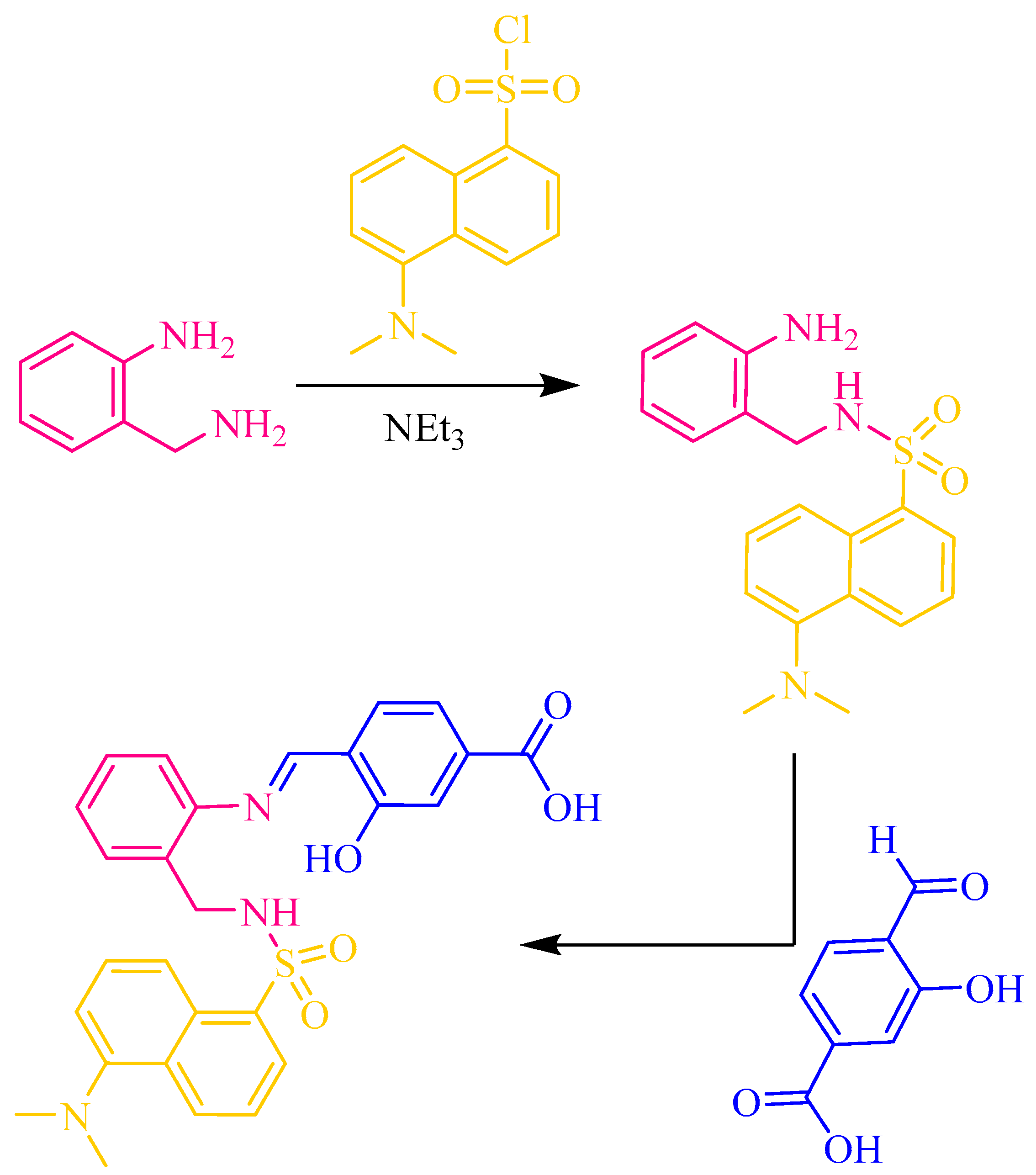
Abstract
:1. Introduction
2. Results and Discussion
3. Conclusions
4. Experimental Section
- Materials and Methods
- N-(2-aminobenzyl)-5-(dimethylamino)naphthalene-1-sulfonamide
- H3L
Author Contributions
Funding
Acknowledgments
Conflicts of Interest
References
- Laborda, F.; Bolea, E.; Cepria, G.; Gómez, M.T.; Jiménez, M.S.; Pérez-Arantegui, J.; Castillo, J.R. Detection, characterization and quantification of inorganic engineered nanomaterials: A review of techniques and methodological approaches for the analysis of complex samples. Anal. Chim. Acta 2016, 904, 10–32. [Google Scholar] [CrossRef] [PubMed] [Green Version]
- Yan, A.; Chen, Z. Detection methods of nanoparticles in plant tissues. In New Visions in Plant Science; Çelik, Ö., Ed.; Intechopen: London, UK, 2018; Chapter 6. [Google Scholar]
- Navratilova, J.; Praetorius, A.; Gondikas, A.; Fabienke, W.; Kammer, F.; Hofmann, T. Detection of engineered copper nanoparticles in soil using single particle ICP-MS. Int. J. Environ. Res. Public Health 2015, 12, 15756–15768. [Google Scholar] [CrossRef] [PubMed] [Green Version]
- Medina-Sánchez, M.; Miserere, S.; Marín SAragay, G.; Merkoçi, A. On-chip electrochemical detection of CdS quantum dots using normal and multiple recycling flow through modes. Lab. Chip 2012, 12, 2000–2005. [Google Scholar] [CrossRef] [PubMed]
- Chatterjee, A.; Santra, M.; Won, N.; Kim, S.; Kim, J.K.; Kim, S.B.; Ahn, K.H. Selective fluorogenic and chromogenic probe for detection of silver ions and silver nanoparticles in aqueous media. J. Am. Chem. Soc. 2009, 131, 2040–2041. [Google Scholar] [CrossRef] [PubMed]
- Cayuela, A.; Soriano, M.L.; Valcárcel, M. Reusable sensor based on functionalized carbon dots for the detection of silver nanoparticles in cosmetics via inner filter effect. Anal. Chim. Acta 2015, 872, 70–76. [Google Scholar] [CrossRef] [PubMed]
- Rebe Raz, S.; Leontaridou, M.; Bremer, M.G.E.G.; Peters, R.; Weigel, S. Development of surface plasmon resonance-based sensor for detection of silver nanoparticles in food and the environment. Anal. Bioanal. Chem. 2012, 403, 2843–2850. [Google Scholar] [CrossRef] [PubMed] [Green Version]
- Sanmartín-Matalobos, J.; García-Deibe, A.M.; Fondo, M.; Zarepour-Jevinani, M.; Domínguez-González, M.R.; Bermejo-Barrera, P. Exploration of an easily synthesized fluorescent probe for detecting copper in aqueous samples. Dalton Trans. 2017, 46, 15827–15835. [Google Scholar] [CrossRef]
- Sanmartín-Matalobos, J.; García-Deibe, M.; Zarepour-Jevinani, M.; Aboal-Somoza, M.; Bermejo-Barrera, A.M.; Fondo, P. Exploring the Chelating Potential of an Easily Synthesized Schiff Base for Copper Sensing. Crystals 2020, 10, 235. [Google Scholar] [CrossRef] [Green Version]
- Srivastava, P.; Ali, R.; Razi, S.S.; Shahid, M.; Patnaik, S.; Misra, A. A simple blue fluorescent probe to detect Hg2+ in semiaqueous environment by intramolecular charge transfer mechanism. Tetrah. Lett. 2013, 54, 3688–3693. [Google Scholar] [CrossRef]
- Van Beijnen, A.J.M.; Nolte, R.J.M.; Naaktgeboren, A.J.; Zwikker, J.W.; Drenth, W. Helical Configuration of Poly (iminomethylenes). Synthesis and CD spectra of Polymers derived from optically active isocyanides. Macromolecules 1983, 16, 1679–1689. [Google Scholar] [CrossRef] [Green Version]


Publisher’s Note: MDPI stays neutral with regard to jurisdictional claims in published maps and institutional affiliations. |
© 2021 by the authors. Licensee MDPI, Basel, Switzerland. This article is an open access article distributed under the terms and conditions of the Creative Commons Attribution (CC BY) license (https://creativecommons.org/licenses/by/4.0/).
Share and Cite
Sanmartín-Matalobos, J.; Bermejo-Barrera, P.; Alves-Iglesias, Y.; García-Deibe, A.M.; Fondo, M. Synthesis and Characterization of a Dansyl-Based Fluorescent Probe for Analytical Purposes. Chem. Proc. 2022, 8, 76. https://doi.org/10.3390/ecsoc-25-11775
Sanmartín-Matalobos J, Bermejo-Barrera P, Alves-Iglesias Y, García-Deibe AM, Fondo M. Synthesis and Characterization of a Dansyl-Based Fluorescent Probe for Analytical Purposes. Chemistry Proceedings. 2022; 8(1):76. https://doi.org/10.3390/ecsoc-25-11775
Chicago/Turabian StyleSanmartín-Matalobos, Jesús, Pilar Bermejo-Barrera, Yeneva Alves-Iglesias, Ana M. García-Deibe, and Matilde Fondo. 2022. "Synthesis and Characterization of a Dansyl-Based Fluorescent Probe for Analytical Purposes" Chemistry Proceedings 8, no. 1: 76. https://doi.org/10.3390/ecsoc-25-11775