
Synthesis and Structural Study of a New β-Turn Inducer Peptidomimetic Incorporating 1-Amino-1-Aminomethylcyclohexane †

 , and
, and
Abstract
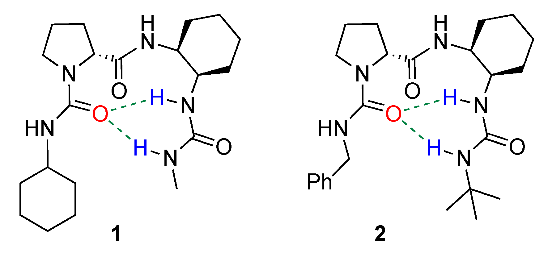
:1. Introduction
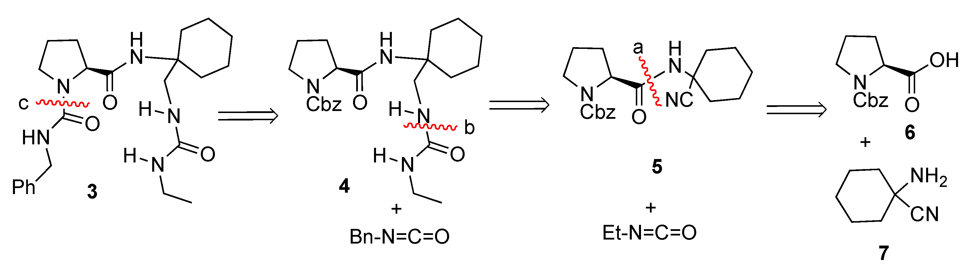
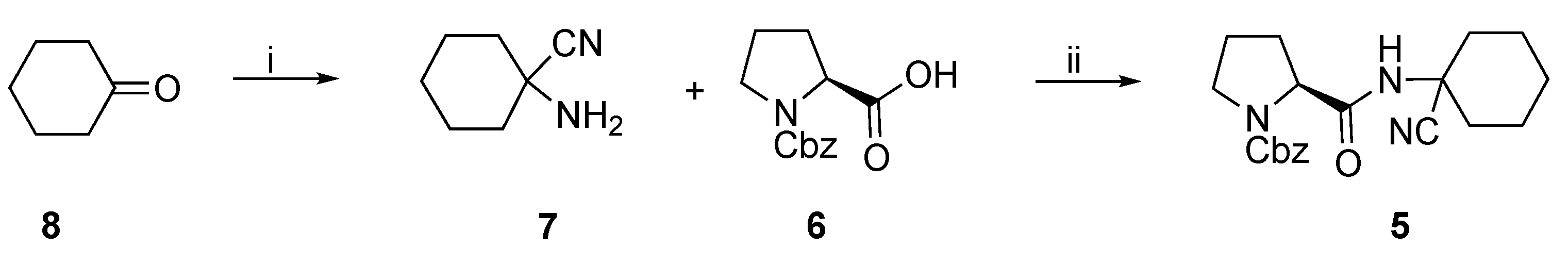
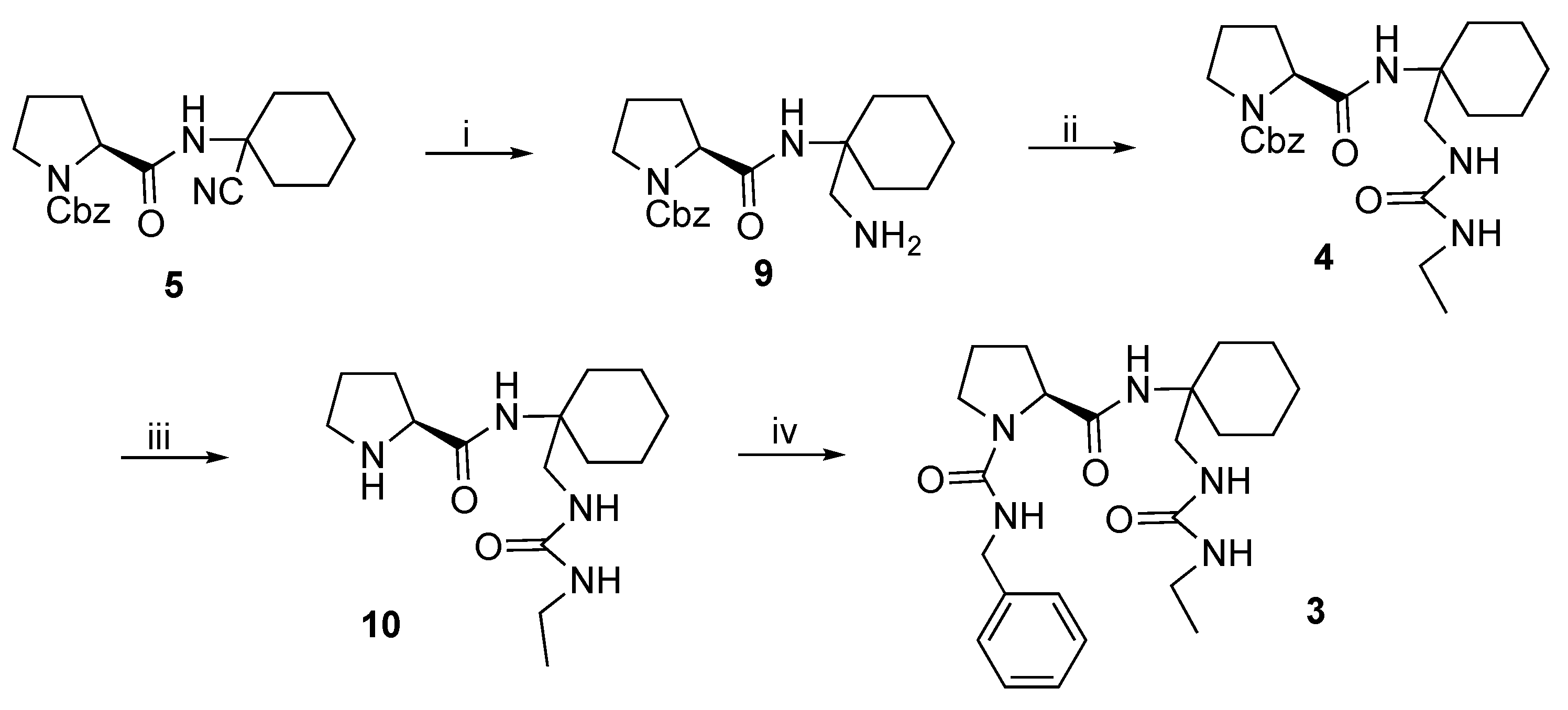
- Coupling of α-aminonitrile 7 with commercial N-Cbz-L-proline 6, to form the strategic bond “a” of compound 5.
- Reduction of the cyano group to amine and subsequent coupling with ethyl isocyanate to form the strategic bond “b” to generate compound 4.
- Finally, removal of the Cbz protecting group and coupling with benzyl isocyanate, leading to the formation of the strategic bond “c” and thus generating the desired diurea 3.
2. Results and Discussion
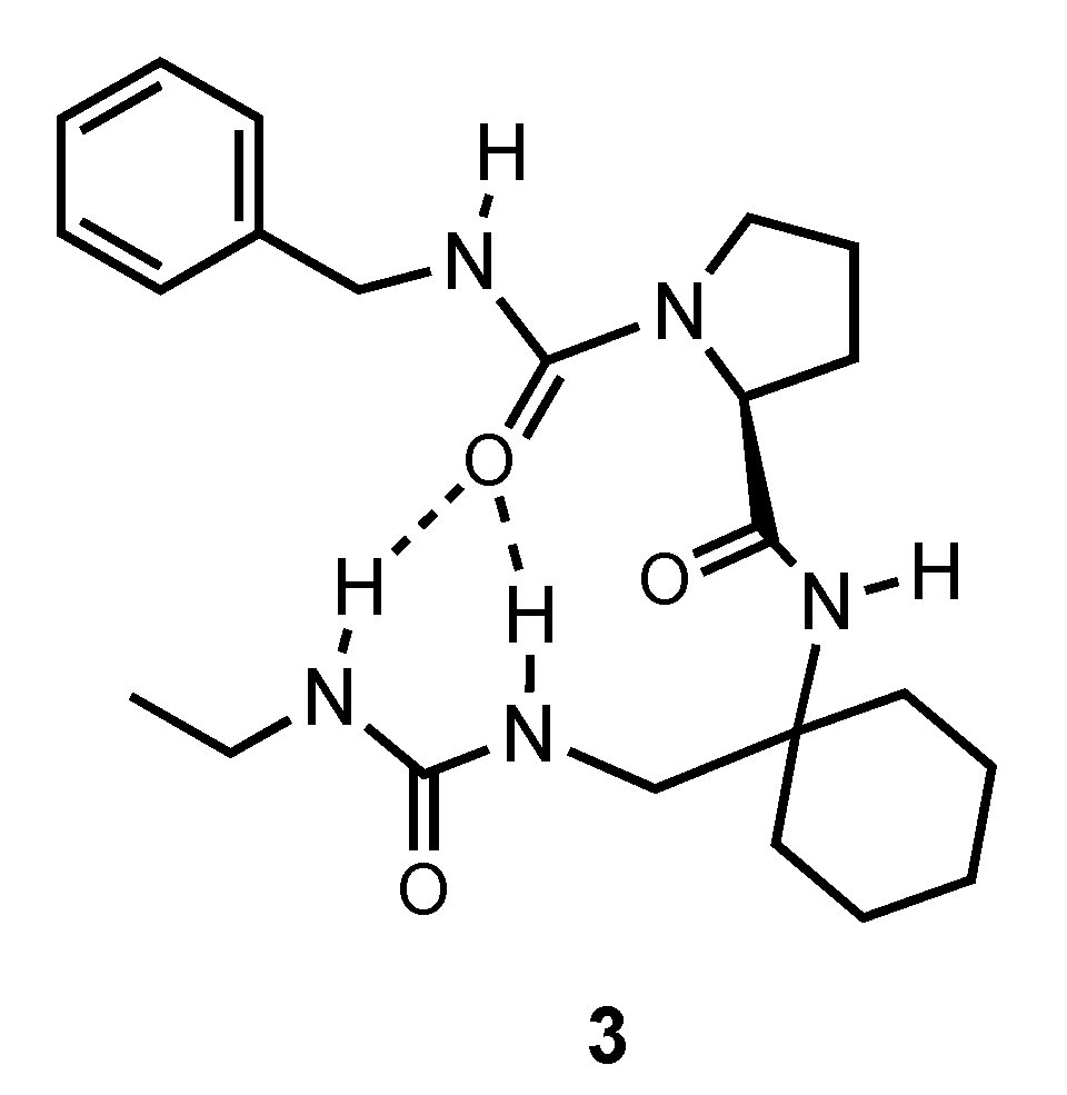
2.1. Synthesis of Compound 3
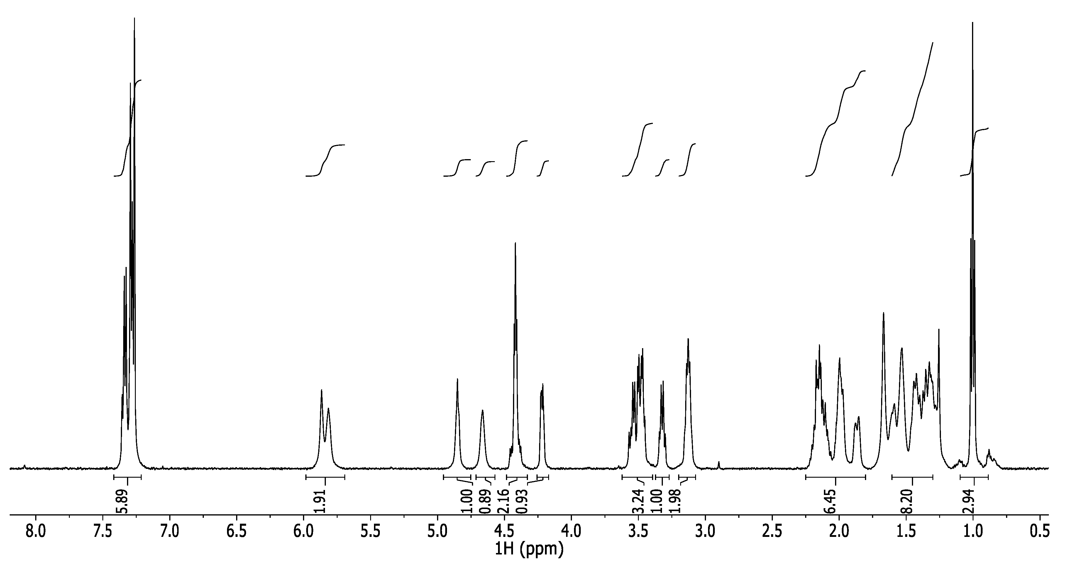
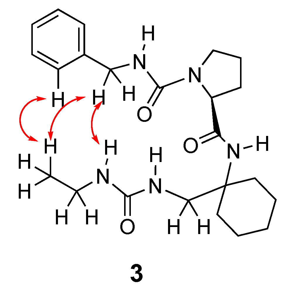
2.2. Structural Study of Compound 3
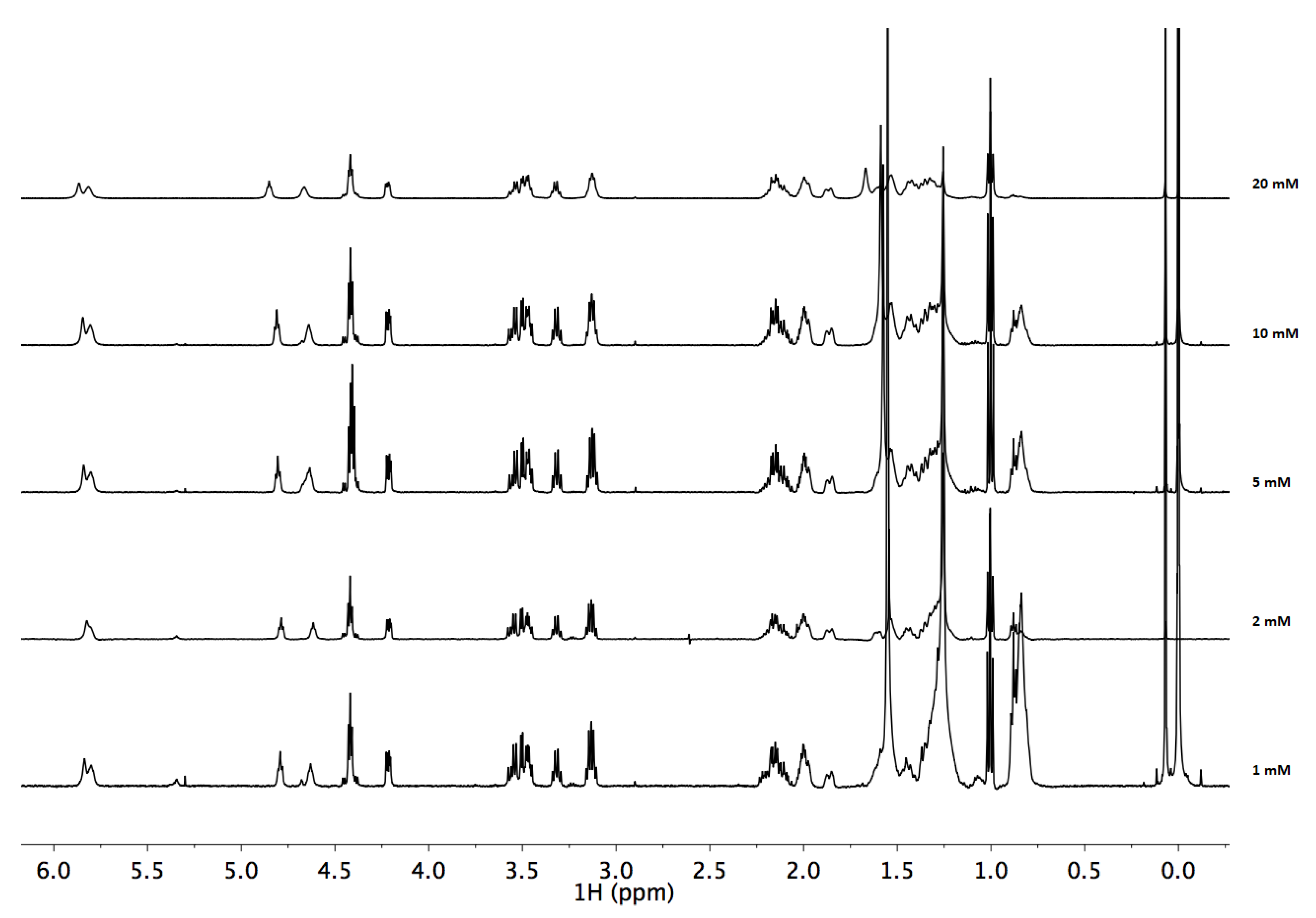
- Serial dilution
- There is little change with concentration.
- Between 1 and 5 mM there are hardly any changes, so it can be concluded that at these concentrations there is no aggregation and the molecules are found as monomers in solution.
- When going from 5 to 10 and 20 mM, some signal shifts, which may mean that above 10 mM aggregation does occur.
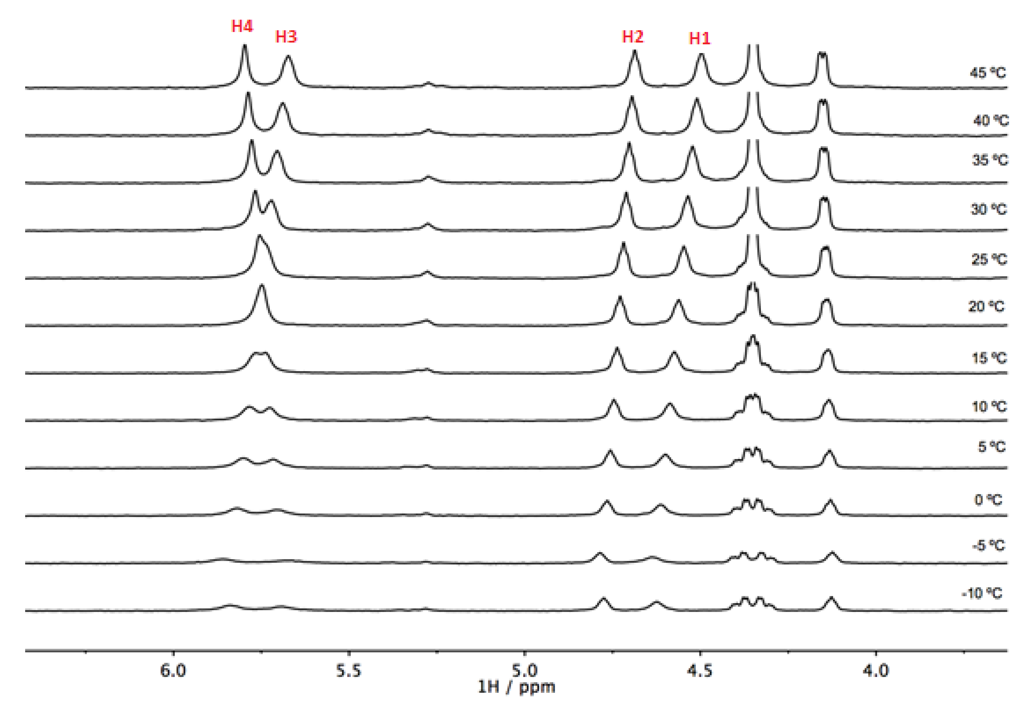
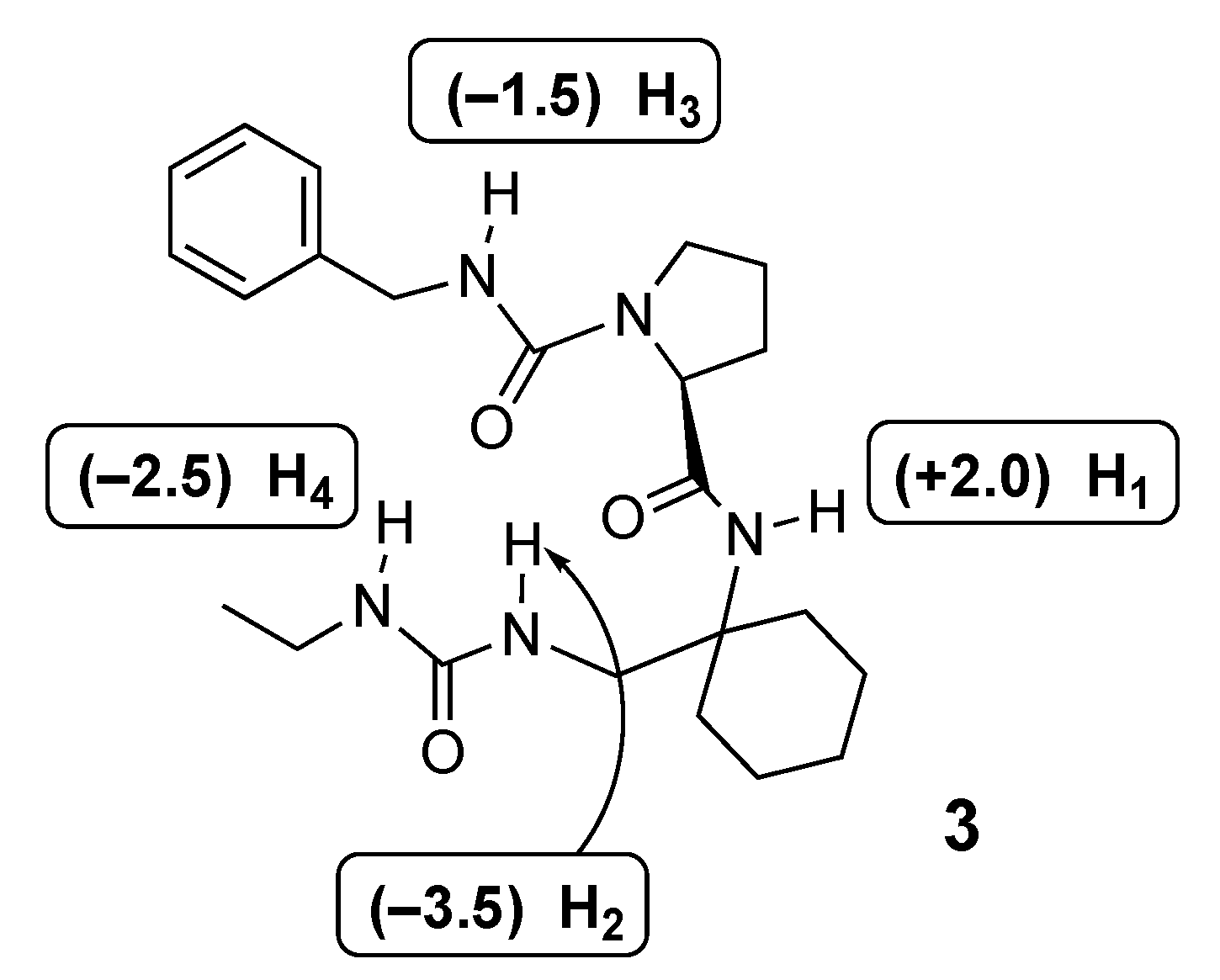
- Temperature coefficients (T-coef)
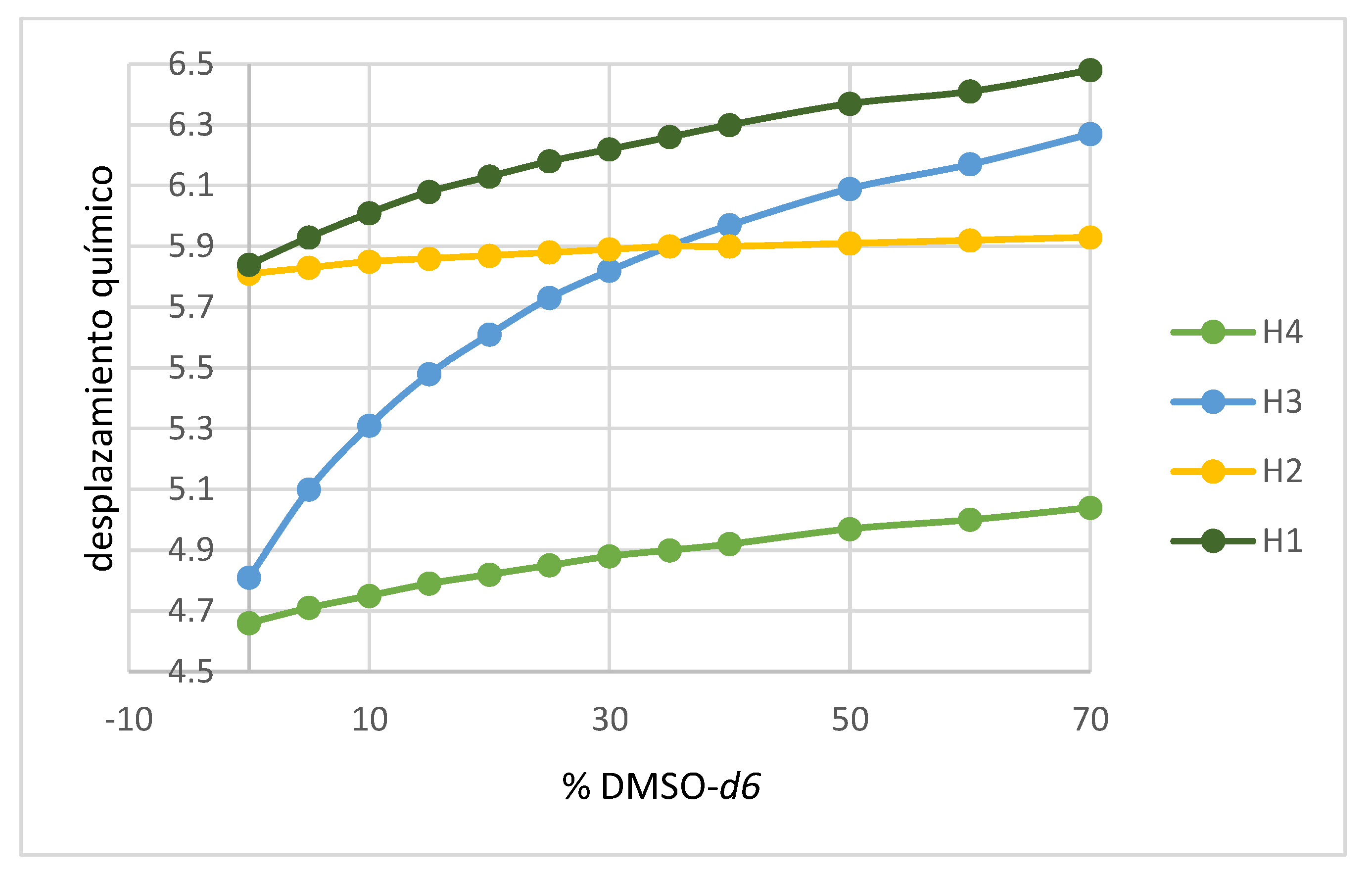
- Titration with DMSO-d6
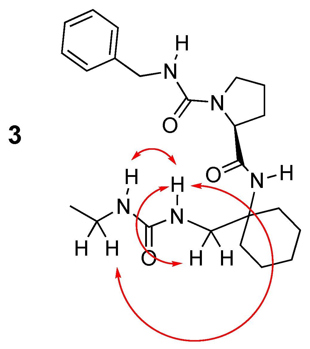
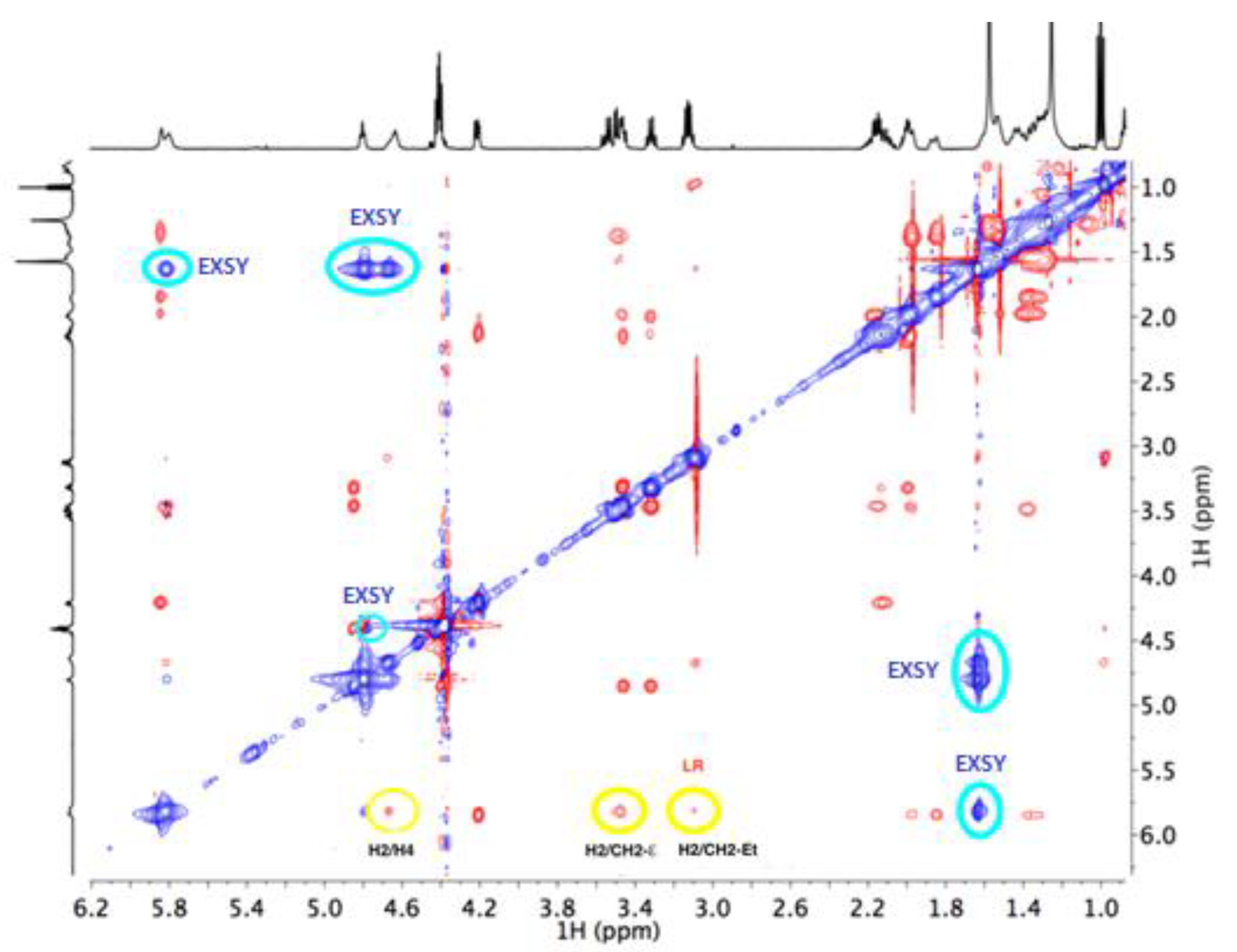
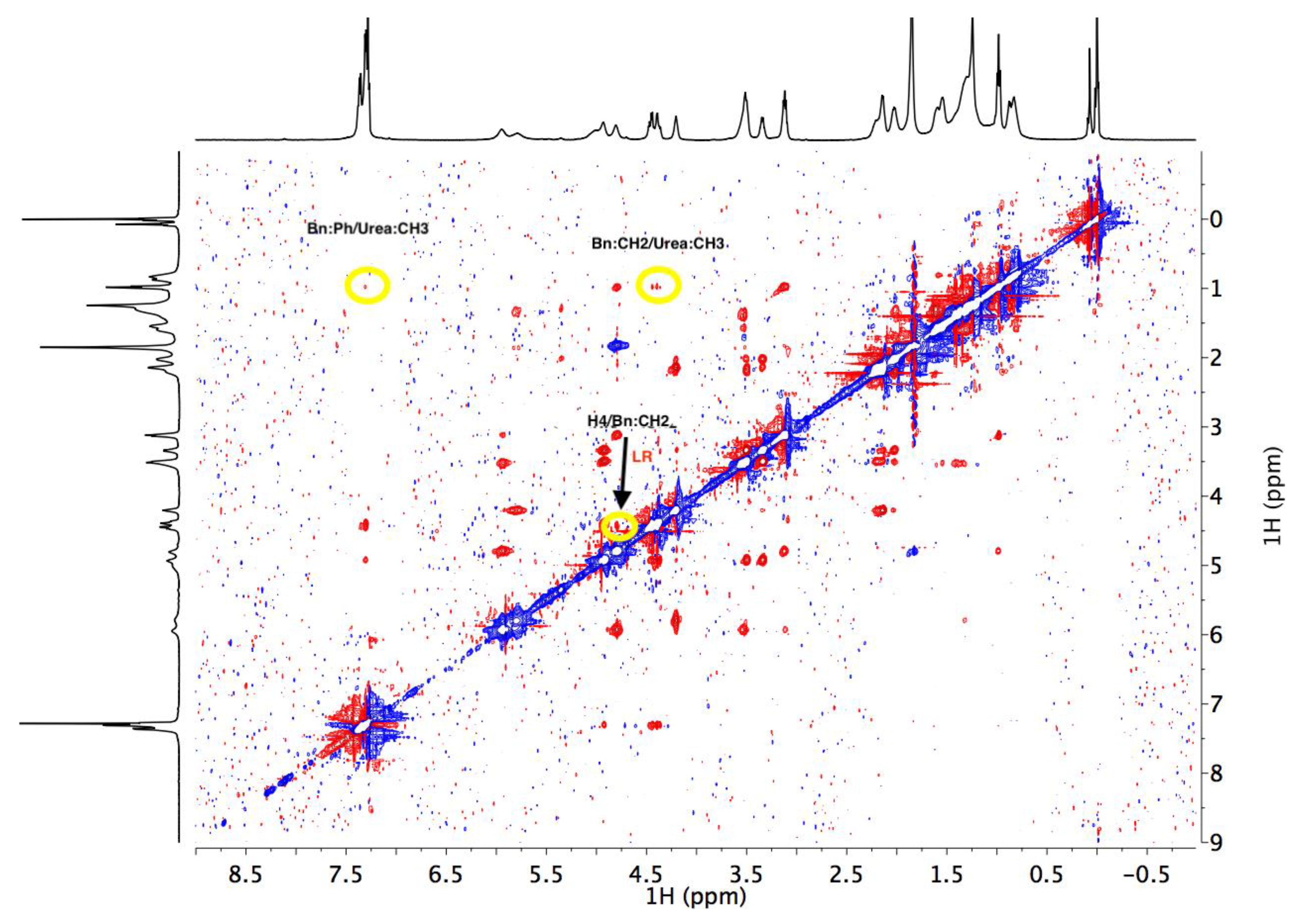
- NOESY
- H2/H4 and H2/CH2 NOE of the ethyl chain.
- Sequential H2/CH2 exocyclic NOE of the Cy unit.
- The aromatic protons of phenyl/CH3 of ethyl urea.
- Benzyl CH2/CH3 of ethyl urea.
- Benzyl CH2/H4.
Author Contributions
Funding
Institutional Review Board Statement
Informed Consent Statement
Data Availability Statement
Conflicts of Interest
References
- Medda, A.K.; Park, C.M.; Jeon, A.; Kim, H.; Sohn, J.-H.; Lee, H.-S. A nonpeptidic reverse-turn scaffold stabilized by urea-based dual intramolecular hydrogen bonding. Org. Lett. 2011, 13, 3486–3489. [Google Scholar] [CrossRef] [PubMed]
- Fischer, L.; Dierjecan, C.; Jolibois, F.; Semetey, V.; Lozano, J.M.; Briand, J.-P.; Marraud, M.; Poteau, R.; Guichard, G. Propensity for local folding induced by the urea fragment in short-chain oligomers. Org. Biomol. Chem. 2008, 6, 2596–2610. [Google Scholar] [CrossRef] [PubMed]


| Atom | δ1H (ppm) |
|---|---|
| Bn: Ph | 7.22–7.42 (m, 5H) |
| Bn: CH2 | 4.31–4.51 (mAB, 2H) |
| Bn: HN | 4.85 (t, J = 5.7 Hz, 1H, NH) |
| Pro: α | 4.22 (dd, J = 7.8, 3.7 Hz, 1H) |
| Pro: β | 1.81–2.23 (m, 2H) |
| Pro: H | 1.81–2.23 (m, 2H) |
| Pro: δ1 | 3.32 (q, J = 7.3 Hz, 1H) |
| Pro: δ2 | 3.43–3.63 (m, 1H) |
| Cy: β | 1.29–1.61 (m, 2H) |
| Cy: β | 1.81–2.23 (m, 2H) |
| Cy: γ | 1.81–2.23 (m, 2H) |
| Cy: γ | 1.81–2.23 (m, 2H) |
| Cy: δ | 1.81–2.23 (m, 2H) |
| Cy: ɛ | 3.43–3.63 (m, 2H) |
| Cy: α-HN | 5.87 (s, 1H, NH) |
| Cy: ɛ-HN | 5.82 (s, 1H, NH) |
| UREA: Et-CH2 | 3.12 (dt, J = 11.7, 6.0 Hz, 2H) |
| UREA: Et-CH3 | 1.00 (t, J = 7.2 Hz, 3H) |
| UREA: Et-HN | 4.61–4.74 (m, 1H) |
| Name | δ-max (+45C) | δ-min (−10C) | range T/°C | T-coef (ppb/K) |
|---|---|---|---|---|
| H4 | 4.49 | 4.63 | 55 | −2.5 |
| H3 | 4.69 | 4.77 | 55 | −1.5 |
| H2 | 5.67 | 5.86 | 55 | −3.5 |
| H1 | 5.80 | 5.69 | 55 | +2.0 |
Publisher’s Note: MDPI stays neutral with regard to jurisdictional claims in published maps and institutional affiliations. |
© 2021 by the authors. Licensee MDPI, Basel, Switzerland. This article is an open access article distributed under the terms and conditions of the Creative Commons Attribution (CC BY) license (https://creativecommons.org/licenses/by/4.0/).
Share and Cite
Campos, M.; Sánchez-Pedregal, V.M.; López-Carracedo, P.; Estévez, R.J.; Estévez, J.C. Synthesis and Structural Study of a New β-Turn Inducer Peptidomimetic Incorporating 1-Amino-1-Aminomethylcyclohexane. Chem. Proc. 2022, 8, 67. https://doi.org/10.3390/ecsoc-25-11684
Campos M, Sánchez-Pedregal VM, López-Carracedo P, Estévez RJ, Estévez JC. Synthesis and Structural Study of a New β-Turn Inducer Peptidomimetic Incorporating 1-Amino-1-Aminomethylcyclohexane. Chemistry Proceedings. 2022; 8(1):67. https://doi.org/10.3390/ecsoc-25-11684
Chicago/Turabian StyleCampos, María, Victor M. Sánchez-Pedregal, Pablo López-Carracedo, Ramón J. Estévez, and Juan C. Estévez. 2022. "Synthesis and Structural Study of a New β-Turn Inducer Peptidomimetic Incorporating 1-Amino-1-Aminomethylcyclohexane" Chemistry Proceedings 8, no. 1: 67. https://doi.org/10.3390/ecsoc-25-11684