Proceeding Paper

Design, Development, and In Silico Study of Pyrazoline-Based Mycobactin Analogs as Anti-Tubercular Agents †

Department of Pharmaceutical Sciences and Technology, Birla Institute of Technology, Mesra, Ranchi 835215, India
*
Author to whom correspondence should be addressed.
Presented at the 25th International Electronic Conference on Synthetic Organic Chemistry, 15–30 November 2021; Available online: https://ecsoc-25.sciforum.net/.
Chem. Proc. 2022, 8(1), 62; https://doi.org/10.3390/ecsoc-25-11767
Published: 14 November 2021

Abstract

:
The pathogenicity and virulence of Mycobacterium tuberculosis has further potentiated its infectiousness, making it a killer disease, as is evident from the WHO database. Eradicating the TB epidemic by 2030 is amongst the major health targets of the United Nations Sustainable Development Goals (SDGs). The increase in multidrug-resistant TB (MDR-TB) cases has challenged and prompted the scientific community to develop novel chemotherapeutic agents with novel mechanisms of action. The goal can be achieved by “conditionally essential target” (CET)-based drug design. The research pertaining to the mycobactin biosynthesis pathway (MBP) relating to iron acquisition is in a nascent stage; the pathway serves as a promising endogenous target for novel lead compound discoveries (non-specific MBP inhibitors). As a continuation of our previous research, reported by Stirret et al., 2008 and Ferreras et al., 2011, in this study we further aim to explore the structural diversity of the previously identified active molecules as this could lead to the discovery of a more potent analog. Eventually, we designed a small library of mycobactin analogs retaining diaryl-substituted pyrazoline (DAP) as the basic scaffold and tested their in silico stabilities by molecular docking (AutoDock 4.2.6). Docking of the designed molecules was performed in the active site of the MbtA receptor (by analogy with the related structure, PDB: 1MDB) to evaluate the binding modes and inhibitory profiles. The lowest energy conformation of each docked ligand was analyzed with the BIOVIA Discovery Studio Visualizer. The docking results identified GM08 and GM09 as potent inhibitors which could therefore serve as good leads. The ADMET profile also revealed satisfactory results. Furthermore, what remains to be seen is the stability of each molecule by employing MD simulation along with intracellular activity.

1. Introduction

Tuberculosis (TB) is a contagious/infectious disease transmitted through the air that is caused by the fatal pathogen/bacillus Mycobacterium tuberculosis (Mtb). It has been afflicting humans for years and is now causing a global health crisis [1]. According to a report released by the World Health Organization (WHO) in 2020, tuberculosis caused around 1.4 million fatalities and over 10 million people were ill with the disease in 2019, signifying a devastating influence on global mortality and morbidity rates [2]. As of today, the WHO believes that one out of every four people has a confirmed tuberculosis infection. MDR TB is a growing pandemic, and the emergence of extended drug-resistant tuberculosis (XDR tuberculosis) offers a new global hazard because it is likely incurable with current medicines. The United Nations Sustainable Development Goals (SDGs) include a health goal of ending the tuberculosis epidemic by 2030 [3]. To tackle the TB pandemic, innovative treatment medicines with novel modes of action are urgently needed.
The goal can be achieved by employing “conditionally essential target” (CET)-based drug design. M. tuberculosis needs iron to colonise and proliferate as well as to maintain its metabolic machinery [4]. In human serum and bodily fluids, free iron is severely restricted/significantly low (about 10−24 M). When faced with a lack of iron in the host, mycobacteria re-uptake tiny molecules known as mycobactins (mycobacterial siderophores/iron chelators) to chelate and ingest this critical trace element from host iron-binding proteins [5]. The mycobactin production pathway has long been regarded as a promising source of anti-tubercular targets for the creation of new probes. Mycobactin T is highly lipophilic and stays attached to mycobacterial cell walls, whereas carboxymycobactin, which is more polar due to its short carboxylic acid side chain, is released into the extracellular media by the pathogen to chelate the valuable iron element. The main structures of mycobactins are made up of a 2-hydroxyphenyloxazolidine moiety coupled to an acylated -N-hydroxylysine residue esterified with a 3-hydroxybutyric acid at the -carboxyl. The latter forms an amide bond with a second -N-hydroxylysine, which is then cyclised to provide a (seven-membered) lactam [6]. Following the annotation of the M. tuberculosis genome sequence [7], a ten-gene locus (mbtA-J) encoding a non-ribosomal peptide synthetase–polyketide synthase (NRPS–PKS) system was shown to be important for the synthesis of the mycobactin peptide core. MbtA, an aryl-adenylating enzyme, catalyses the first two steps of mycobactin biosynthesis. MbtA activates salicylic acid (adenylation step) by producing Sal-AMP, which is subsequently loaded (via an acylation step) onto MtbB’s phosphopantetheinylation domain, which is also part of the NRPS–PKS cluster [8]. MbtA has no human homologues and has been chemically verified as a target for the development of novel anti-TB agents [9]. M. tuberculosis mutants that are unable to manufacture mycobactins or import these siderophores with chelated iron have been demonstrated to have reduced virulence and proliferation in the lungs and macrophages in vitro and in vivo. Furthermore, nucleoside antibiotics that selectively block MbtA enzymatic activity, such as 5′-O-[N-(salicyl)sulfamoyl)adenosine (Sal-AMS), were found to successfully stop M. tuberculosis growth and pathogenicity [10]. Taken together, our findings suggest that drugs targeting the mycobactin production pathway would be effective in the treatment of tuberculosis [11].
Stirrett et al. [12] generated a library of small compounds with structural similarities to the mycobactin framework and assessed them against Mtb in 2008, putting the concept of (CET)-based drug design into effect. The 3,5-diaryl-1-carbothioamide-pyrazoline motif, which resembled the hydroxyphenyl-oxazoline unit of the mycobactin and carboxymycobactin siderophores, served as their basic structural scaffold. The compounds were tested for their capacity to suppress Mtb growth in both iron-deficient and iron-rich environments, as well as their ability to block a salicylation enzyme targeted by Sal-AMS. In iron-depleted circumstances, the bactericidal compound (1) had the highest antitubercular activity in the series, with IC50 and MIC90 values of 8 and 21 M, respectively. Analog 1 had no cytotoxicity against HeLa cells (CD50 = 398 M), and it was inactive against Mtb (MIC90 = 333 M) in iron-rich conditions (GASTD + Fe), showing its involvement in mycobactin system functions, with a remarkable selectivity index (SIMtb = IC50GASTD/IC50GAST) value of 15. Analog 1’s structure proved to be a suitable platform for lead design against CETs as well as research into the mycobactin synthesis pathway.
Chemproc 08 00062 i001
Ferreras et al. [13] created a library of mycobactin analogues, including diaryl-substituted pyrazoline (DAP), in 2011 as a follow-up to their previous work. Mtb and Yersinia pestis were tested using DAP derivatives. Compounds 2 and 3 were the most effective against Mtb (IC50 = 47 M, MIC = 16 M) among the active DAP derivatives. However, the majority of active compounds had anti-tubercular activity in both iron-depleted (GAST) and iron-replete (GAST + Fe) media, implying that the compounds’ targets are linked to mycobacterial activities that are important in both low- and high-iron environments.
Chemproc 08 00062 i002
In line with these previous studies, the goal of this work was to identify novel anti-tubercular drugs with a high affinity for MbtA, the adenylating enzyme that catalyses the first step in mycobactin production and which is only expressed by mycobacteria. In this regard, we attempted to design a library of 12 compounds by altering the pyrazoline moiety (depicted in red) of Ferreras et al.’s reported potent molecules (2 and 3) and modifying the R and R1 positions, respectively. The designed molecules along with their structures are presented in Table 1.
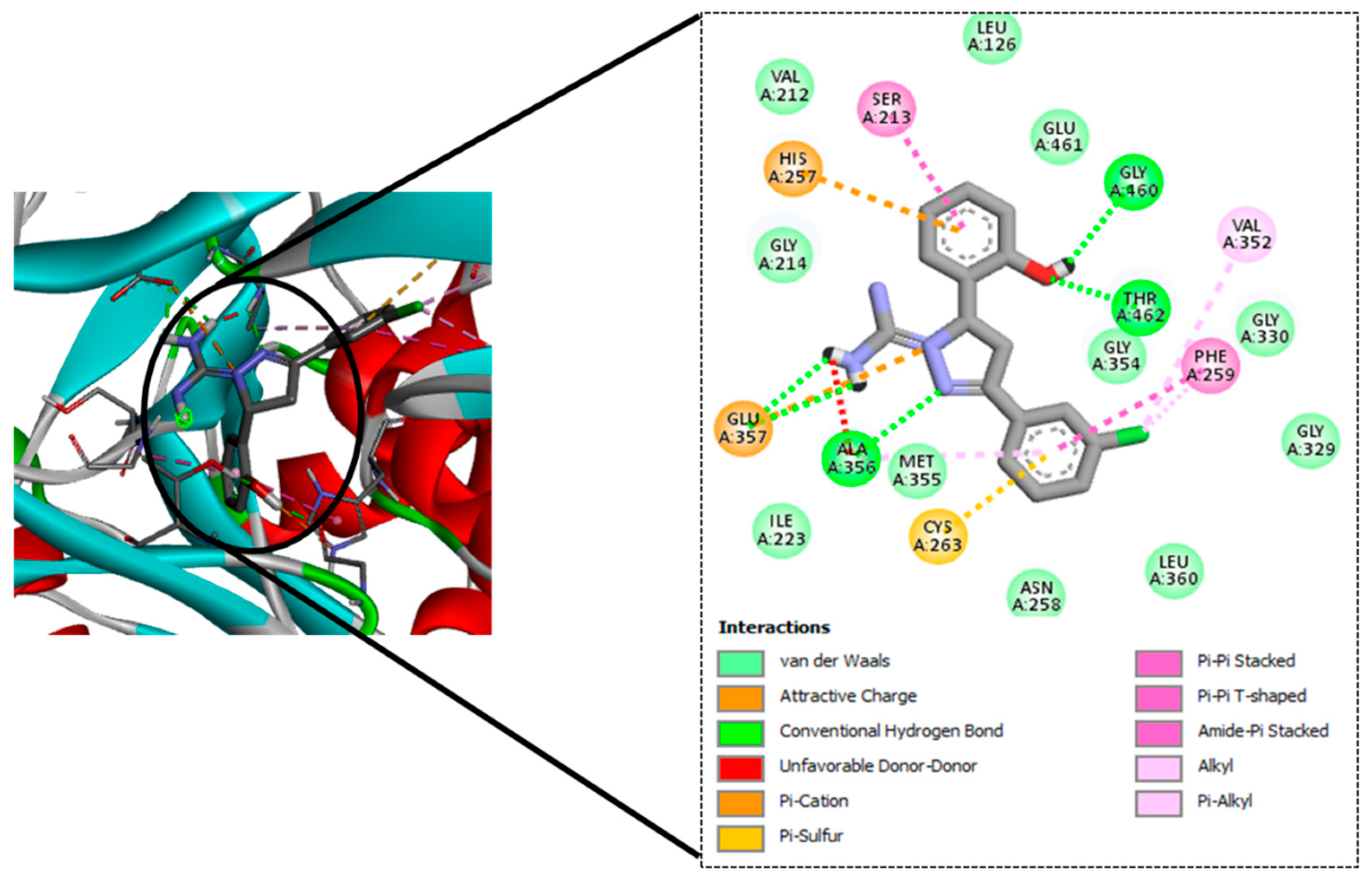
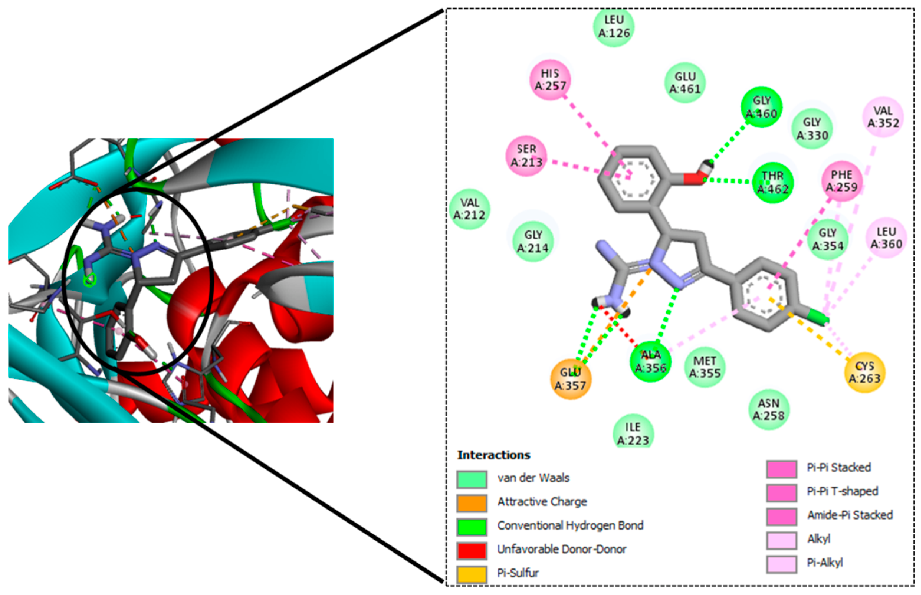
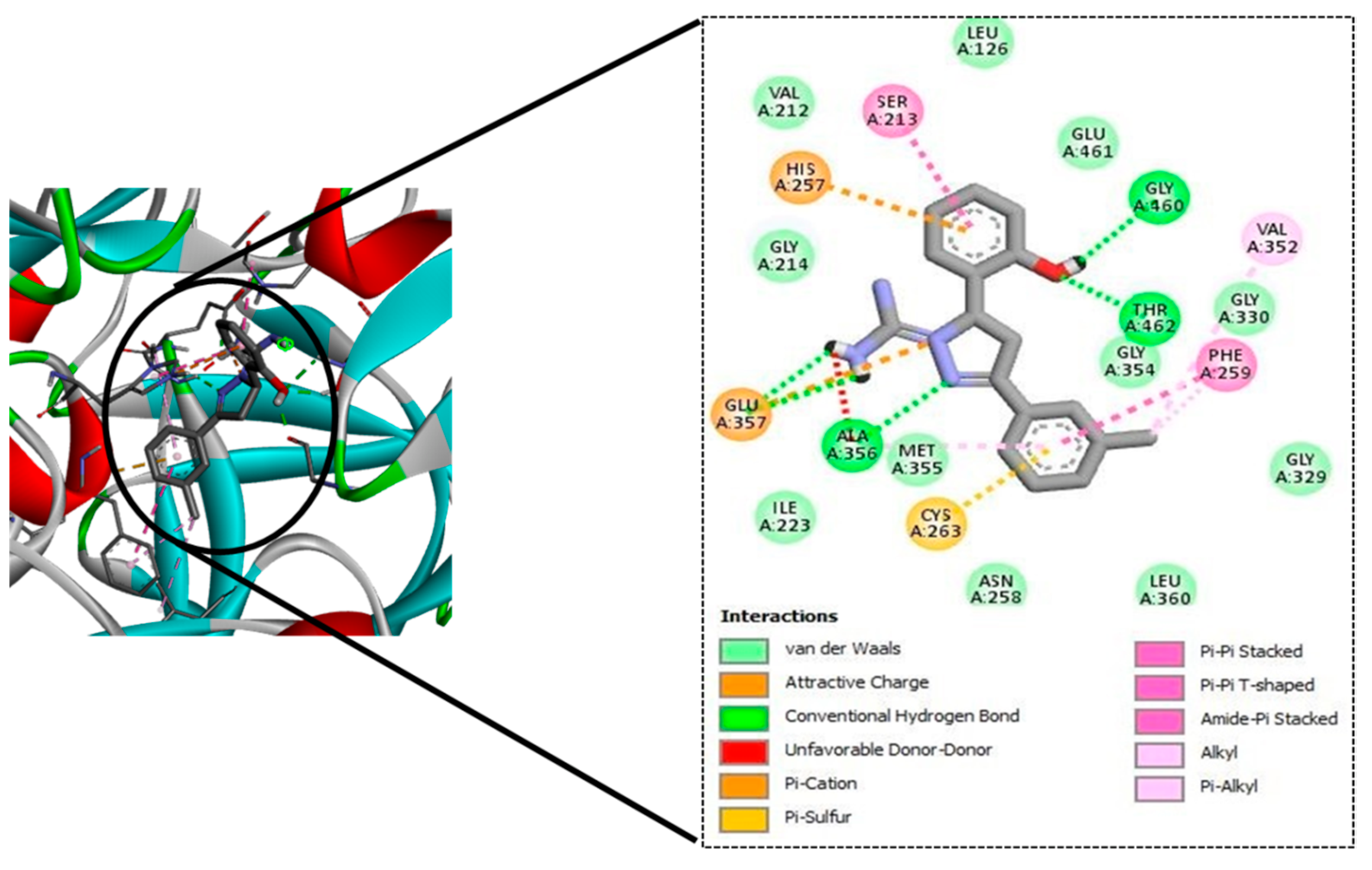
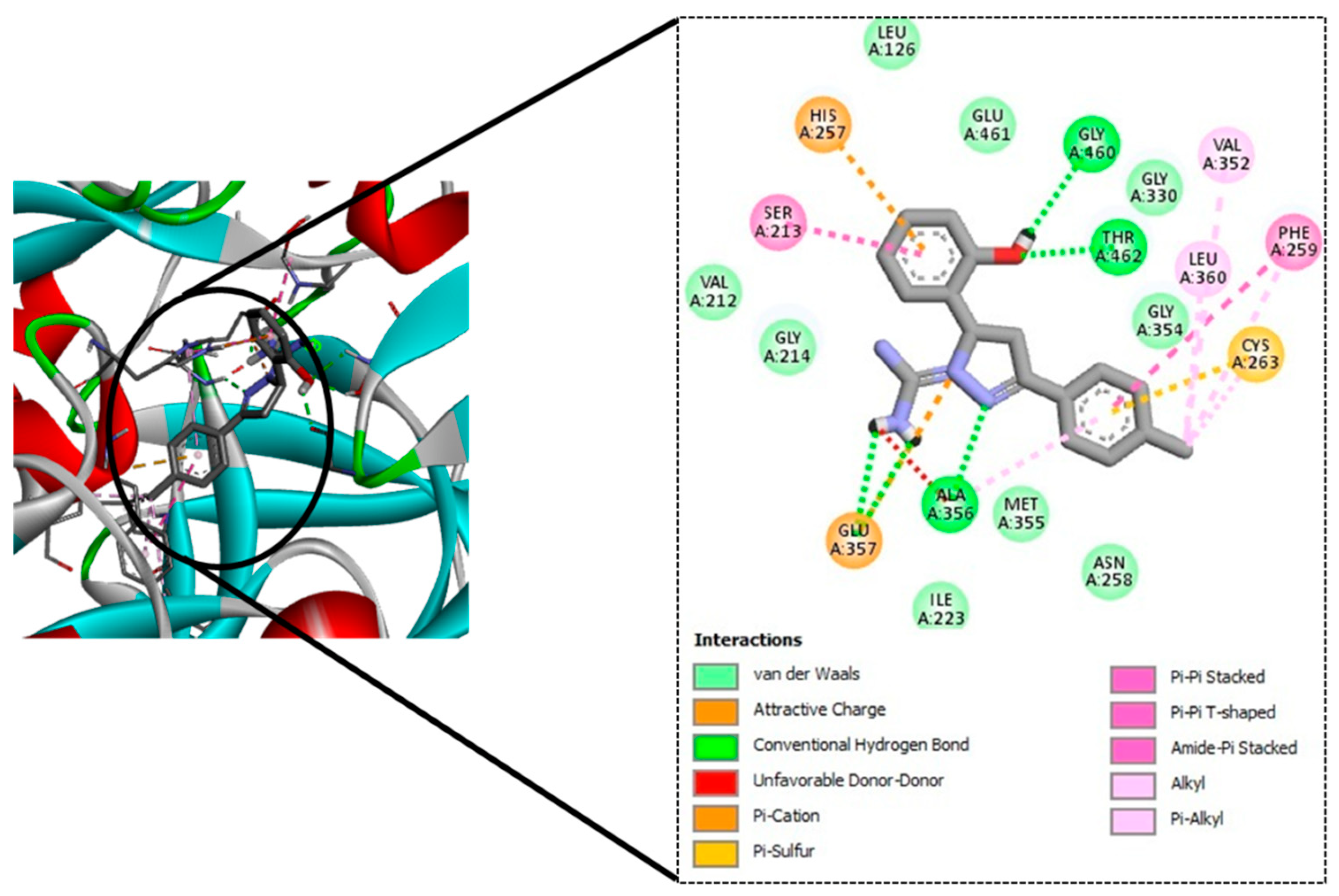
To this end, a 12-member library of designed molecules was tested in silico to test the ability to bind to MbtA. These molecules were docked into the active site of the MbtA crystal structure. The top-scoring molecules were taken up for ADME and toxicity studies.

2. Materials and Methods

2.1. Hardware and Software Employed

The docking simulations of the current study were performed using an HP workstation equipped with Windows 8 single language (64-bit operating system), Intel (R) Core™ i3-3110M CPU @ 2.40 GHz processor system, installed memory (RAM) of 4GB, and a hard disk drive of 1TB. The software used was autodock-4.2.6 program for the docking simulations [14] and Chemdraw 19.0 (PerkinElmer) for the sketching and preparation of the ligands. Visualizations were carried out using UCSF Chimera 1.13.1 [15]. The BIOVIA Discovery Studio Visualizer program was used for the generation of 2D ligand–protein interaction diagrams [16].

2.2. Docking Simulations

2.2.1. Protein Structure Preparation

The crystal structure of Gene: mbtA consisting of Protein Salicyl-AMP ligase/salicyl-S-ArCP synthetase with UniProt ID: P71716 was selected for the study. The 3D X-ray crystallographic structure/PDB file was obtained from the AlphaFold Protein Structure Database. The protein.pdb file was opened in Autodock Tools (ADT), the solvent and ions were removed, and the resulting structure was saved as a .pdbqt file for use in Autodock.

2.2.2. Ligand Preparation

The designed small molecule ligands were prepared by sketching the 2D structures in ChemDraw 19.1. The 2D representations were converted into 3D structures using Chem3D 19.1 and energy-minimized using the integrated MM2 module with default settings. The final stabilized structures were saved in .pdb format for protein–ligand docking.

2.2.3. Protein–Ligand Docking Simulations

The Autodock-4.2.6 program (ADP) was used for all molecular docking studies. The docking algorithm used was the Lamarckian Genetic Algorithm. ADP tools were used to prepare the protein and ligands. The active site was found using UCSF Chimera 1.13.1, and a binding site box (grid box) that was 60 × 60 × 60 in the x, y, and z dimensions was centered on the nucleotide binding pocket (by analogy with the homologous structure, PDB: 1MDB). The default values for all other options were used, i.e., the population size was 150 and the number of genetic algorithm (GA) runs was 50. The maximum number of evaluations was 2,500,000. The final procedure involved the running of the AutoGrid and AutoDock. AutoGrid-4.2 was used for generating map files and AutoDock-4.2 was used for running the molecular docking of each ligand to its respective protein. From the results file (.dlg), the lowest energy conformation of each docked ligand was retrieved. All docking data were evaluated and visualizations of various structures were generated using Autodock-4.2.

2.3. ADME Prediction

The ADME (absorption, distribution, metabolism, and excretion) of the molecule under investigation, which could be employed as a future lead molecule for drug development, is an important factor in predicting its pharmacodynamics. SWISSADME is a web server built and maintained by the Swiss Institute of Bioinformatics’ (SIB) molecular modelling group (https://www.swissadme.ch; accessed on 20 September 2021) [17]. Already created structures of ligands/molecules were uploaded individually in the Marvin JS portion of the website (http://swissadme.ch/index.php; accessed on 20 September 2021) to compute ADME parameters. Structures were automatically translated to SMILES format, and the server predicted ADME. The collected results were stored for further investigation.

2.4. Toxicity Prediction

Toxicology prediction is a crucial feature of all compounds. PkCSM is a web server database that allows users to analyze molecules by either sketching them graphically or providing them in SMILES format [18]. The toxicity information on the web server database includes AMES toxicity, maximum tolerated dose, hepatotoxicity, skin sensitivity, and hERG I and II inhibitors. After logging into the website, the SMILES of the top-scoring compounds after docking were searched and submitted, and toxicity was chosen in prediction mode.

3. Results

3.1. Docking Simulation Studies

The docking investigation of all the ligands with MbtA proteins showed favorable binding energies and inhibition constants. The top-scoring compounds, namely, GM08 (−9.90, 55.54 nM), GM09 (−9.83, 62.70 nM), GM02 (−9.72, 74.65 nM), and GM03 (−9.64, 85.71 nM), indicated a high affinity for the binding pocket and had high negative binding energies. The binding energies/docking scores and inhibition constants of all the molecules are given in Table 2.
The binding conformations of the four top-scoring compounds in the active site/binding pocket involved H-bond interactions with residues of the interacting protein. The details of the residues involved in bonding with ligands, i.e., H-bond interaction residues, are given in Table 3 and the docking images are shown in Figure 1, Figure 2, Figure 3 and Figure 4.

3.2. ADME Prediction

3.2.1. Results of Drug-Likeness, Bioavailability, Synthetic Feasibility, and Alerts for PAINS and Brenk Filters

The likelihood of a compound becoming an oral drug in terms of its bioavailability is referred to as its drug-likeness. The drug-likenesses of our twelve query compounds were calculated using five distinct filters, as shown in Table 4. The results showed that all of the compounds tested (GM08, GM09, GM02, and GM03) had excellent drug-likeness scores and no violations of drug-likeness rules, as well as good lead-likeness scores. To identify possible uncertain fragments that could result in false-positive biological outputs, the PAINS and Brenk methods were used. As a result of the inclusion of fragments, all compounds were found to be in violation. Along with the synthetic accessibility evaluation, the lead-likeness scores for the compounds were computed. Since their scores were in the range of 3.52–3.65, the obtained data suggested that these four compounds might be easily synthesized. The Abbot Bioavailability score predicts whether a chemical has 10% oral bioavailability (in rats) or a measurable Caco-2 cell line permeability assay, and is defined by a feasibility value of 11 percent, 17 percent, 56 percent, or 85 percent. All the compounds were predicted at 56 percent, indicating good bioavailability.

3.2.2. In Silico Evaluation of Pharmacokinetic Compliance

The success of a drug’s trip throughout the body is measured in terms of ADME (absorption, distribution, metabolism, and elimination). By computing the different physico-chemical and bio-pharmaceutical features, the ADME parameters for the substances under research, GM08, GM09, GM02, and GM03, were derived. Molar refractivity, which accounts for the overall polarity of the molecules, was 95.36 (GM08 and GM09) and 95.31 (GM02 and GM03), in the acceptable range (30–140). For all compounds, the topological polar surface area (TPSA) was 85.702 Å2. These findings indicate that the molecules are unable to pass the blood–brain barrier (BBB). The capacity of a molecule to dissolve itself in a lipophilic medium is referred to as solubility class lipophilicity and it correlates with various representations of drug properties that affect ADMET, such as permeability, absorption, distribution, metabolism, excretion, solubility, plasma protein binding, and toxicity. The iLOGP and SILICOS-IT results showed that the iLOGP values of the four molecules under investigation (GM08 = 2.05, GM09 = 2.02, GM02 = 2.02, and GM03 = 2.10) were within the acceptable range (−0.4 to +5.6), while the SILICOS-IT values (GM08 = 2.57, GM09 = 2.57, GM02 = 2.44, and GM03 = 2.44) were in the most favourable range. These compounds had a high rate of intestinal absorption. The solubility of a medicine in water is an essential factor in its absorption and distribution. The molecule’s solubility in water at 25°C is represented by log S calculations. The computed log S values through the ESOL model should not exceed 6 for appropriate solubility. The log S value for GM08 and GM09 was −3.57, whereas the value for GM02 and GM03 was −3.28, indicating low solubility. The data suggest that these compounds have a good balance of permeability and solubility and that they would have acceptable bioavailability when given orally. For all compounds, predicted GI absorption was high. Permeability predictions aid in the comprehension of ADMET and cell-based bioassay results. The permeability over human skin for GM08 and GM09 was −6.45 cm/s, and −6.51 cm/s for GM02 and GM03, both of which values were within acceptable limits. As previously stated, none of these substances demonstrated the ability to penetrate the BBB. Drug–drug interactions and drug bioavailability are sometimes caused by metabolism. Drug-metabolizing enzymes can only bind to the free form of the drug. The interaction of our main compounds with cytochrome P450 enzymes (CYPs), the most well-known class of metabolising enzymes, is critical for understanding their metabolic behaviour. All four compounds were tested for their ability to inhibit CYPs (CYPs of human liver microsomes (HLMs)). Detailed analyses are mentioned in Table 5.

3.3. Toxicity Prediction

The toxicities of the identified compounds GM08, GM09, GM02, and GM03 were investigated in silico. The maximum tolerated dosage (human) range for all of the molecules was found to be between −0.127 and −0.199 Log mg/kg/day. No hERGI (human ether-a-go-go-related Gene) inhibition was found; however, all drugs inhibited hERG II. The results revealed no intracellular buildup of phospholipids (known to cause QT prolongation, myopathy, hepatotoxicity reaction, nephrotoxicity, and pulmonary dysfunction). The software predicted no hepatotoxicity and cutaneous hypersensitivity in any of the compounds. All the predicted toxicity results for GM08, GM09, GM02, and GM03 molecules are mentioned in Table 6.

4. Conclusions

TB remains a substantial health burden in underdeveloped nations, despite significant progress in clinical drug candidate development for TB treatment during the last 10–15 years. The search for therapeutic candidates that inhibit novel targets is still a hot topic in science. Studies aimed at gaining better knowledge of Mtb biology have yielded results, including the discovery of new therapeutic targets. It has been established that mycobacterial virulence and survival in the host are directly affected by impairments in mycobactin production and iron uptake. The rational design of MbtI and MbtA inhibitors based on structure has so far yielded encouraging results. In pursuance of this goal, we used CET-based drug design to find M. tuberculosis inhibitors that can bind to a well-defined target, namely, MbtA. The tubercular enzyme MbtA, a newly discovered TB target that catalyses the initial two-step reaction of mycobactin production, was found to be highly interactive with our top four designed compounds (GM08, GM09, GM02, and GM03). They also showed acceptable pharmacokinetic profiles and nominal toxicity profiles. Furthermore, based on docking scores and predicted pharmacokinetic profiles, it could be concluded that GM08 and GM09 could serve as good leads for future optimization. Exploring intrinsic interactions between potential drugs and their potential therapeutics could open the way for unique and modern antibiotic discovery methodologies to be developed and implemented.

Author Contributions

The conceptualization, supervision, and project administration was done by V.J. The formal analysis, methodology, software investigation, resources, data curation, preparation of the original draft, visualization was done by G.R. S.M. contributed to the software investigation, data curation, and reviewing the correctness of the manuscript. All authors have contributed to the writing, reviewing and editing of all versions of the manuscript. All authors have read and agreed to the published version of the manuscript.

Funding

This research received no external funding.

Institutional Review Board Statement

Not applicable.

Informed Consent Statement

Not applicable.

Data Availability Statement

Not applicable.

Acknowledgments

We would like to express our sincere gratitude to our Department of Pharmaceutical Sciences and Technology, Birla Institute of Technology, Mesra, Ranchi, for providing the necessary software and supporting this research. All authors have consented to the acknowledgement.

Conflicts of Interest

The authors declare no conflict of interest.

References

  1. Daniel, T.M.; Bates, J.H.; Downes, K.A. History of tuberculosis. Tuberc. Pathog. Prot. Control 1994, 13–24. [Google Scholar] [CrossRef]
  2. World Health Organization. Global Task Force on TB Impact Measurement. 2019. Available online: https://www.who.int/groups/global-task-force-on-tb-impact-measurement (accessed on 20 September 2021).
  3. World Health Organization. Tuberculosis; Key Facts. 2021. Available online: https://www.who.int/news-room/fact-sheets/detail/tuberculosis (accessed on 20 September 2021).
  4. Shyam, M.; Shilkar, D.; Verma, H.; Dev, A.; Sinha, B.N.; Brucoli, F.; Bhakta, S.; Jayaprakash, V. The Mycobactin biosynthesis pathway: A prospective therapeutic target in the battle against tuberculosis. J. Med. Chem. 2020, 64, 71–100. [Google Scholar] [CrossRef] [PubMed]
  5. Lamb, A.L. Breaking a pathogen’s iron will: Inhibiting siderophore production as an antimicrobial strategy. Biochim. Biophys. Acta (BBA)-Proteins Proteom. 2015, 1854, 1054–1070. [Google Scholar] [CrossRef] [PubMed] [Green Version]
  6. de Voss, J.J.; Rutter, K.; Schroeder, B.G.; Barry, C.E., III. Iron acquisition and metabolism by mycobacteria. J. Bacteriol. 1999, 181, 4443–4451. [Google Scholar] [CrossRef] [PubMed] [Green Version]
  7. Cole, S.T.; Brosch, R.; Parkhill, J.; Garnier, T.; Churcher, C.; Harris, D.; Gordon, S.V.; Eiglmeier, K.; Gas, S.; Barry, C.E., III; et al. Deciphering the biology of Mycobacterium tuberculosis from the complete genome sequence. Nature 1998, 396, 190. [Google Scholar] [CrossRef]
  8. de Voss, J.J.; Rutter, K.; Schroeder, B.G.; Su, H.; Zhu, Y.; Barry, C.E. The salicylate-derived mycobactin siderophores of Mycobacterium tuberculosis are essential for growth in macrophages. Proc. Natl. Acad. Sci. USA 2000, 97, 1252–1257. [Google Scholar] [CrossRef] [PubMed] [Green Version]
  9. Lun, S.; Guo, H.; Adamson, J.; Cisar, J.S.; Davis, T.D.; Chavadi, S.S.; Warren, J.D.; Quadri, L.E.N.; Tan, D.S.; Bishai, W.R. Pharmacokinetic and in vivo efficacy studies of the mycobactin biosynthesis inhibitor salicyl-AMS in mice. Antimicrob. Agents Chemother. 2013, 57, 5138–5140. [Google Scholar] [CrossRef] [PubMed] [Green Version]
  10. Reddy, P.V.; Puri, R.V.; Chauhan, P.; Kar, R.; Rohilla, A.; Khera, A.; Tyagi, A.K. Disruption of mycobactin biosynthesis leads to attenuation of Mycobacterium tuberculosis for growth and virulence. J. Infect. Dis. 2013, 208, 1255–1265. [Google Scholar] [CrossRef] [PubMed] [Green Version]
  11. Duckworth, B.P.; Nelson, K.M.; Aldrich, C.C. Adenylating enzymes in Mycobacterium tuberculosis as drug targets. Curr. Top. Med. Chem. 2012, 12, 766–796. [Google Scholar] [CrossRef] [PubMed]
  12. Stirrett, K.L.; Ferreras, J.A.; Jayaprakash, V.; Sinha, B.N.; Ren, T.; Quadri, L.E.N. Small molecules with structural similarities to siderophores as novel antimicrobials against Mycobacterium tuberculosis and Yersinia pestis. Bioorg. Med. Chem. Lett. 2008, 18, 2662–2668. [Google Scholar] [CrossRef] [PubMed] [Green Version]
  13. Ferreras, J.A.; Gupta, A.; Amin, N.D.; Basu, A.; Sinha, B.N.; Worgall, S.; Jayaprakash, V.; Quadri, L.E.N. Chemical scaffolds with structural similarities to siderophores of nonribosomal peptide–polyketide origin as novel antimicrobials against Mycobacterium tuberculosis and Yersinia pestis. Bioorg. Med. Chem. Lett. 2011, 21, 6533–6537. [Google Scholar] [CrossRef] [PubMed] [Green Version]
  14. Morris, G.M.; Huey, R.; Lindstrom, W.; Sanner, M.F.; Belew, R.K.; Goodsell, D.S.; Olson, A.J. AutoDock4 and AutoDockTools4: Automated docking with selective receptor flexibility. J. Comput. Chem. 2009, 30, 2785–2791. [Google Scholar] [CrossRef] [PubMed] [Green Version]
  15. Pettersen, E.F.; Goddard, T.D.; Huang, C.C.; Couch, G.S.; Greenblatt, D.M.; Meng, E.C.; Ferrin, T.E. UCSF Chimera—A visualization system for exploratory research and analysis. J. Comput. Chem. 2004, 25, 1605–1612. [Google Scholar] [CrossRef] [PubMed] [Green Version]
  16. Dassault Systèmes. BIOVIA Discovery Studio Visualizer; Dassault Systèmes: San Diego, CA, USA, 2021. [Google Scholar]
  17. Daina, A.; Michielin, O.; Zoete, V. SwissADME: a free web tool to evaluate pharmacokinetics, drug-likeness and medicinal chemistry friendliness of small molecules. Sci. Rep. 2017, 7, 1–13. [Google Scholar] [CrossRef] [PubMed] [Green Version]
  18. Pires, D.E.V.; Blundell, T.L.; Ascher, D.B. pkCSM: Predicting small-molecule pharmacokinetic and toxicity properties using graph-based signatures. J. Med. Chem. 2015, 58, 4066–4072. [Google Scholar] [CrossRef] [PubMed]
Figure 1. Docking interaction of GM08 in the binding pocket of MbtA, showing three hydrogen bonds.
Chemproc 08 00062 g001
Figure 2. Docking interaction of GM09 in the binding pocket of MbtA, showing three hydrogen bonds.
Chemproc 08 00062 g002
Figure 3. Docking interaction of GM02 in the binding pocket of MbtA, showing three hydrogen bonds.
Chemproc 08 00062 g003
Figure 4. Docking interaction of GM03 in the binding pocket of MbtA, showing three hydrogen bonds.
Chemproc 08 00062 g004
Table 1. Tabular representation of the designed molecules.
Chemproc 08 00062 i003
Sl. No.CodeRR1
01GM01 Chemproc 08 00062 i0042-CH3
02GM02 Chemproc 08 00062 i0053-CH3
03GM03 Chemproc 08 00062 i0064-CH3
04GM04 Chemproc 08 00062 i0072-OCH3
05GM05 Chemproc 08 00062 i0083-OCH3
06GM06 Chemproc 08 00062 i0094-OCH3
07GM07 Chemproc 08 00062 i0102-Cl
08GM08 Chemproc 08 00062 i0113-Cl
09GM09 Chemproc 08 00062 i0124-Cl
10GM10 Chemproc 08 00062 i0132-OH
11GM11 Chemproc 08 00062 i0143-OH
12GM12 Chemproc 08 00062 i0154-OH
Table 2. Details of docking-based parameters of the designed compounds in the binding pocket of the target MbtA protein.
Sl No.CodeDock ScoreInhibition Constant
01GM01−9.23171.39 nM
02GM02−9.7274.65 nM
03GM03−9.6485.71 nM
04GM04−9.19182.87 nM
05GM05−9.41126.09 nM
06GM06−9.49110.58 nM
07GM07−9.32148.14 nM
08GM08−9.9055.54 nM
09GM09−9.8362.70 nM
10GM10−8.58510.97 nM
11GM11−9.04235.3 nM
12GM12−8.86317.69 nM
Table 3. Details of the top-scoring identified compounds showing H-bond interacting residues in the binding pocket of MbtA.
Sl No.Ligand CodeH-Bond Residues
1.GM08Gly460, Thr462, Ala356
2.GM09Gly460, Thr462, Ala356
3.GM02Gly460, Thr462, Ala356
4.GM03Gly460, Thr462, Ala356
Table 4. Tabular representation of different drug-likeness rules, bioavailability, lead-likeness, synthetic accessibility, and alerts for PAINS and Brenk filters.
Sl No.Compound CodeDrug-Likeness RulesAlertsLead-LikenessSynthetic Accessibility
Lipinski (Pfizer)Ghose (Amgen)Veber (GSK)Egan (Pharmacia)Muege (Bayer)Bioavailability ScorePAINSBrenk
1.GM08YesYesYesYesYes0.5512Yes3.52
2.GM09YesYesYesYesYes0.5512Yes3.52
3.GM02YesYesYesYesYes0.5512Yes3.65
4.GM03YesYesYesYesYes0.5512Yes3.63
Table 5. Details of in silico ADMET profiles of four selected compounds using the SwissADME online server.
ADMET PROFILE GM08GM09GM02GM03
Physiochemical ParametersFormulaC16H15ClN4OC16H15ClN4OC17H18N4OC17H18N4O
Molecular Weight314.77 g/mol314.77 g/mol294.35 g/mol294.35 g/mol
Mol. Refractivity95.3695.3695.3195.31
TPSA85.70 Å285.70 Å285.70 Å285.70 Å2
LipophilicityILOGP2.052.022.022.10
SILICOS-IT2.572.572.442.44
Water SolubilityLog S (ESOL), Class−3.57−3.57−3.28−3.28
Log S (Ali), Class−3.93−3.93−3.66−3.66
SILICOS-IT, Class−4.64−4.64−4.42−4.42
PharmacokineticsGI AbsorptionHighHighHighHigh
BBB PermeantNoNoNoNo
Log Kp (Skin Permeant)−6.45 cm/s−6.45 cm/s−6.51 cm/s−6.51 cm/s
CYP1A2YesYesNoNo
CYP2C19NoNoNoNo
CYP2C9NoNoNoNo
CYP2D6NoNoNoNo
CYP3A4NoNoNoNo
Table 6. Tabular representation of the data on the predicted toxicity of the top four compounds.
Model NameUnitsGM08GM09GM02GM03
AMES ToxicityYes/NoYesNoYesYes
Max. Tolerated Dose (Human)Log mg/kg/day−0.127−0.177−0.15−0.199
hERG I inhibitorYes/NoNoNoNoNo
hERG II inhibitorYes/NoYesYesYesYes
Oral Rat Chronic Toxicity (LD50)Mol/kg3.2723.2733.2573.258
Oral Rat Chronic ToxicityLog mg/kg_bw/day1.4981.5661.4151.519
HepatotoxicityYes/NoNoNoNoNo
Skin SensitizationYes/NoNoNoNoNo
T. Pyriformis ToxicityLog ug/L0.2640.2740.2620.271
Minnow ToxicityLog mM1.1431.3961.3611.614
Publisher’s Note: MDPI stays neutral with regard to jurisdictional claims in published maps and institutional affiliations.

