Proceeding Paper

Resorufin-Based Colorimetric and Fluorescent Probe for Selective Detection of Mercury (II) †

by
Milind Shamrao Thakare
1,
Dipak B. Patil
2,
Siddhant V. Kokate
2 and
Nilesh S. Pawar
1,*
1
Department of Chemistry, Pratap College, Amalner 425401, Maharashtra, India
2
Departamento de Química, División de Ciencias Naturales y Exactas, Campus Guanajuato, Universidad de Guanajuato, Noria Alta S/N, Guanajuato 36050, Mexico
*
Author to whom correspondence should be addressed.
Presented at the 25th International Electronic Conference on Synthetic Organic Chemistry, 15–30 November 2021; Available online: https://ecsoc-25.sciforum.net/.
Chem. Proc. 2022, 8(1), 50; https://doi.org/10.3390/ecsoc-25-11779
Published: 14 November 2021

Abstract

:
Environmental pollution crisis, particularly mercury ions (Hg2+) contamination, seriously threatens the health of all living organisms. Many studies have shown that even extremely low concentrations of Hg2+ can rigorously damage living organisms. Therefore, it is very much needed for real-time detection of Hg2+. To tackle mercury contamination and for its detection, we herewith proposed an intelligent design of a new fluorescent ‘turn-on’ probe, which was prepared based on the mercury-promoted hydrolysis of vinyl ether moiety. The probe rapidly reacted with mercury ions and showed good selectivity over other metal ions.

1. Introduction

Mercury is an extremely toxic and biological non-essential element that subsists naturally in the environment. Environmental pollution problems, particularly mercury ions (Hg2+) pollution, seriously threaten the health of organisms. Currently, mercury is primarily used in the production of many chemical drugs and the manufacture of electronic or electrical appliances, resulting in the release of mercury-containing wastewater or waste [1,2,3]. Irrespective of the source and primary location of the deposit, eventually, mercury (II) mixes with freshwater and marine ecosystems, causing various environmental issues in aspects related to plants, animals, and even humans [4]. Many acute poisonings have occurred. For example, the famous Minamata disease in Japan occurred due to the pollution of mercury in the Agano Tributary, and a striking epidemic in Iraq occurred because polluted seeds were used in bread [5]. The poisonousness of mercury depends on the form and the severity of the exposure. The forms of mercury include metallic mercury, inorganic mercury, and organic mercury. Among them, organic mercury is much more toxic due to its strong fat solubility. Thus, it can easily penetrate the cell membrane and pass through the blood–brain barrier. When it gathers in the brain tissue, it will cause severe brain impairment. Methyl mercury, the most famous one among different types of organic mercury, is the most harmful to the human body [6,7,8,9,10]. Methyl mercury was the culprit in the watery disease epidemic that broke out in Japan in the 1950s. The US Environmental Protection Agency stipulates that the upper limit of mercury (II) in drinking water is 2 ppb (10 nM).
Many studies have shown that even extremely low concentrations of Hg2+ can severely damage organisms. Therefore, real-time detection of Hg2+ is essential. Fluorescent probe technology, with its great advantages, has become the preferred technique for environmental detection and in vivo analysis of Hg2+. In recent years, many contributions have been made to design and synthesise novel fluorescent probes for the detection of environmental pollutant Hg2+ analysis.

2. Previous Research

2.1. Fluorescent Probes for Hg2+ Analysis Based on Ring-Opening Reactions

Li et al. [11] designed a simple rhodamine derivative probe bearing a hydrophilic carboxylic acid group. The fluorescent probe selectively responded to Hg2+ in 100% aqueous solution with 42-fold fluorescence intensity enhancement within the pH range from 5.0 to 8.0. The fluorescent intensity change followed the concentration of Hg2+ in a linear range covering from 3.0 × 10⁻⁷ to 1.0 × 10⁻⁵ M, and the detection limit was found to be 9.7 × 10⁻⁸ M.
Chemproc 08 00050 i001

2.2. Fluorescent Probes for Hg2+ Analysis Based on Ring Opening, Followed by Cyclisation

Ge et al. [12] designed a novel pyrido[1,2-a] benzimidazole- rhodamine-based ratiometric fluorescent probe for Hg2+. The probe showed high sensitivity, with a detection limit of 18.8 nM, and also exhibited satisfying selectivity, with the maximum emission shifting from 464 nm to 584 nm. Remarkably, the ratiometric fluorescent probe presented an about 200 nm Stokes shift, and such a large Stokes shift could avoid auto-fluorescence interference, serious self-quenching, and fluorescence detection errors. With the addition of Hg2+, the molecular ring-opening reaction of the spironolactone resulted in a fluorescence resonance energy transfer (FRET) effect that brought about emission shifting and fluorescence changing from blue to red. Furthermore, the application of detecting Hg2+ in Glioma cells demonstrated the potential of this study.
Chemproc 08 00050 i002

2.3. Fluorescent Probes for Hg2+ Analysis Based on S-Atom Complexation

Zhou et al. [13] developed a BODIPY-based sensitive fluorescent probe that utilised the carboxyl-thiol metal bonding receptor to recognise the Hg2+ cations in a neutral aqueous solution via the PET mechanism. There was an about 630-fold fluorescence enhancement in the reaction of the probe with Hg2+. Fluorescent probes showed a selective response towards Hg2+ over other relevant competing metal ions. For sensitivity, the sensing limit of the probe was 5.7 nM, which met the detective requirement at the ppb level. Noticeably, the response time towards Hg2+ was below 30 s, which made the detection more convenient and avoided the time effects on probe performance and sample properties.
Chemproc 08 00050 i003
Jiao et al. [14] developed and synthesised a novel selective and sensitive fluorescent chemosensor, which was based on coumarin Schiff’s base. They reported that the X-ray diffraction single-crystal structure analysis of the probe showed that the probe crystallises in a monoclinic system, and two aromatic groups of the compound were almost in the same plane, providing the explanation for the detection mechanism. Mercury ions form bonds with heteroatoms of the probe due to which the intramolecular charge transfer (ICT) effect was cut off, which resulted in the fluorescent intensity enhancement at 530 nm. The probe indicated a good selectivity over other common metal ions, and the lower limit of detection was calculated as 1 ppb.
Chemproc 08 00050 i004

2.4. Fluorescent Probes for Hg2+ Analysis Based on Other Mechanisms

Wu et al. [15] designed and synthesised a probe comprising 7-hydroxy-4-methylcoumarin as a fluorophore and a vinyl ether group as a recognition unit. After treating with Hg2+ for 10 min, the sensor revealed a 110-fold fluorescence enhancement at 450 nm, in HEPES buffer. The detection limit of probe was calculated as 0.12 μM, and the response time was less than 10 min.
Chemproc 08 00050 i005

2.5. Fluorescent Probes for Hg2+ Analysis Based on Deprotection of Dithioacetals
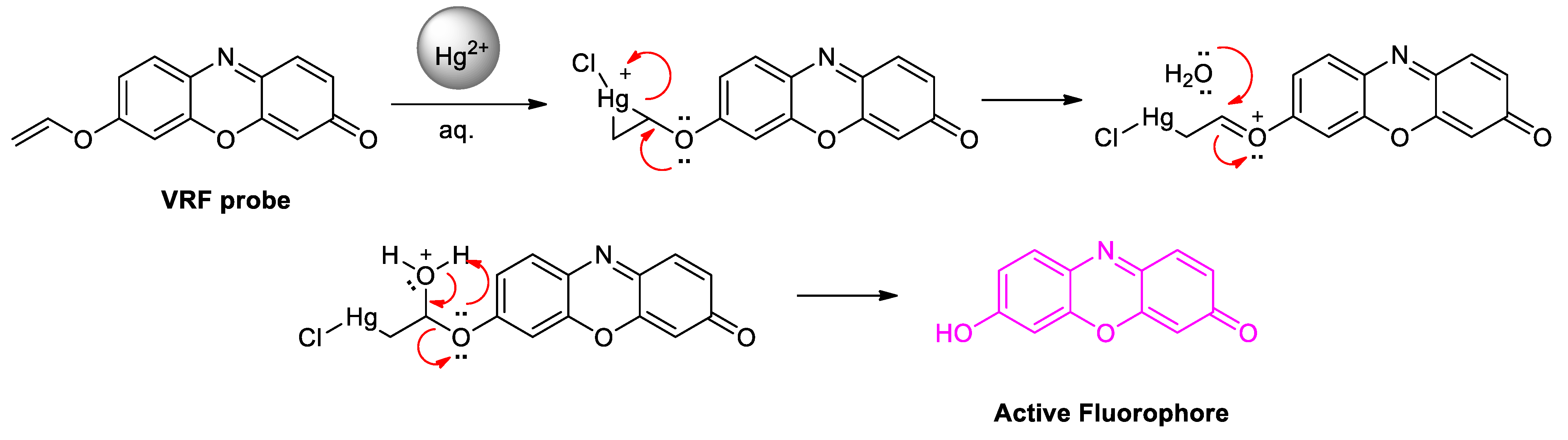
Zhou et al. [16] reported a new ratiometric fluorescent probe with an electron-deficient dithioacetal group on the three-site, which would be removed by Hg2+ to afford a ratiometric fluorescent signal. The ratiometric signal indicated a gradual change in the emission peak from 465 nm to 545 nm when exposed to Hg2+, without being affected by the microenvironment. This kind of ratiometric tool is more appreciated than intensity-based fluorescent sensors. The emission intensity presented about a 12-fold enhancement in emission ratio, with good linearity, and the lower limit of detection was calculated to be 5.8 nM. Hg2+-induced conversion of 1,3-dithiane to carbaldehyde is an efficient umpolung reaction, which can favour the formation of the intramolecular charge transfer (ICT) mechanism in compounds.
Chemproc 08 00050 i006

3. Hypothesis

With the above literature background, it was envisioned that an organic molecule with vinyl ether entity type would be activated by Hg2+. Keeping this in mind, we designed and synthesised a new VRF probe comprising resorufin as a fluorophore and a vinyl ether group as a recognition unit, suggesting that the strong fluorescent appearance is attributed to the free form of resorufin, the Hg2+-promoted hydrolysis reaction product, as depicted in the proposed mechanism. The reaction thus envisaged would liberate highly fluorescent fluorophore, and therefore the designed molecules would serve as a probe for sensing mercury. Thus, we hypothesised a new approach that involves masking and unmasking the fluorophore. It can be judged from the given mechanism that the fluorescence of fluorophore can be quenched or turned off by anchoring with the organic substrate. Once the mercury has been sensed, the probe would liberate highly fluorescent fluorophore with the formation of organic product. We herewith disclose a reaction-dependent strategy that involves the masking and unmasking of resorufin-engineered fluorophore for selective sensing of mercury. Considering the ability of Hg2+ to activate alkene functionality, a dormant fluorophore was designed for the sensing of mercury, leading to cascade and delivery of active fluorophore.

4. Materials and Instrumentations

All chemicals were either borrowed or obtained from commercial suppliers and used as received without further purification. For performing all reactions, we used oven-dried screw-cap vials with magnetic stirrers and nitrogen, to maintain an inert atmosphere. Solvents, which were dried, as well as the liquid reagents, were transferred using sterile syringes or hypodermic syringes. Coated aluminium sheet silica plates (TLC) were used for monitoring and analysing the progress of the reactions. TLC plates plate were observed under the UV light to locate and analyse the position of sample spots. For further confirmation, the spots were exposed to KMnO4 and visualised after charring on a hot plate.
1H NMR and 13C NMR spectra were measured on a Bruker AV-400/500 spectrometer, with chemical shifts reported in ppm (in DMSO-d6 or CDCl3, with TMS as the internal standard). Data for 1H NMR are reported as follows: chemical shift (ppm), multiplicity (s, singlet; d, doublet; t, triplet; q, quartet; m, multiplet), integration, and coupling constant (Hz). ESI mass spectra were carried out on an HPLC–MS spectrometer (Agilent 6100). Measurement of Fluorescence spectra was performed with a PerkinElmer LAMBDA 950 UV–Vis Spectrophotometer and a Photon Technology International, quanta Master 400 Spectrofluorometer, respectively, in degassed spectral grade solvents.

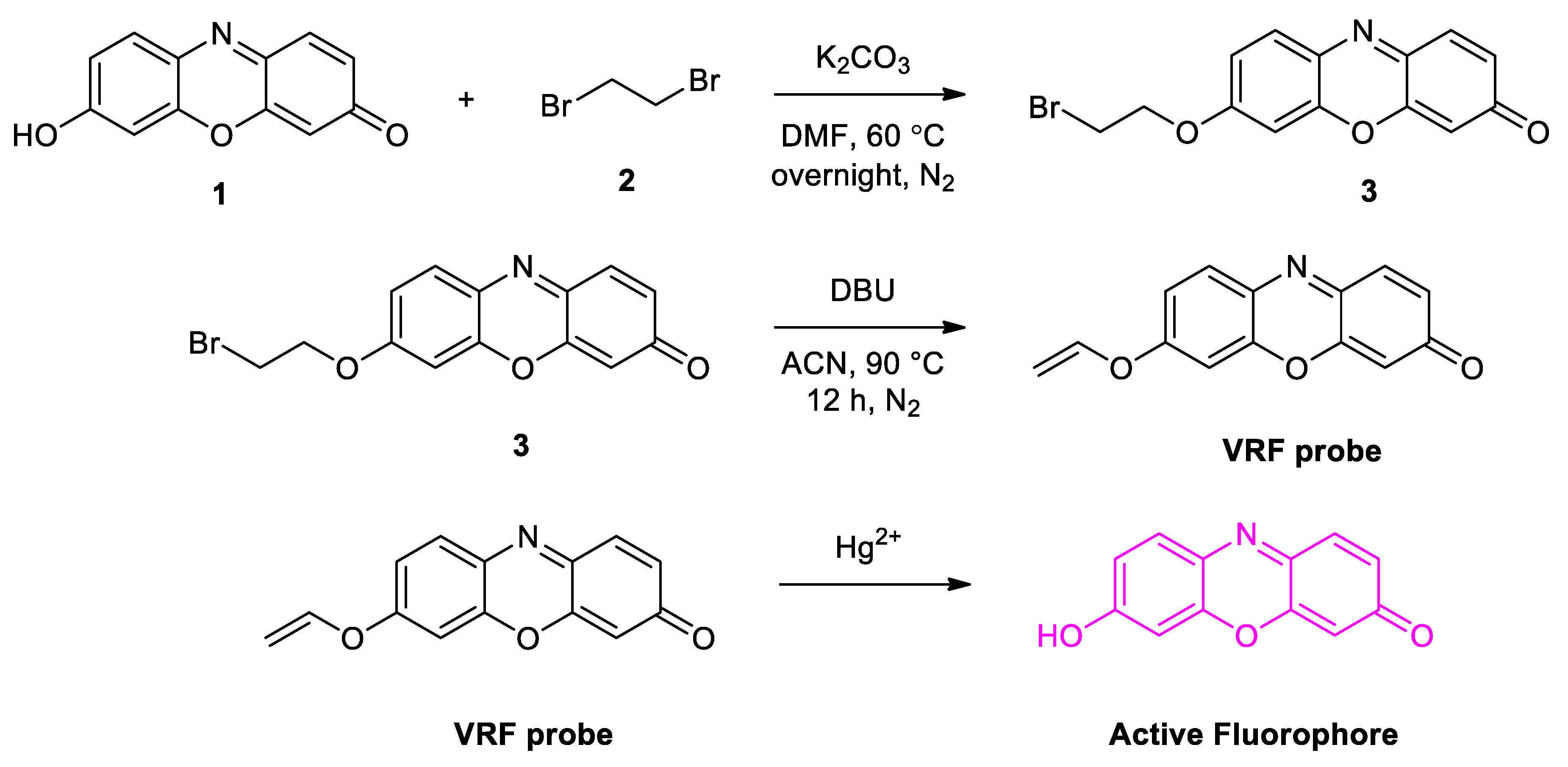
5. Design and Synthesis of Probe

The probe design consists of alkylation of resorufin (Step-1), followed by a base-catalysed dehydrohalogenation (Step-2) reaction, offering the desired probe which on mercury promoted hydrolysis converts the non-fluorescent molecule to fluorescent molecule.
The overall scheme (Scheme 1) for the synthesis of the VRF probe is shown below.

General Procedure for the Synthesis of VRF Probe
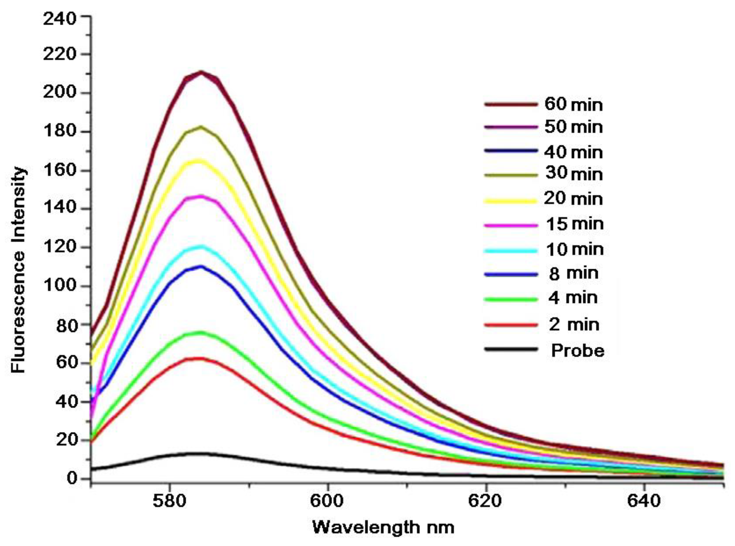
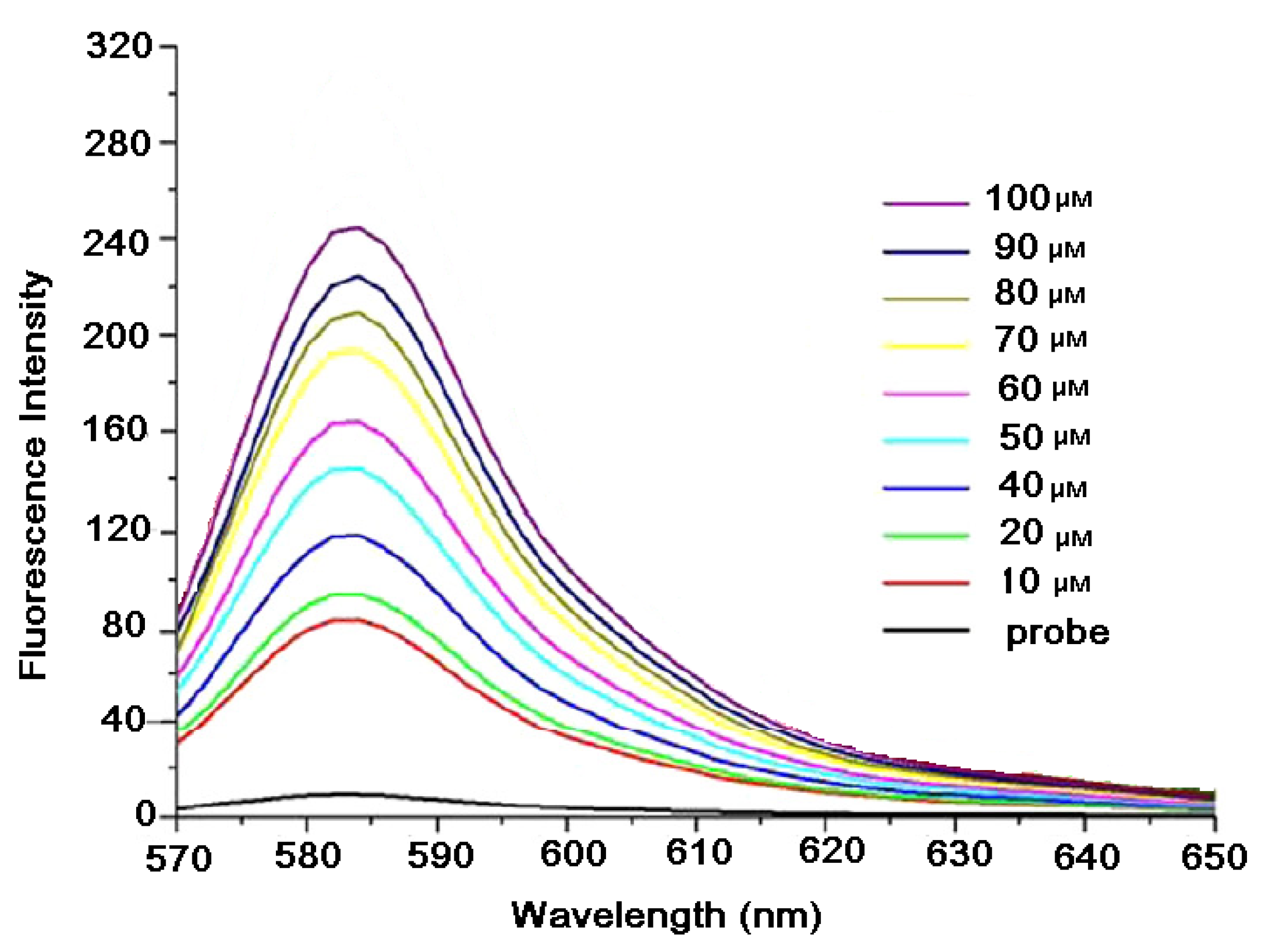
The VRF probe was prepared in two steps as follows:
Chemproc 08 00050 i007
Step 1—Synthesis of 7-(2-bromoethoxy)-3Hphenoxazin-3-one 3: To a solution of resorufin 1 (1 mmol) and K2CO3 (2 mmol) in DMF, 1,2-dibromoethane 2 (1 mmol) was added in one portion, under an inert atmosphere of N2. The reaction mixture was stirred at 60 °C overnight. After evaporation of the solvent, the resulting light brown material was dissolved in DCM (100 mL). After removal of insoluble materials by Celite filtration, the filtrate was concentrated in vacuo. The crude product was purified by silica-gel column chromatography (eluent: ethyl acetate/hexanes = 1:1) to obtain the desired product, in 60% yield. 1H NMR (CDCl3, 500 MHz): δ 3.63 (2 H, t, J = 6.3 Hz), 3.72 (2 H, t, J = 5.7 Hz), 6.33 (1 H, d, J = 1.8 Hz), 6.82-6.86 (2 H, m), 6.95 (1 H, dd, J = 9.1, 1.8 Hz), 7.43 (1 H, d, J = 9.7 Hz), 7.72 (1 H, d, J = 9.1 Hz); 13C NMR (CDCl3, 125 MHz): δ (ppm) = 29.6, 66.5, 100.9, 107.0, 114.1, 128.7, 131.8, 134.5, 134.9, 145.8, 145.9, 150.0, 162.8, 186.5; HRMS (C14H10BrNO3, [M + Na]+) calcd. 353.0, found 353.1.
Chemproc 08 00050 i008
Step 2—Synthesis of 7-(vinyloxy)-3h-phenoxazin-3-one (VRF probe): To a stirred solution of 7-(2-bromoethoxy)-3H-phenoxazin-3-one 3 (1 mmol) in CH3CN (10 mL), DBU (1,8-diazabicyclo[5.4.0]undec-7-ene) was added (2 mmol). The mixture was stirred at 90 °C for 12 h under inert atmosphere. The solvent was evaporated under vacuum. Water (20 mL) was added to the crude, and the solution was extracted thrice with DCM (20 mL). The combined organic layers were washed with brine solution, dried over Na2SO4, and evaporated under vacuum. The residue was purified by silica-gel column chromatography (eluent: 20% ethyl acetate/hexanes) to obtain the desired product in 40% yield. 1H NMR (500 MHz, DMSOd6): δ 7.77 (d, J = 8.8 Hz, 1H), 7.46 (d, J = 9.9 Hz, 1H), 7.05 (dd, J = 8.8, 2.6 Hz, 1H), 6.95 (d, J = 2.6 Hz, 1H), 6.88 (dd, J = 9.8, 2.0 Hz, 1H), 6.71 (dd, J = 13.6, 5.9 Hz, 1H), 6.36 (d, J = 2.0 Hz, 1H), 5.03 (dd, J = 13.6, 2.0 Hz, 1H), 4.74 (dd, J = 6.0, 2.0 Hz, 1H); 13C NMR (125 MHz, CDCl3) δ 186.24, 153.57, 152.40, 149.26, 144.37, 135.24, 134.80, 131.31, 131.23, 130.65, 120.14, 118.54, 109.09, 107.33; HRMS (C14H9NO3, [M + Na]+): calcd 262.04, found 262.13.

6. Sensing Study

Mercury promoted cleavage of vinylic ether to yield active fluorophore.
Chemproc 08 00050 i009
Proposed mechanistic route of vinyl ether deprotection using Hg2+ (Scheme 2) is shown below.

7. Results and Discussion

Synthesis: The synthesis of the VRF probe is relatively straightforward; it was synthesised in two steps. The first step involved the reaction of the hydroxy group of resorufin 1 and 1,2-dibromoethane 2 in presence of a base, to give 3. The second step involved the treatment of 3 with DBU, which underwent 1–2 elimination to produce desired VRF probe in moderate yield (Scheme 1). The possible reaction mechanism of VRF towards mercury (II) is shown in Scheme 2. The obtained VRF probe was then characterised by NMR and mass spectroscopic techniques, which showed its characteristic vinylic protons at 5.03 δ, 4.74 δ, and its mass peak at 262.23.

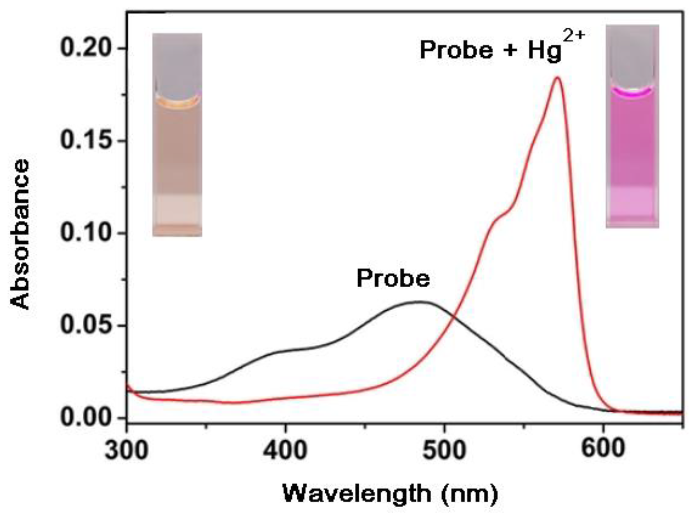
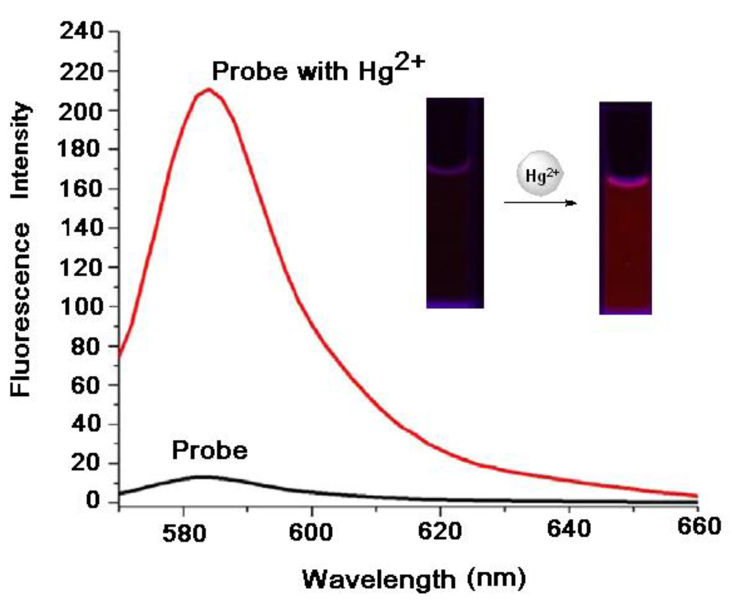
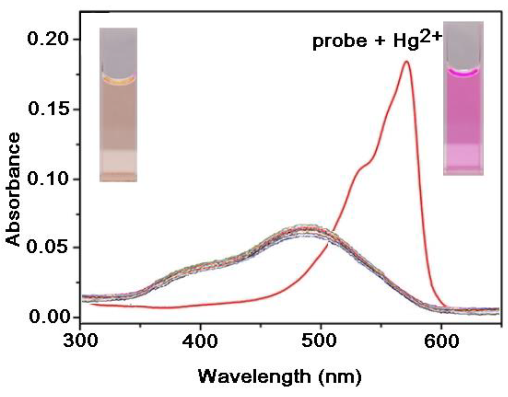
7.1. Effects of Mercury (II) on Absorption and Emission Spectroscopic Properties of VRF Probe

The absorption and emission spectroscopic properties of this probe in the absence and presence of Hg2+ were studied. The absorption spectrum of the VRF probe in the absence of Hg2+ exhibited a very weak broad absorption band at 480 nm (Figure 1). After treatment with Hg2+, the absorption band maxima showed a bathochromic shift from 480 to 575 nm, as well as a colour change from colourless to pink (Figure 1). The fluorescence spectrum of the VRF probe in the absence of Hg2+ showed a very weak emission at 582 nm, which is attributed to the strong quenching effect of the vinylic ether unit. However, the reaction of the VFR probe with Hg2+ resulted in a remarkable ‘turn-on’ fluorescence change from no fluorescence to strong reddish-brown fluorescence, which was directly observed by the naked eye under a UV lamp (365 nm). The fluorescence spectrum of the probe in presence of Hg2+ (100 μM) showed a remarkable more intense emission peak at 585 nm (Figure 2). The fluorescence enhancement of the probe was attributed to the mercury-triggered cleavage reaction, causing the release of free resorufin 1. The changes in absorption and fluorescence spectra were observed due to the deprotection of the vinyl ether moiety and the formation of resorufin 1.
The time-dependent fluorescence response of the VRF probe (10 μM) with Hg2+ (100 μM) in CH3CN/HEPES buffer (1:1, 0.1 M, pH = 7.4) was measured at 25 °C (Figure 3). The fluorescence spectrum of the VRF probe in the absence of Hg2+ showed a very weak emission at 582 nm (Figure 2). Then, the fluorescence intensities of the VRF probe in the presence of Hg2+ (100 μM) from 2 to 60 min were recorded. It was observed that immediately after 2 min the fluorescence spectrum showed a notable increase in the intensity of fluorescence. From 2 to 60 min, there was a rapid increase in fluorescence intensity, and then it saturated. The highest fluorescence intensity was observed at 60 min. We also found that the proton NMR spectrum of the product of VRF + Hg2+ reaction was similar to that of free resorufin 1.
The concentration-dependent fluorescence response for the VRF probe (10 µM) upon addition of Hg2+ (10–100 µM) in CH3CN/HEPES buffer (1:1, 0.1 M, pH = 7.4) was measured at 25 °C (Figure 4). The fluorescence spectrum of the VRF probe in the absence of Hg2+ (0 µM) showed very weak emission at 582 nm. However, with the addition of Hg2+ (10 µM), a notable enhancement in the fluorescence intensity at 585 nm was observed. The fluorescence response of the VRF probe showed excellent linearity with an increase in the concentration of Hg2+ from 10 to 100 μM. The fluorescence intensities were measured every 60 min for each addition of Hg2+. The highest fluorescence intensity was recorded at an Hg2+ concentration of 100 μM.

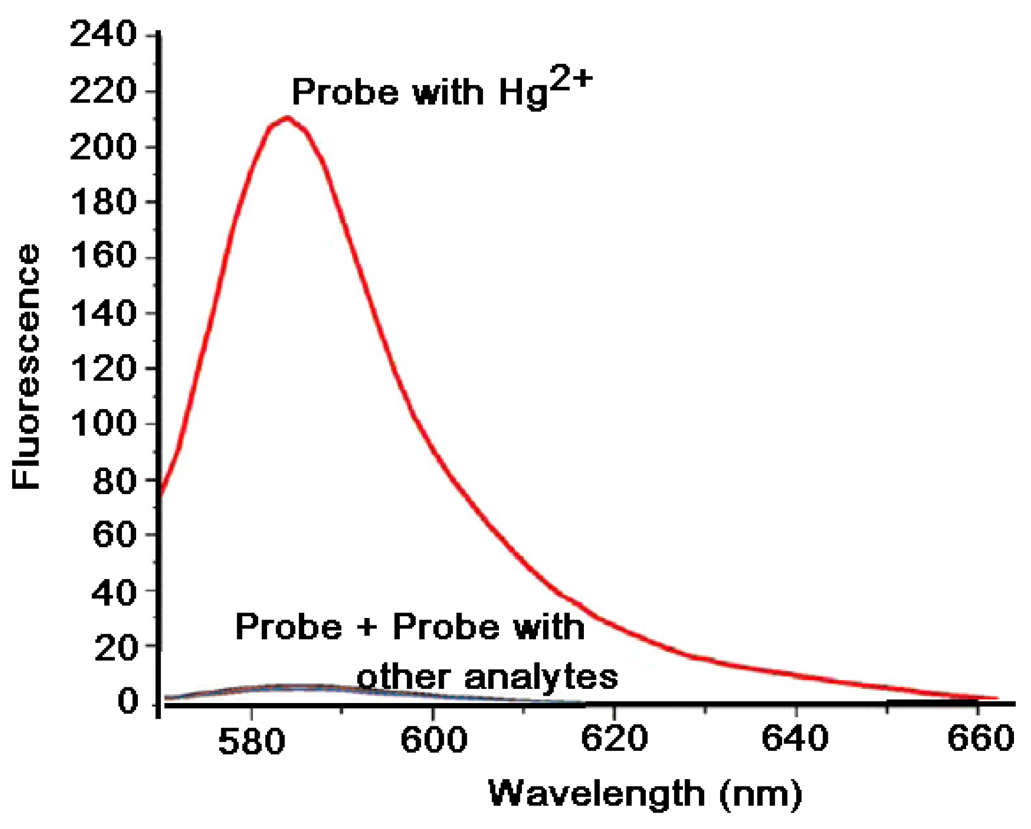
7.2. Response of VRF Probe to Mercury and Other Metal Ions

Selectivity is a very important parameter for evaluating the performance of the probe. To investigate the selectivity of the VRF probe, ions such as Hg2+, Co2+, Zn2+, Fe3+, Mg2+, Mn2+, K+, Ni2+, Ca2+, Fe2+, and Ag+ were selected. The absorption spectra of the VRF probe (10 μM) in the presence of Hg2+ and other metal ions (100 μM) in CH3CN/HEPES buffer solution (1:1, v/v, 0.1 M, pH = 7.4) were recorded (Figure 5). Similar to what we observed in Figure 1, the probe also exhibited a very weak broad absorption band here, at 480 nm. The treatment of the probe with metal ions other than Hg2+ did not cause any noteworthy changes in the absorption profile. However, the absorption spectrum of the probe with Hg2+ showed a remarkable enhancement in the absorption with absorption maxima at 575 nm, as well as a colour change from colourless to pink (Figure 5).
The emission spectra of the VRF probe (10 μM) in the presence of Hg2+ and other metal ions (100 μM) in CH3CN/HEPES buffer solution (1:1, v/v, 0.1 M, pH = 7.4) were also recorded (Figure 6). Similar to what we observed in Figure 2, the probe also exhibited a very weak emission here, at 582 nm. This low background signal is extremely desirable for the sensitive detection of metal ions. The treatment of the VRF probe with metal ions other than Hg2+ did not cause any noteworthy changes in the emission profile. However, the reaction of the probe with Hg2+ resulted in a remarkable ‘turn-on’ fluorescence change, from no fluorescence to strong reddish-brown fluorescence, which was directly observed by the naked eye under a UV lamp (365 nm). The fluorescence spectrum of the probe in presence of Hg2+ (100 μM) showed a remarkable, more intense emission peak at 585 nm (Figure 6). This observed and distinct change in fluorescence colour of the reaction system in the absence and presence of Hg2+ is highly convenient for the rapid detection of Hg2+ (Figure 2). These all results reveal that the VRF probe shows high selectivity towards Hg2+.

8. Conclusions

In conclusion, we proposed a new chromogenic and fluorescent probe based on the protection and deprotection of fluorophore for the recognition of Hg (II), which is currently a widely spread contaminant in ecosystems. The present probe is highly sensitive and selective enough for the determination of mercury. The probe selectively displayed drastic changes in absorption and emission intensities for Hg (II). Moreover, this ‘OFF–ON’ fluorescent probe showed a noteworthy fluorescence increase. The VRF probe displayed a substantial colour change from colourless to pink, as well as noticeable fluorogenic signalling performance entirely towards Hg2+ ions. Selective Hg2+- signalling by the VRF probe was unaltered by the presence of other metal ions. We strongly believe that this approach for the detection of notorious Hg (II) will definitely raise interest among the scientific community; thus, many probes based on this strategy may appear in the near future. We expect this probe to be further useful in the identification of Hg (II) at a cellular level.

Author Contributions

Synthesis, M.S.T. and D.B.P.; Methodology, M.S.T.; Characterisation, M.S.T.; Optical data study, S.V.K.; Writing—original draft, M.S.T.; Writing—review and editing, S.V.K.; Conceptualisation, N.S.P.; Supervision, N.S.P. All authors have read and agreed to the published version of the manuscript.

Funding

This research received no external funding.

Institutional Review Board Statement

Not applicable.

Informed Consent Statement

Not applicable.

Data Availability Statement

Not applicable.

Acknowledgments

Authors are very thankful to NTP, IISER Bhopal, and Aslam Shaikh, for analytical support, and also to J. S. Rane, Pratap College, Amalner, for providing necessary laboratory facilities.

Conflicts of Interest

The authors declare no conflict of interest. The funders had no role in the design of the study; in the collection, analyses, or interpretation of data; in the writing of the manuscript; or in the decision to publish the results.

References

  1. Nendza, M.; Herbst, T.; Kussatz, C.; Gies, A. Potential for Secondary Poisoning and Biomagnification in Marine Organisms. Chemosphere 1997, 35, 1875–1885. [Google Scholar] [CrossRef]
  2. Renzoni, A.; Zino, F.; Franchi, E. Mercury Levels along the Food Chain. Environ. Pollut. 1998, 77, 68–72. [Google Scholar] [CrossRef]
  3. Mason, F.M.M.; Reinfelder, R.P.; Morel, J.R. Bioaccumulation of Mercury and Methylmercury. Water Air Soil Pollut. 1995, 80, 915–921. [Google Scholar] [CrossRef]
  4. Boening, D.W. Ecological Effects, Transport, and Fate of Mercury: A General Review. Chemosphere 2000, 40, 1335–1351. [Google Scholar] [CrossRef]
  5. Kim, K.H.; Kabir, E.; Jahan, S.A. A Review on the Distribution of Hg in the Environment and Its Human Health Impacts. J. Hazard. Mater. 2016, 306, 376–385. [Google Scholar] [CrossRef] [PubMed]
  6. Kraepiel, A.M.L.; Keller, K.; Chin, H.B.; Malcolm, E.G.; Morel, F.M.M. Sources and Variations of Mercury in Tuna. Environ. Sci. Technol. 2003, 37, 5551–5558. [Google Scholar] [CrossRef] [PubMed]
  7. Burger, J.; Gochfeld, M. Mercury in Canned Tuna: White versus Light and Temporal Variation. Environ. Res. 2004, 96, 239–249. [Google Scholar] [CrossRef] [PubMed]
  8. Kuwabara, J.S.; Arai, Y.; Topping, B.R.; Pickering, I.J.; George, G.N. Mercury Speciation in Piscivorous Fish from Mining-Impacted Reservoirs. Environ. Sci. Technol. 2007, 41, 2745–2749. [Google Scholar] [CrossRef]
  9. Kiefer, A.M.; Seney, C.S.; Boyd, E.A.; Smith, C.; Shivdat, D.S.; Matthews, E.; Hull, M.W.; Bridges, C.C.; Castleberry, A. Chemical Analysis of Hg0-Containing Hindu Religious Objects. PLoS ONE 2019, 14, e0226855. [Google Scholar] [CrossRef] [Green Version]
  10. Guo, Q.; Zhang, Y.; Lin, Z.H.; Cao, Q.Y.; Chen, Y. Fluorescent Norbornene for Sequential Detection of Mercury and Biothiols. Dye Pigment 2020, 172, 107872. [Google Scholar] [CrossRef]
  11. Li, D.; Li, C.Y.; Li, Y.F.; Li, Z.; Xu, F. Rhodamine-Based Chemodosimeter for Fluorescent Determination of Hg(2+) in 100% Aqueous Solution and in Living Cells. Anal. Chim. Acta 2016, 934, 218–225. [Google Scholar] [CrossRef]
  12. Ge, Y.; Liu, A.; Ji, R.; Shen, S.; Cao, X. Detection of Hg2þ by a FRET Ratiometric Fluorescent Probe Based on a Novel Pyrido[1,2-a]Benzimidazole-Rhodamine System. Sens. Actuator B Chem. 2017, 251, 410–415. [Google Scholar] [CrossRef]
  13. Zhou, B.; Qin, S.; Chen, B.; Han, Y. A New BODIPY-Based Fluorescent “Turn-on” Probe for Highly Selective and Rapid Detection of Mercury Ions. Tetrahedron Lett. 2018, 59, 4359–4363. [Google Scholar] [CrossRef]
  14. Jiao, Y.; Zhou, L.; He, H.; Yin, J.; Duan, C. A New Fluorescent Chemosensor for Recognition of Hg2+ Ions Based on a Coumarin Derivative. Talanta. 2017, 162, 403–407. [Google Scholar] [CrossRef] [PubMed]
  15. Wu, C.; Wang, J.; Shen, J.; Bi, C.; Zhou, H. Coumarin-Based Hg2þ Fluorescent Probe: Synthesis and Turn-on Fluorescence Detection in Neat Aqueous Solution. Sens. Actuator B Chem. 2017, 243, 678–683. [Google Scholar] [CrossRef]
  16. Zhou, Y.; He, X.; Chen, H.; Wang, Y.; Xiao, S.; Zhang, N.; Li, D.; Zheng, K. An ESIPT/ICT Modulation Based Ratiometric Fluorescent Probe for Sensitive and Selective Sensing Hg2+. Sens. Actuator B Chem. 2017, 247, 626–631. [Google Scholar] [CrossRef]
Scheme 1. The overall scheme for the synthesis of VRF probe and its mercury promoted cleavage to yield active fluorophore.
Chemproc 08 00050 sch001
Scheme 2. The possible reaction mechanism of VRF probe towards mercury (II).
Chemproc 08 00050 sch002
Figure 1. The absorption spectra of VRF (10 μM) in the absence and presence of Hg2+ (100 μM) in CH3CN/HEPES buffer solution (1:1, v/v, 0.1 M, pH 7.4). Inset: the change in the colour of VRF in the absence and presence of Hg2+ (colourless to pink).
Chemproc 08 00050 g001
Figure 2. Fluorescence spectra of VRF probe (10 μM) in the absence and presence of Hg2+ (100 μM) in CH3CN/HEPES buffer solution (1:1, v/v, 0.1 M, pH 7.4); excitation wavelength = 560 nm, the spectrum was acquired 60 min after HgCl2 addition at 25 °C. Inset: fluorescence colour change observed under UV light at 365 nm.
Chemproc 08 00050 g002
Figure 3. Time-dependent fluorescence response of VRF probe (10 μM) with Hg2+ (100 μM) in CH3CN/HEPES buffer (1:1, 0.1 M, pH = 7.4) at 25 °C; excitation wavelength = 560 nm, emission wavelength = 585 nm. The reaction was completed within one hour.
Chemproc 08 00050 g003
Figure 4. Concentration-dependent fluorescence response for VRF probe (10 µM) upon addition of Hg2+ (10–100 µM) in CH3CN:HEPES buffer (1:1, 0.1 M, pH = 7.4) at 25 °C; excitation wavelength = 560 nm, emission wavelength = 585 nm.
Chemproc 08 00050 g004
Figure 5. Absorption spectra of VRF probe (10 μM) in the presence of Hg2+ and other metal ions (100 μM) in CH3CN/HEPES buffer solution (1:1, v/v, 0.1 M, pH = 7.4). Inset: the change in the colour of VRF in the absence and presence of Hg2+ (colourless to pink).
Chemproc 08 00050 g005
Figure 6. Fluorescence response of VRF probe (10 µM) in the presence of Hg2+ and other metals ions (100 µM) in CH3CN/HEPES buffer solution (1:1, v/v, 0.1 M, pH = 7.4); excitation wavelength = 560 nm, emission wavelength = 585 nm.
Chemproc 08 00050 g006
Publisher’s Note: MDPI stays neutral with regard to jurisdictional claims in published maps and institutional affiliations.

