1. Introduction
Solanum glaucophyllum belongs to the Solanaceae family and is widely distributed in the flooded areas of natural grasslands in the northeast of Argentina. Since 1960, its consumption by cattle has been associated with vitamin D
3 intoxication, known as enzootic calcinosis. This particular disease is related to alteration in the absorption of vitamin D
3, which produces calcium depositions in the muscle tissues and joints of cattle. It is a progressive disease with signs such as stiffness, painful gait, and loss of body condition, and it is responsible for significant mortality in affected ruminants that nowadays produces important economic losses for Argentine livestock [
1,
2].
Concerning bibliographic background, vitamin D
3 is known to be present in species belonging to the Solanaceae family. The principal hydroxylated metabolite found is 1α,25-(OH)
2D
3, known as calcitriol, and is mainly conjugated with carbohydrates as 1α,25-(OH)
2D
3-glycoside [
3]. The biologically most active metabolite, calcitriol, is associated with pathological signs of the mentioned disease and has been identified in
S. glaucophyllum from the enzymatic hydrolysis of the water: ethanol extract obtained from the leaves of this species [
4]. In cattle, calcitriol is released through the enzymatic hydrolysis of 1α,25-(OH)
2D
3-glycoside at the ruminal level after
S. glaucophyllum leaf ingestion [
1].
In addition, vitamin D
3 in
S. glaucophyllum, showed a similar to vertebrate’s photodependent process of synthesis, but there is few researching regarding quantitative studies about the vitamin D
3 biosynthetic pathway of the plant [
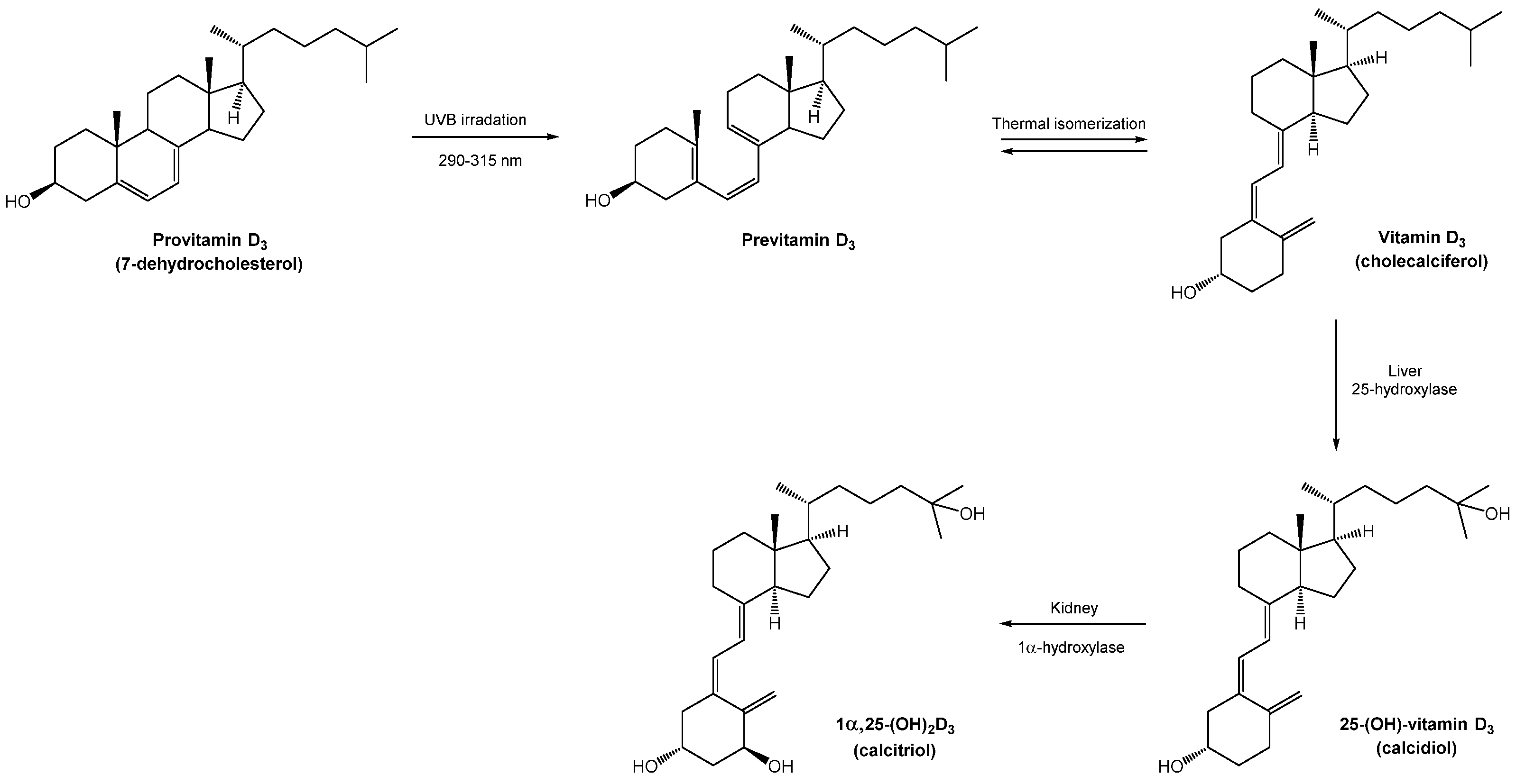
5]. In vertebrates, vitamin D
3 is synthesized upon UVB irradiation. The UVB irradiation of provitamin D
3 (7-dehydrocholesterol) in the skin breaks the B-ring to form previtamin D
3, which rearranges itself in vitamin D
3 (cholecalciferol). Vitamin D
3 is transported to the liver where it is enzymatically hydroxylated at C-25 by the 25-hydroxylase enzyme, producing 25-OHD
3 (calcidiol). The 25-OHD
3 is hydroxylated for a second time at C-1 in the kidneys to the active metabolite 1α,25-(OH)
2D
3 (calcitriol) (
Scheme 1).
Inspired by thrusting forward the study of the biosynthetic pathway of vitamin D3 in S. glaucophyllum, the aim of the present work is to provide knowledge of vitamin D3 toxicity related to concentration in the mentioned species located throughout the Río Salado basin. High-performance liquid chromatography (HPLC) allows qualitative and quantitative analyses in order to know the presence and content of related compounds in S. glaucophyllum with highly accurate and sensitive results. Herein, we present the development of a reverse-phase high-performance liquid chromatography (RP-HPLC) method for the determination of vitamin D3 and its hydroxylated metabolites. So far, the present work contributes to validating the optimal starting amount of S. glaucophyllum leaves to be hydrolyzed, the chemical hydrolysis conditions, and the method of analysis of the main metabolites in the plant leaf materials.
2. Materials and Methods
2.1. General
The solvents used for extraction and chromatography were previously distilled. The HPLC analysis was performed using isopropanol (HPLC gradient grade for liquid chromatography, LiChrosolv), acetonitrile (HPLC gradient grade for liquid chromatography, LiChrosolv), and ultrapurified Milli-Q water (Millipore, Billerica, MA, USA). All the solvents were degassed by simultaneous sonication and filtration through 0.2 µm PTFE membranes prior to use. Calcitriol (1α,25-(OH)2D3) and calcidiol (25-OHD3) analytical standards were obtained from Sigma-Aldrich (St. Louis, MO, USA). For column chromatography, neutral aluminum oxide Fluka Typ 507C (100–125 mesh) was used. The chromatographies were monitored with thin-layer chromatography (TLC) on silica gel plates (60F-254) and visualized under UV light or by using a p-anisaldehyde solution (5 mL p-anisaldehyde, 5 mL H2SO4 concentered, 1mL acetic acid, and 90 mL ethanol).
The HPLC analysis was conducted on an LS-MS-Thermo Scientific -UltiMate 3000-MSQ PLUS HPLC system equipped with an Agilent Zorbax SB-Aq stable bond analytical C18 reverse-phase column and a fixed wavelength UV detector. Calcitriol (1α,25-(OH)2D3) and calcidiol (25-OHD3) were monitored at an absorbance of 265 nm. UV spectra for maximal wavelength standards determination were recorded with an Agilent Cary 60 UV–Vis spectrophotometer. Stock calcitriol and calcidiol standard mixtures were generated using a molar extinction coefficient of 18,300 AU M–1 L–1 at 265 nm in HPLC-grade isopropanol. The prepared stock standard mixtures were perfused with nitrogen and stored at −20 °C.
2.2. Plant Material
Solanum glaucophyllum (Solanaceae) plant specimens were collected at Dolores in the Buenos Aires province of Argentina from the La Quebrada location (36°17′21.99″ south latitude and 57°36′14.76″ west longitude) in April 2021.
2.3. Harvest and Extraction
The plant material was harvested by hand, preferably at noon to avoid dew and excessive humidity. The stems were separated, and the leaves were spread on the ground indoors to dry superficially, separate from the rest of the soil and foreign bodies. After 24 h, the samples were placed in an oven at 36 °C until reaching a constant weight; this took approximately 3 days.
Finely ground dry leaves of S. glaucophyllum (163 g) were extracted with a solution (1250 L) of water:ethanol (80:20) at 40 °C for 8 h. The extract was concentrated under reduced pressure, giving 36.7 g (3.7%).
2.4. Chemical Hydrolysis
In a 250 mL two-necked round-bottomed flask equipped with a condenser loaded with S. glaucophyllum extract (0.7 g), 2 N HCl solution (140 mL) was added. The mixture was stirred for 13 h at 85 °C and for an additional 18 h at room temperature. Then, the reaction mixture was extracted with ethyl acetate (5 × 50 mL). The organic layer was dried over MgSO4 and concentrated under reduced pressure to give 0.126 g of hydrolyzed extract.
2.5. Purification of Hydrolyzed Extract
Chromatography on neutral aluminum oxide (100–125 mesh) of a portion of the hydrolyzed extract (50.0 mg) eluted with dichloromethane:methanol (100:0 to 95:5, step-gradient system) yielded forty tubes. Chromatographic separation was monitored by TLC using a mixture of dichloromethane:methanol (90:10) as mobile phase. Two major compounds, 1 and 2, were observed in tubes 1 and 25, respectively, weakly seen by their stain color on the plate.
2.6. Calcitriol (1α,25-(OH)2D3) and Calcidiol (25-OHD3) Identification
Hydrophilic extract pellets obtained from S. glaucophyllum leaves were previously purified by chromatography before HPLC analysis. All the tubes containing analytes in purified extract fractions were vacuum-dried and dissolved in isopropanol (HPLC grade). The prepared solutions were filtered through a 0.2-μm PTFE syringe filter and placed in brown 2 mL HPLC autosampler vials with Teflon-coated lids. The HPLC analysis was conducted with an LS-MS-Thermo Scientific -UltiMate 3000-MSQ PLUS HPLC system equipped with a fixed wavelength UV detector. Calcitriol (1α,25-(OH)2D3) and calcidiol (25-OHD3) were monitored at an absorbance of 265 nm. HPLC was performed with a flow rate of 0.7 mL/min−1 by binary pumps at 25 °C. The mobile phase consisted of acetonitrile (HPLC gradient grade for liquid chromatography, LiChrosolv) and Milli-Q water. Ultrapurified water was prepared with a Milli-Q Advantage system (Millipore, Billerica, MA, USA), giving a product with a resistivity of ~18.5 MΩ/cm-1. After loading the column with each extract purified fraction dissolved in isopropanol HPLC, the mobile phase was programmed with an isocratic ratio of 90:10 acetonitrile:water solution over 18 min. Between each sample injection, an isopropanol blank (HPLC grade) was run. The peak retention times of 1α,25-(OH)2D3 and 25-OHD3 analytical standards were employed for the identification of these metabolites in the samples.
3. Results and Discussion
In the present study, 1α,25-(OH)
2D
3 and 25-OHD
3 free aglycone metabolites in
S. glaucophyllum leaves were identified by HPLC analysis [
7,
8,
9,
10]. The exhaustive screening of the RP-HPLC method allowed us to perform both metabolite identifications in the same elution procedure, starting from individual stock calcitriol and calcidiol standards. The HPLC-optimized conditions for the analysis of vitamin D
3 metabolites are shown in
Table 1.
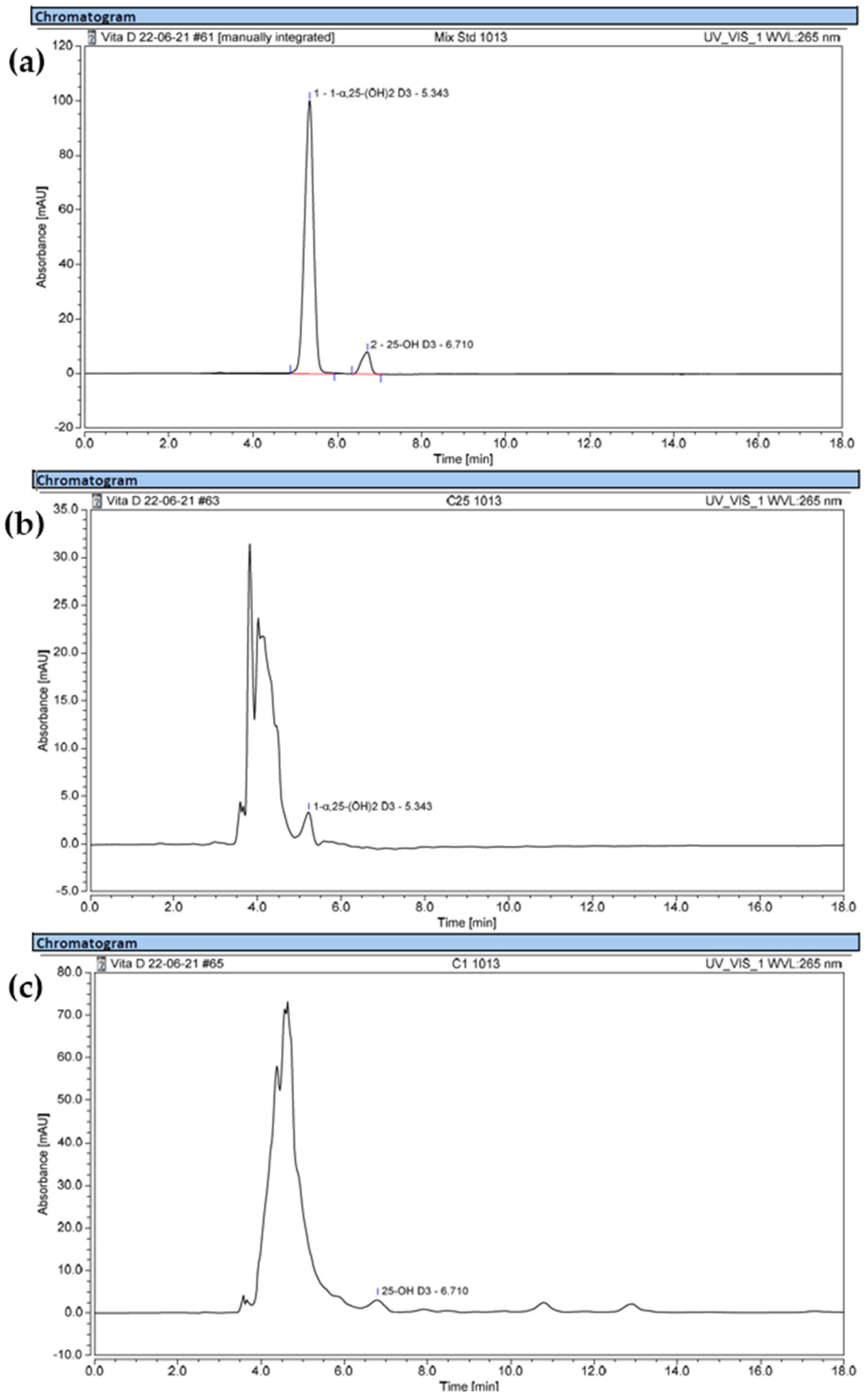
The HPLC chromatogram of the 1α,25-(OH)
2D
3 and 25-OHD
3 standard mixtures are shown in
Figure 1a with peak retention times of 5.343 min and 6.710 min, respectively. All the purified extracts obtained by the chemical hydrolysis of
S. glaucophyllum leaves starting as hydrophilic extract were analyzed. The 1α,25-(OH)
2D
3 and 25-OHD
3 peaks from purified sample tubes 25 and 1 were respectively assigned by comparing their retention times with those of the pure standards.
Figure 1b shows the 1α,25-(OH)
2D
3 peak from purified sample tube 25 with a retention time of 5.343 min.
Figure 1c shows the 25-OHD
3 peak from purified extract tube 1 with a retention time of 6.710 min.


 ,
, 

