Article

The Potential Role of Polyelectrolyte Complex Nanoparticles Based on Cashew Gum, Tripolyphosphate and Chitosan for the Loading of Insulin

by
Janira M. N. A. Bezerra
1,
Antônia C. J. Oliveira
1,
Edson C. Silva-Filho
2,
Patricia Severino
3,4,5,
Selma B. Souto
6,
Eliana B. Souto
7,8,*,
Mônica F. La R. Soares
1 and
José L. Soares-Sobrinho
1,*
1
Quality Control Core of Medicines and Correlates (NCQMC), Department of Pharmaceutical Sciences, Federal University of Pernambuco, Recife 50740-520, PE, Brazil
2
Interdisciplinary Laboratory for Advanced Materials–LIMAV, Federal University of Piaui, Teresina 64049-550, PI, Brazil
3
Biotechnological Postgraduate Program, University of Tiradentes (Unit), Av Murilo Dantas, 300, Aracaju 49010-390, SE, Brazil
4
Nanomedicine and Nanotechnology Laboratory (LNMed), Institute of Technology and Research (ITP), Av Murilo Dantas, 300, Aracaju 49010-390, SE, Brazil
5
Tiradentes Institute, 150 Mt Vernon St, Dorchester, MA 02125, USA
6
Department of Endocrinology, Hospital São João, Porto, Alameda Prof. Hernâni Monteiro, 4200-319 Porto, Portugal
7
Department of Pharmaceutical Technology, Faculty of Pharmacy, University of Coimbra, Pólo das Ciências da Saúde, Azinhaga de Santa Comba, 3000-548 Coimbra, Portugal
8
CEB-Centre of Biological Engineering, Campus de Gualtar, University of Minho, 4710-057 Braga, Portugal
*
Authors to whom correspondence should be addressed.
Diabetology 2021, 2(2), 107-116; https://doi.org/10.3390/diabetology2020009
Submission received: 28 February 2021 / Revised: 27 April 2021 / Accepted: 26 May 2021 / Published: 8 June 2021

Abstract

:
Polyelectrolytic complexation has stood out due to its application in the development of drug delivery systems using biopolymers as raw materials. The formation of complexes between cashew gum and chitosan can be intermediated by cross-links, mediated by the action of the sodium tripolyphosphate crosslinking agent. These polymers have been used in the nanotechnological development of formulations to protect peptide drugs, such as insulin, allowing their oral administration. In this work, we describe the development of polyelectrolytic complexes from cashew gum and chitosan as biopolymers for oral administration of insulin. The obtained complexes showed a mean particle size of 234 nm and polydispersity index of 0.2. The complexes were 234 nm in size, PDI 0.2, zeta potential −4.5 mV and 22% trapping. The obtained complexes demonstrated considerable and promising characteristics for use as oral insulin delivery systems.

1. Introduction

The use of biopolymers for the production of drug delivery systems is receiving increasing attention because of their specific attributes in the production of nanoparticles [1]. Such interest is attributed to their degradability, biocompatibility and non-toxic profile. Besides this, when combined with other polymers, a range of structures, such as gels, layer-by-layer films, excipients and/or even nanometric scale drug delivery systems, can be obtained with improved properties when compared to synthetic polymers [2,3,4,5].
Cashew gum (CG) is a polysaccharide extracted from the exudate of the species Anacardium occidentale L., popularly known as cashew, a tree widely cultivated in the regions of northeastern Brazil and some areas of the African continent [6,7]. Its structural composition has a variety of monosaccharides, differing in quantities and proportion by repeated units of β-D-galactopyranose (72%), α-D-glucopyranose (14%), α-L-arabinofuranose (4.6%), acid β-D-glucuronic (4.5%) and α-L-ramnopyranose (3.2%) [8].
The presence of available groups of glucuronic acids and hydroxyls in its framework, even in small percentages, adds a negative surface charge to the material, making it possible to carry out reactions with different polymers through different molecular interactions [9]. CG shows promising properties to be used in the formation of layer-by-layer films applied to nanobiomedical devices [10], as Pickering emulsions stabilizers [11], in aceclofenac toothpastes [12] and also showing antinociceptive and anti-inflammatory activity [13].
The characteristics present in the conformation of CG can be added to those of other polymers, such as chitosan (CH) [14]. CH is obtained by deacetylation of chitin, present in the shells of insects, crustaceans and in some fungi [15]. It is a biopolymer with a linear conformation structure also of repeated units of β-(1–4) N-Acetylglycosamine and D-Glucosamine [15], and it has several positively charged amine groups in its structural composition [16,17]. The polyelectrolytic complexation method is a technique in which biopolymers can interact spontaneously from differences in their charges, by electrostatic interactions, hydrogen bonds and ionic bonds, with the use of organic solvents or exposure to high temperatures not being necessary [18,19,20]. Peptide drugs, such as insulin, play important roles in a range of biological processes and reactions, being of great importance for glycemic control in patients affected by the diabetes mellitus pathology [21]. Insulin is usually administered subcutaneously, which can result in constant discomfort in its use and may be subject to incomplete distribution. However, due to the physicochemical features of the molecule and biopharmaceutical characteristics, the subcutaneous route still constitutes the main form of treatment [22]. A possible alternative to overcome the limitations of this drug is the use of polyelectrolytic complexes [1,4]. Therefore, the present study aims to produce polyelectrolytic complexes through the interaction between GC and CH, intermediated by the use of the reticulating agent sodium tripolyphosphate (TPP).

2. Materials and Methods

2.1. Materials

Cashew gum (CG) (Mw = 2.35 × 10⁴ g/mol) was isolated from the exudate obtained from collections of trees found in the region of Parnaíba (Piauí, Brazil) and purified [8]. Chitosan (CH) (50 kDa) with a 78% deacetylation degree was purchased from Sigma-Aldrich (Darmstadt, Germany). Other used products included the solvents: hydrochloric acid (HCl) (Dinâmica®, Sao Paulo, Brazil), lactic acid (Puratec®, Rio de Janeiro, Brazil) sodium tripolyphosphate (TPP) (Sigma Aldrich, Darmstadt, Germany) and syringe filters with 0.46 µm Millipore® (Burlington, MA, USA) porosity. Humulin R® Insulin (INS) (100 IU/mL) was purchased from Lilly pharmaceutical company (São Paulo, Brazil). A syringe with a 21 G needle attached was used. All water used in the processes was ultrapure obtained from the Milli-Q system (Smart®, Ultrapure water system, Heal force®, Shanghai, China).

2.2. Preparation of CG, CH and TPP

The CG solution was prepared at a concentration of 0.5% (w/v). CH solution was prepared at a concentration of 0.2% (w/v), dissolved in a solution of lactic acid at 0.1% (v/v), with agitation of 600 rpm overnight. The 13.5 mM TPP solution was prepared by dissolving it in ultrapure water. The pH of the CG, CH and TPP solutions were checked to be 6.2, 3.4 and 9.1, respectively. All solutions were filtered through a 0.45 µm Millipore® (Burlington, MA, USA) porosity filter.

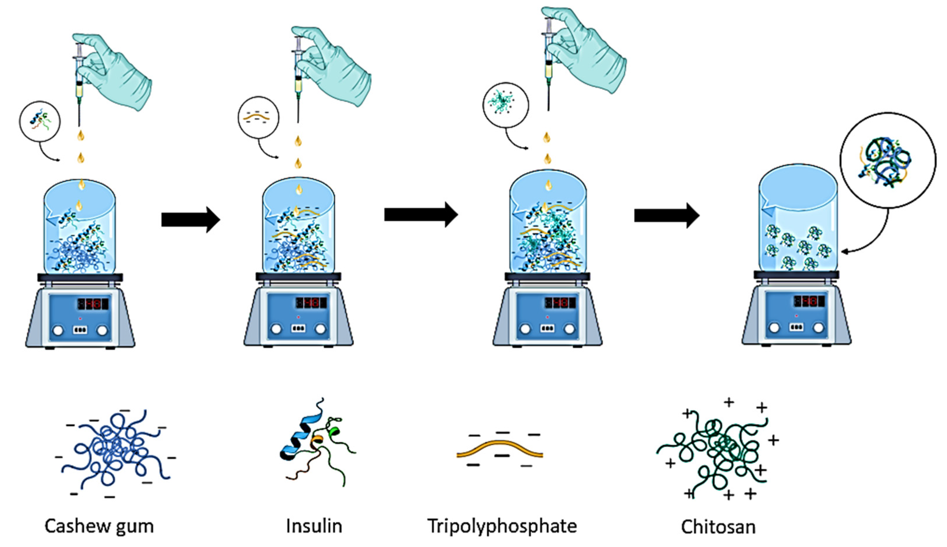
2.3. Preparation of CG/INSULIN/TPP/CH Nanoparticles

Complexes without insulin were formed by polyelectrolytic complexation. Different volumes of TPP (0.4, 0.6, 0.7 and 0.8 mL) were injected into a CG solution (10.0 mL). Afterwards, the CH solution (0.6 mL) was dripped for 30 min, stirring for another 30 min (Figure 1). The nanospheres loaded with insulin were prepared as follows: insulin (0.05–0.005%) was added to the aqueous CG solution (10.0 mL). After 30 min, TPP (0.8 mL) was added and stirred for 30 min. After that time, CH (0.6 mL) was added and stirred for another 30 min. All nanoparticles were prepared in triplicate.

2.4. Characterization of Nanoparticles

2.4.1. Particle Size Determination, Polydispersity Index (PDI) and Zeta-Potential of Nanoparticles

The size, PDI and zeta potential analyzes were performed on the Zetasizer Nano ZS equipment (Malvern Instruments Ltd., Malvern, UK). The particle size distribution and PDI were obtained by dynamic light scattering (DLS), with an angle of 90°. The zeta potential was calculated mathematically based on the Smoluchowski equation. The stability of the complexes was evaluated at predetermined times. The formulations were stored in a refrigerator (6 °C to 10 °C). All measurements were recorded in triplicate at 25 ± 2 °C.

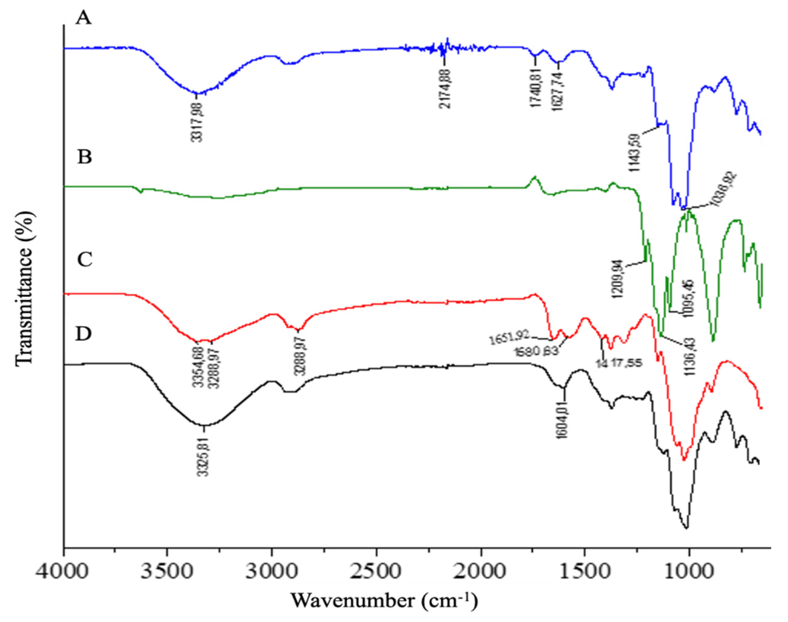
2.4.2. FTIR Spectroscopy Analysis

CG, CH, TPP and electrolytic complexes were characterized by infrared spectroscopy, using a PerkinElmer FTIR, spectrum 400, in the ATR module, in the range of 650 and 4000 cm−1.

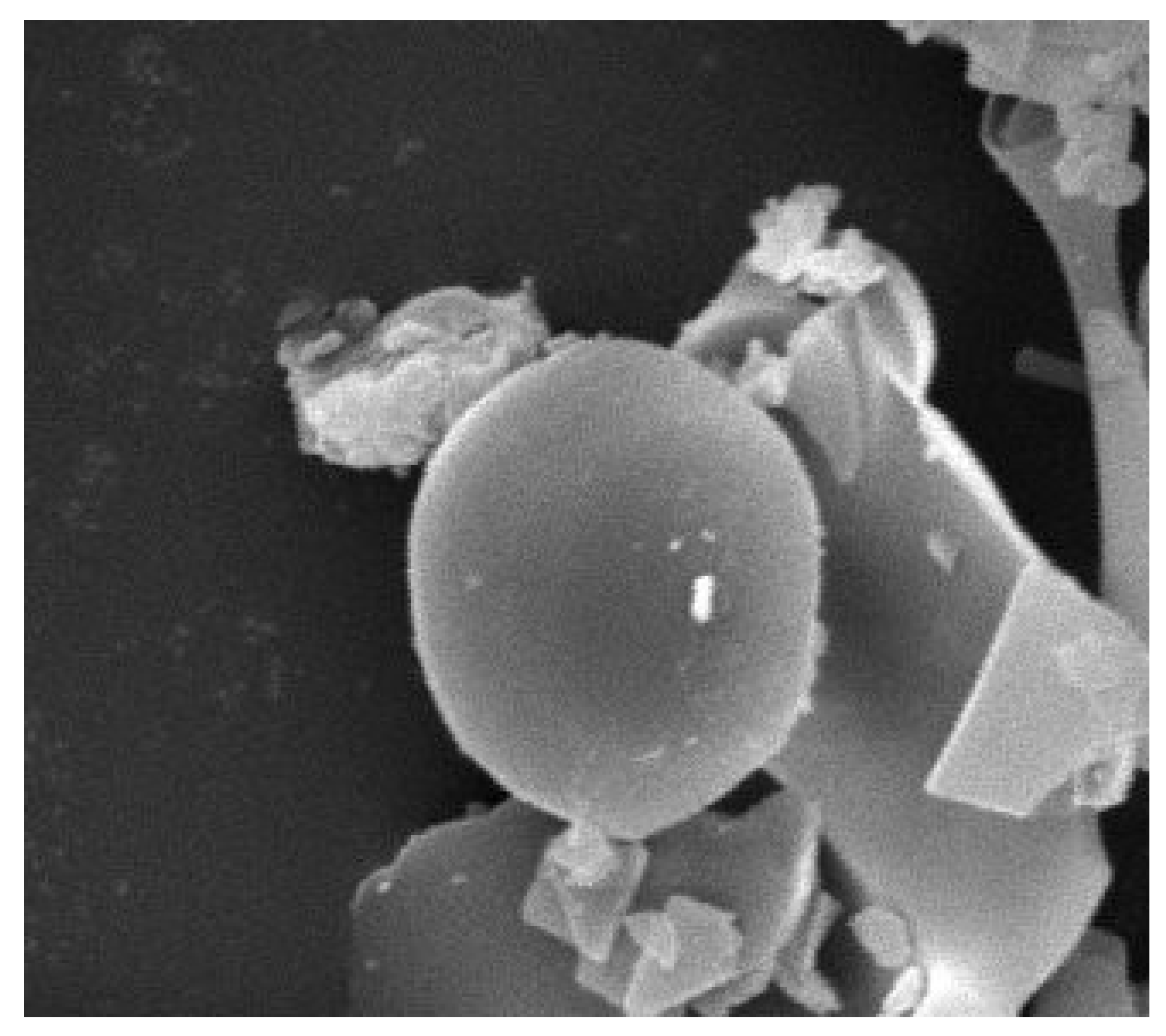
2.4.3. Morphological Characterization of Scanning Electron Microscopy (SEM)

SEM analyses were performed on the SS-550 Sperscan® equipment (Shimadzu, Tokyo, Japan). The samples were coated with a gold film of approximately 20 nm, for about two minutes, at an amperage of 5 mA (model SC-701 Quick Coater®, Tokyo, Japan) to facilitate the conduction and observe the morphology of the nanoparticles.

2.4.4. Determination of Insulin Encapsulation Efficiency

The insulin encapsulation efficiency was determined by the difference between the total amount of insulin used to prepare the nanoparticles and the amount of free insulin divided by the total amount of insulin used. To analyze the amount of free insulin, 1.5 mL of the nanoparticles were subjected to centrifugation at 12,000 RPM for 50 min at 4 °C. The supernatant containing free insulin was collected and quantified in triplicate by high-performance liquid chromatography (HPLC).

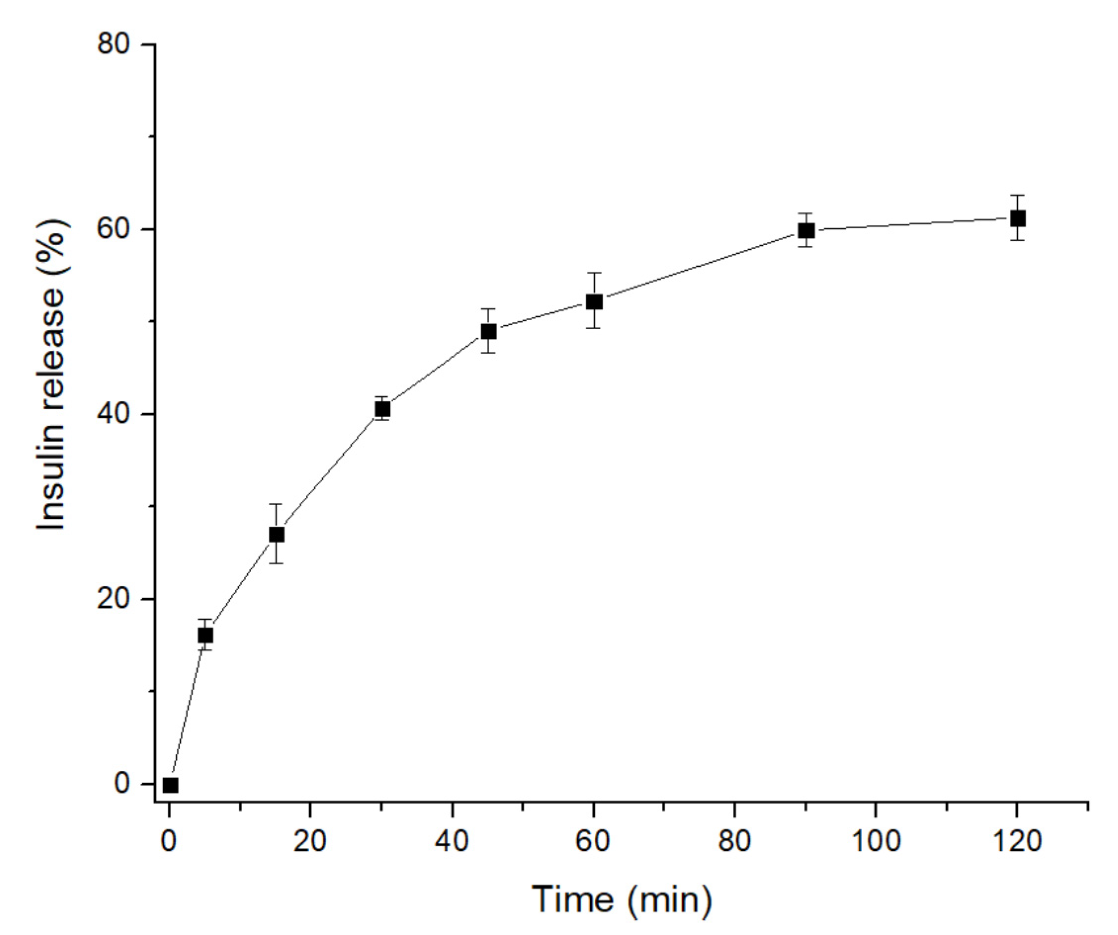
2.4.5. In Vitro Release Study

The tests were performed by the dialysis method, using a dialysis membrane (MWCO 14 kDa, Sigma Aldrich, Darmstadt, Germany), to determine the release of insulin from the complexes under different conditions of enzyme-free gastrointestinal pHs. The complex preparations were submerged in simulated gastric fluid (SFG) without pepsin (USP34-NF29) at 37 °C for 120 min with stirring. The insulin samples released were collected at certain times, and the same volume that was removed was replaced with buffered medium under the same conditions to keep the volume constant. To determine the release of insulin from the complexes, a fixed volume collection was carried out and subsequently centrifuged at 12,000 rpm for 30 min at 4 °C; the supernatants were analyzed by HPLC. The analyses were performed in triplicate.

2.4.6. Chromatographic Conditions for the Determination of Insulin by HPLC

For the determination of insulin, an Ascentis®-C18 column (250 × 4.6 mm, 5 µm, Supelco®, Bellefonte, PA, USA), a mobile phase of 52% 20 mM aqueous buffer KH2PO4 (buffer at pH 3.1 ± 0.1, adjusted using phosphoric acid), 31% acetonitrile and 7% methanol, flow of 1 mL·min−1 with an injection volume of 20 µL and a column temperature 35 °C were used. A wavelength of 214 nm and isocratic elution were also used. Chromatograms were recorded, and the peak area (responses) was measured using an automatic integrator.

3. Results

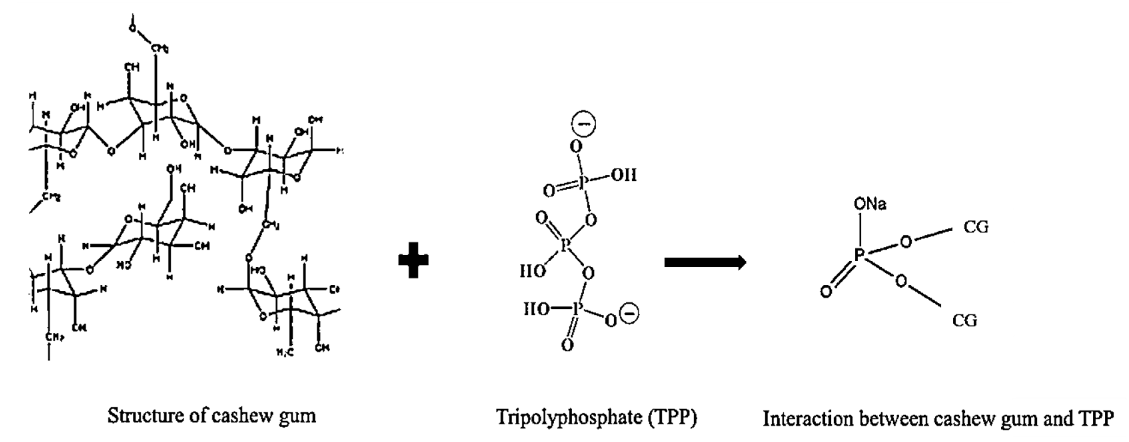
3.1. Preparation of Cashew Gum–TPP–Chitosan Polyelectrolyte Complex

The nanoparticles were produced using the polyelectrolytic complexation method [1]. The volumes of CG and CH were fixed, and the volume of TPP was varied in order to investigate its influence on the formation of complexes. Table 1 shows the hydrodynamic size, PDI and zeta potential data of the colloidal systems obtained in the four formulations. Polyelectrolytic complexes composed of CG and CH were prepared; however, they did not show viability. The formation of heterogeneous systems with sizes ranging from 150 nm to 300 nm was evidenced, obtaining a polymodal population. In addition, precipitate formation was observed.

3.2. Preparation and Physicochemical Characterization of the Cashew Gum–Insulin–TPP–Chitosan Polyelectrolyte Complex

Based on the results shown in Table 1, the NP-4 formulation was selected as it depicted the lowest PDI, indicating improved uniformity over the remaining formulations. NP-4 was thus chosen to be loaded with insulin. Insulin concentrations ranging from 0.05% to 0.005% were used for the preparation of the complexes. The system that contained a higher percentage of insulin had a high size and PDI. The use of a reduced amount of the protein resulted in a complex with a single population, with a size of 234.5 nm, PDI 0.27 and zeta potential of −4.5 mV. The insulin trapped inside the nanoparticles was 22%. Both complexes showed opalescent coloring, indicating the formation of nanoparticles. The study of complex release without insulin under simulated gastric fluid (SFG) conditions without enzymes was carried out. In the SFG medium, about 3% of the insulin released was observed in the first moments. After 2 h of testing, 60% of insulin release was observed, as shown in Figure 2. The colloidal stability of the complexes with and without insulin were monitored for 1 year, with no changes in particle size, as seen in Table 2.

3.3. Characterization of the Polyelectrolyte Complex

FTIR spectra of the isolated materials and the polyelectrolytic complex are shown in Figure 3. The CG bands were observed at 2174.88 cm−1 and 3317.98 cm−1, which referred to groups C-H and O-H, respectively. Stretching of C-O-C vibrations was observed due to glycosidic bonds in the region of 1036.92 cm−1 [23,24]. The main bands referring to the CH molecule were attributed to an elongation of the O-H and N-H groups by 3354.68 cm−1 and 3288.97 cm−1, respectively [25]. The C-H pyramid structures were at 1417.55 cm−1, and the C-H stretch vibrations were at 2872.07 cm−1 [17]. Stretching vibrations classified as amide I (C-H) and amide II (N-H) in the bands of 1651.92 cm−1 and 1580.63 cm−1, respectively, were also observed [26]. The amide functional groups present on the CH surface are responsible for interactions with other molecules [17]. In the TTP molecule, the intense absorption bands at 1136.43 cm−1, 1095.45 cm−1 and 1209.94 cm−1 referred to the groups P=O, P-O-P and =O, respectively.
In the spectrum of the polyelectrolytic complex, changes were observed in regions in which they suggested interactions between the materials used for its formation. In the 3325.81 cm−1 region, the band became wider and shifted, reducing the wavelength, indicating an increase in hydrogen interactions [26]. There was a lack of the band related to the glucuronic acid group of cashew gum in the complex (1740.81 cm−1, C=O), showing that there was an electrostatic interaction with the chitosan. A reduced change of direction was observed in the amide I region in the spectrum from 1651.92 cm−1 to 1604.01 cm−1, related to the CH/TPP interaction [17,26].
In the region of the amide II band of CH, the interaction of the N-H groups, either with TPP or CG, was observed. This was also confirmed by the reduction of the peak by 1127.63 cm−1, which indicated the possible electrostatic interaction between the sodium tripolyphosphate molecule and the positively charged amine group of chitosan [27].
The morphological analysis of the polyelectrolytic complex was observed by SEM as shown in Figure 4. A smooth and spherical shape was observed, proving the formation of the complex between CG/TPP/CH.

4. Discussion

For the treatment of diabetes, currently available therapeutic options include several oral and injectable antidiabetic drugs (GLP1 analogs) and insulin. In this work, insulin has been proposed to be loaded into nanoparticles obtained from cashew gum and chitosan, through a method requiring sodium tripolyphosphate as a crosslinking agent [1]. Insulin is a hormone produced by pancreatic β cells, with a peptidic structure consisting of 51 amino acids and composed of two chains, A and B, linked by disulfide bonds. Its key role in the treatment of Diabetes mellitus is linked to its capacity to promote the uptake and use of glucose at the cellular level, the synthesis of glycogen in the liver and muscle, the synthesis of triglycerides in adipose tissue and the liver, the storage of triglycerides and the synthesis of proteins. Besides this, insulin inhibits glycogenolysis, lipolysis, gluconeogenesis and ketogenesis. The sensitive character of this peptide, and its low bioavailability, compromises its administration through the oral route, while efforts have been made to promote its paracellular and/or transcellular pathways in the ileum and colon. The cross-linking of nanoparticles has been proposed to strengthen their resistance in the gut.
To evaluate the capacity of nanoparticles for the loading of insulin and their suitability for oral administration, a regular synthetic molecule was used experimentally only as a model compound that structurally resembled the same physiologically produced molecule.
The CG “in nature” has a low surface negative charge due to a lower percentage of acid groups of around 5%. Its composition contains about 60 to 72% of galactose [8]. Biopolymers with high levels of galactose and mannose present surface charges between −13.7 and −2.1 mV, which hinders their interaction with other molecules [23].
CH is a hydrophilic polycathion at an acidic pH, and it has structurally charged amides that give the molecule a high positive charge [17]. Its interaction with GC alone for the formation of complexes by the ionotropic gelling process is not sufficient due to the structural characteristics and the low negative charge present in GC, which results in the formation of heterogeneous, polydispersed and unstable systems [23]. In order to improve the anionic properties of GC, a crosslinking with TPP was performed. CG has functional groups available to make bonds to TPP atoms. TPP can interact via intermolecular or intramolecular bonds with the CG. The chemical interactions may be the result of hydrogen and ionic bonds between the susceptible oxygen in the TPP molecule with the -OH groupings of the CG monosaccharide units (Figure 5).
The effects of changes in the proportion of PPT for formation of CG/CH complexes were analyzed. Possible variations related to hydrodynamic size, PDI and surface load were evaluated. Four formulations were prepared (NP-1, NP-2, NP-3 and NP-4). The NP-1 and NP-2 systems had a bimodal and PDI population formation above 0.3, indicating highly polydispersed formulations and demonstrating that the amount of tripolyphosphate added was not enough to interact and mediate as a link between biopolymers. The amount of CG loads and the action of the reticulant agent were not enough to neutralize the positive QT loads [17], not completing the interaction, besides the presence of precipitated in the formulation, showing amounts of polysaccharide without interaction [16]. The surface load of NP-1 and NP-2 were less negative when compared to formulations of NP-3 and NP-4. The formulations (NP-3 and NP-4) used the highest proportion of TPP, which provided a greater neutrality of CH loads, and were also used because of the reticulant action of TPP.
A strong influence on size, PDI and payload was observed in relation to the amount used in formulations loaded with insulin. Heterogeneous and unstable systems were obtained with a high concentration of insulin. On the other hand, a stable and monomodal system was obtained with a reduced amount of the protein. An influence of the size increase of the complexes with the presence of insulin was observed, when compared to those produced without the drug. This phenomenon may have been due to the simultaneous competition between the negative charges present in CG, TPP and insulin interacting with the positive charges of CH, leading to an increase of the particle Alteration in the surface load of the complex was also observed. The complex with insulin presented a greater anionic character. This finding was related to the negative charge of insulin. The release profile may have been related to free insulin or weakly bound insulin on the complex surface. Based on FTIR spectra, electrostatic interactions, hydrogen bonds and ionic interactions between the biopolymer groups and the reticulating agent were observed, which resulted in the formation of the complex.

5. Conclusions

The present study described the production and characterization of polyelectrolytic complexes, composed of CG, CH and TTP, for the transport of the insulin polypeptide. The structural and electrostatic characteristics of these colloids were studied, which led to the formation of nanoparticles with an average size ranging from 143 to 204 nm. Several processing parameters and the polyelectrolytic nature of the biopolymers and TPP used influenced the characteristics of the nanoparticles. The process of formation of stable particles depended on the volume of TPP used. The potential use of these CG/TP/CH complexes has been applied to carry the insulin polypeptide. A stable system with a size of 243 nm was obtained. Thus, the formulation can be used as a promising carrier for oral insulin administration for the management of diabetes.

Author Contributions

J.M.N.A.B.: formal analysis, methodology, investigation, writing—review and editing, visualization, validation. A.C.J.O.: writing—review and editing, visualization. E.C.S.-F.: methodology, formal analysis, writing—review and editing. S.B.S.: visualization, formal analysis, writing—review and editing; P.S.: conceptualization, validation, formal analysis, investigation, writing—review and editing, visualization; supervision, resources, project administration. E.B.S.: conceptualization, validation, formal analysis, investigation, writing—review and editing, visualization; supervision, resources, project administration. M.F.L.R.S.: conceptualization, validation, formal analysis, investigation, writing—review and editing, visualization; supervision, resources, project administration. J.L.S.-S.: conceptualization, validation, formal analysis, investigation, writing—review and editing, visualization; supervision, resources, project administration. All authors have read and agreed to the published version of the manuscript.

Funding

This research was funded by the Fundação de Amparo à Ciência e Tecnologia do Estado de Pernambuco (FACEPE), Banco do Nordeste (grant FUNDECI/2016.0015), Portuguese Science and Technology Foundation (FCT/MCT) and from European Funds (PRODER/COMPETE) through the project UIDB/04469/2020 (strategic fund), co-financed by FEDER, under the Partnership Agreement PT2020.

Institutional Review Board Statement

Not applicable.

Informed Consent Statement

Not applicable.

Data Availability Statement

Data are available from authors upon request to the corresponding authors.

Conflicts of Interest

The authors declare no conflict of interest.

References

  1. Severino, P.; da Silva, C.F.; da Silva, M.A.; Santana, M.H.A.; Souto, E.B. Chitosan Cross-Linked Pentasodium Tripolyphosphate Micro/Nanoparticles Produced by Ionotropic Gelation. Sugar Tech 2016, 18, 49–54. [Google Scholar] [CrossRef]
  2. Souto, E.B.; da Ana, R.; Souto, S.B.; Zielińska, A.; Marques, C.; Andrade, L.N.; Horbańczuk, O.K.; Atanasov, A.G.; Lucarini, M.; Durazzo, A.; et al. In Vitro Characterization, Modelling, and Antioxidant Properties of Polyphenon-60 from Green Tea in Eudragit S100-2 Chitosan Microspheres. Nutrients 2020, 12, 967. [Google Scholar] [CrossRef] [PubMed] [Green Version]
  3. Oliveira, D.M.L.; Rezende, P.S.; Barbosa, T.C.; Andrade, L.N.; Bani, C.; Tavares, D.S.; da Silva, C.F.; Chaud, M.V.; Padilha, F.; Cano, A.; et al. Double membrane based on lidocaine-coated polymyxin-alginate nanoparticles for wound healing: In vitro characterization and in vivo tissue repair. Int. J. Pharm. 2020, 591, 120001. [Google Scholar] [CrossRef] [PubMed]
  4. Shimojo, A.A.M.; da Silva Santos Duarte, A.; Lana, J.F.S.D.; Luzo, A.C.M.; Fernandes, A.R.; Sanchez-Lopez, E.; Souto, E.B.; Santana, M.H.A. Association of Platelet-Rich Plasma and Auto-Crosslinked Hyaluronic Acid Microparticles: Approach for Orthopedic Application. Polymers 2019, 11, 1568. [Google Scholar] [CrossRef] [PubMed] [Green Version]
  5. Ataide, J.A.; Gerios, E.F.; Cefali, L.C.; Fernandes, A.R.; Teixeira, M.D.C.; Ferreira, N.R.; Tambourgi, E.B.; Jozala, A.F.; Chaud, M.V.; Oliveira-Nascimento, L.; et al. Effect of Polysaccharide Sources on the Physicochemical Properties of Bromelain-Chitosan Nanoparticles. Polymers 2019, 11, 1681. [Google Scholar] [CrossRef] [PubMed] [Green Version]
  6. Porto, B.C.; Cristianini, M. Evaluation of cashew tree gum (Anacardium occidentale L.) emulsifying properties. LWT-Food Sci. Technol. 2014, 59, 1325–1331. [Google Scholar] [CrossRef]
  7. De Oliveira, W.Q.; Wurlitzer, N.J.; de Oliveira Araújo, A.W.; Comunian, T.A.; do Socorro Rocha Bastos, M.; de Oliveira, A.L.; Magalhães, H.C.R.; Ribeiro, H.L.; de Figueiredo, R.W.; de Sousa, P.H.M. Complex coacervates of cashew gum and gelatin as carriers of green coffee oil: The effect of microcapsule application on the rheological and sensorial quality of a fruit juice. Food Res. Int. 2020, 131, 109047. [Google Scholar] [CrossRef] [PubMed]
  8. de Paula, R.C.M.; Heatley, F.; Budd, P.M. Characterization of Anacardium occidentale exudate polysaccharide. Polym. Int. 1998, 45, 27–35. [Google Scholar] [CrossRef]
  9. Abreu, F.O.M.S.; Oliveira, E.F.; Paula, H.C.B.; de Paula, R.C.M. Chitosan/cashew gum nanogels for essential oil encapsulation. Carbohydr. Polym. 2012, 89, 1277–1282. [Google Scholar] [CrossRef] [Green Version]
  10. Araújo, I.M.; Zampa, M.F.; Moura, J.B.; dos Santos, J.R., Jr.; Eaton, P.; Zucolotto, V.; Veras, L.M.; de Paula, R.C.; Feitosa, J.P.; Leite, J.R.; et al. Contribution of the cashew gum (Anacardium occidentale L.) for development of layer-by-layer films with potential application in nanobiomedical devices. Mater. Sci. Eng. C 2012, 32, 1588–1593. [Google Scholar] [CrossRef]
  11. Cardial, M.R.L.; Paula, H.C.B.; da Silva, R.B.C.; da Silva Barros, J.F.; Richter, A.R.; Sombra, F.M.; de Paula, R.C.M. Pickering emulsions stabilized with cashew gum nanoparticles as indomethacin carrier. Int. J. Biol. Macromol. 2019, 132, 534–540. [Google Scholar] [CrossRef]
  12. Hasnain, M.S.; Rishishwar, P.; Rishishwar, S.; Ali, S.; Nayak, A.K. Extraction and characterization of cashew tree (Anacardium occidentale) gum; use in aceclofenac dental pastes. Int. J. Biol. Macromol. 2018, 116, 1074–1081. [Google Scholar] [CrossRef]
  13. Silva, F.; Torres, L.; Silva, L.; Figueiredo, R.; Garruti, D.; Araújo, T.; Duarte, A.; Brito, D.; Ricardo, N. Cashew gum and maltrodextrin particles for green tea (Camellia sinensis var Assamica) extract encapsulation. Food Chem. 2018, 261, 169–175. [Google Scholar] [CrossRef]
  14. Wu, D.; Zhu, L.; Li, Y.; Zhang, X.; Xu, S.; Yang, G.; Delair, T. Chitosan-based Colloidal Polyelectrolyte Complexes for Drug Delivery: A Review. Carbohydr. Polym. 2020, 238, 116126. [Google Scholar] [CrossRef] [PubMed]
  15. Qin, Y.; Li, P.; Guo, Z. Cationic chitosan derivatives as potential antifungals: A review of structural optimization and applications. Carbohydr. Polym. 2020, 236, 116002. [Google Scholar] [CrossRef] [PubMed]
  16. Sharkawy, A.; Barreiro, M.F.; Rodrigues, A.E. Chitosan-based Pickering emulsions and their applications: A review. Carbohydr. Polym. 2020, 250, 116885. [Google Scholar] [CrossRef]
  17. Mukhopadhyay, P.; Sarkar, K.; Chakraborty, M.; Bhattacharya, S.; Mishra, R.; Kundu, P.P. Oral insulin delivery by self-assembled chitosan nanoparticles: In vitro and in vivo studies in diabetic animal model. Mater. Sci. Eng. C 2013, 33, 376–382. [Google Scholar] [CrossRef] [PubMed]
  18. Kizilay, E.; Kayitmazer, A.B.; Dubin, P.L. Complexation and coacervation of polyelectrolytes with oppositely charged colloids. Adv. Colloid Interface Sci. 2011, 167, 24–37. [Google Scholar] [CrossRef]
  19. Luo, Y.; Wang, Q. Recent development of chitosan-based polyelectrolyte complexes with natural polysaccharides for drug delivery. Int. J. Biol. Macromol. 2014, 64, 353–367. [Google Scholar] [CrossRef] [PubMed]
  20. Masood, F. Polymeric nanoparticles for targeted drug delivery system for cancer therapy. Mater. Sci. Eng. C 2016, 60, 569–578. [Google Scholar] [CrossRef]
  21. Wong, C.Y.; Al-Salami, H.; Dass, C.R. Recent advancements in oral administration of insulin-loaded liposomal drug delivery systems for diabetes mellitus. Int. J. Pharm. 2018, 549, 201–217. [Google Scholar] [CrossRef]
  22. Lauterbach, A.; Müller-Goymann, C.C. Applications and limitations of lipid nanoparticles in dermal and transdermal drug delivery via the follicular route. Eur. J. Pharm. Biopharm. 2015, 97, 152–163. [Google Scholar] [CrossRef]
  23. Soares, P.A.; Bourbon, A.I.; Vicente, A.A.; Andrade, C.A.; Barros, W., Jr.; Correia, M.T.; Pessoa, A., Jr.; Carneiro-da-Cunha, M.G. Development and characterization of hydrogels based on natural polysaccharides: Policaju and chitosan. Mater. Sci. Eng. C 2014, 42, 219–226. [Google Scholar] [CrossRef] [Green Version]
  24. Batista, R.A.; Espitia, P.J.P.; Vergne, D.M.C.; Vicente, A.A.; Pereira, P.A.C.; Cerqueira, M.A.; Teixeira, J.A.; Jovanovic, J.; Severino, P.; Souto, E.B.; et al. Development and Evaluation of Superabsorbent Hydrogels Based on Natural Polymers. Polymers 2020, 12, 2173. [Google Scholar] [CrossRef]
  25. Ailincai, D.; Mititelu-Tartau, L.; Marin, L. Citryl-imine-PEG-ylated chitosan hydrogels-Promising materials for drug delivery applications. Int. J. Biol. Macromol. 2020, 162, 1323–1337. [Google Scholar] [CrossRef] [PubMed]
  26. Rampino, A.; Borgogna, M.; Blasi, P.; Bellich, B.; Cesàro, A. Chitosan nanoparticles: Preparation, size evolution and stability. Int. J. Pharm. 2013, 455, 219–228. [Google Scholar] [CrossRef] [PubMed]
  27. Li, J.; Huang, Q. Rheological properties of chitosan-tripolyphosphate complexes: From suspensions to microgels. Carbohydr. Polym. 2012, 87, 1670–1677. [Google Scholar] [CrossRef]
Figure 1. Production of nanoparticles by polyelectrolytic interactions between cashew gum (CG), insulin, sodium tripolyphosphate (TPP) and chitosan (CH).
Diabetology 02 00009 g001
Figure 2. In vitro release profile of nanoparticles simulated gastric fluid for 120 min.
Diabetology 02 00009 g002
Figure 3. FTIR spectra of (A) cashew gum, (B) chitosan, (C) sodium tripolyphosphate and (D) polyelectrolyte complex.
Diabetology 02 00009 g003
Figure 4. Morphology of polyelectrolytic complexes by scanning electron microscopy (500×).
Diabetology 02 00009 g004
Figure 5. Interaction between cashew gum (CG) and tripolyphosphate (TPP).
Diabetology 02 00009 g005
Table 1. Size, polydispersity index and zeta potential of the different polyelectrolytic complexes.
FormulationCashew Gum (mL)TPP (mL)Chitosan (mL)Size (nm)PDIZeta (mV)
NP-110.00.40.6143.90.59−6.47
NP-210.00.60.6161.60.35−5.12
NP-310.00.70.6169.30.29−4.23
NP-410.00.80.6204.80.24−2.94
Table 2. Colloidal stability of polyelectrolytic complexes with and without insulin.
2 Days7 Days15 Days1 Month1 Year
NP without insulinSize (nm)206.8206.4207.2210.0205.0
PDI0.240.20.20.20.3
Zeta(mV)Zeta(mV)−7.17−6.74−4.60−3.43
NP with insulinSize (nm)260.4266.0268.3272.4311.8
PDI0.270.20.20.20.2
Zeta (mV)Zeta (mV)−8.21−5.07−4.62−1.81
Publisher’s Note: MDPI stays neutral with regard to jurisdictional claims in published maps and institutional affiliations.

