Article

Posterosuperior Segments of the Liver: Comparison of Short-Term Outcomes between Open and Minimally Invasive Surgery Performed by a Single Surgeon

Department of General and Specialized Surgery, Parma University Hospital, 43126 Parma, Italy
*
Author to whom correspondence should be addressed.
Livers 2023, 3(4), 674-686; https://doi.org/10.3390/livers3040044
Submission received: 21 August 2023 / Revised: 23 October 2023 / Accepted: 8 November 2023 / Published: 16 November 2023

Abstract

:
Laparoscopic posterosuperior liver segment resection is considered technically challenging. This is a retrospective single-center single-surgeon study. The aim of the present study is to investigate the short-term outcomes in a single institution between laparoscopic (LLR) and open (OLR) posterosuperior liver segments (PSSs) resections performed by a single surgeon at Parma University Hospital. The patients were divided into Group 1 (OLR) and Group 2 (LLR) and stratified in two different time settings according to the experience of the surgeon (2010–2015 and 2016–2021). A total 112 patients were included in the study. The 75.3% of OLR were performed in the first period, while 70.2% of LLR were carried out during the second period (2016–2021). The Iwate score was significantly (p < 0.001) higher in OLR group compared to the LLR group. Most of the advanced (77%) and expert (100%) LLRs were performed during the second period. LOS was shorter in LLR group comparing to OLR group (p < 0.001). The postoperative morbidity rate was similar in both groups (p > 0.05). The presence of liver cirrhosis and multiple lesions were identified as risk factors for severe postoperative complications. PSS-LLR has become much safer and more effective due to increasing surgeon’s expertise along with the implementation of cutting-edge technology and innovative surgical techniques.

1. Introduction

Laparoscopic liver resection (LLR) has been exploited due to its advantage of minimal invasiveness, resulting in early recovery and short hospital stay. LLR was first introduced in 1991 for the treatment of benign cystic lesions [1].
The initial experience of minimally invasive surgery of the liver was limited to benign, easily accessible lesions. Through the years, the indications have evolved. In the first international statement in 2008, LLR was recommended for hepatic lesions smaller than 5 cm, solitary and located in the anterolateral segments [2,3]. The Morioka international consensus conference followed by [4] the Southampton Consensus Guidelines for Laparoscopic Liver Surgery [5] expanded surgical indications to malignancy. Growing evidence has supported the safety and feasibility of LLR also for lesions located in the PSS [6,7].
Several LLR difficulty scores were developed to predict the likelihood of achieving a proper liver resection through laparoscopy and to guide the surgical planning, taking into account a variety of patient and tumor factors, such as liver function, and tumor size and location [8,9,10,11].
The original difficulty scoring system described by Ban et al. [12] was updated in 2014, becoming a relatively complex four-level difficulty classification system, best known as the Iwate criteria. It still stands as the most employed difficulty scoring system. The Iwate Score considers different factors: tumor size and location, proximity to major vessels, the extent of liver resection, the eventual need of hand-assisted laparoscopy, and liver function [13].
Other difficult scoring systems have been introduced. The Institut Mutualiste Montsouris (IMM) group introduced a new, simple three-level classification system based on the type of procedure [11].
Different difficulty scoring systems are accepted world-wide and have been well validated in several studies [12,13,14,15]
PSS-LLR (segments 4s, 6s 7, 8 and 1) is considered technically challenging due to the need for precise trocar placement, the limited exposure of the operative field, the curvilinear resection surfaces and the difficulty in controlling major bleeding [10,11,12].
Despite minimally invasive approaches having been accepted worldwide for liver surgery, the laparoscopic treatment of posterosuperior segments remains challenging with a limited adherence compared to other laparoscopic liver resections [16].
Technological development and the learning curve have allowed for the resection of lesions located in posterosuperior segments. Several studies evaluated the role of laparoscopic posterosuperior liver segment resection, but no one is focused on only one surgeon.
We decided to review our experience retrospectively with the aim of comparing the intraoperative and immediate postoperative results of LLR and OLR in patients operated on for lesions located in posterosuperior segments by a single surgeon in a single institution.

2. Methods

This is a retrospective single-center single-surgeon study. All consecutive adult (age > 18 years) patients who underwent PSS resection performed by R.D.V. at the Division of General Surgery of the Parma University Hospital were included. The study timeline spanned from 1 January 2010 to 31 December 2021.
Inclusion criteria were as follows: adult patients > 18 years; and patients who underwent only posterosuperior liver resections with open or laparoscopic approach.
Exclusion criteria were as follows: patients who underwent major liver resections according to the Brisbane 2000 nomenclature [17]; and patients who underwent associated major abdominal procedures or concomitant liver resection of other segments.
The study has been approved by our institution independent ethics committee (Comitato etico AVEN (area vasta Emilia nord)). Informed consent was obtained from each patient included in the study. The paper was written according to the STROBE statement [18].
The patients were divided into Group 1, who underwent OLR, and Group 2, who underwent LLR. The two groups were analyzed in two different time settings according to the experience of the surgeon (2010–2015 and 2016–2021).

2.1. Variables and Definitions

Age, sex, BMI and comorbidities were summarized by the Charlson Comorbidity Index [19].
Preoperative liver function was assessed using both the model for end-stage liver disease score (MELD) and the Child–Pugh score.
Biochemical values of bilirubin, albumin, platelets, creatinine, and prothrombin time, and the international normalized ratio (INR) were also recorded at baseline.
The American Society of Anesthesiologists (ASA) class was assessed during the preoperative patient evaluation [20].
The characteristics of the lesions (number, location, size and the presence of vascular invasion) were assessed through preoperative triple-phase CT scan or magnetic resonance imaging and confirmed by intraoperative ultrasound. The nature of the lesions (hepatocellular carcinoma (HCC), cholangiocarcinoma (CCC), colorectal liver metastasis (CRLM), liver metastasis (LM), benign lesion) and preoperative treatment were assessed. The difficulty of the resection was graded according to the Iwate score and Institute Mutualiste Montsouris (IMM) scoring system. These scores were also calculated for OLR to evaluate the changes through the years [12,21]. The extension of the resection was defined according to the Brisbane nomenclature [17].
Other surgery-related variables include the following: minimally invasive approach, overall clamping time, intraoperative blood loss and transfusion, laparoscopy-to-open-conversion rate, and duration of surgery.
The LOS (length of stay) was calculated from the day of the operation to hospital discharge. Postoperative complication grading was recorded according to Clavien–Dindo Classification [22].
Postoperative bile leakage, liver failure (bile leakage, bleeding and liver failure were graded according to the ISGLS classifications), ascites, bleeding and infections, and pulmonary, cardiovascular and cerebrovascular complications were evaluated [23,24,25].
Postoperative mortality was calculated as the number of deaths occurring within 90 days from surgery.

2.2. Surgical Techniques

The surgical technique (laparoscopic or laparotomic) was chosen according to the learning curve of the operator through the years. The initial laparoscopic approach was reserved for the left liver procedures. A step-by-step learning curve was performed with the progressive treatment of complex laparoscopic resections including laparoscopic resection of posterosuperior liver segments.
In our center, the treatment modality is always discussed among a multidisciplinary team including hepatologists, interventionists, radiologists and medical oncologists. Whether to perform surgery and the extent of liver resection are also discussed based on the patient’s general condition, liver cirrhosis, and tumor size and location. The final decision on whether to perform LLR or OLR is made by the operating surgeon.

2.2.1. OLR Technique

The patient was placed in a supine position. The right arm was tagged along the body while the left stretched at 90°. A reversed L-shaped incision with lateral extension beyond the mid-clavicular line up to the right flank was performed. The manual palpation of the liver was performed to potentially identify known or undiagnosed lesions. The mobilization of the right liver was carried out using monopolar, bipolar and ultrasound energy-based devices (Thunderbeat™, Olympus Medical Systems Corp., Tokyo, Japan). The manual retraction of the liver was performed to ease ligaments exposure. The round and falciform ligaments were taken down, and the right lobe mobilization was completed thanks to the section of the right triangular and coronary ligaments as far back as the suprahepatic inferior vena cava (IVC). Multiple short Spigelian veins between the IVC and the posterior surface of the liver were ligated and divided. In case of significant sizable right inferior hepatic vein (RIHV) it was preserved whenever possible.
The hepatic pedicle was consistently prepared for Pringle Maneuver [26]. After the liver mobilization, IOUS was always performed to identify liver lesions and the transection line was marked. The parenchymal transection was carried out with the Kelly clamp crushing technique in the first years and then using an ultrasonic dissector. Vasculo-biliary structures were managed selectively using clips, sutures, ties or linear staplers.

2.2.2. LLR Techniques

LLR techniques used at our hospital have been described elsewhere [3,27,28,29].
Under general anesthesia, the patient was placed in the modified Lloyd-Davies position with leg stirrups and tilted to 30° reverse Trendelenburg position (the first surgeon stood between the patient’s legs with one assistant on each side).
The left lateral decubitus was commonly employed for lesions in segment 6, 7 and dorsal aspect of segment 8 [30,31].
Initial access for a 12 mm camera port was placed in the supra-umbilical or in the right para-rectal position, the pneumoperitoneum was established, and the pressure was maintained under 12 mmHg. Four ports were positioned in a reverse J configuration [32].
The round and falciform ligaments were dissected and then a right lobe mobilization was completed through the section of right triangular and coronary ligaments as far back as the suprahepatic IVC using Thunderbeat™.
Multiple short Spigelian veins between the IVC and posterior surface of the liver were ligated and divided when necessary.
Laparoscopic Pringle Maneuver approach changed through the years. In the initial laparoscopic experience extracorporeal Pringle Maneuver was performed, and involved taping the hepatoduodenal ligament with cotton tape through a 10 mm Goldfinger Dissector. A 5 mm port was inserted in the left flank and then the end of the cotton tape was externalized through a 20-Fr drain tube tourniquet that was pushed inside the abdominal cavity up to the hepatic pedicle.
In the last 3 years, the extracorporeal Pringle Maneuver was abandoned in favor of the intracorporeal Pringle Maneuver performed using a 16 Fr Huang’s loop [33].
Such as for open liver resections after liver mobilization and hepatoduodenal pedicle preparation, IOUS was always performed using a flexible ultrasound transducer. The parenchymal transection was achieved with Thunderbeat™ for shallow parenchymal transection and switching ultrasonic dissector, bipolar energy-based devices and Thunderbeat™. Vasculo-biliary structures were managed selectively using clips, ties or staplers. The specimen was placed into a retrieval bag and removed via a slightly enlarged port site or for a large specimen via a Pfannenstiel incision.

2.3. Study Endpoints

The primary endpoint was to compare perioperative outcomes across the two groups and the changes through the two study periods (2010–2015 and 2016–2021).
All patients were followed up using local protocols, which included blood test, abdominal ultrasound, CT or magnetic resonance imaging, and office visits.

2.4. Statistical Analysis

Data analysis was performed using Jamovi. Univariate and multivariate analyses were performed.
The sample description was performed using the median and interquartile range (IQR) for numeric variables, and number and proportion for categorical variables. Mann–Whitney and Fisher tests were used to compare baseline patient characteristics between the two treatment groups, respectively.
Differences between the groups were assessed using a log-rank test. All factors deemed to be statically significant for a p-value of less than 5% (p < 0.05).

3. Results

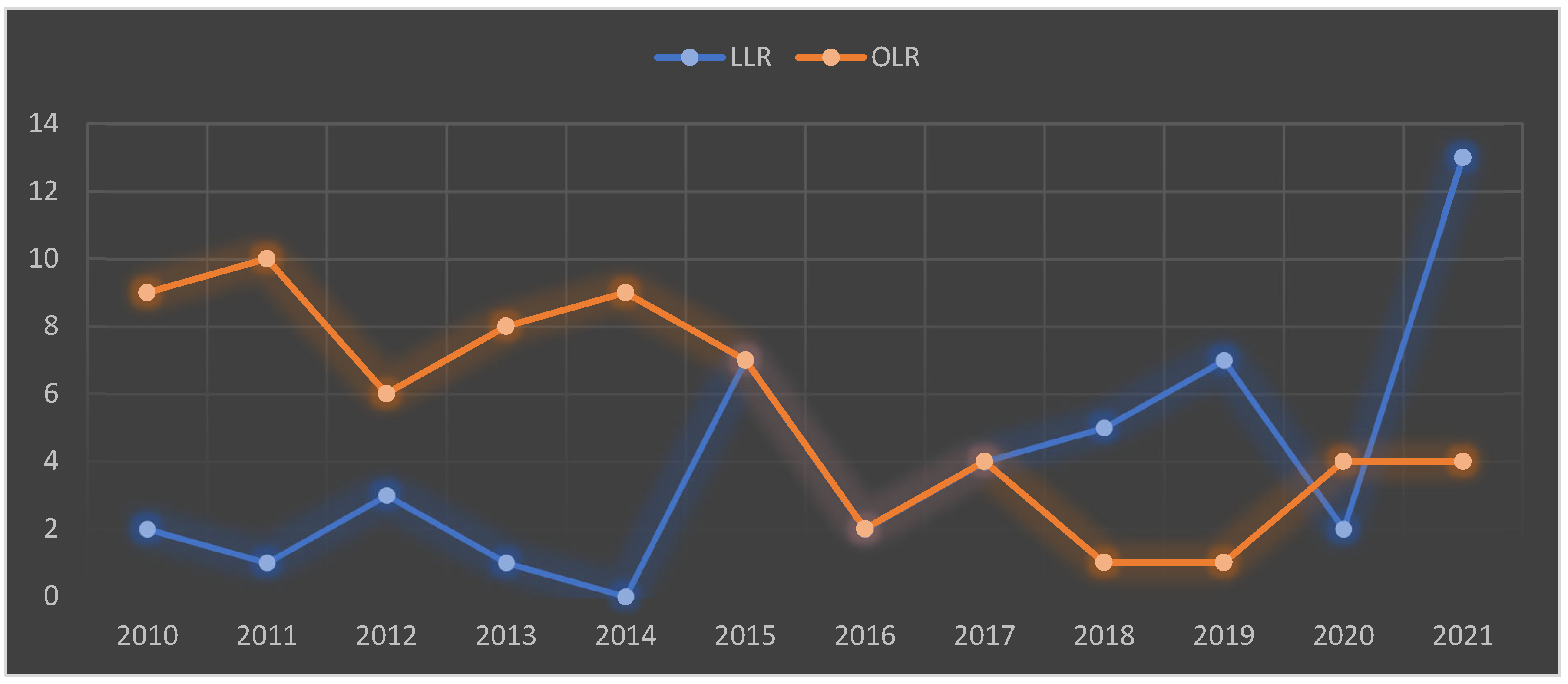
3.1. General Characteristics

A total 112 patients who underwent posterosuperior liver segment resection were included in the study.
The baseline characteristics of each group are summarized in Table 1.
According to the time of surgery, most of the OLRs were performed in the early period (2010–2015) and most of the LLRs were performed in the late period (2016–2021). Overall, 75.3% of the OLRs (49/63) were performed in the early period, and 70.2% (33/49) of the LLRs were performed in the late period. Laparoscopy demonstrated significant procedure-specific increases over the second period, with more LLR than OLR.  Figure 1. p < 0.001.
Previous open hepatectomies were reported in 12 patients who underwent OLR (18.4%) and only in 2 who underwent LLR (4.2%).
No differences for the Charlson Comorbidity Index were observed among the two groups (p > 0.05). Hepatocarcinoma was the most common etiology in the LLR group (55.1%) and CRLM in the OLR group(50.7%).
Among the 48 patients with CRLM, 34 (62.5%) underwent preoperative chemotherapy.
Among the 51 patients with HCC, prior locoregional treatments were performed in five cases (9.8%).
The Iwate score was significantly higher in the OLR group than the LLR group (p < 0.001). Most of the OLRs (81.5%) were at an advanced or expert Iwate difficulty level compared to 51% of LLRs.
According to Iwate difficulty levels, most of the advanced (77%) and all the expert (100%) LLRs were performed only in the second period of the study.
The difficulty level of IMM showed no differences between OLR and LLR (p > 0.05).

3.2. Surgical Characteristics

Anatomical resection was performed 77 times (68.7%), and non-anatomical resections were performed 35 times (31.2%). Anatomic OLR was performed 53/65 times (81.5%). Anatomic LLR was performed 24/47 times (51.0%). The bisegmentectomy of segments 6 and 7 was the most common OLR in 20/65 (30.7%) cases. The wedge resection of segment 7 was the most common LLR in 13/47 (27.6%) cases. Hepato-duodenal lymphadenectomy was performed in 13 patients (11.6%), in every CCC case, and in six additional patients. The Pringle Maneuver was performed in 89 cases (79.4%), and the median Pringle Maneuver time was 30 min (14.2–47.2). The median estimate blood loss was 150 mL (100–200), and only two patients required intraoperative blood transfusion (1.7%).
Operative time was significantly higher in LLR than OLR (p = 0.012).
Conversion rate was 31.9%. Among the 15 conversions, 11 (73.3%) were performed during the early period (2010–2015), and only 4 (26.7%) were performed during the late period (2016–2021); a conversion was performed in three patients to control major bleeding.
Surgical procedure details are reported in Table 2.

3.3. Postoperative Outcomes

The LOS was 8 days (7–11) in the LLR group and 11 days (9–21) in the OLR group (p < 0.001).
Severe postoperative complications (Clavien–Dindo > 3a) were reported in four (6.1%) patients who underwent OLR and only in one (2.1%) who underwent LLR.
Grade A bile leakages were found in five (7.6%) OLR patients and in one (2.1%) LLR patient. Grade B bile leakages were found in one (1.5%) OLR patient. Grade C bile leakages were not found. Transient liver failure was found in one (1.5%) OLR patient. No grade B and C bleeding was reported.
Readmission within 90 days was reported for 12 (10.7%) patients. Mortality within 90 days was reported for one patient (0.8%).
Postoperative details and complications are summarized in Table 3.
The presence of liver cirrhosis and multiple lesions were identified as risk factors for severe postoperative complications, as shown in Table 4.
In the multivariate analysis, liver cirrhosis (odds ratio (OR) 2.4; 95% confidence interval (CI) 0.48–4.38; p = 0.016) and multiple lesions > 2 (odds ratio (OR) 1.98; 95% confidence interval (CI) −0.24–4.2; p = 0.045) were identified as independent risk factors for severe postoperative complications.

4. Discussion

PSS-LLR have been considered technically challenging and graded as a major procedure in several studies due to the several anatomical issues of posterosuperior segments [10,11,12].
The difficulties of PSS LLR lie in the limited operative field due to the space-occupying liver. The anatomical area of PSSs is closed by the diaphragm and the rib cage. The depth of PSSs, the curvilinear resection surfaces and the limited anatomical exposure led to major difficulties in performing IOUS and in controlling major bleeding. The operative field is far from the conventional abdominal trocar site. In this context, when the trocars are not correctly placed, the laparoscopic instruments often fail to reach the lesion as they are too short, making the transection of the liver and the bleeding control very difficult [11,14,17,19].
Despite the difficulties behind the laparoscopic treatment of posterosuperior segments several factors contributed to the adoption of this approach. The number and the complexity of performed LLRs has grown worldwide through the years, as also reported in our experience.
The results of the present study showed that LLR is a viable option for the treatment of posterosuperior segment lesions, with good postoperative outcomes when compared to OLR.
Before mastering PSS LLRs, a steep learning curve, starting with easier LLRs, is required. Several studies showed that 20 to 60 cases are needed to reach proficiency in major LLRs [6,10,34,35]. In our study, laparoscopic liver resection was performed in 43.7% cases, with most of the LLRs performed during the second period and with a constant growth through the years.
We also observed that fewer surgical procedures (43.7%) were performed during the second period (2016–2021), especially in 2020, due to the COVID-19 pandemic.
The growing surgeon’s expertise and experience, combined with the growing literature evidence, expanded the indications for LLR, including the treatment of posterosuperior segments, as evidenced by our results.
The difficulty behind the LLR of posterosuperior segments also affected type of resection. Different previous literature reports described non-anatomical PSS LLR resection to be as difficult as anatomical resection, such as right hepatectomy or right posterior hepatectomy. Several reports also showed better postoperative outcomes of anatomical PSS resection over non-anatomical one [36,37,38].
However, the recent adaptation of advanced approaches has facilitated the performance of LLR for lesions located in PSSs. In our study, no differences were found among anatomical and non-anatomical LLR outcomes.
In our study, as reported in a previous literature report [15], the Iwate score was significantly higher in the OLR group than the LLR group. Most of the OLRs (81.5%) were at an advanced or expert Iwate difficulty level compared to the 51% of LLRs. In the second period, most of the advanced (77%) and all the expert (100%) LLRs, according to Iwate difficulty levels, were performed. This indicates that OLR may be more challenging from a technical perspective and justify the increase in difficult LLR during the second period.
The conversion rate during our experience was similar to other previous studies [39,40]. The conversion rate was higher during the initial experience of PSS LLR, where 73.3% of all conversions took place. Various factors, such as inappropriate patient selection, poor tumor location, difficulty in bleeding control, and difficulty in ensuring an adequate resection margin, may be related to the higher rate of conversion during the first period [40,41]. Conversion from laparoscopy to laparotomy for bleeding is the main cause of conversion during LLR [40]. Among the four patients who required conversion during the second period, in three cases (75%), conversion occurred to control major bleeding. The improved surgical skills and consequent reduction of errors related to inexperience, may justify the lower conversion rate during the second period where conversion was mostly related to major bleeding.
Despite the technical difficulties of PSS-LLR, the overall outcomes of this study showed similar results in both groups [3,7,42]. Patients who underwent PSS LLR showed shorter LOS and less severe postoperative complications as reported in previous studies [7,43,44,45].
OLR for PSS often requires a large incision due to the deep and closed location resulting in a delayed physical recovery due to postoperative pain and discomfort [46]. This suggests that LLR may result in faster recovery and reduced hospitalization time.
Pulmonary complications (pleural effusion), cut surface complications (effusion, infection and bile leak) ascites and wound infection are reported as common complications after PSS.
Fluid collection in the right subdiaphragmatic space are the most common complications after PSSs [47] and often leads to other related thoracic complications (reactive pleural effusion, atelectasis and pneumonia) [29,45,48].
In the present study, no significant difference in postoperative morbidity was noted; we observed higher non-significant pulmonary complications in the OLR group (23%) than the LLR (17%) group and a higher non-significant difference in postoperative ascites in the OLR group (13.8%) than the LLR group (4.2%).
In previous studies, the postoperative ascites rate was reported to be lower in patients with cirrhosis after LLR than after OLR due to reduced manipulation and damage to collateral circulations during LLR, which could prevent the decompensation of a cirrhotic liver. [25,27,45,49].
No differences were observed among the two groups for postoperative bile leak, liver failure, bleeding and infection (p > 0.05).
We observed a much worse postoperative outcome in patients with multiple lesions and cirrhosis, no matter the surgical approach with both OLR and LLR. This suggests that patients with these conditions may be at higher risk of experiencing complications after PSS resection.
However, patients who underwent PSS LLR had a shorter hospital stay and less overall morbidity than patients in the OLR group.
This study has several limitations. First, it is a retrospective single-center study, which may limit the generalizability of the findings to other settings. Second, the study focused on short-term outcomes and did not assess long-term oncological outcomes. Further research is needed to evaluate the long-term effects of LLR and OLR on patient outcomes.

5. Conclusions

PSS-LLR has become safer and more effective thanks to the implementation of newer techniques and to the developed surgical expertise, which helped to overcome the anatomical boundaries and the very unique pitfalls of the PSS-LLR. The short-term outcomes of PSS resections are similar between LLR and OLR, while postoperative LOS and recovery appeared to be favored by the laparoscopic approach. Further research is needed to evaluate the long-term effects of LLR and OLR on patient outcomes.

Author Contributions

Conceptualization, M.G. and R.D.V.; methodology, M.G.; software, M.G.; validation, M.G. and M.I.; formal analysis, M.G.; investigation, M.I.; data curation, M.G.; writing—original draft preparation, M.G.; writing—review and editing, M.I.; visualization, R.D.V.; supervision, R.D.V. All authors have read and agreed to the published version of the manuscript.

Funding

The authors received no financial support for the research, authorship, and/or publication of this article.

Institutional Review Board Statement

The study has been approved by independent ethics committee (Comitato etico AVEN (area vasta Emilia nord)).

Informed Consent Statement

Informed consent was obtained from each patient included in the study.

Data Availability Statement

The data that support the findings of this study are available from the corresponding author, [M.G.], upon reasonable request.

Conflicts of Interest

The authors declare no conflict of interest.

Abbreviations

OLROpen liver resection
LLLLaparoscopic liver resection
PSSPosterosuperior segment
LOSLength of stay

References

  1. Reich, H.; McGlynn, F.; DeCaprio, J.; Budin, R. Laparoscopic excision of benign liver lesions. Obstet. Gynecol. 1991, 78 Pt 2, 956–958. [Google Scholar] [PubMed]
  2. Buell, J.F.; Cherqui, D.; Geller, D.A.; O’rourke, N.; Iannitti, D.; Dagher, I.; Koffron, A.J.; Thomas, M.; Gayet, B.; Han, H.S.; et al. The International Position on Laparoscopic Liver Surgery: The Louisville Statement, 2008. Ann. Surg. 2009, 250, 825–830. [Google Scholar] [CrossRef] [PubMed]
  3. Cho, J.Y.; Han, H.S.; Yoon, Y.S.; Shin, S.H. Feasibility of laparoscopic liver resection for tumors located in the posterosuperior segments of the liver, with a special reference to overcoming current limitations on tumor location. Surgery 2008, 144, 32–38. [Google Scholar] [CrossRef]
  4. Wakabayashi, G.; Cherqui, D.; Geller, D.A.; Buell, J.F.; Kaneko, H.; Han, H.S.; Asbun, H.; O’rourke, N.; Tanabe, M.; Koffron, A.J.; et al. Recommendations for laparoscopic liver resection: A report from the second international consensus conference held in Morioka. Ann. Surg. 2015, 261, 619–629. [Google Scholar]
  5. Abu Hilal, M.; Aldrighetti, L.; Dagher, I.; Edwin, B.; Troisi, R.I.; Alikhanov, R.; Aroori, S.; Belli, G.; Besselink, M.; Briceno, J.; et al. The Southampton Consensus Guidelines for Laparoscopic Liver Surgery: From Indication to Implementation. Ann. Surg. 2018, 268, 11–18. [Google Scholar] [CrossRef]
  6. Lee, W.; Han, H.-S.; Yoon, Y.-S.; Cho, J.Y.; Choi, Y.; Shin, H.K.; Choi, H.; Jang, J.S.; Kwon, S.U. Comparison of laparoscopic liver resection for hepatocellular carcinoma located in the posterosuperior segments or anterolateral segments: A case-matched analysis. Surgery 2016, 160, 1219–1226. [Google Scholar] [CrossRef]
  7. Guro, H.; Cho, J.Y.; Han, H.S.; Yoon, Y.S.; Choi, Y.; Jang, J.S.; Kwon, S.U.; Kim, S.; Choi, J.K. Laparoscopic liver resection of hepatocellular carcinoma located in segments 7 or 8. Surg. Endosc. 2018, 32, 872–878. [Google Scholar] [CrossRef]
  8. Tanaka, S.; Kubo, S.; Kanazawa, A.; Takeda, Y.; Hirokawa, F.; Nitta, H.; Nakajima, T.; Kaizu, T.; Kaneko, H.; Wakabayashi, G. Validation of a Difficulty Scoring System for Laparoscopic Liver Resection: A Multicenter Analysis by the Endoscopic Liver Surgery Study Group in Japan. J. Am. Coll. Surg. 2017, 225, 249–258.e1. [Google Scholar] [CrossRef]
  9. Russolillo, N.; Maina, C.; Fleres, F.; Langella, S.; Lo Tesoriere, R.; Ferrero, A. Comparison and validation of three difficulty scoring systems in laparoscopic liver surgery: A retrospective analysis on 300 cases. Surg. Endosc. 2020, 34, 5484–5494. [Google Scholar] [CrossRef]
  10. Halls, M.C.; Berardi, G.; Cipriani, F.; Barkhatov, L.; Lainas, P.; Harris, S.; D’Hondt, M.; Rotellar, F.; Dagher, I.; Aldrighetti, L.; et al. Development and validation of a difficulty score to predict intraoperative complications during laparoscopic liver resection. Br. J. Surg. 2018, 105, 1182–1191. [Google Scholar] [CrossRef]
  11. Kawaguchi, Y.; Fuks, D.; Kokudo, N.; Gayet, B. Difficulty of Laparoscopic Liver Resection: Proposal for a New Classification. Ann. Surg. 2018, 267, 13–17. [Google Scholar] [CrossRef] [PubMed]
  12. Ban, D.; Tanabe, M.; Ito, H.; Otsuka, Y.; Nitta, H.; Abe, Y.; Hasegawa, Y.; Katagiri, T.; Takagi, C.; Itano, O.; et al. A novel difficulty scoring system for laparoscopic liver resection. J. Hepatobiliary Pancreat. Sci. 2014, 21, 745–753. [Google Scholar] [CrossRef] [PubMed]
  13. Wakabayashi, G. What has changed after the Morioka consensus conference 2014 on laparoscopic liver resection? HepatoBiliary Surg. Nutr. 2016, 5, 281–289. [Google Scholar] [CrossRef]
  14. Tanaka, S.; Kawaguchi, Y.; Kubo, S.; Kanazawa, A.; Takeda, Y.; Hirokawa, F.; Nitta, H.; Nakajima, T.; Kaizu, T.; Kaibori, M.; et al. Validation of index-based IWATE criteria as an improved difficulty scoring system for laparoscopic liver resection. Surgery 2019, 165, 731–740. [Google Scholar] [CrossRef]
  15. Tripke, V.; Huber, T.; Mittler, J.; Lang, H.; Heinrich, S. Prediction of complexity and complications of laparoscopic liver surgery: The comparison of the Halls-score to the IWATE-score in 100 consecutive laparoscopic liver resections. J. Hepatobiliary Pancreat. Sci. 2020, 27, 380–387. [Google Scholar] [CrossRef]
  16. Hibi, T.; Cherqui, D.; Geller, D.A.; Itano, O.; Kitagawa, Y.; Wakabayashi, G. Expanding indications and regional diversity in laparoscopic liver resection unveiled by the International Survey on Technical Aspects of Laparoscopic Liver Resection (INSTALL) study. Surg. Endosc. 2016, 30, 2975–2983. [Google Scholar] [CrossRef]
  17. Strasberg, S.M.; Belghiti, J.; Clavien, P.A.; Gadzijev, E.; Garden, J.O.; Lau, W.Y.; Makuuchi, M.; Strong, R.W. The Brisbane 2000 Terminology of Liver Anatomy and Resections. HPB 2000, 2, 333–339. [Google Scholar] [CrossRef]
  18. Von Elm, E.; Altman, D.G.; Egger, M.; Pocock, S.J.; Gøtzsche, P.C.; Vandenbroucke, J.P. The Strengthening the Reporting of Observational Studies in Epidemiology (STROBE) Statement: Guidelines for Reporting Observational Studies. PLoS Med. 2007, 4, 1623–1627. [Google Scholar] [CrossRef]
  19. Charlson, M.E.; Pompei, P.; Ales, K.L.; MacKenzie, C.R. A new method of classifying prognostic comorbidity in longitudinal studies: Development and validation. J. Chronic Dis. 1987, 40, 373–383. [Google Scholar] [CrossRef]
  20. Doyle, D.J.; Hendrix, J.M.; Garmon, E.H. American Society of Anesthesiologists Classification. Available online: https://europepmc.org/article/NBK/nbk441940 (accessed on 4 June 2023).
  21. Barron, J.O.; Orabi, D.; Moro, A.; Quintini, C.; Berber, E.; Aucejo, F.N.; Sasaki, K.; Kwon, C.-H.D. Validation of the IWATE criteria as a laparoscopic liver resection difficulty score in a single North American cohort. Surg. Endosc. 2021, 36, 3601–3609. [Google Scholar] [CrossRef]
  22. Dindo, D.; Demartines, N.; Clavien, P.-A. Classification of Surgical Complications. Ann. Surg. 2004, 240, 205–213. [Google Scholar] [CrossRef] [PubMed]
  23. Koch, M.; Garden, O.J.; Padbury, R.; Rahbari, N.N.; Adam, R.; Capussotti, L.; Fan, S.T.; Yokoyama, Y.; Crawford, M.; Makuuchi, M.; et al. Bile leakage after hepatobiliary and pancreatic surgery: A definition and grading of severity by the International Study Group of Liver Surgery. Surgery 2011, 149, 680–688. [Google Scholar] [CrossRef] [PubMed]
  24. Rahbari, N.N.; Garden, O.J.; Padbury, R.; Brooke-Smith, M.; Crawford, M.; Adam, R.; Koch, M.; Makuuchi, M.; Dematteo, R.P.; Christophi, C.; et al. Posthepatectomy liver failure: A definition and grading by the International Study Group of Liver Surgery (ISGLS). Surgery 2011, 149, 713–724. [Google Scholar] [CrossRef] [PubMed]
  25. Rahbari, N.N.; Garden, O.J.; Padbury, R.; Maddern, G.; Koch, M.; Hugh, T.J.; Fan, S.T.; Nimura, Y.; Figueras, J.; Vauthey, J.-N.; et al. Post-hepatectomy haemorrhage: A definition and grading by the International Study Group of Liver Surgery (ISGLS). HPB 2011, 13, 528–535. [Google Scholar] [CrossRef] [PubMed]
  26. Pringle, J.H.V. Notes on the arrest of hepatic hemorrhage due to trauma. Ann. Surg. 1908, 48, 541–549. [Google Scholar] [CrossRef]
  27. Abu Hilal, M.; Tschuor, C.; Kuemmerli, C.; López-Ben, S.; Lesurtel, M.; Rotellar, F. Laparoscopic posterior segmental resections: How I do it: Tips and pitfalls. Int. J. Surg. 2020, 82, 178–186. [Google Scholar] [CrossRef]
  28. Machado, M.A.C.; Makdissi, F.F.; Galvão, F.H.; Machado, M.C. Intrahepatic Glissonian approach for laparoscopic right segmental liver resections. Am. J. Surg. 2008, 196, e38–e42. [Google Scholar] [CrossRef]
  29. Cheng, K.-C.; Yeung, Y.-P.; Ho, K.-M.; Chan, F.K.-M. Laparoscopic Right Posterior Sectionectomy for Malignant Lesions: An Anatomic Approach. J. Laparoendosc. Adv. Surg. Tech. 2015, 25, 646–650. [Google Scholar] [CrossRef]
  30. Ishizawa, T.; Gumbs, A.A.; Kokudo, N.; Gayet, B. Laparoscopic segmentectomy of the liver: From segment I to VIII. Ann. Surg. 2012, 256, 959–964. [Google Scholar] [CrossRef]
  31. Teramoto, K.; Kawamura, T.; Takamatsu, S.; Noguchi, N.; Nakamura, N.; Arii, S. Laparoscopic and Thoracoscopic Partial Hepatectomy for Hepatocellular Carcinoma. World J. Surg. 2003, 27, 1131–1136. [Google Scholar] [CrossRef]
  32. Fiorentini, G.; Ratti, F.; Cipriani, F.; Catena, M.; Paganelli, M.; Aldrighetti, L. Tips and Tricks for a Laparoscopic Approach to Paracaval Liver Segments. Ann. Surg. Oncol. 2018, 25, 1695–1698. [Google Scholar] [CrossRef] [PubMed]
  33. Huang, J.-W.; Su, W.-L.; Wang, S.-N. Alternative Laparoscopic Intracorporeal Pringle Maneuver by Huang’s Loop. World J. Surg. 2018, 42, 3312–3315. [Google Scholar] [CrossRef] [PubMed]
  34. Tomassini, F.; Scuderi, V.; Colman, R.; Vivarelli, M.; Montalti, R.; Troisi, R.I. The single surgeon learning curve of laparoscopic liver resection: A continuous evolving process through stepwise difficulties. Medicine 2016, 95, e5138. [Google Scholar] [CrossRef] [PubMed]
  35. Vigano, L.; Laurent, A.; Tayar, C.; Tomatis, M.; Ponti, A.; Cherqui, D. The Learning Curve in Laparoscopic Liver Resection: Improved feasibility and reproducibility. Ann. Surg. 2009, 250, 772–782. [Google Scholar] [CrossRef] [PubMed]
  36. Cucchetti, A.; Cescon, M.; Ercolani, G.; Bigonzi, E.; Torzilli, G.; Pinna, A.D. A Comprehensive Meta-regression Analysis on Outcome of Anatomic Resection Versus Nonanatomic Resection for Hepatocellular Carcinoma. Ann. Surg. Oncol. 2012, 19, 3697–3705. [Google Scholar] [CrossRef]
  37. Li, S.; Huang, T.; Shen, S.; Hua, Y.; Hu, W.; Kuang, M.; Peng, B.; Liang, L. Anatomical versus non-anatomical liver resection for hepatocellular carcinoma exceeding Milan criteria. Br. J. Surg. 2017, 104, 118–127. [Google Scholar] [CrossRef]
  38. Moris, D.; Tsilimigras, D.I.; Kostakis, I.D.; Ntanasis-Stathopoulos, I.; Shah, K.N.; Felekouras, E.; Pawlik, T.M. Anatomic versus non-anatomic resection for hepatocellular carcinoma: A systematic review and meta-analysis. Eur. J. Surg. Oncol. 2018, 44, 927–938. [Google Scholar] [CrossRef]
  39. Li, L.; Xu, L.; Wang, P.; Zhang, M.; Li, B. The risk factors of intraoperative conversion during laparoscopic hepatectomy: A systematic review and meta-analysis. Langenbeck’s Arch. Surg. 2022, 407, 469–478. [Google Scholar] [CrossRef]
  40. Yoon, Y.-S.; Han, H.-S.; Cho, J.Y.; Ahn, K.S. Total laparoscopic liver resection for hepatocellular carcinoma located in all segments of the liver. Surg. Endosc. 2010, 24, 1630–1637. [Google Scholar] [CrossRef]
  41. Berardi, G.; Aghayan, D.; Fretland, Å.A.; Elberm, H.; Cipriani, F.; Spagnoli, A.; Montalti, R.; Ceelen, W.P.; Aldrighetti, L.; Abu Hilal, M.; et al. Multicentre analysis of the learning curve for laparoscopic liver resection of the posterosuperior segments. Br. J. Surg. 2019, 106, 1512–1522. [Google Scholar] [CrossRef]
  42. Buell, J.F.; Thomas, M.J.; Doty, T.C.; Gersin, K.S.; Merchen, T.D.; Gupta, M.; Rudich, S.M.; Woodle, E.S. An initial experience and evolution of laparoscopic hepatic resectional surgery. Surgery 2004, 136, 804–811. [Google Scholar] [CrossRef] [PubMed]
  43. Bueno, A.; Rotellar, F.; Benito, A.; Martí-Cruchaga, P.; Zozaya, G.; Hermida, J.; Pardo, F. Laparoscopic limited liver resection decreases morbidity irrespective of the hepatic segment resected. HPB 2014, 16, 320–326. [Google Scholar] [CrossRef] [PubMed]
  44. Aldrighetti, L.; Guzzetti, E.; Pulitanò, C.; Cipriani, F.; Catena, M.; Paganelli, M.; Ferla, G. Case-matched analysis of totally laparoscopic versus open liver resection for HCC: Short and middle term results. J. Surg. Oncol. 2010, 102, 82–86. [Google Scholar] [CrossRef] [PubMed]
  45. Scuderi, V.; Barkhatov, L.; Montalti, R.; Ratti, F.; Cipriani, F.; Pardo, F.; Tranchart, H.; Dagher, I.; Rotellar, F.; Abu Hilal, M.; et al. Outcome after laparoscopic and open resections of posterosuperior segments of the liver. Br. J. Surg. 2017, 104, 751–759. [Google Scholar] [CrossRef] [PubMed]
  46. Fuks, D.; Gayet, B. Laparoscopic surgery of postero-lateral segments: A comparison between transthoracic and abdominal ap-proach. Updates Surg. 2015, 67, 141–145. [Google Scholar] [CrossRef]
  47. Morise, Z.; Kawabe, N.; Tomishige, H.; Nagata, H.; Kawase, J.; Arakawa, S.; Isetani, M. How Far Can We Go with Laparoscopic Liver Resection for Hepatocellular Carcinoma? Laparoscopic Sectionectomy of the Liver Combined with the Resection of the Major Hepatic Vein Main Trunk. BioMed Res. Int. 2015, 2015, 960752. [Google Scholar] [CrossRef]
  48. Cipriani, F.; Ratti, F.; Paganelli, M.; Reineke, R.; Catena, M.; Aldrighetti, L. Laparoscopic or open approaches for posterosuperior and anterolateral liver resections? A propensity score based analysis of the degree of advantage. HPB 2019, 21, 1676–1686. [Google Scholar] [CrossRef]
  49. Taesombat, W.; Nonthasoot, B.; Sutherasan, M.; Nivatvongs, S.; Sirichindakul, B. Long-term outcomes of laparoscopic versus open liver resection for hepatocellular carcinoma: Retrospective case-matched study. Int. J. Surg. Open 2020, 24, 12–17. [Google Scholar] [CrossRef]
Figure 1. Trends of laparoscopic and open surgeries over twelve years OLR: Open liver resections. LLR: Laparoscopic liver resections.
Livers 03 00044 g001
Table 1. Baseline characteristics of patients who underwent PPS resection stratified based on surgical approach (OLR vs. LLR).
Baseline CharacteristicsTotalOLRLLRp-Value
Period of resection <0.001
2010–201565 (56.2%)49 (75.3%)16 (24.6%)
2016–202147 (43.7%)14 (29.7%)33 (70.2%)
Median age (IQR), years70 (58.7–75.2)70 (61–74)70 (57.5–76)0.701
Male sex, n (%)88 (78.5%)52 (80.0%)36 (76.5%)0.669
Median BMI (IQR)25.4 (23.7–28.3)25.5 (23.8–28.4)25.4 (23.2–27.7)0.618
Previous liver surgery, n (%)14 (12.5%)12 (18.4%)2 (4.2%)0.025
Preoperative ascites8 (7.0%)5 (7.6%)3 (4.5%)0.797
Charlson Comorbidity Index, median (IQR)5 (4–8)5 (3–8)6 (4–7.5)0.612
ASA score, n (%)0.0140
11/112 (2.6%)2/65 (3.1%)1/47 (2.1%)
261/112 (54.4%)39/65 (60.0%)22/47 (46.8%)
348/112 (42.8%)24/65 (36.9%)24/47 (51.0%)
40 (0.0%)0 (0.0%)0 (0.0%)
Etiology, n (%)0.470
Cholangiocarcinoma7 (6.2%)6 (9.5%)1 (2.1%)
HCC51 (45.5%)24 (38.0%)27 (55.1%)
CRLM48 (42.8%)32 (50.7%)16 (32.6%)
LM2 (1.7%)1 (1.5%)1 (2.1%)
Benign disease4 (3.5%)2 (3.0%)2 (4.2%)
Child–Pugh score, median (IQR)6 (5–7)6 (5–7) 6 (5–7)0.147
Child–Pugh n (%)0.103
A74 (66.0%)47 (72.3%)27 (57.4%)
B38 (33.9%)18 (27.6%)20 (42.5%)
Meld score, median (IQR)8 (6–10)7 (6–10)8 (7–12)0.034
Median tumor size, mm (IQR)31 (22–43)32 (24–47)26 (21–41) 0.103
N lesions, median (IQR)1 (1–2)1 (1–2)1 (1–1.5)0.026
Tumor location, n (%)0.441
I1 (0.8%)1 (1.5%)0 (0.0%)
IVa4 (3.5%)2 (3.0%)2 (4.2%)
VIs10 (8.9%)6 (9.2%)4 (8.5%)
VII57 (50.8%)35 (53.8%)22 (46.8%)
VIII40 (35.7%)21 (32.3%)19 (40.4%)
Iwate score, Difficulty Index,
median (IQR)
7 (6–10)8 (7–10)7 (5–9) <0.001
Iwate, Difficulty Level <0.001
Low3 (2.6%)1 (1.5%)2 (4.2%)
Intermediate32 (28.5%)11 (16.9%)21 (44.6%)
Advanced45 (40.1%)26 (40.0%)19 (40.4%)
Expert32 (28.5%)27 (41.5%)5 (10.6%)
IMM, Difficulty Level0.352
I 49 (43.7%)23 (48.9%)26 (40.0%)
II10 (8.9%)4 (8.5%)6 (9.2%)
III53 (47.3%)20 (42.5%)33 (50.7%)
IQR: Interquartile range.
Table 2. Surgical details.
Intraoperative DetailesTotalOLRLLRp-Value
Operative time, median (IQR)300 (223.7–248.7)315 (240–360)250 (210–315)0.012
Tipe of resection, n (%)0.121
Anatomical resection55/112 (50.8%)36/65 (55.4%)19/47 (40.4%)
Atypical resection57/112 (49.2%)29/65 (44.6%)28/47 (59.6%)
Pringle Manuever time, median (IQR)30.0 (14.2–47.2)30.0 (12.0–45.0)30.0 (15.5–52.0)0.678
Blood loss (mL), median (IQR)150 (100–200)150 (100–300)100 (100–200)0.194
Conversion rate, n (%)15/47 (31.9%)/15/47 (31.9%)/
IQR: Interquartile range.
Table 3. Postoperative outcomes.
Postoperative OutcomesTotalOLRLLRp-Value
LOS, days (IQR)10 (7.75–14.5)11 (9–21)8 (7–11)<0.001
ICU LOS, days (IQR)1 (1–2.25)2 (1–3)1(0–2)0.011
Severe complications, Clavien–Dindo > 3b5 (4.4%)4 (6.1%)1 (2.1%)0.315
Clavien–Dindo, n (%)<0.001
146/112 (41.0%)35/65 (53.8%)11/47 (23.4%)
239/112 (34.8%)27/65 (41.5%)12/47 (25.5%)
3a18/112 (16.0%)16/65 (24.6%)2/47 (4.2%)
3b4/112 (3.5%)3/65 (4.6%)1/47 (2.1%)
4a0/112 (0.0%)0/65 (0.0%)0/47 (0.0%)
4b1/112 (0.8%)1/65 (1.5%)0/47 (0.0%)
51/112 (0.8%)1/65 (1.5%)0/47 (0.0%)
Bile leakage, n (%)0.129
A6 (5.3%)5 (7.6%)1 (2.1%)
B1 (0.8%)1 (1.5%)0 (0.0%)
Liver failure, n (%)1 (0.8%)1 (1.5%)0 (0.0%)NA
Postoperative ascites, n (%)11 (9.8%)9 (13.8%)2 (4.2%)0.095
Bleeding, n (%)5 (4.4%)4 (6.1%)1 (2.1%)0.315
Pulmonary complications, n (%)23 (20.5%)15 (23.0%)8 (17.0%)0.438
Postoperative infection, n (%)19 (16.9%)9 (13.8%)10 (21.2%)0.305
Cardiovascular complications, n (%)12 (10.7%)7 (10.7%)5 (10.6%)0.987
Cerebrovascular complication, n (%)1 (0.8%)1 (1.5%)0 (0.0%)NA
Readmission within 90 days, n (%)12 (10.7%)8 (12.3%)4 (8.5%)0.527
Mortality within 90 days, n (%)1 (0.8%)1 (1.5%)0 (0.0%)NA
NA: Not available. IQR: Interquartile range.
Table 4. Univariate analysis for severe postoperative complications.
VariableNo Severe Complication
(n = 107)
Severe Complication (n = 5)p-Value
Age ≥ 65 y67 (62.6%)3 (60.0%)0.907
Gender, male83 (77.5%)5 (100.0%)NA
BMI > 25 kg/m261 (57.0%)3 (60.0%)0.902
ASA score ≥ 345 (42.0%)3 (60.0%)0.435
Charlson Comorbidity Index, n (%)0.524
0–16 (5.60%)0 (0.0%)
2–321 (19.6%)0 (0.0%)
4–530 (28.0%)2 (40.0%)
6–720 (18.6%)2 (40.0%)
>830 (28.0%)1 (20.0%)
Liver chirrosis, n (%)6 (5.6%)2 (40.0%)0.004
Child–Pugh grade B, n (%)37 (34.5%)1 (20.0%)0.508
MELD score, median (IQR)8 (6–10)8 (5–9)0.910
Tumor size ≥ 3 cm, n (%)54 (50.4%)3 (60.0%)0.684
Tumor location0.446
Segment 11 (0.9%)0 (0.0%)
Segment 4s4 (3.7%)0 (0.0%)
Segment 6s9 (8.4%)1 (20.0%)
Segment 756 (52.3%)1 (20.0%)
Segment 837 (34.5%)3 (60.0%)
Multiple lesions, n (%)38 (35.0%)4 (80.0%)0.046
Proximity to the major hepatic vessels, n (%)58 (54.2%)4 (80.0%)0.262
Segmentectomy, n (%)52 (48.5%) 3 (60.0%)0.625
LLR, n (%)46 (42.9%)1 (20.0%)0.315
NA: Not available.
Disclaimer/Publisher’s Note: The statements, opinions and data contained in all publications are solely those of the individual author(s) and contributor(s) and not of MDPI and/or the editor(s). MDPI and/or the editor(s) disclaim responsibility for any injury to people or property resulting from any ideas, methods, instructions or products referred to in the content.

