Article

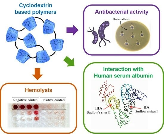
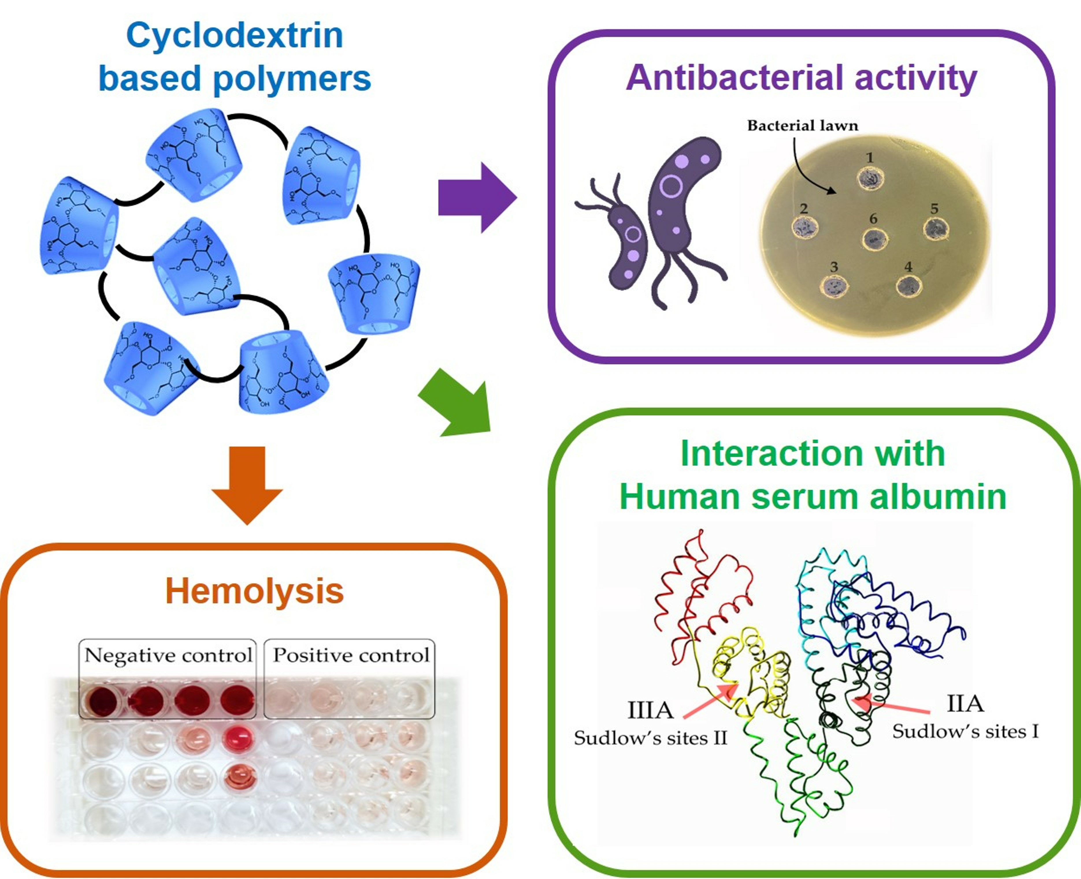
In Vitro Biological Properties of Cyclodextrin-Based Polymers: Interaction with Human Serum Albumin, Red Blood Cells and Bacteria

by
Linara R. Yakupova
,
Anna A. Skuredina
*,
Tatina Yu. Kopnova
and
Elena V. Kudryashova
Chemistry Department, Lomonosov Moscow State University, 119991 Moscow, Russia
*
Author to whom correspondence should be addressed.
Polysaccharides 2023, 4(4), 343-357; https://doi.org/10.3390/polysaccharides4040020
Submission received: 1 August 2023 / Revised: 10 September 2023 / Accepted: 23 September 2023 / Published: 28 September 2023

Abstract

:
The aim of this work was to investigate the physico-chemical and biological properties of cyclodextrin-based polymers by the example of interaction with human serum albumin, erythrocytes, and bacteria to understand the prospects of their application as drug delivery systems. We synthesized polymers based on one of cyclodextrin derivatives with nonpolar (-CH3) or polar (-CH2CH(OH)CH3) substituents by crosslinking with 1,6-hexamethylene diisocyanate or succinic anhydride. The polymers form particles with an average size of ~200 nm in the aqueous solutions; their structures were confirmed by FTIR and 1H NMR. Cyclodextrin derivatives and their polymers did not affect the secondary structure content of human serum albumin, which might mean a mild effect on the structural and functional properties of the main blood plasma protein. Polymers extract drug molecules from albumin + drug complex by 8–10%, which was demonstrated using ibuprofen and bromophenol blue as model bioactive molecules for site I and site II in protein; thus, the nanoparticles might slightly change the drug’s pharmacokinetics. Using the hemolysis test, we found that polymers interact with red blood cells and can be assigned to non-hemolytic and slightly hemolytic groups as biocompatible materials, which are safe for in vivo use. The cyclodextrins and their polymers did not extract proteins from bacterial cell walls and did not demonstrate any antibacterial activity against Gram-positive and Gram-negative strains. Thus, the cyclodextrin-based polymers possess variable properties depending on the substituent in the monomer and linker type; demonstrated biocompatibility, biodegradability, and negligible toxicity that opens up prospects for their application in biomedicine and ecology.

Graphical Abstract

1. Introduction

Polymer carriers are widely used for the delivery of small-drug molecules. The designed carriers provide a multifunctional platform to enhance the treatment efficacy and reduce the adverse effect risks by the controlled drug release, increased solubility and stability, improved bioavailability, reduced toxicity, etc. The application of such polymers in drug delivery obviously has revolutionized the field of medicine and biotechnology [1]. Nevertheless, researchers continue to advance the existing carriers as well as provide novel ones to respond to modern society’s issues.
Polymers used in drug delivery possess a number of different physico-chemical and biological properties due to various paths of synthesis, types of linkers, and other conditions [2]. For the development of beneficial carriers with specific properties and functions, oligosaccharides torus-shaped cyclodextrins (CDs) are frequently used. Among other drug delivery systems, CDs stand out due to their biocompatibility, low cost, numerous derivatives, and ability to form non-covalent complexes with “host” molecules. CDs encapsulate a whole hydrophobic drug or its nonpolar fragments into the cavity, protecting them from degradation and improving their solubility and bioavailability [3,4].
CDs are commonly used in the design of various polymeric structures such as crosslinked CD networks [5], which are also known as CD-nanosponges [6], CD-based polyrotaxanes [7,8], and CD-grafted polymers and CD-based multi-arm polymers [9,10]. CD-based polymers are superior to native CDs as drug carriers due to their variable molecular structure, formation of supramolecular architecture, and unique properties. Moreover, CD-based polymers can be appropriately constructed to demonstrate biocompatibility, biodegradability, responsiveness to stimuli, and targeting specific organs. These characteristics are optimal for the design of highly effective drug delivery systems [1].
Crosslinked CD (CDpols) are particularly fascinating among all CD-based polymers because they contain two drug-binding sites: the hydrophobic cavities of CDs and the functional groups of the linker. The latter depends on the type of crosslinker. Authors use bi- or multifunctional epoxy, diisocyanate, and anhydrous compounds [1,6]. The formation of a polymer matrix provides sustained drug release that might contribute to the reduction of the drug’s dose, optimizing the drug regimen, and reducing side effects. Hydrophilic CDpols can enhance drug absorption through biological barriers in immediate-release dosage forms, while hydrophobic polymers are used to formulate sustained-release dosage forms for water-soluble drugs, including proteins and peptides [6].
CDpols are really promising as they could increase binding constants between drug molecules and carriers compared to free CDs [11,12]. For instance, Wankar et al. showed that ethionamide binds to polymeric β-CD nanoparticles (produced by β-CD crosslinking under strongly alkaline conditions in the presence of epichlorohydrin) two times stronger than to free β-CD. The encapsulation of ethionamide into polymeric β-CD nanoparticles inhibits drug crystallization and enhances its solubility [13]. Moreover, recently we have shown that polymers based on sulfo butyl β-CD crosslinked with 1,6-hexamethylenediisocyanate increase moxifloxacin affinity up to 100 times compared to free CD derivatives [14]. The increase in binding constants might be dramatically important for drug forms used for intravenous and oral administration as the significant dilution of the system is critical for non-covalent complexes.
Since the rising interest in CDpols as drug delivery systems, it is crucial to thoroughly examine the carrier’s safety and its interaction with different biological macromolecules. Recently, we studied CDpols in terms of structural aspects and their interactions with fluoroquinolone drugs [14]. Here, we focus on intravenous administration since blood environments are more susceptible to the integration of foreign molecules compared to acidic conditions in oral administration. We are interested in whether CDpols might interact with human serum albumin (HSA)—the main bloodstream protein. HSA is a large molecule that regulates plasma oncotic pressure and fluid distribution between body compartments [15]. HSA has a high capacity for binding to various compounds, making it an important carrier and depot for endogenous and exogenous substances. It plays a significant role in the metabolism and modification of ligands, as well as the detoxification of potential toxins. Thus, CDpol interaction with HSA might affect the drug’s pharmacokinetics [16]. It is essential to ensure CDpols’ safety, so we examined their interaction with erythrocytes (hemolysis process). We also investigated the antibacterial properties of CDpols on Gram-positive and Gram-negative bacterial strains to ensure that the carrier might be antimicrobial and contribute to the drug’s action [17].
Thus, we conducted comprehensive research on CDpols’ biological properties (interaction with HSA and bacteria, the possibility of drug extraction from the drug-HSA complex, and the hemolysis process). One can successfully develop safe and efficient drug delivery systems based on CDs.

2. Materials and Methods

2.1. Materials

2-hydroxypropyl-β-cyclodextrin (HPCD) and methyl-β-cyclodextrin (MCD) with the substitution degree of hydrogen in one D-glucopyranose link equal to 0.5–1.3 and 1.5–2.1 respectively, monosodium phosphate (NaH2PO4), sodium chloride (NaCl), succinic anhydride, 1,6-hexamethylenediisocyanate, dimethyl sulfoxide (DMSO) and human serum albumin (HSA) were obtained from Sigma-Aldrich (St. Louis, MO, USA). Hydrochloric acid (HCl) was purchased from Reakhim (Moscow, Russia). Tablets for the sodium-phosphate buffer solution (pH 7.4) were purchased from Pan-Eco (Moscow, Russia).

2.2. Methods

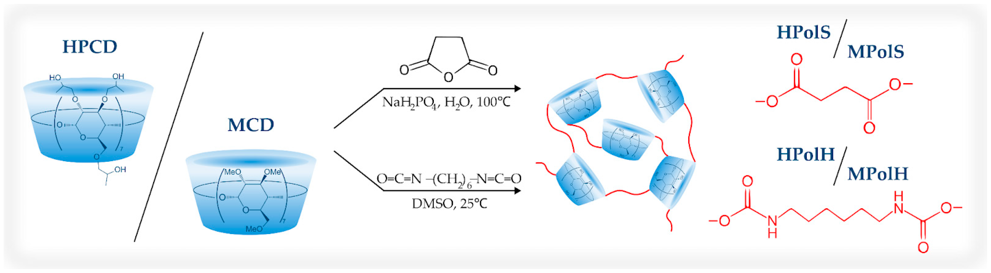
2.2.1. Synthesis of HPCD and MCD Polymers Linked by Succinic Anhydride or 1,6-Hexamethylene Diisocyanate

The procedure for synthesizing polymers with succinic anhydride as a linker was carried out according to our previous work [18]. Briefly, 300 mg of 2-hydroxypropyl-β-cyclodextrin or methyl-β-cyclodextrin were dissolved in distilled water. Next, 75 mg of catalyst (NaH2PO4) was added to the CD’s solution (60 mg/mL) and heated to 100 °C. An aqueous solution of the linker was added dropwise to achieve a molar ratio of 1:8 (for both CDs). The final solutions (6 mL) were stirred for 1.5 h with heating at 100 °C.
The synthesis of CD polymers using a 1,6-hexamethylene diisocyanate linker was conducted according to one of our early works [14]. Initially, HPCD’s or MCD’s warm aqueous solutions (100 mg/mL) were mixed with an estimated amount of DMSO (volume ratio H2O: DMSO was 1:1 in the final solution). Then, the required amount of linker solution in DMSO was added dropwise (molar ratio CD: linker = 1:3), and the mixtures were stirred for an hour.
The purification of MCD and HPCD polymers from unreacted reagents, organic solvent, and catalyst was performed by dialysis using a 3.5 kDa membrane (Serva, Heidelberg, Germany) for 4 h at room temperature with regular external solution replacement using distilled water (1.2–1.5 L in total). Then, the samples were dried in sterile Petri dishes for 24 h at 25 °C on a thermo-controlled shaker-incubator (Biosan, Riga, Latvia).
The yield of the polymer synthesis reaction was determined by dividing the mass of the product dried at 120 °C overnight by the mass of the initial reagents and multiplied by 100%.
The concentration of CD tori in the polymers was determined using FTIR spectroscopy and a calibration curve based on 1032 cm−1 and 1042 cm−1 for HPCD and MCD, respectively, related to the C-O-C bond vibration of cyclodextrin.

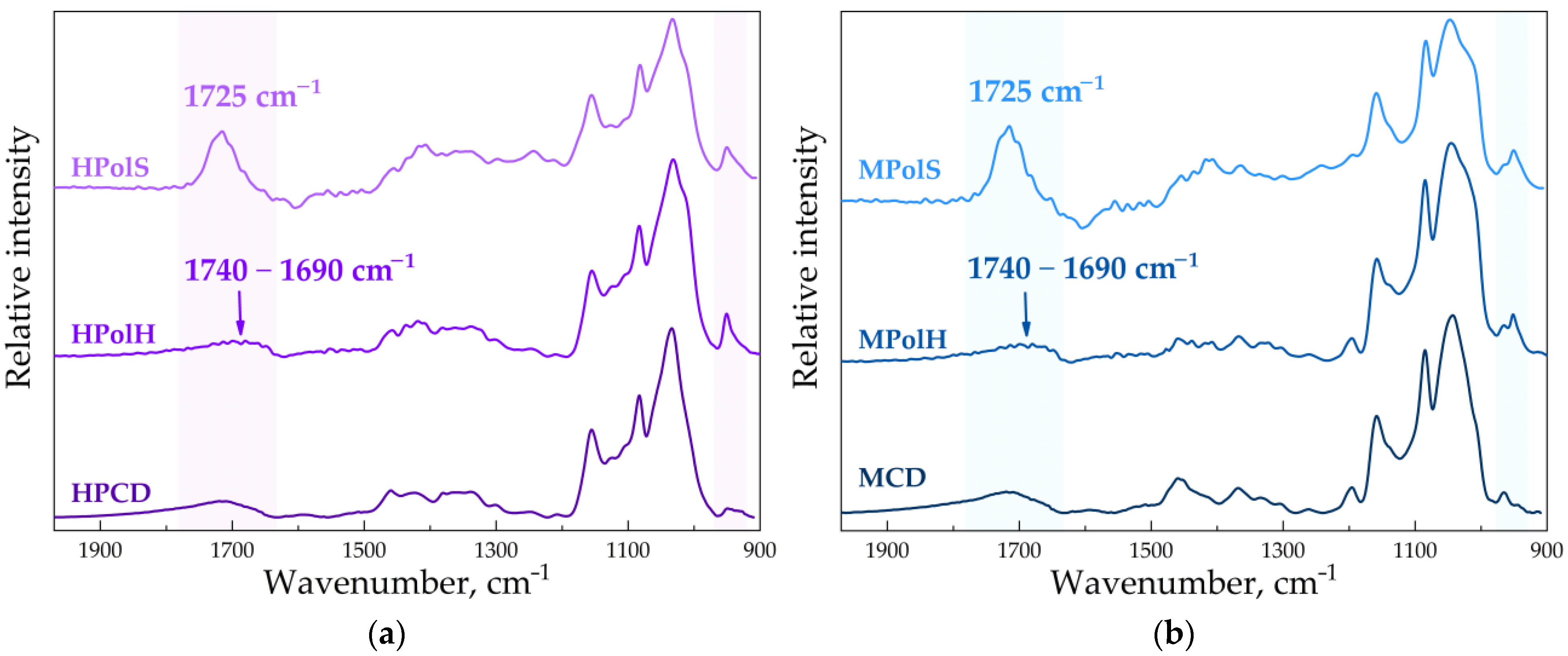
2.2.2. FTIR-Spectroscopy

To obtain the FTIR spectra, a Tensor 27 spectrometer from Bruker in Ettlingen, Germany, was utilized. The spectrometer contained an attenuated total reflection cell from Bruker in Ettlingen, Germany, which held a ZnSe single-flection crystal. The spectrometer had an MCT detector cooled with liquid nitrogen and was equipped with a thermostat from Huber in Offenburg, Germany. FTIR spectra were recorded three times with a resolution of 1 cm−1 and 70 scans each, in the range of 2200–850 cm−1, using a sample volume of 40 μL at room temperature (22 °C). The system was purged with dry air using an air compressor from Jun-Air in Munich, Germany, and a background spectrum of buffer solution was obtained similarly. The spectra were analyzed using Opus 7.0 software from Bruker in Ettlingen, Germany.
The secondary structure of both the HSA and HSA-CDpols’ complexes was determined by analyzing the spectra in the range of 1700–1600 cm−1. The primary bands were detected using the second derivative of the spectrum, and the spectra were deconvoluted using the Levenberg–Marquardt algorithm. The residual RMS error was found to be below 0.00001.
The determination of the degree of substitution (DS) by FTIR spectroscopy was conducted as DS = S1/S2, where S1 is the area of the appeared absorbance peaks that corresponds to the linkers (~1725 cm−1) and S2 is the area of the peak corresponding to the monomer (1033 cm−1 for HPCD and cm−1 for MCD). The areas were calculated using Opus 7.0 software.

2.2.3. UV-Spectroscopy

UV spectra were recorded using the Ultrospec 2100 pro equipment from Amersham Biosciences in the UK. The spectra were obtained in a quartz cell with an optical path length of 1 cm from Hellma Analytics in Jena, Germany, within the wavelength range of 200–450 nm.

2.2.4. Fluorescence Spectroscopy

Fluorescence measurements were performed using a Varian Cary Eclipse spectrophotometer (Agilent Technologies, Santa Clara, CA, USA). The emission spectra of HSA were recorded at a temperature of 37 ± 0.1 °C in a 10 mm thick quartz cuvette. The excitation wavelength was set to 280 nm, and the emission range was from 290 to 555 nm with a step size of 1 nm. The protein concentration in all samples was maintained at 0.02 mM in a sodium-phosphate buffer solution with a pH of 7.4.

2.2.5. NMR-Spectroscopy

The 1H-NMR spectra were obtained using a Bruker Avance 400 spectrometer (Reinshtetten, Germany) operating at a frequency of 400 MHz. The spectra were recorded in D2O and referenced to HDO at 4.75 ppm. Approximately 10–15 mg of the sample was utilized for each spectrum. For DS determination, we normalized the spectra by the H1 peak and compared the integral intensities of the peaks in accordance with the number of protons in the monomer and linker.

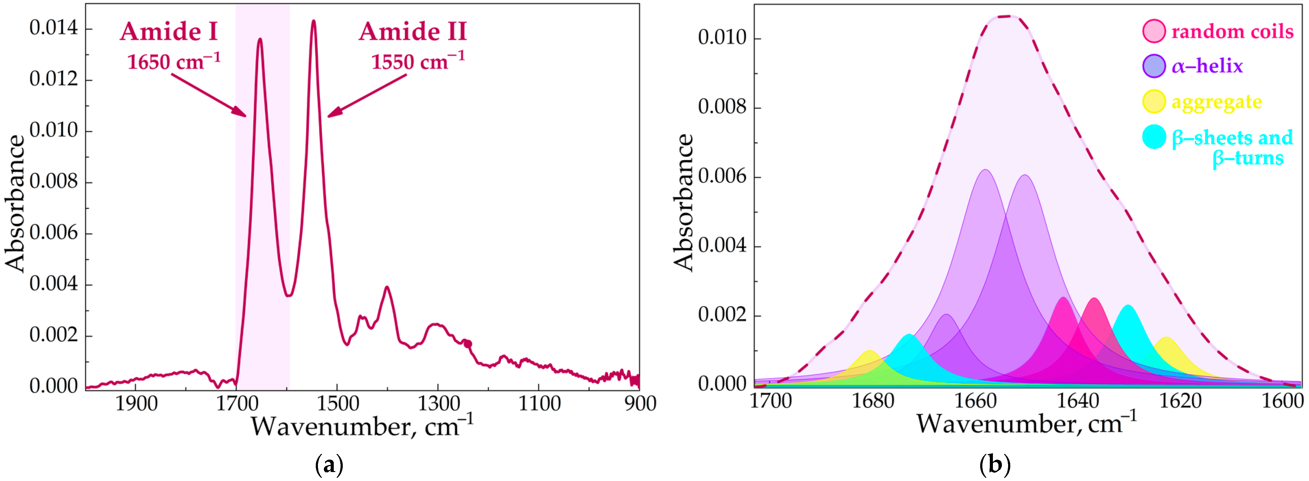
2.2.6. Circular Dichroism Spectroscopy

Circular dichroism spectra were registrated using a J-815 spectrometer from the company “Jasco” (Tokyo, Japan), which was equipped with a thermostatable cell. The measurements were conducted at 25 °C in a quartz cuvette with a path length of 1 mm. The wavelength range was set from 200 to 260 nm. The spectra were scanned five times at an interval of 1 nm, and the concentration of human serum albumin was 0.02 mM.

2.2.7. Dynamic Light Scattering (DLS)

The ζ-potentials and hydrodynamic diameters of the CDpols were determined using the Zetasizer Nano S “Malvern” (4 mW He-Ne laser, 633 nm; Malvern, UK) at 25 °C. Each sample was analyzed three times. The data are presented with the standard deviation.

2.2.8. Nanoparticle Tracking Analysis (NTA)

The sizes of the obtained polymers were also determined by NTA (Nanosight LM10-HS, Nanosight Ltd., Salisbury, UK). The aqueous solution of each polymer was diluted 10–500 times to achieve satisfactory particle concentration (~108 particles/mL) with purified water (Milli Q).

2.2.9. Atomic Force Microscope

The size and homogeneity of the obtained samples were controlled using the Atomic Force Microscope NTEGRA II (NT-MDT, Zelenograd, Moscow, Russian Federation).

2.2.10. The CDpols Stability Studies

CDpols stability studies at pH 7.4 (that model blood plasma conditions) were carried out using the sample’s aqueous solution stored at 37 °C for 48 h. The FTIR spectra of the initial solution were compared to the FTIR spectra of the same solution stored for 6, 12, 24, and 48 h.
We also studied the enzymatic degradation of CDpols by model enzymes trypsin and chymotrypsin that might hydrolase particles. The proteins were preliminarily adsorbed in a 96-well plate in various quantities: 10, 50, or 100 µL of enzymes (3 mg/mL, pH 4.0; trypsin or a combination of trypsin with chymotrypsin) were applied into the wells, and incubated for 40 min (T = 25 °C, 120 rpm). The amount of the adsorbed enzyme was determined by comparing the initial enzyme concentration with the concentration of unabsorbed protein (UV-spectroscopy, λ = 280 nm). The glutaraldehyde (3 mg/mL, pH = 8.5) was added to the molar ratio enzyme: glutaraldehyde = 1: 2, and the planchet was incubated for another 1 h (T = 25 °C, 120 rpm). We washed the wells three times with a sodium-phosphate buffer solution (pH 7.4). After 200 µL of CDpol was added to the wells of the 96-well plate, the system was incubated at T = 37 °C (120 rpm) for 24 h. We measured the turbidity (600 nm) and FTIR spectra to investigate the changes in the samples that might appear due to the CDpols hydrolysis.

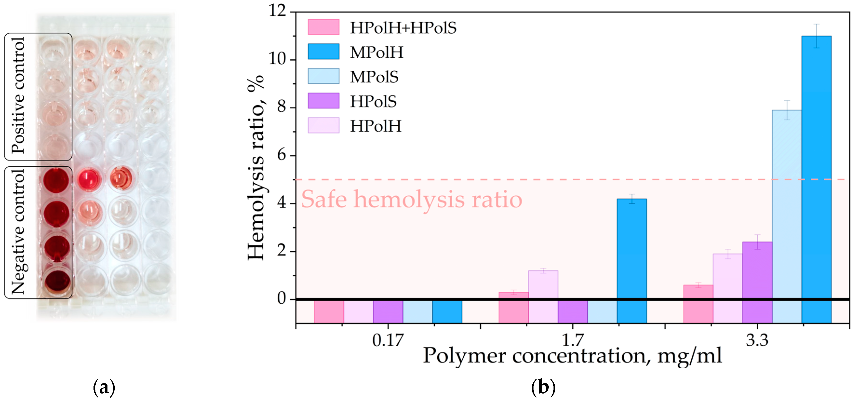
2.2.11. Hemolysis Assay

Hemolysis ratios of the polymers were determined using purified erythrocytes from human blood, provided voluntarily by one of the article’s authors. Then, 3 mL of blood stored in a tube with 0.3 mL 2% EDTA was centrifuged at 1000 rpm for 10 min. Then, the erythrocyte mass was washed with 0.15 M NaCl solution and centrifuged again. These steps were repeated 4 times. Then, 40 µL of erythrocyte suspension in 0.15 M NaCl was added to the 40 µL of polymer solution in the same saline solution in the required concentrations (final concentrations were 0.17, 1.70, and 3.30 mg/mL for each polymer). For 100% hemolysis (negative control), 6 µL of 2% Triton X-100 was added to 200 µL erythrocyte solution, and for 0% hemolysis (positive control), 40 µL of the saline solution was added. The samples were incubated for 2 h at 37 °C, then centrifuged at 1000 rpm for 10 min. The absorbance of the supernatant solutions was measured by a UV spectrophotometer at the wavelength 540 nm to determine the amount of released hemoglobin. Hemolysis ratios were calculated using Formula (1):
H e m o l y s i s r a t i o % = A s a p m l e A ( p o s i t i v e ) A n e g a t i v e A ( p o s i t i v e ) × 100

2.2.12. In Vitro Studies

The overnight culture of Escherichia coli ATCC 25922 and Bacillus subtilis ATCC 6633 in Luria Bertuni medium was prepared and diluted to a 0.5 McFarland standard.
For antibacterial testing, the agar well diffusion method was used. For this, a nutrient medium was poured into petri dishes, on which overnight bacterial cultures were applied and distributed with a spatula. Then, six wells with a diameter of 9 mm were made in the agar surface, and 50 μL of the sample (sterile buffer as negative control) was added to each well. The Petri dishes were then incubated for 24 h at 37 °C.
To explore the polymer’s effect on the bacterial cell wall, microorganisms’ cultures were washed from the nutrient medium with a sodium-phosphate buffer solution using centrifugation (9000 rpm for 5 min). Then 300 μL of CDs or their polymer solutions (2 mg/mL) were added to the washed cell mass; the samples were incubated for 1 h at 37 °C. UV spectra of the supernatant solutions were measured.

3. Results and Discussion

The great interest in CDpols requires safety studies and the investigation of their interaction with biological macromolecules. Therefore, the aim of this study was to examine the physico-chemical and mainly biological characteristics of CDpols, in particular, their effect on human serum albumin, red cells, and bacteria.
We chose methyl-β-cyclodextrin (MCD) and 2-hydroxypropyl-β-cyclodextrin (HPCD) as monomers for CDpols synthesis. These CD derivatives are widely applied in medicine due to their high solubility and availability [19]. Succinic anhydride (S) and hexamethylene diisocyanate (H) were selected as crosslinkers because of their capacity to form biodegradable covalent bonds with the CDs (ester and urethane correspondingly). These linkers provide additional fragments in the polymer network with specific physico-chemical properties: hydrophobic pores in the case of H and charged unreacted carboxylic groups in the case of S.
The CDpol synthesis involved the reaction of the CDs with the crosslinkers, according to Figure 1. The reaction parameters, such as temperature, reaction time, and molar ratio of reactants, were chosen in accordance with our previous research [14,18] to achieve the desired polymer properties that will be discussed in detail below. The yields of polymer synthesis reactions were approximately 52 ± 10 and 82 ± 14 % for the polymers linked by succinic anhydride and 1,6-hexamethylene diisocyanate correspondently. The polymers’ abbreviations were established as follows: the first letter corresponds to the type of monomeric cyclodextrin (MCD or HPCD correspond to M and H, respectively), followed by the designation of the polymer (Pol), and the last letter indicating the linker used (succinic anhydride or 1,6-hexamethylene diisocyanate correspond to S and H, respectively).

3.1. Physico-Chemical Properties of CD-Based Polymers

As expected, synthesized CDpols exhibited different physico-chemical characteristics depending on the type of crosslinker used and the CD’s substituent (Table 1). The introduction of succinic anhydride (S) led to the production of a transparent glassy polymer with good solubility in water. On the other hand, the hexamethylene diisocyanate (H) led to a white powder formation with limited solubility in aqueous solutions, which was expected according to higher hydrophobicity of methylene groups in H compared to S’s multiple -COO.
The CDpol’s parameters were determined using various methods, including Fourier-transform infrared spectroscopy (FTIR), nuclear magnetic resonance (NMR) spectroscopy, dynamic light scattering (DLS), and nanoparticle tracking analysis (NTA). Using the latter method, the ζ-potentials and hydrodynamic diameters of each polymer were obtained. The CDpols form particles with an average size ranging from 100 to 300 nm (Figure S1). The particle sizes and homogeneity were also confirmed with AFM.
The ζ-potential value is an important parameter that provides valuable information about the surface charge of colloidal particles. It uncovers the potential difference between the dispersion medium and the stationary layer of fluid surrounding the particle. This value dramatically depends on different factors such as pH, ionic strength, and surface functionalization. In colloidal systems, the ζ-potential value is used to predict the stability and behavior of particles, as described in the work of Midekessa et al. [20].
When H was used as the crosslinker, CDpols exhibited a strong positive ζ-potential. We suppose this effect is explained by the hydrolysis of unreacted –N=C=O groups into primary amines, demonstrated earlier in the articles of Freichels et al. and Paiphansiri et al. [21,22]. CDpols synthesized using S demonstrated ζ-potential values close to neutral. The MCD-based polymer exhibits a more significant positive ζ-potential in comparison to HPCD. This is likely attributed to the reduced number of hydroxyl groups available in MCD for the linker and suggests a preference for the hydrolysis of linker end groups.
As known, particles with high ζ-potential values (either positive or negative) tend to repel each other, resulting in a stable dispersion. On the other hand, particles with low ζ-potential values are more likely to aggregate and form larger clusters. Thus, aggregation is not inherent to polyurethane nanoparticles, whereas polymers with an S linker might be prone to aggregation.

3.2. The Structure of CDpols

To investigate the chemical structure of the CD-based polymers, we applied FTIR and 1H NMR spectroscopy. For CD derivatives in the ATR-FTIR spectra, we observed typical absorption bands in the range of 1100−1000 cm−1 that correspond to the C-O-C glycoside bonds (Figure 2) [23]. It is worth stressing that in the CDpols’ case, the expressed shoulder appears in this band, which indicates a voltage in the CD’s toruses. The formation of polymers linked by S resulted in the appearance of absorption bands at 1725 cm−1 and 1226 cm−1 that can be attributed to the C=O bonds in carboxylic and ester groups, as well as the C-O-C bonds in ester groups, respectively [24].
For the H crosslinker, the formation of a polyurethane bond was expected, which would be indicated by a band around 1740–1700 cm−1 in the FTIR spectra corresponding to the oscillation of the C=O group [25]. Indeed, for this type of polymer (HPolH and MPolH), a band in the region of 1740–1690 cm−1 was observed. Additionally, a band at 951 cm−1 appeared, indicating the presence of a C–N bond in primary amines [26]. This functional group may have formed through the hydrolysis of unreacted isocyanate groups in aqueous media, as previously demonstrated for crosslinked hydroxyethyl starch nanocapsules [27]. As discussed earlier, the positive ζ-potential of the polymers linked by 1,6-hexamethylenediisocyanate confirms the presence of -NH2 groups on the surface of the particles.
The 1H NMR spectra of CDpols provide evidence for the formation of the polymers, as indicated by the presence of signals corresponding to the D-glucopyranose units (HPCD or MCD) and the linkers (Figure S2). Briefly, the multiplets at δ 3.40–4.11 ppm corresponds to H2–H5 of the D-glucopyranose units, while the signals at δ 5.01–5.31 ppm are attributed to H1 of the D-glucopyranose units. We found these signals in all 1H NMR spectra of CD polymers. For the HPCD-based polymers, the doublets at δ = 1.13 ppm with a coupling constant of 6.2 Hz are observed, which corresponds to the protons of the hydroxypropyl substituent. Furthermore, in the 1H NMR spectra of all CD polymers, the detected signals corresponded to linkers. This suggests that the CD polymers are composed of repeating units of the CD monomer and the linker molecule.
We obtained the degree of substitution (DS) by NMR and FTIR data (Table S1). The DS values range from 0.5 to 3.5. The polymers obtained using succinic acid demonstrate three times higher DS values compared to CDpols crosslinked with 1,6-hexamethylendiisocyanate.
Moreover, we conducted the CDpols stability studies at physiological pH and in the presence of model enzymes (trypsin and chymotrypsin). During the storage of all CDpols in sodium-phosphate buffer solution (pH 7.4) for 48 h, we did not detect any changes in the FTIR spectra of the samples. This finding indicates that CDpols are stable and do not hydrolyze. Furthermore, the CDpols storage with the enzymes (trypsin and chymotrypsin) demonstrated that H crosslinked polymers undergo hydrolysis by less than 20% per day (pH = 7.4, T = 37 °C).
Overall, the FTIR and NMR spectroscopy analyses provide strong evidence for the formation of CD-based polymers (polyurethanes and polyesters), highlighting their diverse characteristics that might impact their interaction with biological structures.

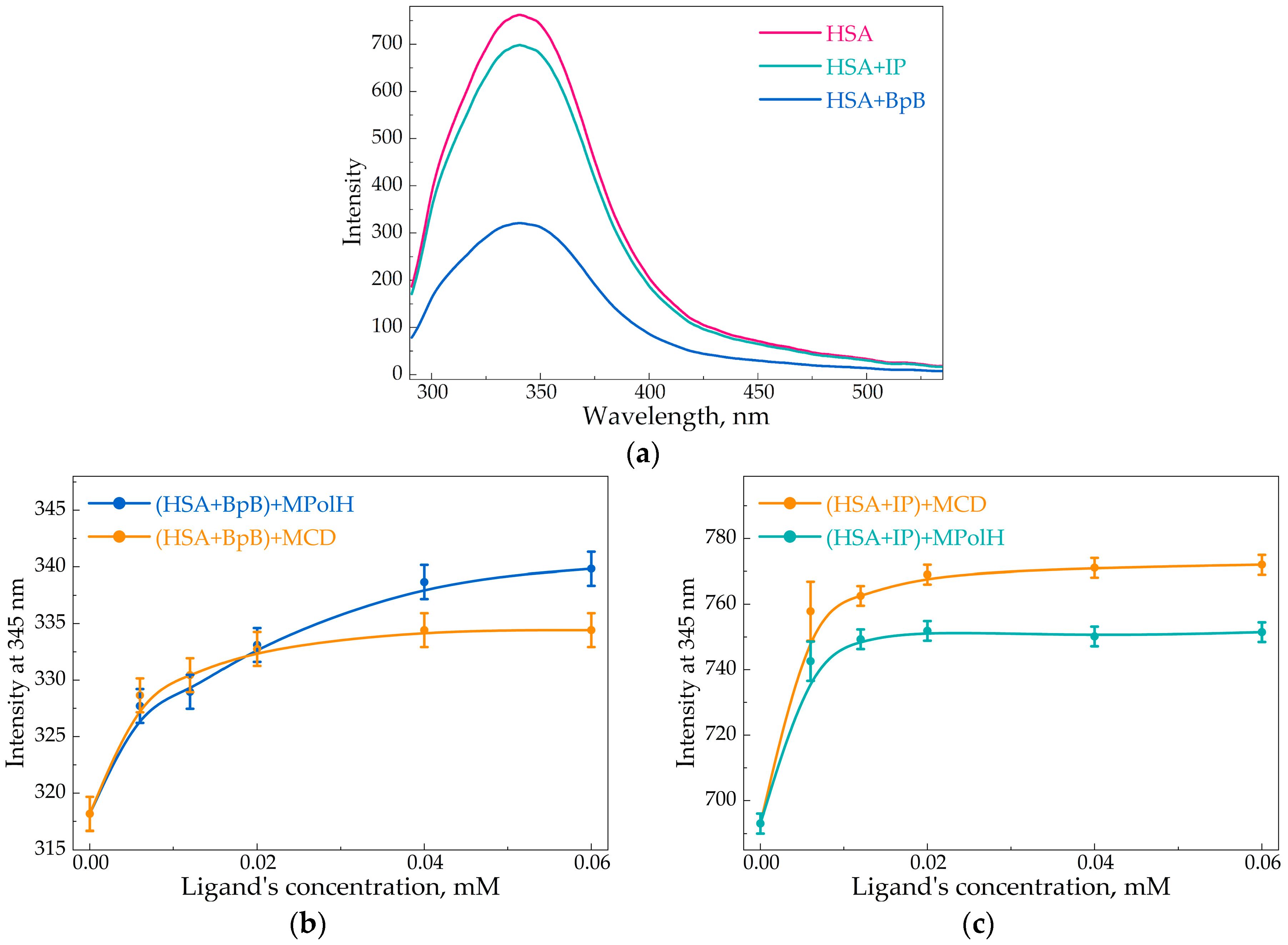
3.3. Interaction of HSA with CD-Based Polymers

Several studies have shown that the formation of polymer-HSA complexes or adsorption of HSA on the surface of polymers led to changes in the secondary structure of albumin [28,29]. For example, Suvarna et al. studied how nanoparticles fabricated using low molecular weight chitosan oligosaccharide (∼20 kDa) and high molecular weight chitosan (∼85−90 kDa) influence the secondary structure of HSA [30]. They showed that larger nanoparticles cause weaker protein conformational changes as well as nanoparticles obtained from high molecular weight chitosan. Treuel et al. examined how polystyrene nanoparticles (PSN) interact with serum albumin [31]. The PSN were slightly negatively charged and caused almost no change in the secondary structure of the albumin until PSN’s concentration reached 1 × 10¹¹ particles per mL. The increase in the PSN’s concentration to 2.4 × 10¹¹ NPS/mL leads to a complete loss of alpha helices in the HSA’s secondary structure. The results are confirmed by Ragi et al., who studied poly(ethyleneglycol) (PEG) [32]. The authors reported that 0.1 mM PEG causes no alterations in the protein secondary structure, while at high concentration (1 mM), the reduction of α-helices content from 59 to 53% takes place. To sum up, high concentrations of polymers could dramatically alter HSA’s secondary structure (that might cause the changes of albumins’ functional properties), while small concentrations of polymers do not cause significant changes. Thus, we expected that our CDpols at concentration 0.1 mM would not notably alter HSA’s secondary structure.
We applied FTIR spectroscopy to examine the alterations in albumin’s secondary structure content after the formation of CDpol-HSA complexes (Figure 3a,b). We consider the Amide I band in the FTIR spectra since it is sensitive to changes in the protein secondary structure [32,33]. The Amide I band (1600–1700 cm−1) represents the oscillation of ν(C=O)~80% and ν(C-N)~15%. By deconvoluting the Amide I band, we were able to determine the content of α-helix, β-structures (β-turns and β-sheets), and random coils, providing valuable insights into the HSA’s secondary structure changes (Table 2).
For all CDpol-HSA complexes, we observed almost no changes in the HSA’s secondary structure content (Table 2) as well as CDs. We determined only a slight α-helix content decrease and an increase in the proportion of β-structures. Similar results were achieved when the polymer carbon nanodots interacted with HSA [29] or tyrosine-modified polyethyleneimines adsorbed on the surface of HSA [28]. They reported that for linear polymers, the changes in the secondary structure of HSA are minimal, while for branched polymers, they could be quite significant.
Moreover, the secondary structure of HSA was analyzed using circular dichroism to investigate the impact of CDpols. The data confirmed the results obtained by the FTIR-spectroscopy (Table 2).

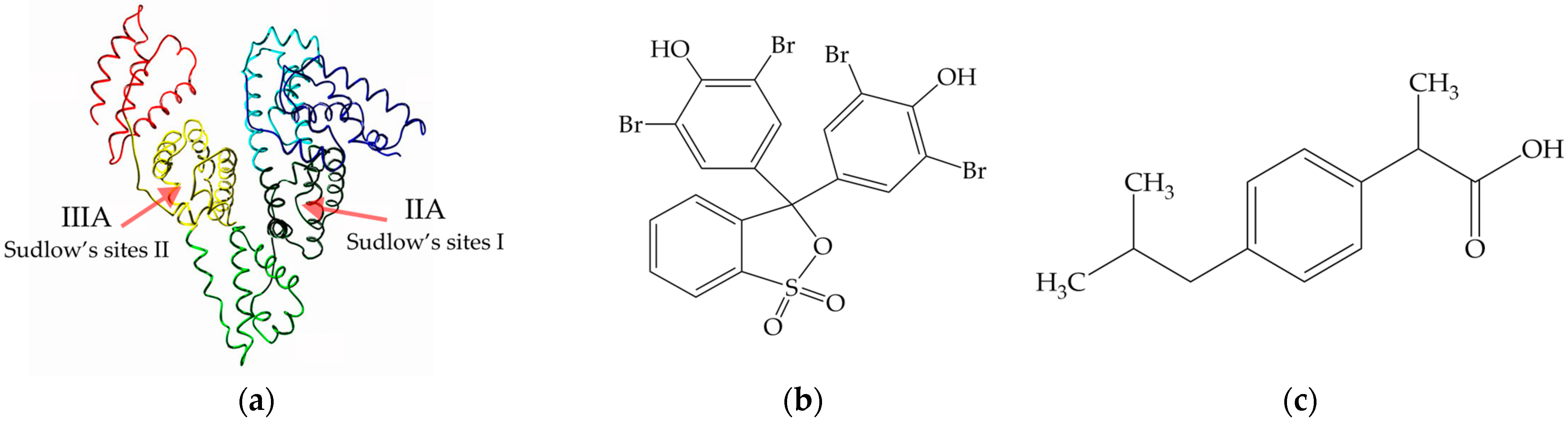
3.4. Influence of CD-Based Polymers on Binding HSA with Drugs

Next, we investigated if CDpols cause the dissociation of HSA-drug complexes, as this effect might influence the drug’s pharmacokinetics. HSA has fluorescence spectra corresponding to the presence of specific amino acids, such as tryptophan, phenylalanine, and tyrosine [34]. We applied a fluorescence study to examine the protein’s interactions with model drug molecules and the effect of CDpols on these interactions. First, we found that the investigated polymers based on CD did not significantly affect the intensity of HSA’s fluorescent emission (changes in the order 2%) in the studied concentration range up to a molar excess of polymer equal to 10.
The majority of ligands bind in HSA in the hydrophobic cavities of albumin’s subdomains IIA and IIIA (Sudlow’s sites I and II, respectively) [16,35]. These sites are known to have a high affinity for drugs and are critical for drug transport and distribution in the body. Site II is one of the most active regions for the binding of many ligands, such as ibuprofen (prototypical ligand), digitoxin, and tryptophan [36]. Site I prefers to bind bulky heterocyclic anions, for instance, warfarin [37]. Warfarin shares this binding site with a range of other drugs, including indomethacin, phenylbutazone, and tolbutamide, and competing with them for binding to HSA. Some bioactive molecules demonstrate almost equal distributions between both binding sites in the IIA and IIIA subdomains, such as aspirin and levofloxacin (Figure 4a) [36].
The use of such specific molecules (markers) allows the identification of binding sites of other drugs: since the marker interacts with a specific site, the studied drug can either compete with the marker for the same site or interact freely with another, which is reflected in the values of dissociation constants [38]. Furthermore, markers can be applied as models to study the effect of drug delivery systems on the interaction between protein and drug molecules. In this work, the markers bromophenol blue (BpB) and ibuprofen (IP) were used, which are specific to binding sites I and II, respectively (Figure 4b,c) [39].
In the presence of markers, quenching of the HSA’s fluorescence emission is observed, which indicates the binding process (Figure 5a) [40]. The aim of this experiment was the investigation of the polymers’ effect on the binding of drugs to HSA by adding them to the HSA-marker complex. We were interested in whether CDpols would increase the protein’s fluorescence emission, indicating the drug’s extraction from the albumin. The results demonstrate a slight increase (about 8–12%) in fluorescence intensity upon the addition of all types of polymers or corresponding CD monomers, suggesting that the binding affinity between the drug and HSA is weakened due to polymer and protein competition for drug binding. The phenomenon of drug extraction from the protein binding site is more pronounced in the case of ibuprofen for the monomer CD and in the case of bromophenol blue for CDpols. CD monomer is a small complexing agent compared to CDpols, which can lead to a stronger interaction with the ibuprofen molecule due to the well-correlated sizes. Hergert et al. showed that ibuprofen forms the inclusion complex with CD, which is characterized by a binding constant of 1.4 × 10⁴ M−1 [41]. On the contrary, the inclusion complexes of bromophenol blue with CD are not described in the literature. Thus, we suggested that the changes in the fluorescence intensity in ternary systems HSA-ibuprofen-CD (compared to HSA-bromophenol-CD) should be more significant as CD could form an inclusion complex with ibuprofen. It was proved in our experiments (Figure 5b,c). Figure 5b shows that bromophenol blue induces an increase in HSA’s emission intensity by 5%, while ibuprofen increases the intensity by 12% (Figure 5c). On the other hand, the larger size of the CDpols may provide better placement of the bromophenol blue molecule, which would lead to more efficient extraction. Thus, the presented polymers do not produce significant changes in the extraction of drugs from protein, these findings have important implications for drug development, as they suggest that the use of polymers may alter the pharmacokinetics and efficacy of drugs by affecting their binding to HSA (Figure 5b,c).

3.5. Blood Compatibility of CD-Polymers

The blood compatibility of materials exposed to blood is primarily determined by their potential for hemolysis. In order to assess the damaging effects of particles on red blood cells, we conducted an in vitro hemolysis test on purified erythrocyte mass isolated from human blood. Biomaterials are considered non-hemolytic if they exhibit a hemolysis ratio of 0–2%, slightly hemolytic in the range of 2–5%, and hemolytic at a hemolysis ratio of 5–100% [42,43]. In the present work, a negative control was used for 100% hemolysis and a positive control for 0% hemolysis; Triton X-100 and saline solution were used, respectively (Figure 6a).
The HPolH and HPolS particles can be identified as non-hemolytic up to a concentration of 1.7 mg/mL as well as their combinations in the entire studied concentration range (Table 3). The HPolH and HPolS particles in a concentration of 3.3 mg/mL can be considered as slightly hemolytic. Based on the results, these particles are safe and hemocompatible, making them promising components for the development of drug forms for intravenous administration since biocompatibility is an important characteristic of drug carriers. Polymers based on MCD (crosslinked with succinic anhydride and 1,6-hexamethylenediisocyanate) demonstrate higher hemolysis ratio values. However, it is still safe up to a concentration of 1.7 mg/mL. According to the literature data, some components of human blood, particularly albumins, demonstrate a protective function and significantly reduce the cytotoxic effects of CDs. Thus, it can be expected that due to the protective properties of blood components, hemolysis in vivo for drug delivery systems will decrease [44].

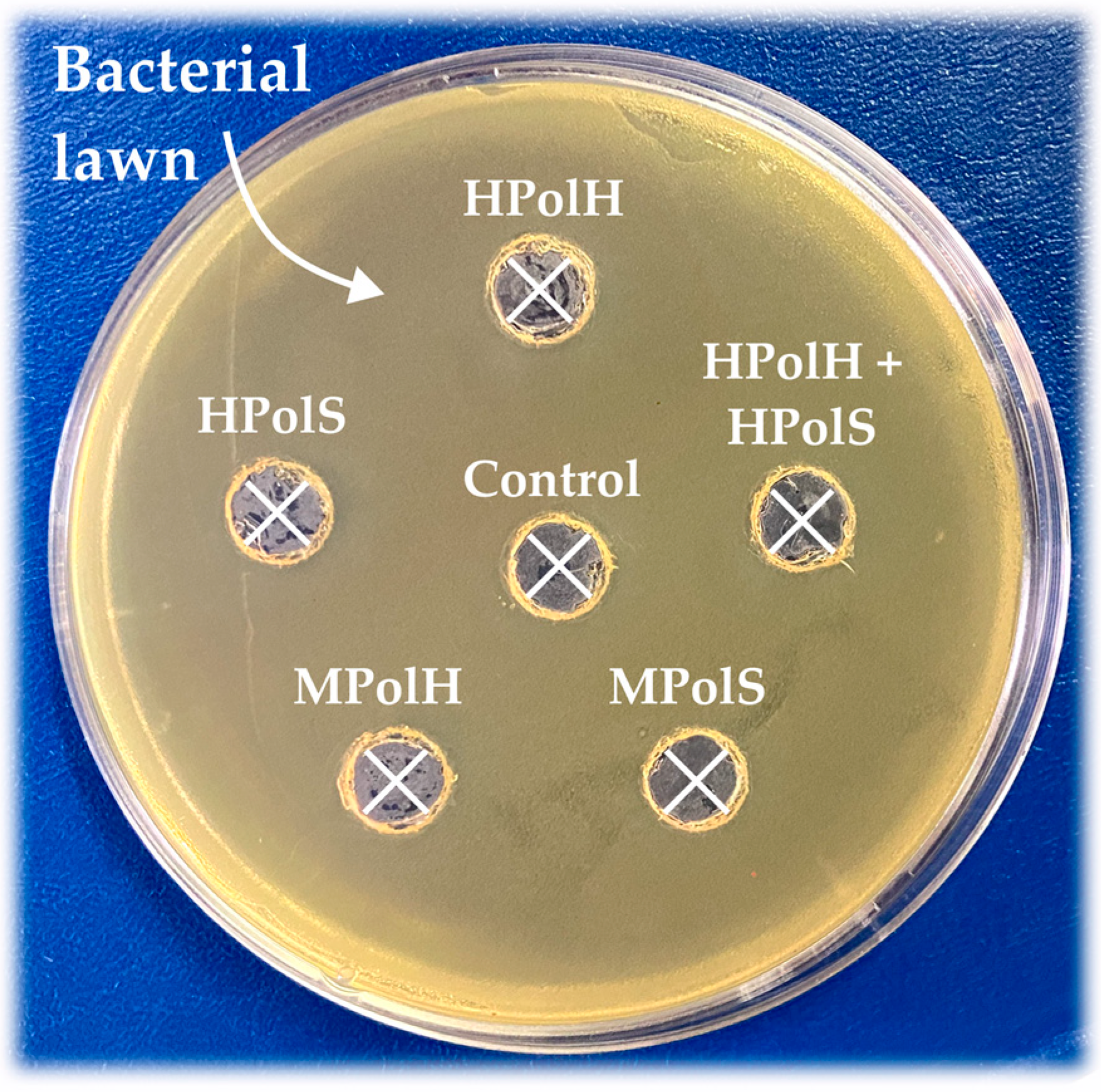
3.6. Antibacterial Activity of CD-Polymers

In this work, the effect of CDpols on bacterial cells was investigated using Gram-negative bacteria Escherichia coli ATCC 25922 and Bacillus subtilis ATCC 6633 using the fast and reliable agar–well diffusion method (Figure 7) [45]. No inhibition zones were found on the Petri dishes around wells with polymer solutions, which indicates that CDpols do not affect the bacterial culture growth up to a concentration of 5 mg/mL, which is really high. These results correlate with our previous studies [17] in which CDs also demonstrated no inhibition zones. The lack of antibacterial activity in CD-based polymers is not crucial for the development of drug delivery systems since many drug carriers do not possess antimicrobial activity.
Moreover, the possibility of interactions between CDpols and the bacteria cell surface was investigated. In our previous work [17,46], we demonstrated that CD-particles adsorb on bacterial cells. Here, we examined whether such interactions would affect surface cell structures, particularly the bacterial cell wall. Since we did not detect any antibacterial activity, it can be concluded that the cell membrane is still intact in the presence of the CDpolymers, and a considerable amount of proteins have not been released into the surrounding environment. Consequently, we used high concentrations of CDpolymers to explore the possibility of disrupting the protein makeup of the cell wall. We incubated the bacteria with the polymers and studied the supernatant using UV spectroscopy. We detected the absence of the peak at 280 nm, indicating no protein extraction from bacteria (up to CDpols concentration 2 mg/mL). Thus, no effect on the bacterial cell wall is confirmed. This characteristic makes them promising for further investigation and development in various biomedical applications, including drug delivery systems and tissue engineering.

4. Conclusions

The development of delivery systems for low-molecular-weight drugs with multifunctional properties that enhance the characteristics of drug molecules remains a relevant task. In this work, polymers based on cyclodextrins were investigated in terms of their potential to be used for intravenous administration. The structures of the polymers (polyurethanes and polyesters) were confirmed by FTIR and 1H NMR; these methods allowed us to characterize the structure of the products fully. Moreover, dynamic light scattering analysis revealed that polymers form particles of approximately 200 nm in aqueous solutions.
CD derivatives and their polymers demonstrate no significant effect on the secondary structure content of human serum, which means the carriers will not alter the protein’s structure or functions. Nevertheless, they might extract bioactive molecules from the albumin + drug complex by 8–12% from both protein binding sites. This data must be considered for further development of drug delivery systems based on cyclodextrin’s polymers, as the finding might lead to drug pharmacokinetics changes. Hemolysis tests uncovered that the particles are non-hemolytic or slightly hemolytic, which is important in vivo use. Cyclodextrins and their polymers do not extract proteins of bacterial cell walls or exhibit any other antibacterial activity mechanism against Gram-positive or Gram-negative strains in vitro. The CD-based polymers appeared to be nontoxic, biodegradable, and biocompatible, which opens up prospects for their application in biomedicine and ecology.

Supplementary Materials

The following supporting information can be downloaded at: https://www.mdpi.com/article/10.3390/polysaccharides4040020/s1, Figure S1: The size distribution of the sample HPolS obtained by NTA in Milli Q water, 25 °C. Figure S2. 1H NMR of HPCD, succinic anhydride, HPolH, HPolS, MPolH and MPolS, D2O, 400 MHz. Table S1: The degree of substitution (DS) for CD-based polymers.

Author Contributions

Conceptualization: A.A.S., L.R.Y. and E.V.K.; Experimental work, data analysis and interpretation: L.R.Y. and A.A.S.; Writing—Original draft preparation: L.R.Y., T.Y.K. and A.A.S.; Writing—Review and editing: A.A.S., L.R.Y. and E.V.K.; Supervision: A.A.S. and E.V.K. All authors have read and agreed to the published version of the manuscript.

Funding

This work was supported by the Russian Science Foundation grant number 22-24-00604.

Institutional Review Board Statement

Not applicable.

Data Availability Statement

Not applicable.

Acknowledgments

The work was performed using equipment (FTIR spectrometer Bruker Tensor 27, Jasco J-815 CD Spectrometer, AFM microscope NTEGRA II) from the program for the development of Moscow State University. The work was supported by the Scholarship of the President of the Russian Federation for young scientists and postgraduates (for A.A.S.). The authors thank Markov P.O. for the NMR spectroscopy experiments and data interpretation.

Conflicts of Interest

The authors declare no conflict of interest.

References

  1. Liu, Z.; Ye, L.; Xi, J.; Wang, J.; Feng, Z. Cyclodextrin polymers: Structure, synthesis, and use as drug carriers. Prog. Polym. Sci. 2021, 118, 101408. [Google Scholar] [CrossRef]
  2. Tiwari, G.; Tiwari, R.; Bannerjee, S.; Bhati, L.; Pandey, S.; Pandey, P.; Sriwastawa, B. Drug delivery systems: An updated review. Int. J. Pharm. Investig. 2012, 2, 2. [Google Scholar] [CrossRef] [PubMed]
  3. Del Valle, E.M.M. Cyclodextrins and their uses: A review. Process Biochem. 2004, 39, 1033–1046. [Google Scholar] [CrossRef]
  4. Stella, V.J.; He, Q. Cyclodextrins. Toxicol. Pathol. 2008, 36, 30–42. [Google Scholar] [CrossRef]
  5. Arslan, M.; Sanyal, R.; Sanyal, A. Cyclodextrin embedded covalently crosslinked networks: Synthesis and applications of hydrogels with nano-containers. Polym. Chem. 2020, 11, 615–629. [Google Scholar] [CrossRef]
  6. Sherje, A.P.; Dravyakar, B.R.; Kadam, D.; Jadhav, M. Cyclodextrin-based nanosponges: A critical review. Carbohydr. Polym. 2017, 173, 37–49. [Google Scholar] [CrossRef]
  7. Hashidzume, A.; Yamaguchi, H.; Harada, A. Cyclodextrin-Based Rotaxanes: From Rotaxanes to Polyrotaxanes and Further to Functional Materials. Eur. J. Org. Chem. 2019, 2019, 3344–3357. [Google Scholar] [CrossRef]
  8. Qie, S.; Hao, Y.; Liu, Z.; Wang, J.; Xi, J. Advances in Cyclodextrin Polymers and Their Applications in Biomedicine. Acta Chim. Sin. 2020, 78, 232. [Google Scholar] [CrossRef]
  9. Seidi, F.; Jin, Y.; Xiao, H. Polycyclodextrins: Synthesis, functionalization, and applications. Carbohydr. Polym. 2020, 242, 116277. [Google Scholar] [CrossRef]
  10. Yao, X.; Huang, P.; Nie, Z. Cyclodextrin-based polymer materials: From controlled synthesis to applications. Prog. Polym. Sci. 2019, 93, 1–35. [Google Scholar] [CrossRef]
  11. van de Manakker, F.; Vermonden, T.; van Nostrum, C.F.; Hennink, W.E. Cyclodextrin-Based Polymeric Materials: Synthesis, Properties, and Pharmaceutical/Biomedical Applications. Biomacromolecules 2009, 10, 3157–3175. [Google Scholar] [CrossRef] [PubMed]
  12. Simões, S.M.N.; Rey-Rico, A.; Concheiro, A.; Alvarez-Lorenzo, C. Supramolecular cyclodextrin-based drug nanocarriers. Chem. Commun. 2015, 51, 6275–6289. [Google Scholar] [CrossRef]
  13. Wankar, J.; Salzano, G.; Pancani, E.; Benkovics, G.; Malanga, M.; Manoli, F.; Gref, R.; Fenyvesi, E.; Manet, I. Efficient loading of ethionamide in cyclodextrin-based carriers offers enhanced solubility and inhibition of drug crystallization. Int. J. Pharm. 2017, 531, 568–576. [Google Scholar] [CrossRef] [PubMed]
  14. Skuredina, A.A.; Le-Deygen, I.M.; Belogurova, N.G.; Kudryashova, E.V. Effect of cross-linking on the inclusion complex formation of derivatized β-cyclodextrins with small-molecule drug moxifloxacin. Carbohydr. Res. 2020, 498, 108183. [Google Scholar] [CrossRef] [PubMed]
  15. Ghosh, S.; Paul, B.K.; Chattopadhyay, N. Interaction of cyclodextrins with human and bovine serum albumins: A combined spectroscopic and computational investigation. J. Chem. Sci. 2014, 126, 931–944. [Google Scholar] [CrossRef]
  16. Fanali, G.; di Masi, A.; Trezza, V.; Marino, M.; Fasano, M.; Ascenzi, P. Human serum albumin: From bench to bedside. Mol. Aspects Med. 2012, 33, 209–290. [Google Scholar] [CrossRef]
  17. Skuredina, A.A.; Kopnova, T.Y.; Tychinina, A.S.; Golyshev, S.A.; Le-Deygen, I.M.; Belogurova, N.G.; Kudryashova, E.V. The New Strategy for Studying Drug-Delivery Systems with Prolonged Release: Seven-Day In Vitro Antibacterial Action. Molecules 2022, 27, 8026. [Google Scholar] [CrossRef]
  18. Yakupova, L.R.; Skuredina, A.A.; Markov, P.O.; Le-Deygen, I.M.; Kudryashova, E.V. Cyclodextrin Polymers as a Promising Drug Carriers for Stabilization of Meropenem Solutions. Appl. Sci. 2023, 13, 3608. [Google Scholar] [CrossRef]
  19. Davis, M.E.; Brewster, M.E. Cyclodextrin-based pharmaceutics: Past, present and future. Nat. Rev. Drug Discov. 2004, 3, 1023–1035. [Google Scholar] [CrossRef]
  20. Midekessa, G.; Godakumara, K.; Ord, J.; Viil, J.; Lättekivi, F.; Dissanayake, K.; Kopanchuk, S.; Rinken, A.; Andronowska, A.; Bhattacharjee, S.; et al. Zeta Potential of Extracellular Vesicles: Toward Understanding the Attributes that Determine Colloidal Stability. ACS Omega 2020, 5, 16701–16710. [Google Scholar] [CrossRef]
  21. Freichels, H.; Wagner, M.; Okwieka, P.; Meyer, R.G.; Mailänder, V.; Landfester, K.; Musyanovych, A. (Oligo)mannose functionalized hydroxyethyl starch nanocapsules: En route to drug delivery systems with targeting properties. J. Mater. Chem. B 2013, 1, 4338. [Google Scholar] [CrossRef] [PubMed]
  22. Paiphansiri, U.; Dausend, J.; Musyanovych, A.; Mailänder, V.; Landfester, K. Fluorescent Polyurethane Nanocapsules Prepared via Inverse Miniemulsion: Surface Functionalization for Use as Biocarriers. Macromol. Biosci. 2009, 9, 575–584. [Google Scholar] [CrossRef] [PubMed]
  23. Aleem, O.; Kuchekar, B.; Pore, Y.; Late, S. Effect of β-cyclodextrin and hydroxypropyl β-cyclodextrin complexation on physicochemical properties and antimicrobial activity of cefdinir. J. Pharm. Biomed. Anal. 2008, 47, 535–540. [Google Scholar] [CrossRef] [PubMed]
  24. Zhao, D.; Zhao, L.; Zhu, C.-S.; Huang, W.-Q.; Hu, J.-L. Water-insoluble β-cyclodextrin polymer crosslinked by citric acid: Synthesis and adsorption properties toward phenol and methylene blue. J. Incl. Phenom. Macrocycl. Chem. 2009, 63, 195–201. [Google Scholar] [CrossRef]
  25. Wolińska-Grabczyk, A.; Kaczmarczyk, B.; Jankowski, A. Investigations of hydrogen bonding in the poly(urethane-urea)-based membrane materials by using FTIR spectroscopy. PJCT 2008, 10, 53–56. [Google Scholar] [CrossRef]
  26. Stewart, J.E. Vibrational Spectra of Primary and Secondary Aliphatic Amines. J. Chem. Phys. 1959, 30, 1259–1265. [Google Scholar] [CrossRef]
  27. Baier, G.; Baumann, D.; Siebert, J.M.; Musyanovych, A.; Mailänder, V.; Landfester, K. Suppressing Unspecific Cell Uptake for Targeted Delivery Using Hydroxyethyl Starch Nanocapsules. Biomacromolecules 2012, 13, 2704–2715. [Google Scholar] [CrossRef]
  28. Kubczak, M.; Grodzicka, M.; Michlewska, S.; Karimov, M.; Ewe, A.; Aigner, A.; Bryszewska, M.; Ionov, M. The effect of novel tyrosine-modified polyethyleneimines on human albumin structure—Thermodynamic and spectroscopic study. Colloids Surf. B Biointerfaces 2023, 227, 113359. [Google Scholar] [CrossRef]
  29. Bhattacharya, A.; Das, S.; Mukherjee, T.K. Insights into the Thermodynamics of Polymer Nanodot–Human Serum Albumin Association: A Spectroscopic and Calorimetric Approach. Langmuir 2016, 32, 12067–12077. [Google Scholar] [CrossRef]
  30. Suvarna, M.; Dyawanapelly, S.; Kansara, B.; Dandekar, P.; Jain, R. Understanding the Stability of Nanoparticle–Protein Interactions: Effect of Particle Size on Adsorption, Conformation and Thermodynamic Properties of Serum Albumin Proteins. ACS Appl. Nano Mater. 2018, 1, 5524–5535. [Google Scholar] [CrossRef]
  31. Treuel, L.; Malissek, M.; Gebauer, J.S.; Zellner, R. The Influence of Surface Composition of Nanoparticles on their Interactions with Serum Albumin. ChemPhysChem 2010, 11, 3093–3099. [Google Scholar] [CrossRef] [PubMed]
  32. Ragi, C.; Sedaghat-Herati, M.R.; Ouameur, A.A.; Tajmir-Riahi, H.A. The effects of poly(ethylene glycol) on the solution structure of human serum albumin. Biopolymers 2005, 78, 231–236. [Google Scholar] [CrossRef] [PubMed]
  33. Tretiakova, D.; Le-Deigen, I.; Onishchenko, N.; Kuntsche, J.; Kudryashova, E.; Vodovozova, E. Phosphatidylinositol Stabilizes Fluid-Phase Liposomes Loaded with a Melphalan Lipophilic Prodrug. Pharmaceutics 2021, 13, 473. [Google Scholar] [CrossRef]
  34. Ghuman, J.; Zunszain, P.A.; Petitpas, I.; Bhattacharya, A.A.; Otagiri, M.; Curry, S. Structural Basis of the Drug-binding Specificity of Human Serum Albumin. J. Mol. Biol. 2005, 353, 38–52. [Google Scholar] [CrossRef] [PubMed]
  35. Sudlow, G.; Birkett, D.J.; Wade, D.N. The Characterization of Two Specific Drug Binding Sites on Human Serum Albumin. Mol. Pharmacol. 1975, 11, 824–832. [Google Scholar] [PubMed]
  36. Trynda-Lemiesz, L. Paclitaxel-HSA interaction. Binding sites on HSA molecule. Bioorganic Med. Chem. 2004, 12, 3269–3275. [Google Scholar] [CrossRef] [PubMed]
  37. Abu, T.M.M.; Ghithan, J.; Abu-Taha, M.I.; Darwish, S.M.; Abu-hadid, M.M. Spectroscopic approach of the interaction study of ceftriaxone and human serum albumin. J. Biophys. Struct. Biol. 2014, 6, 1–12. [Google Scholar] [CrossRef]
  38. Zhou, Z.; Hu, X.; Hong, X.; Zheng, J.; Liu, X.; Gong, D.; Zhang, G. Interaction characterization of 5−hydroxymethyl−2−furaldehyde with human serum albumin: Binding characteristics, conformational change and mechanism. J. Mol. Liq. 2020, 297, 111835. [Google Scholar] [CrossRef]
  39. Wang, Y.; Wang, X.; Wang, J.; Zhao, Y.; He, W.; Guo, Z. Noncovalent Interactions between a Trinuclear Monofunctional Platinum Complex and Human Serum Albumin. Inorg. Chem. 2011, 50, 12661–12668. [Google Scholar] [CrossRef]
  40. Marković, O.S.; Cvijetić, I.N.; Zlatović, M.V.; Opsenica, I.M.; Konstantinović, J.M.; Terzić Jovanović, N.V.; Šolaja, B.A.; Verbić, T.Ž. Human serum albumin binding of certain antimalarials. Spectrochim. Acta Part A Mol. Biomol. Spectrosc. 2018, 192, 128–139. [Google Scholar] [CrossRef]
  41. Hergert, L. Spectrofluorimetric study of the β-cyclodextrin–ibuprofen complex and determination of ibuprofen in pharmaceutical preparations and serum. Talanta 2003, 60, 235–246. [Google Scholar] [CrossRef]
  42. Sahiner, N.; Sagbas, S.; Sahiner, M.; Blake, D.A.; Reed, W.F. Polydopamine particles as nontoxic, blood compatible, antioxidant and drug delivery materials. Colloids Surfaces B Biointerfaces 2018, 172, 618–626. [Google Scholar] [CrossRef]
  43. Sahiner, N.; Sagbas, S.; Aktas, N. Preparation of macro-, micro-, and nano-sized poly(Tannic acid) particles with controllable degradability and multiple biomedical uses. Polym. Degrad. Stab. 2016, 129, 96–105. [Google Scholar] [CrossRef]
  44. Szente, L.; Singhal, A.; Domokos, A.; Song, B. Cyclodextrins: Assessing the Impact of Cavity Size, Occupancy, and Substitutions on Cytotoxicity and Cholesterol Homeostasis. Molecules 2018, 23, 1228. [Google Scholar] [CrossRef]
  45. Balouiri, M.; Sadiki, M.; Ibnsouda, S.K. Methods for in vitro evaluating antimicrobial activity: A review. J. Pharm. Anal. 2016, 6, 71–79. [Google Scholar] [CrossRef] [PubMed]
  46. Skuredina, A.A.; Tychinina, A.S.; Le-Deygen, I.M.; Golyshev, S.A.; Kopnova, T.Y.; Le, N.T.; Belogurova, N.G.; Kudryashova, E.V. Cyclodextrins and Their Polymers Affect the Lipid Membrane Permeability and Increase Levofloxacin’s Antibacterial Activity In Vitro. Polymers 2022, 14, 4476. [Google Scholar] [CrossRef] [PubMed]
Figure 1. The scheme of 2-hydroxypropyl-β-cyclodextrin (HPCD) and methyl-β-cyclodextrin (MCD) polymers’ synthesis linked by succinic anhydride and 1,6-hexamethylenediisocyanate.
Polysaccharides 04 00020 g001
Figure 2. Normalized FTIR spectra of: (a) HPCD, HPolH, and HPolS; (b) MCD, MPolH, and MPolS in the range of 2000–900 cm−1, H2O, 22 °C.
Polysaccharides 04 00020 g002
Figure 3. (a) The FTIR spectra of HSA, pH 7.4; (b) the deconvolution of HSA FTIR spectra, CHSA = 0.06 mM, simulated spectra are marked with a dotted line, pH 7.4.
Polysaccharides 04 00020 g003
Figure 4. (a) The domain organization of HSA [16]; (b) the structure of bromophenol blue (model ligand for Site I); (c) the structure of ibuprofen (model ligand for Site II).
Polysaccharides 04 00020 g004
Figure 5. (a) Fluorescence emission spectra of HSA, its complex with ibuprofen (IP) and bromophenol blue (BpB); graph of changes in HSA’s fluorescence emission in complex with (b) bromophenol blue or (c) ibuprofen, depending on the concentration of MCD or MPolH present in the solution, CHSA = 0.02 mM, pH 7.4, T = 37 °C.
Polysaccharides 04 00020 g005
Figure 6. (a) The photo of the 96-well tablet with hemolysis under the action of CD-polymers; (b) the diagram of the hemolysis ratio values of CD-polymers, expressed as a percentage (%).
Polysaccharides 04 00020 g006
Figure 7. The results for the antibacterial activity of CD polymers, CCDpolymers = 5 mg/mL, control is a sodium-phosphate buffer solution, 37 °C, agar–well diffusion method, E. coli ATCC 25922. The crosses indicate the absence of bacterial growth inhibition.
Polysaccharides 04 00020 g007
Table 1. Physico-chemical properties of CD-based polymers.
CD TypeLinkerAbbreviationζ-Potential, mV
HPCDsuccinic anhydride1,
6-hexamethylene diisocyanate
HPolS
HPolH
–0.6 ± 0.1
13.8 ± 0.7
MCDsuccinic anhydride1,
6-hexamethylene diisocyanate
MPolS
MPolH
2.4 ± 1.2
31.6 ± 0.4
Table 2. The content of secondary structures in HSA, CHSA = CCDpol = 0.1 mM, pH 7.4, SD (n = 3), expressed as a percentage (%).
α-Helixβ-StructuresRandom Coil
HSA62 ± 221 ± 117 ± 1
HSA + HpolH60 ± 223 ± 117 ± 1
HSA + HPolS59 ± 223± 118 ± 1
HSA + MPolH59 ± 222 ± 119 ± 1
HSA + MPolS59 ± 225 ± 116 ± 1
Table 3. The hemolysis ratio values of CD-polymers expressed as a percentage (%).
Concentration, mg/mLType of Polymers
HPolHMPolHHPolSMPolSHPolH–HPolS
3.301.9 ± 0.211.0 ± 0.52.4 ± 0.37.9 ± 0.40.6 ± 0.1
1.701.2 ± 0.14.2 ± 0.2000.3 ± 0.1
0.1700000
Disclaimer/Publisher’s Note: The statements, opinions and data contained in all publications are solely those of the individual author(s) and contributor(s) and not of MDPI and/or the editor(s). MDPI and/or the editor(s) disclaim responsibility for any injury to people or property resulting from any ideas, methods, instructions or products referred to in the content.

