Review

Low-Energy Electron Generation for Biomolecular Damage Inquiry: Instrumentation and Methods

by
Elahe Alizadeh
1,*,
Dipayan Chakraborty
2 and
Sylwia Ptasińska
2,3
1
Department of Medicine, Queen’s University, Kingston, ON K7L 3J9, Canada
2
Radiation Laboratory, University of Notre Dame, Notre Dame, IN 46556, USA
3
Department of Physics & Astronomy, University of Notre Dame, Notre Dame, IN 46556, USA
*
Author to whom correspondence should be addressed.
Biophysica 2022, 2(4), 475-497; https://doi.org/10.3390/biophysica2040041
Submission received: 11 October 2022 / Revised: 10 November 2022 / Accepted: 11 November 2022 / Published: 17 November 2022
(This article belongs to the Special Issue Biological Effects of Ionizing Radiation)

Abstract

:
Technological advancement has produced a variety of instruments and methods to generate electron beams that have greatly assisted in the extensive theoretical and experimental efforts devoted to investigating the effect of secondary electrons with energies approximately less than 100 eV, which are referred as low-energy electrons (LEEs). In the past two decades, LEE studies have focused on biomolecular systems, which mainly consist of DNA and proteins and their constituents as primary cellular targets of ionizing radiation. These studies have revealed that compared to other reactive species produced by high-energy radiation, LEEs have distinctive pathways and considerable efficiency in inducing lethal DNA lesions. The present work aims to briefly discuss the current state of LEE production technology and to motivate further studies and improvements of LEE generation techniques in relation to biological electron-driven processes associated with such medical applications as radiation therapy and cancer treatment.

1. Introduction—Importance of Electron Interactions with Biological Systems

The detection of the “cathode ray”, composed of negatively charged elementary particles so-called electrons, by J.J. Thomson in 1897 [1] was a pivotal discovery that shaped our fundamental knowledge of the atomic structure of matter, and then revolutionized technological developments, including those in such therapeutic areas as medical device sterilization, radiation therapy (RT) and imaging techniques [2]. In particular, high-energy electron (HEE) beam radiation is highly effective in such applications, and is frequently superior to conventional methods. For example, autoclaving or sterilizing gases, which are standard methods, often cannot be used for the sterilization of complex and highly functionalized medical devices and implants, as they are usually temperature sensitive, consist of polymers, or contain electronic components such as microchips and semiconductors [3]. Consequently, medical production facilities have adopted on-site irradiation sterilization setups using accelerated electron beams with energies between 150 and 300 keV for the rapid decontamination of surfaces and layers (within seconds) or to inactivate bacteria and pathogens in liquid solutions with limited penetration depth (less than 150 μm). Importantly, the electron penetration depth can be precisely controlled so as to prevent damage to electronic components and crystalline materials, which are extremely sensitive to HEE beam irradiation [4,5].
In RT, the external beam radiation most often takes the form of an electron beam or other particulate beams (e.g., proton, neutron or alpha particles) or as photons (X-rays). When treating tumors in a particular part of a patient’s body, HEEs and X-rays beams are most commonly supplied by a medical linear accelerator (LINAC). Most LINACs can produce both electrons and X-rays, as electron beams can be converted into X-ray photons after bombarding a high-density target and generating bremsstrahlung. Thus, with a LINAC in X-ray mode, if the target is withdrawn from the beam path (and the beam current decreases), a HEE beam is obtained. HEE beams notably produce a maximum dose deposition near the surface of the target without appreciable effect on deeper structures. This is because electrons are rapidly attenuated by soft tissues, causing the dose to decrease quickly with depth after only a few centimeters (typically <3 cm). Accordingly, while X-rays beams (~500–1000 keV) with a greater penetration depth, are typically used to treat deep-seated tumors (to a depth of approximately 5 cm), the shallow penetration depth of HEE beams limits their use to superficial tumors on the skin or near the surface. For instance, tumors of the eyelid, nose, and ear can be treated successfully with an electron beam of 4 MeV while preserving nearby normal tissues. HEE beams are also used immediately after surgical excision in the treatment of difficult keloids [6].
LINACs in both X-ray and electron modes with an energy greater than 10 MeV produce secondary particle contamination, particularly an undesirable dose of neutrons [7]. These high penetration thermal and fast neutrons may be generated via photo-nuclear and electro-nuclear reactions of incident photons and electrons, respectively, with the nuclei of any high-Z materials used in LINACs head, primarily in collimators and flattening filters. Consequently, neutron-absorbing materials such as concrete and borated polyethylene products are commonly employed as shields in maze and RT rooms to protect radiation workers [8].
With the more than five decades that have passed since their introduction, there are currently over 10,000 electron LINACs in use worldwide for RT applications, most with nominal energies in the high-energy range of 5–15 MeV [9]. These instruments are applied in diverse therapeutic modalities such as volumetric modulated arc therapy (VMAT) [10] and intra-operative radiotherapy (IORT) [11,12] to achieve the main objective of RT, i.e., killing cancer cells while protecting normal tissues and minimizing risk of secondary cancers. They are equipped with electron applicators, specially designed diaphragms that collimate the electron beam at the skin surface in order to prevent electron scattering by the air before reaching the patient and to achieve a flat and symmetric dose profile in the targeted tissue. Over the last 20 years, the idea of also using very high-energy electrons (VHEEs) in the range of 60–250 MeV for RT [13] has gained interest as a promising alternative to photons for the treatment of deep-seated tumors [14,15]. Due to their high relativistic inertia, VHEE beams penetrate in inhomogeneous tissue deeply and without significant scattering. Compared with the 5–15 MeV clinical LINACs, VHEE accelerators are relatively large and expensive; however, after the past three decades of research into linear colliders, the development of new technologies such as X-band radio-frequency accelerators [16] and laser-plasma wakefield accelerators [17,18] should enable compact and cost-effective VHEE sources to be available.
Two of the above-mentioned applications, sterilization and RT, benefit from the damaging impact of the HEE beam in living tissues or biological specimens; however, that same effect is undesirable for electron microscopy imaging [19], particularly when sensors and materials highly sensitive to electron beams are imaged at atomic resolutions. Notably, the effects of the HEE beam are attributable to both direct ionization by the primary electron beam and secondary electrons (SEs) produced in the irradiated specimens. Although production of SEs is inevitable, the yields of various physical processes involved in the action of both primary and secondary electrons vary, especially at energies above and below the ionization threshold. It is commonly accepted in many studies that SEs with less energy than the ionization energy of water (~12.5 eV) are low-energy electrons (LEEs) [20]. In this work, we refer to nonthermal SEs with initial kinetic energies below approximately a few tens to 100 eV as LEEs; these are an abundant transient species created in irradiated cells that thermalize through successive multiple energy loss events. The role of these SEs has newly become of interest, together with their great ability to induce chemical reactions and biological damage. Since 2000 in particular, when the ground-breaking study on the DNA damage induced by LEEs was reported, exploration of the role of electrons by the physics community has rapidly expanded [21]. Our understanding of fundamental processes driven by LEEs in biological systems has thus advanced immensely in the last two decades [22]. In particular, studies have been conducted to provide molecular understanding and elucidate the mechanisms involved in high-energy radiation-induced lethal lesions, including those at the cellular level that perturb or stop cellular function and those that affect the human genome. In the cellular environment, the damage is usually attributed to the sum of two indiscriminately destructive processes, the direct interaction between radiation and individual DNA moieties [23,24] and the indirect interaction with the reactive species produced from molecules that surround DNA such as proteins, oxygen, and especially water [25,26]. Thus, numerous intermediate species may be produced, including secondary LEEs, ions, organic radicals, and reactive oxygen species (ROS), which can themselves be involved in a wide variety of radiation-induced chemical reactions within the cell [27]; as such, the specific mechanisms by which damage is induced in the genomic DNA under cellular conditions are difficult to explicate.
Moreover, because living cells contain 70–80% water, cellular DNA lies in an essentially aqueous medium [28], and most of the inelastic scattering processes between the incoming high-energy radiation and biological tissues occur within water. Earlier experiments by Hearst et al. [29] and then Saenger et al. [30] revealed that cellular DNA binds water molecules at a ratio of approximately 20 molecules per nucleotide, which causes some contiguous surface water molecules to be bounded more tightly to the DNA, in addition to the more loosely bound bulk water nearby [31]. Accordingly, experimental results on the fundamental interactions between LEEs and isolated DNA under ultra-high vacuum (UHV) do not necessarily correspond to what would be observed in the cellular aqueous environment. Development of instrumentation to probe electron reactivity and chemical specificity is ongoing [32], particularly for the biological and medical applications mentioned above. This technological development is intended primarily to characterize the energetics and dynamics of electron processes, as well as the final molecular products formed, which occur when LEEs access the dissociative electronic excited state of the target. Therefore, these technologies have been altered in the direction of understanding how these fundamental mechanisms are adapted in living cells [33]. However, in the present work, we focus instead on another technological aspect, which are sources of LEEs that can be utilized to produce LEE beams for experiments closely related to the cellular conditions.
To date, the majority of LEE studies that involve biomolecules have been carried out using a so-called hot cathode, which is an electrode electrically heated by passing a current through it that causes electron emission. The energy, intensity, and resolution of the emitted electron beam can be tuned depending on the design that is summarized in Section 2.1. While numerous significant research findings have been obtained using sources operated based on thermionic cathodes, there is still an ongoing search for alternative sources that can produce electron beams with improved features or that possess specific characteristics. Section 2.2 gives a brief overview of these sources and techniques. It is worth noting that the LEE sources described in Section 2 are used in studies that involve biomolecules under vacuum conditions. Such studies have revealed the fundamental information that is an essential first step in efforts to understand LEE roles in medical applications. Extending beyond vacuum studies, several experimental attempts have been made to assess the contribution of LEEs to biomolecular damage in controlled environments. Two approaches that serve as a bridge between vacuum and cellular conditions will be described in Section 3. Then, Section 4 details several selected case studies that have employed innovative routes to generate LEEs in cellular systems.

2. Sources of Low Energy Electrons (LEEs) in Vacuum

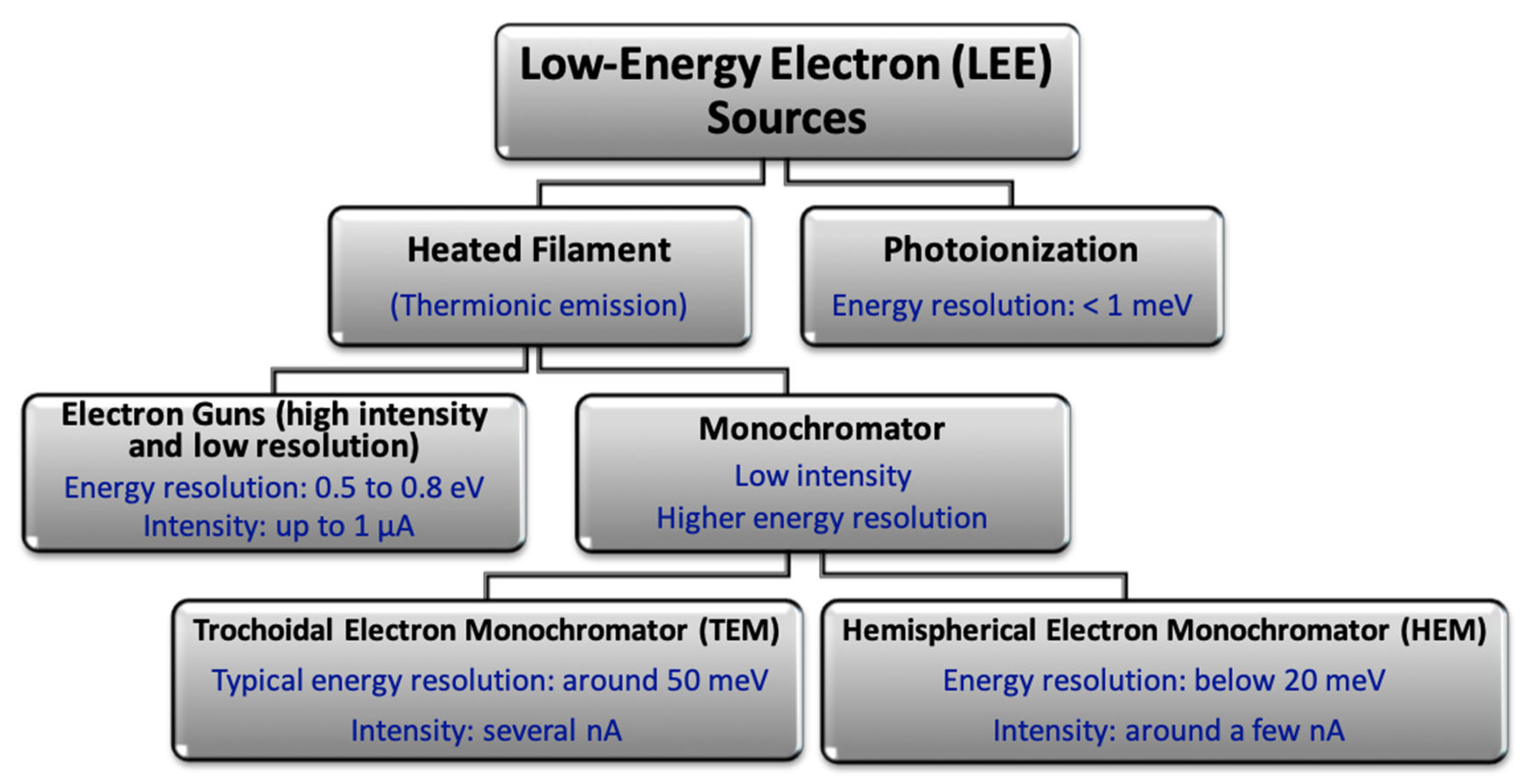
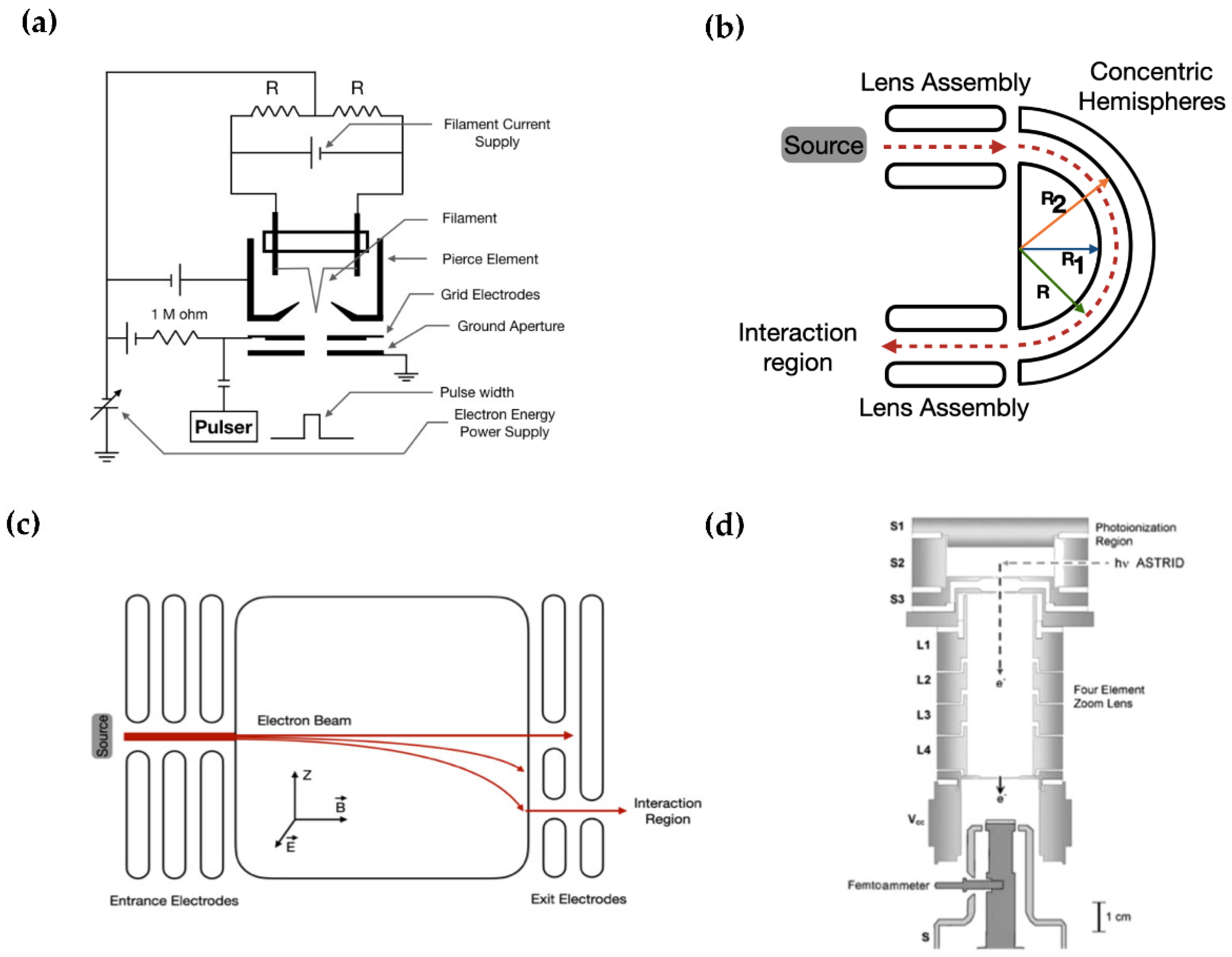
2.1. Common Sources of LEEs

The classic technique used to study the way LEEs interact with molecules, including biomolecules, is by analyzing electron scattering by molecules either isolated in the gas phase or within the thin films in the condensed phase. In such studies, a beam of LEEs generated by an electron source is delivered to the target and then the tracks of scattered electrons and any possible fragments formed are analyzed by several methods. The fragments produced include direct products, as well as those generated from subsequent molecular processes such as electron excitation, attachment, ionization, and consequent dissociation. Depending on the required energy resolution and the electron beam intensity, two different types of sources are used for such collisional studies; these are broadly summarized in Figure 1. The first category is a hot cathode, i.e., an electrically heated filament, wherein free electrons are produced through the thermionic emission process. Due to the high temperature and the electric potential drop across the filament, such electrons possess a broad energy distribution, usually between 0.5 and 1 eV. From this distribution, the monochromator cuts out a small width and produces a beam with a typical resolution of 20–150 meV. Depending upon the operational need, electric and magnetic fields are used to control the energy resolution and the beam intensity. In the second type, high-resolution (<1 meV) electron beams are produced by photoionization. Although this is a standard method of electron production, this type of source has not been utilized widely for biomolecules and will be presented in Section 2.2. More details on the different types of electron guns are briefly discussed in the following subsections.
Standard electron guns—Several electron-collision setups are currently in use that adopt a heated filament as the electron source incorporated into a system of a small number of electrodes and an external magnetic field to collimate the electron beam. The electron beam parameters vary with the type of filament, but the typical resolution is about 0.5 to 1 eV and the current intensity can be within the µA range, making this source appropriate for studies in which high beam intensity is required. High-intensity electron sources are able to produce a sufficient number of secondary ions, which can be further investigated for their metastable decay pathways and kinetic energy release in sector-field analyzers [34]. However, the most effective use of this type of source is in the cross-beam time of flight (TOF) mass spectroscopy that measures the mass-to-charge ratio of fragment ions.
To illustrate the operating principle briefly, a schematic of a pulsed electron gun is provided in Figure 2a. A constant current source for the thermionic emission of the electrons from a heated filament is connected to an external power supply, which applies a constant negative voltage and controls the kinetic energy of the emitted electrons. An electrode, referred to as the “Pierce element”, is used to focus the electron beam after the cathode. A grid electrode, which is connected to the same external power supply through a high resistance circuit (typically in MΩ order), is used for the capacitive pulsing mechanism. A pulse generator is also connected to the grid electrode, which delivers a positive output pulse with an amplitude greater than the applied negative grid voltage. To define the energy of the emitted electrons, a ground aperture is used after the grid electrode. Changing the pulse width and the voltage of the power supply allows precise control of the electron beam width and the energy of the emitted electrons, respectively, within the above-mentioned energy resolution [35]. This type of electron gun has been widely used for LEE impact studies with biomolecules both in the gas phase and the solid phase, particularly as thin films of DNA [36,37]. In a recent study, Panelli and co-workers [38] reported experimental results from three-dimensional momentum-imaging measurements of anion fragments (i.e., H, O and NH2) formed via dissociative electron attachment (DEA) to gas-phase formamide. As formamide is composed of many precursors of complex biomolecules, including an amide bond, it is an archetypal model molecule used to investigate the interaction of LEEs with proteins and nucleic acids [39].
Hemispherical electron monochromator (HEM)—A hemispherical electron monochromator (HEM) is a type of electron source that is generally exploited for high-energy resolution applications. Again, an electron beam is generated through the thermionic emissions of an electrically heated filament, but in HEM, a series of electrostatic lenses select and direct only a minor section of the emitted electron beam with a narrow energy resolution [40] typically less than 20 meV and with an intensity up to nA range. As shown in the schematic in Figure 2b, a HEM consists of two concentric hemispheres in which the outer radius of the inner hemisphere is R1, and the inner radius of the outer hemisphere is R2, and along with the lens assemblies that focus the diverging electron beam. During operation, the applied electric field between the two hemispheres can be expressed as E R = Δ V   R 1   R 2 R 2 R 1 . 1 R 2 ; where ΔV denotes the potential difference between the hemispheres. If an electron with initial energy eV0 moves within the hemispheres along a constant potential, then R = R0 leads to V 0 = Δ V R 2 R 1 R 1   R 2   where R0 is the median ray and is defined as R 0 = R 1 + R 2 2 . Thus, V0 depends on the potential difference between the two hemispheres (ΔV) and R1 and R2 are physical constants. It has been shown [41] that for two hemispheres, the configuration has first-order double focusing properties, i.e., conical diverging electrons that enter the entrance aperture are focused onto the exit aperture. The best-reported energy resolution of HEM was approximately 14 meV at the full width at half maximum (FWHM) [42], while the maximum achievable intensity was ~1 nA.
HEMs have been widely used in studies of the electron-biomolecules interaction through various techniques, such as electron loss spectroscopy (ELS), electron ionization measurements, and probing DEA to gas-phase and condensed-phase biomolecules [43,44]. For example, a custom-built HEM that operates at an energy resolution of 70 to 100 meV and an electron current of ~10 nA, was combined with a quadrupole mass spectrometer to investigate DEA to gas-phase amino acids such as proline [45] and radiosensitizers such as 5-nitrouracil [46]. With respect to condensed-phase studies, Panajotović and Sanche [47] employed a high-resolution electron energy loss (HREEL) spectrometer that contains hemispherical sectors placed in a cryogenically pumped UHV chamber to measure the dependence of energy loss in incident electron energy for backscattered electrons from thin films of adenine [47] and thymidine [48]. Adenine (a purine base) and thymidine (a nucleoside) are both fundamental parts of cellular DNA. The apparatus recorded and analyzed the vibrational and electronic excitation from energy loss spectra at incident energy values ranging from 1.5 to 12 eV. These studies revealed that LEEs with energy less than 5 eV have considerable potential to cause complex deformations of the entire thymidine molecule via H and OH release from the sugar moiety, which results in bond cleavage in DNA.
Trochoidal electron monochromator (TEM)—The theory of the crossed-field (trochoidal) electron monochromator (TEM) and its principal parameters were first introduced in detail by Stamatovic and Schulz in 1968 [49,50]. A TEM system employs crossed homogeneous electric and magnetic fields, and electrons emitted from an electrically heated filament are injected parallel to an axial magnetic field, B . A schematic of a TEM system is given in Figure 2c [49]. After initial acceleration and passage through the entrance electrodes, the electrons are directed through an electric field, E , that is generated by applying a potential difference between two parallel electrodes and applied perpendicularly to their path. The electrons thus exhibit a cycloidal motion as they pass through the crossed electric and magnetic field. They drift in a direction perpendicular to both fields (the z-direction) and experience differential dispersion according to their initial velocities. The velocity component is expressed as V z = E × B B .
As electrons are formed through the thermionic emission process, the beam has of a broad energy distribution (approximately 0.5 eV to 0.8 eV). Accordingly, as shown in Figure 2c, the orifice in the exit electrode is displaced from that in the entrance electrode, and thereby selects a small portion of the electron beam that can successfully pass through the region, with a slight energy uncertainty. To maintain a compromise between the energy resolution and intensity of the beam (in several nA), the monochromator also cuts out a small width (typically 50 meV) from the large initial energy distribution of the electrons. After the exit electrode, the resulting mono-energetic electron beam is accelerated to the required energy level by applying a suitable potential difference. Since the concept of TEM was developed, it has been used in a number of LEE impact experiments with biologically important molecules both in the gas phase and within condensed thin films [52,53]. Sanche and co-workers recently applied TEM in an apparatus to measure the absolute cross-sections of LEE damage to condensed macromolecules, such as plasmid DNA using LEE transmission, and ELS techniques under UHV [54].

2.2. Alternative and Unconventional Sources

Despite decades of intense successful studies of low-energy interactions with biomolecules using the conventional electron sources described in previous sections, researchers remain actively engaged in the pursuit of innovative technologies that offer alternative ways to generate LEEs in electron beams with even higher energy resolution (~1 meV [55]) and current. Moreover, instruments that support a wide range of beam durations have been developed from a continuous beam all the way to ultrashort (femtosecond [56]) electron pulses.
Magnetically guided trap-based electron beams—One of the most recently developed techniques reported this year is a novel approach using a Penning–Malmberg buffer-gas trap, also known as a Surko trap [57]. This type of source is able to achieve a high-flux electron beam with an energy resolution of approximately 30 meV. The basic principle and design are similar to a system utilized extensively for positron collision measurements. It consists of a multi-stage (typically three), differentially pumped beamline with an externally provided magnetic field, a tungsten hairpin filament electron source, and a Penning–Malmberg trap. The trap consists of several electrodes with potentials that are individually controlled. Two buffer gases, N2 and CF4, are admitted to different sections of the trap. These gases provide collisional energy loss mechanisms, i.e., the electrons are trapped by N2 and ultimately cooled to room temperature by CF4. This type of system was tested in electron studies investigating the absolute elastic total cross section of helium [57] and electron-impact excitation of the vibrational mode of CF4 [58]. Although the Surko trap has not previously been utilized for LEE studies with biomolecules, it has been suggested that the technique can be applied to those cases in which there are discrepancies between the available experimental data or between observations and theoretical descriptions of vibrational and electronic excitation [57].
Secondary electron emission from metals/insulators—LEEs can be produced during surface bombardment with energetic particles, ions, electrons, and neutrals. The yield of emitted SEs depends on the material properties and topography of the bombarded surface as well as the energy of the primary beam, the angle of incidence, and the film/foil thickness [59]. However, the energy distribution of SEs is independent of the primary beam energy. The typical energy distribution of SEs produced from metals and insulators by the primary beam of electrons is shown in Figure 3; this distribution is characterized in terms of the most probable energy (ESE) and FWHM. Despite the possibility of having electrons emitted with different ESE depending on the type of target bombarded, it would be impractical in biomolecule studies to accelerate the SEs with a weak electric field without affecting the primary beam, as utilized in electron scanning microscopy [59].
In addition, SEs can be produced when a high-energy beam penetrates thin foils; this has been measured and simulated for aluminum foils through which keV electrons were transmitted [60]. Less than 10% of the primary beam was converted to LEEs, and authors suggested that higher efficiency can be achieved for different materials and thicknesses and for lower primary energies [60]. Electron sources from which SEs are emitted due to surface bombardment or passing electrons through the material are more disadvantageous than the conventional sources (see Section 2.1), particularly as their energy can scarcely be tuned. However, they can be beneficial for experiments performed under ambient conditions, as will be described later in Section 3.2.
Polarized electron beams—The production of polarized electron beams in the low energy range for scattering and excitation experiments has been of interest for nearly a century, since the very idea of electron spin was introduced [61]. In such experiments, the polarization of an electron beam is defined by the degree (magnitude) of polarization and the direction of its vector, which is an average of the spin vectors of all electrons in the beam [61]. Alignment of this vector along the direction of the beam’s motion is referred to as longitudinal polarization, and alignment normal to the direction of motion as transverse polarization. Typically, this vector is oriented at some angle to the direction of beam motion [61]. Because the natural source of polarized electrons (beta decay) is not convenient in the laboratory setting, several different technological approaches have been utilized to produce an intense electron beam with a high degree of polarization at low energy. The earliest work reported low-energy Mott scattering, photoionization, and field- and photo-emission from solids as the first developments.
A limited number of laboratories has utilized polarized electron sources in the low energy regime with molecular targets. Most of these experiments were performed with diatomic molecules (in which O2 and NO were the first targets [62]), and with chiral compounds. Previous work has provided a vast amount of essential information on the physical mechanisms involved, but many of the effects observed during polarized electron scattering are still poorly understood. Moreover, further scattering studies with polarized electrons at low energy are needed to assess fundamental predictions of quantum physics [63,64]. Interestingly, one of the motivations of early studies with chiral molecules was to elucidate the origins of biological homochirality in living systems; the L-handedness of amino acids, and D-handedness of sugars. The most common methods of producing polarized electrons nowadays employ solid-state sources, particularly GaAs-based sources [61,65]. A GaAs photocathode generates polarized electrons when subjected to incident light that is right- or left-hand circularly polarized, is monochromatic, and has energy that corresponds to the near-band-gap energy. Such photoemission can yield polarization at or just less than 50%; in contrast, linearly polarized light induces unpolarized electron emission. As with the sources described in Section 2.1, this type of electron source requires UHV conditions. Ambient contaminants such as air components as well as any reactive target molecules or their products can cause chemical degradation of the GaAs surface, which results in poor performance with respect to polarization, quantum efficiency, and lifetime, and eventually leads to device damage. Therefore, particular consideration of an effective pumping system is essential for gas-phase targets with small cross sections. It has been reported that sources that have open structural geometries around the photoemissive cathode are pumped by ion and/or nonevaporable getter pumps with high pumping speed, and the occasional use of cesiation showed the best performance [66]. Further advances in GaAs-based sources to generate polarized electrons have been described previously together with a few potential alternative sources such as plasma afterglow metastable helium, spin filters, field emission tips, and multiphoton ionization processes [66,67]. Although GaAs-based sources have not been used explicitly as a polarized electron source in studies of biomolecules, there are reports about the effect of sub-eV collisions between longitudinally polarized electrons and chiral camphor molecules that could potentially contribute to the understanding of biological homochirality [68,69].
A recent development is low-energy polarized electron sources attributed to the so-called chiral induced spin selectivity (CISS) effect, in which X-ray induced photoelectrons pass through organized oligomer films of chiral molecules, which serve as a spin filter, attached to a metallic substrate [70,71,72,73,74]. In this approach, low-energy photoelectrons are produced from the metallic substrate during X-ray irradiation, in which the effect is similar to the photoelectric effect described in the next section. However, in this case, the emitted electrons are spin-dependent because circularly polarized light is used. Then, the emitted photoelectrons interacted with monolayers of chiral molecules for which stearoyl-lysine was used in the pioneering work [70]. Certain other small chiral molecules and their assemblies have been proposed for use in generating polarized electrons, as well as other materials such as both metal-organic crystals and hybrid organic-inorganic perovskite films, and inorganic crystals and oxides; however, no free electrons can be produced by this method. Rather, the transmission of electrons is realized by tunneling between bound states or by metal-like charge flow [75]. Very recent developments in technology based on CISS include using a magnetized substrate in which the magnetization direction can be turned, and thereby controlling the helicity of the ejected photoelectrons [76,77]. Eventually, controlled spin-selective production of electrons can be realized by applying an external magnetic field [78]. Interestingly, the first study was just recently reported in which the cross-section of damage to chiral molecules (amino acids) depended on the spin of the interacting LEE [76].
Photoionization—As a process of electron production, photoionization has already been utilized for electron scattering processes for several decades, mainly for atomic and small molecular systems; However, sources based on this principle still are under development by several groups [55,79]. In this type of source, the electron beam is not produced through thermionic emission; instead, it is produces through the photoionization of argon atoms. The energy resolution of the electrons depends on the energy resolution of the photon beam; a resolution in the sub-meV range has been reported using this process [80]. A synchrotron light source can also be used for photoionization [51]. To illustrate the concept of the process, a schematic of the apparatus is shown in Figure 2d. In this case, argon gas is used for the photoemission of electrons. The base pressure of the apparatus is about 10−8 Pa and the pressure in the presence of the argon gas is several times 10−5 Pa. Synchrotron radiation with an energy of 15.76 eV interacts with the pure argon photoionization region in Figure 2d, generating photoelectrons with an energy resolution (FWHM) approximately 1.6 meV. These electrons are then focused using a four-element electrostatic zoom lens to guide them into the interaction region. The reported beam intensity is 200 fA. This apparatus is enclosed by a μ-metal shield to exclude the Earth’s magnetic field. Another work reported the use of solid-state heterostructure crystals for photoionization as well [81]. Interestingly, photoionization has also been used in performing electron scattering measurements on optical enantiomers, specifically investigating the interactions of longitudinally polarized electrons from the gas-phase chiral species S- and R-2-butanol [82].
Plasma sources—Plasma mainly is an entirely or partially ionized gas consisting of ions, free electrons, and photons primarily, as well as radicals, which are considered biologically and chemically reactive species. In the laboratory, plasmas are conveniently created by applying an electric field to an introduced gas between two electrodes, or as its admixtures with different percentages of other compounds. The electric field accelerates the electrons and initiates a cascade of collisional processes, including excitation, ionization, and dissociation, which results in producing a diverse range of chemical species [83] and a large number of electrons with energies on the order of and lower than the ionization potential of atoms and molecules [84]. The complex nature and exact mechanisms of the interactions between plasma discharges, particularly low-temperature plasmas, and biological systems have been intensively investigated [83,85,86]. As a source of LEEs, just this year, a novel experimental setup was reported, in which electrons were generated using a DC-biased grid in inductively coupled plasma, but some earlier designs have also used a similar method to obtain the electron beam [87]. In the recent design, electron beams with low energy on the order of a few tens of eV were produced in argon plasma and controlled by the substrate voltage, gas pressure, and source power. However, the extraction of LEEs requires additional experimental arrangements such as using an electric and/or magnetic field to control the electron energy and its resolution, which can lead to certain technical challenges in studies involving biomolecules. Nevertheless, plasmas as a source of LEE within the biological system were recently reported in the study of Houde and co-workers [88], which will be briefly discussed in Section 4.

3. Recent Developments of LEE Sources to Approach Cellular Conditions

Many research groups have recently extended the experimental studies in vacuum and on isolated biomolecules to more complex systems in which the DNA molecule resides in an aqueous environment in the vicinity of cellular components (such as, O2, H2O, histones and DNA-binding proteins, and vitamins) for obtaining data as relevant as possible to actual cell damage [89,90]. Here, as an alternative approach for simulating cellular conditions, we review some of the techniques and experimental setups that are employed under well-defined atmospheric conditions.

3.1. X-ray Photoelectron Spectroscopy at Near Ambient Pressure (XPS-NAP)

X-ray photoelectron spectroscopy (XPS) is among the standard analysis technique used to investigate direct damage by X-rays and LEEs performed on isolated DNA in UHV (p < 1 × 10⁻⁶ Pa) [91,92]. XPS spectra are acquired by assessing the kinetic energy of the photo-emitted electrons in the depth < 10 nm for standard soft X-ray excitation sources and provide qualitative and quantitative information about the elemental composition and chemical state of the surface. As such, this technique is a versatile tool for monitoring the radiation-induced chemical transformation of functional groups on DNA molecules in situ, without post-irradiation treatment. Further, during XPS measurements, not only do the primary monochromatic X-rays produce SEs, which interact with tissue and damage DNA, but also detection of these photoelectrons allows the identification of chemical changes. For instance, McKee et al. [93] used X-rays to eject LEEs from a gold substrate and then probed the resulting damage in condensed nucleotides. This method was further developed by integrating a separate LEE source to cause damage to deoxyadenosine monophosphate that was monitored using XPS [94].
In still further developments, the need for XPS analysis of materials in their natural state has led to the development of near-ambient pressure XPS (NAP-XPS) systems, which serve as a real-time or nearly real-time process monitoring tool at moderate pressures (p > 2500 Pa) [95,96]. This less traditional form of XPS allows electrons, for which the inelastic mean free path is on the order of nanometers at ambient pressure, to reach the detector (which is still maintained under UHV) by employing various stages of differential pumping to successively decrease the pressure in the system from the sample to the detector. Compared with conventional XPS, some NAP-XPS instrumentation can operate at higher working pressures; this significantly reduces sample preparation and pump down times, as samples are directly loaded into the analytical chamber and there is no need for a load lock in the system [97]. A novel ambient pressure XPS instrument [98] has provided the first-time simultaneous induction and in situ monitoring of DNA damage under two different conditions, i.e., the standard UHV conditions, as well as under the H2O and N2 atmospheres at NAP. This technique allows the direct quantification of radiation-induced damage in both dry and fully hydrated DNA molecules by means of XPS measurements performed with Al Kα X-rays of ~1.5 KeV. The UHV-XPS measurements were made using a standard photoelectron spectrometer under pressure below 1 × 10⁻⁶ Pa, and the NAP-XPS measurements at pressures in the NAP regime (0.04–0.14 Pa). Details on experimental procedures and data analysis are provided in Refs. [99,100]. Finally, combining experimental results of NAP-XPS with Monte Carlo particle scattering simulations has been used to distinguish direct damage by photons and secondary LEEs from that caused by hydroxyl radicals or hydration induced modifications of damage pathways [101].

3.2. X-ray Interaction with Metal at Standard Ambient Temperature and Pressure (SATP)

After studying on LEE-DNA interactions under UHV for a decade, many attempts were then made to apply the UHV sources of LEEs to approach an environment more closely resembling closer the nuclei of cells. Ptasińska et al. [102] performed anion desorption experiments stimulated by the impact of 3–20 eV electrons on short single-stranded DNA which was covered by three monolayers of water to mimic aqueous conditions. The apparatus consisted of a load-lock vacuum chamber (~10−7 Pa) with a multiple-sample holder, to which samples could be mounted and transported on a rotary feedthrough into the main chamber under a pressure of ~10−8 Pa. An electron gun in the chamber produced a LEE beam with a kinetic energy below 20 eV and an energy resolution of 0.5 eV (FWHM) that impinged on the sample’s surface, with resulting desorbed anions analyzed by a quadrupole mass spectrometer. To produce water monolayers, the water vapor was introduced through a stainless-steel tube connected to a gas-handling manifold and adsorbed on a film of oligonucleotides cooled to 90 K by liquid nitrogen. The manifold consisted of a precision-leak valve connected to a small expansion chamber with a barometer. This chamber was further connected via an admission valve to a small tube with an opening located in front of the substrate. In other studies, Orlando et al. [103,104] designed and constructed an apparatus to allow introduction of selected DNA sequences thin films bonded to different numbers of water molecules into an UHV environment (~1.3 × 10⁻⁸ Pa). Briefly, the system featured a load lock transfer system magnetically coupled to the UHV chamber, which was differentially pumped and maintained in a nitrogen buffer gas. The UHV system contained a variable temperature sample holder, pulsed LEE beam (5–50 eV), and TOF and quadrupole mass spectrometers. The technique and apparatus were later used for irradiation of a sensitive chemical-vapor-deposited graphene-coated gold platform to show the significant role of water molecules in controlled and enhanced sequence-dependent LEE-induced DNA damage [105].
Subsequently, Sanche and co-workers developed a novel technique for simulating more realistic conditions based on the emission of photo-ejected SEs from a metallic surface exposed to primary X-ray photons as an alternative LEE-source at standard ambient temperature and pressure (SATP). Studies using this technique were initiated by Brun et al. [106], who irradiated DNA thin films deposited on gold and glass substrates and exposed them to Al Kα X-rays of 1.5 keV in air and under vacuum. Owing to the strong attenuation coefficient of soft X-rays and the very short ranges of LEEs [107], the DNA films were prepared by freeze-drying to obtain films as thin and uniform as possible and prevent excessive charging in the targets. For DNA deposited on glass, damage yields are due to X-rays alone, while for DNA on gold, damage results from X-rays in combination with extra low-energy photoelectrons from the gold (average energy of 5.8 eV). The difference in damage yields between the two in samples deposited on substrates is ascribed to the interaction of LEEs with the DNA and its nearby atmosphere. Radiation damage to DNA in the form of strand breaks was quantified with post-irradiation biochemical methods such as agarose gel electrophoresis. The damage yields were expressed in terms of G value, which refer to the number of modified molecules or products per unit of energy absorbed by the target (nmol/J or µmol/J) and thus depends not only on the radiation energy, but also on the type of radiation and target [108]. The obtained G values estimates indicated that in the presence of hydration layers around DNA, LEEs are more efficient than soft (1.5 keV) X-rays in inducing DNA damage as LEEs generate additional OH radicals. The technique and experimental setup were latter advanced by Alizadeh et al. [109], namely by substituting the gold substrate with tantalum and adapting the irradiation chamber. This modified technique then offered a way to investigate the effect of the different hydration layers on LEE-induced DNA damage and allowed the shift from direct to indirect effects. A detailed description of sample preparation and the experimental arrangement can be found in [110] and is briefly described below.

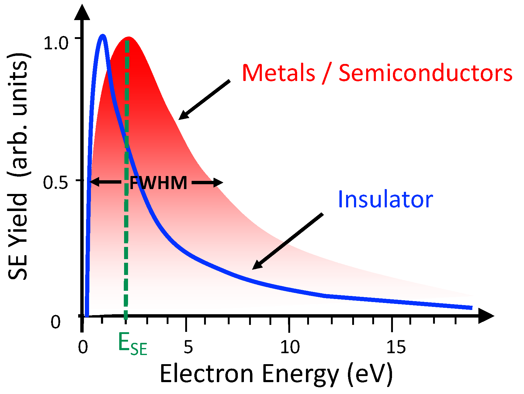
3.2.1. Secondary Electron Emission from Metals

The general features of the emission of SEs from solid surfaces exposed to primary X-ray photons are well understood, as this is a fundamental process in scanning electron microscopy [59,111]. In short, exposing a solid target to high-energy radiations such as X-rays causes bound electrons to be excited to a continuum by photon absorption. The high-energy electrons produced impinge upon the surface and induce successive excitation processes or are inelastically scattered. Thus, the electrons decelerate through electron-electron collision and eject weakly bound valence electrons (in the case of ionically or covalently bonded materials) or conduction band electrons (in the case of metals), which have binding energies of ~1–15 eV to their parent atom(s); this generates SEs above the Fermi level. SEs are 1.5 keV generated by direct injection of electron or ion beams [112], as also mentioned in Section 2.2. Moreover, to support and predict experimental data, Monte Carlo simulations have been widely used to simulate the trajectories and emission mechanisms of SEs [113].
In experiments involving LEE interactions with DNA, tantalum has been identified as a suitable substrate [114,115]. Freeze-drying of DNA on tantalum or glass [106] can produce considerably less damage than when using many other metal substrates, such as gold, are used [109]. This is because the formation of an oxide layer on the metal surface creates a stable and chemically inert surface, ensuring no direct DNA-metal interaction, and thus causing minimal substrate-induced damage to the DNA. In the newly developed technique by Alizadeh et al. [109] (Figure 4a), X-ray photons that traverse a DNA film interact with the tantalum substrate, causing energetic photoelectrons and Auger electrons (AEs) to be emitted inside the metal. These deeper electrons lose energy and hence those produced at the surface essentially consist of LEEs with a specific energy distribution. As indicated in Figure 4b, the SE energy distribution curve from tantalum peaks at 1.4 eV (with an average of ~5.85 eV) and more than 95% of the SEs emitted from tantalum are below 30 eV. From such a distribution, SEs were quantified by measuring their yield which is the ratio of SEs emitted from a specimen to the number of incident primary electrons [109].

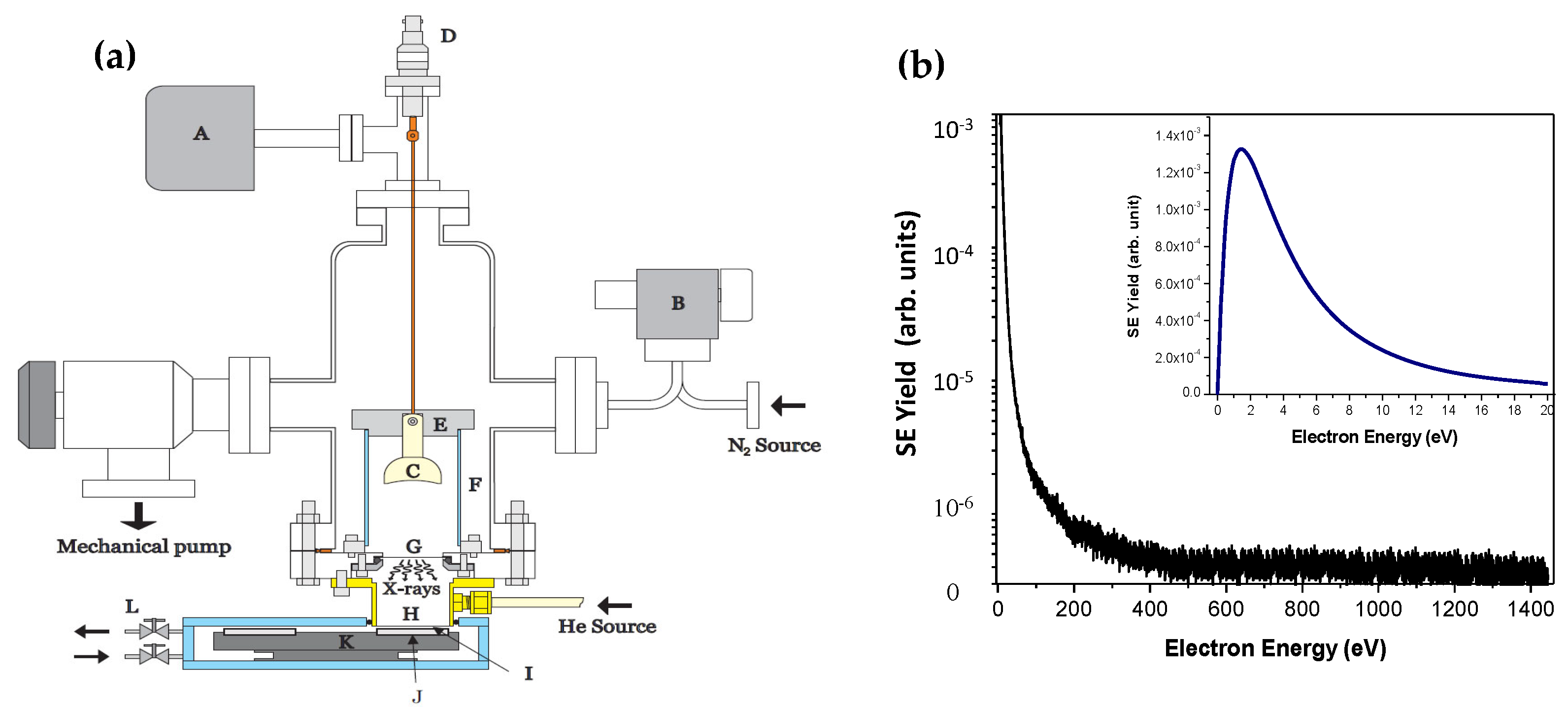
3.2.2. Experimental Setup and Irradiation Conditions

The experiments of Alizadeh et al. [109] were performed with a home-made apparatus depicted schematically in Figure 4a. The construction of the Al Kα X-ray source is based on the original design of Hoshi et al. [116] used in many biological experiments for plant cell irradiation. The new technique and altered experimental setup were applied to investigate the damage induced in plasmid DNA by photo-ejected LEEs and soft X-rays under controlled atmospheres of pure gases such as N2 and O2, and under different levels of hydration at SATP [117]. Experiments performed with liquid water condensed on and within DNA films at different humidity levels by varying the hydration level of DNA allowed a progressive shift from direct to indirect effects of the LEEs. It was observed that for a humid DNA film under an oxygenated environment, the additional LEE-induced damage resulting from the combination of water and oxygen exhibits a supper-additive effect. These findings not only provide insight into the radiosensitization mechanism of O2, but also reveal that LEEs, O2, and H2O contribute synergistically to efficiently enhance the formation of more lethal forms of DNA damage [118]. In similar studies, DNA components like thymidine and synthetic oligonucleotides were irradiated under an N2 atmosphere at SATP [119,120]. Rather than analyzing conformational modifications to DNA as in previous investigations, liquid chromatography (LC) and tandem mass spectrometry (MS/MS) were used to identify the specific products remaining on the surface of the irradiated substrates. Moreover, to investigate the nanoscopic mechanisms responsible for the efficacy of concomitant treatments mechanisms, this technique was employed to study the interactions of LEEs with complex DNA and platinum-based chemotherapeutic agents (Pt-drugs) including cisplatin, carboplatin and oxaliplatin [121]. The results of this study provided information on the role of subexcitation-energy electrons and dissociative electron attachment in the radiosensitization of DNA by Pt-drugs. This is a key step in unraveling the mechanisms of radiosensitization by these agents in chemoradiation cancer therapy and could be an essential step toward the development of optimal clinical protocols [122].

4. Ultimate Sources for Studies under Cellular Conditions

Over the past two decades, considerable research has been devoted to invention of sources of LEEs at the sites of cellular targets (particularly nucleus DNA) to increase dose deposition locally, which constitutes a potential strategy for targeted cancer therapy is increasing the local density of LEEs near DNA. This strategy can be achieved by using either high-Z materials or through AE-emitting radionuclides, in the vicinity of DNA or other vital cellular structures and components in cancer cells. The fundamental mechanisms underlying this strategy are briefly described here.
Gold nanoparticles (AuNPs) and the Auger effect—Molecular compounds containing high-Z materials, such as iodine, platinum, gadolinium and gold, have higher attenuation coefficients, and thus absorb ionizing radiation energy at a significantly higher rate than do low-Z materials like soft tissues. This leads to an increase in the production of extra short-range secondary LEEs via the Auger effect and intermolecular (or interatomic) Coulombic decay (ICD). Among the various high-Z molecules, there is considerable interest in the potential therapeutic use of AuNPs due to their biocompatibility [123,124]. AuNPs are generally associated with two therapeutic modalities, photosensitization and radiosensitization. While radiosensitizers function only in combination with ionizing radiation for radiotherapy, photosensitizers used in photodynamic therapy (PDT) have a strong absorbance peak in the ultraviolet (UV) range so they only act in combination with UV light. PDT has a cytotoxic effect on cancer cells through the formation of ROS generated upon activation of the photosensitizer using UV light, inducing oxidative stress in the cancerous tissue in which the photosensitizer is located, and leads to tumor cell death [125]. Because of the short UV penetration (<0.5 mm), photosensitizers are applied in dermatology to optimize and improve the UV therapy of such skin diseases as psoriasis and mycosis fungoides. On the other hand, radiosensitizers lead to an increase in sensitivity of the hypoxic and radioresistant areas of tumors against high energy radiation; thus, acting on deep-seated tumors [126].
Figure 5 depicts a schematic of the way X-ray interactions with AuNPs within living cells result in the production of LEEs by both Auger and ICD processes, which has been suggested as a potential strategy for targeted cancer treatment [127]. The damage caused by irradiation of AuNPs depends on AE emission, which generates a cascade of LEEs that travel very short distances and deposit their energy locally; as such it is crucial to place the AuNPs on or in the cancer cells. Antosh et al. [128] accordingly conjugated a pH-sensitive tumor-targeting agent, and the pH Low-Insertion Peptide (pHLIP), with 1.4 nm AuNPs, in order to localize and increase their uptake by cancer cells. Monte Carlo simulations were used to predict the particle interactions and energy deposition when irradiated AuNPs were placed in a water or cellular medium. The simulated results helped in estimating the variation in dose enhancement due to AuNPs addition under different radiation energies and with different nanoparticle sizes [129].
Furthermore, Sykes and co-workers [130] described the construction and properties of a novel source that uses AE emitters to produce high fluxes of LEEs. They synthesized one-atom-thick 2D films of the radioisotope 125I on a gold substrate that were stable under ambient conditions. 125I decays by electron capture of a core–shell electron, producing an excited state of 125Te. During de-excitation, a cascade of electronic relaxations leads to the emission of multiple AEs with energies below 500 eV, which are further decelerated by inelastic scattering in the gold film and generate more LEEs. A portion of the scattered and generated electrons are emitted outside of the metal, where their energy distribution can be appraised. Scanning tunnelling microscopy, supported by electronic structure simulations, was used to directly observe the nuclear transmutation of individual 125I atoms into 125Te and explain the surprising stability of the planar film as it underwent radioactive decay. The metal interface geometry induced a 600% amplification of LEE emission compared with atomic 125I. This enhancement of biologically active LEEs may offer a new direction for highly targeted nanoparticle therapies, particularly AuNPs [130].
Intermolecular Coulomb decay (ICD)—Interatomic or intermolecular Coulombic decay (ICD) is a nonlocal electronic decay mechanism in the inner valence levels that occurs in molecular systems with weakly binding forces, such as van der Waals forces or hydrogen bonding. In the ICD process, energy released by electronic relaxation of an excited atom or molecule leads to ionization of a neighboring one via Coulombic electron interactions. Thus, ICD is more efficient when more neighboring ionizable molecules are present. ICD was predicted theoretically in 1997, while its existence was confirmed experimentally ten years later [131,132] and has been explored in many subsequent experiments. While most studies measuring ICD have concentrated on rare gas clusters, recent experimental approaches have reported its occurrence even in water clusters [133] or at condensed-phase interfaces that contain water clusters and dimers [134].
ICD produces reactive radical species and electrons, primarily LEEs with energies < 15 eV, which are important in causing radiation damage to DNA [135]. If ICD contributes significantly to DNA damage, this could be exploited during X-ray treatment of cancer. Initial experimental studies investigating the role of ICD in biological damage were conducted by demonstrating the occurrence of ICD in water clusters and dimers after inner-valence ionization employing synchrotron radiation [133,136]. Dörner and co-workers [132] later revealed the quantitative role of ICD in the production of LEEs by alpha particle impact by colliding He+ ions with isolated Ne atoms and dimers (Ne2). In the latter case, the Ne atoms impacted were embedded in an environment already utilizing ICD as a deexcitation channel. As a consequence, the emission of LEEs dramatically enhanced in ion-atom collisions, suggesting that ICD may have a significant influence on cell death after exposure to ionizing radiation. Additional studies have been performed to characterize the dose enhancement attributed to high-Z-containing nanoparticles. For example, Seo et al. [137] investigated the production of ROS in a gadolinium oxide nanoparticle solution irradiated with 50 keV X-rays and high energy protons. Later work by Ren et al. [138] investigated the electron impact ionization of hydrogen-bonded complexes that consist of tetrahydrofuran (THF) molecules loosely bound to a water molecule as a model for more complex biological system.
Femtosecond laser filamentation (FLF)—Another novel source of LEE was introduced by Houde et al. [139] who exploited intense ultra-short infrared laser pulses to produce high density avalanches of LEEs through the formation of filaments in the so-called inverse Bremsstrahlung process. The physical origin of laser filamentation from a femtosecond laser pulse is well understood [139,140]. Briefly, at sufficiently high power, the Kerr self-focusing of a laser beam is counteracted by the defocusing action of plasma generated on the beam track, resulting in the self-channeling propagation regime known as femtosecond laser filamentation (FLF) [141]. Figure 6 shows a schematic of the experimental setup for generating LEE filaments. This photonic process can lead to the creation of molecular and radical species in liquid media, which can then initiate polymerization in a polymer gel dosimeter. LEEs with the most probable kinetic energy centers approximately 10 eV. In the work by Houde et al. [139] the macroscopic dose deposition from laser-induced filamentation was measured using a Fricke (ferrous sulfate) dosimeter, while filament morphology (the spatial dose distribution, and hence the microscopic energy density) was evaluated with a radiologically tissue-equivalent dosimeter (polyacrylamide gel) and visualized by magnetic resonance imaging (MRI) [142]. Ultimately, the authors discovered that LEE generated from FLF and conventional radiation modalities for RT are radio-chemically equivalent in terms of the type of final damage and their mechanisms of action in aqueous media at the molecular and biomolecular levels. Further, an in vivo study using small-animal cancer model additionally revealed the therapeutic benefits of FLF in radiotherapy, in which an extreme dose of energy was deposited at ultra-high dose rates (up to 1011 Gy/s) deep within a macroscopic volume, and changing the duration of the laser pulse allowed precise control of the distance over which the entrance dose was zero [88].

5. Summary and Conclusions

Extensive theoretical and experimental studies have revealed that compared to other reactive species produced by high-energy radiations, LEEs are highly efficient in inducing lethal DNA lesions. They also have exceptional properties in inducing DNA radio-sensitization to cause lethal damage in cancer cells and hence have potential in concurrent chemoradiation therapy, which is a treatment modality that combines chemotherapeutic agents and radiation. Because the high-energy radiation used in RT produces a large number of LEEs, it is conceptually feasible to enhance the benefit of LEEs and maximize the biological effects of ionizing radiation in cancer treatment through two approaches. Firstly, increasing and controlling the local density of LEEs in close proximity to cellular DNA; and secondly, sensitizing cellular targets to LEEs. The presented work reviewed and summarized the techniques and instrumentation used to generate LEEs and provided a brief overview of each source’s applications in biological media for enhancing the benefits of LEEs.
Considerable multidisciplinary research effort is still required to develop methods for generating and controlling the density of LEEs at cellular targets, specifically in cancer cells. Some innovative experimental setups and novel methods will need to be developed to hybridize LEE generation methods and spectroscopic tools to monitor their products. For instance, combining NAP-XPS with mass spectrometry techniques has provided the opportunity to study oxygen induced fixation of radiation damage [143]. While molecular oxygen plays a critical role in cellular energy biogenesis, it is also potentially dangerous, as reactive species are formed as byproducts of normal oxygen metabolism [144]. NAP-XPS investigation of desorbed products under both aerobic and anaerobic conditions helps in understanding the fundamentals of DNA-protein crosslink formation, which predominantly occurs under low-oxygen concentrations and in cancerous tissue that has high resistance to radiation. Another example of a hybrid methodology is the coupling of a TEM for LEE generation and samples irradiation with an orthogonal reflectron TOF mass spectrometer to detect ionic fragments. Such an assembly allows us to learn about the fragmentation routes that result from LEEs interactions, and thus represents a useful tool in the design and development of novel chemotherapeutic agents and radio-sensitizers. Further, it may help improve strategies in RT and clinical practice and prevent potential adverse effects. Additional technological improvements also need to be considered in order to assess the role and potential effects of LEEs in biological systems under more realistic conditions.

Author Contributions

All authors contributed to writing and editing of the manuscript. All authors have read and agreed to the published version of the manuscript.

Funding

D.C. and S.P. acknowledge the U.S. Department of Energy Office of Science, Office of Basic Energy Sciences under Award Number DE-FC02-04ER15533 (NDRL no: 5383).

Conflicts of Interest

The authors declare no conflict of interest.

Abbreviations

AEAuger electron
AuNPgold nanoparticle
CISSchiral induced spin selectivity
DEAdissociative electron attachment
ELSelectron loss spectroscopy
FLFfemtosecond laser filamentation
FWHMfull width at half-maximum
HEEhigh-energy electron
HEMhemispherical electron monochromator
HREELhigh-resolution electron energy loss
ICDintermolecular Coulomb decay
IORTintra-operative radiotherapy
LCliquid chromatography
LEElow energy electron
LINAClinear accelerator
MS/MStandem mass spectrometry
MRImagnetic resonance imaging
NAPnear ambient pressure
ppressure
PDTphotodynamic therapy
Pt-drugsplatinum-based chemotherapeutic agents
RFradio-frequency
ROSreactive oxygen species
RTradiation therapy
SATPstandard ambient temperature and pressure
SEsecondary electron
TEMtrochoidal electron monochromator
THFtetrahydrofuran
TOFtime of flight
UHVultra-high vacuum
UVultraviolet
VMATvolumetric modulated arc therapy
VHEEvery high-energy electrons
XPS X-ray photoelectron spectroscopy

References

  1. Navarro, J. A History of the Electron: J. J. and G. P. Thomsons, 1st ed.; Cambridge University Press: New York, NY, USA, 2012. [Google Scholar]
  2. Singh, R.; Singh, D.; Singh, A. Radiation sterilization of tissue allografts: A review. World J. Radiol. 2016, 8, 355–369. [Google Scholar] [CrossRef] [PubMed]
  3. Fertey, J.; Thoma, M.; Beckmann, J.; Bayer, L.; Finkensieper, J.; Reißhauer, S.; Berneck, B.S.; Issmail, L.; Schönfelder, J.; Casado, J.P.; et al. Automated application of low energy electron irradiation enables inactivation of pathogen- and cell-containing liquids in biomedical research and production facilities. Sci. Rep. 2020, 10, 12786. [Google Scholar] [CrossRef] [PubMed]
  4. Wetzel, C.; Schönfelder, J.; Schwarz, W.; Funk, R.H.W. Surface modification of polyurethane and silicone for therapeutic medical technics by means of electron beam. Surf. Coat. Technol. 2010, 205, 1618–1623. [Google Scholar] [CrossRef]
  5. Gotzmann, G.; Portillo, J.; Wroski, S.; Kohl, Y.; Gorjup, E.; Schuck, H.; Rögner, F.H.; Müller, M.; Chaberny, I.F.; Schönfelder, J.; et al. Low-energy electron-beam treatment as alternative for on-site sterilization of highly functionalized medical products: A feasibility study. Radiat. Phys. Chem. 2018, 150, 9–19. [Google Scholar] [CrossRef]
  6. Akbarpoor, R.; Khaledi, N.; Wang, X.; Samiei, F. Optimization of low-energy electron beam production for superficial cancer treatments by Monte Carlo code. J Can. Res. Ther. 2019, 15, 475–479. [Google Scholar] [CrossRef]
  7. Amgarou, K.; Lacoste, V.; Martin, A. Experimental characterization of the neutron spectra generated by a high-energy clinical LINAC. Nucl. Instrum. Methods Phys. Res. Section A Accel. Spectrom. Detectors Assoc. Equip. 2011, 629, 329–336. [Google Scholar] [CrossRef]
  8. Banaee, N.; Goodarzi, K.; Nedaie, H.A. Neutron contamination in radiotherapy processes: A review study. J Radia. Res. 2021, 62, 947–954. [Google Scholar] [CrossRef] [PubMed]
  9. Very High Energy Electron Beam Radiotherapy Workshop (VHEE’2020); CERN: Zurich, Switzerland, 2020.
  10. Otto, K. Volumetric modulated arc therapy: IMRT in a single gantry arc. Med. Phys. 2008, 35, 310–317. [Google Scholar] [CrossRef] [PubMed]
  11. Pilar, A.; Gupta, M.; Laskar, S.G.; Laskar, S. Intraoperative radiotherapy: Review of techniques and results. Ecancermedicalscience 2017, 11, 750. [Google Scholar] [CrossRef] [Green Version]
  12. Sethi, A.; Chinsky, B.; Gros, S.; Diak, A.; Emami, B.; Small, W.J. Tissue inhomogeneity corrections in low-kV intra-operative radiotherapy (IORT). Transl. Cancer Res. 2015, 4, 182–188. [Google Scholar] [CrossRef]
  13. Papiez, L.; DesRosiers, C.; Moskvin, V. Very high energy electrons (50–250 MeV) and radiation therapy. Technol. Cancer Res. Treat. 2002, 1, 105–110. [Google Scholar] [CrossRef] [PubMed]
  14. Schüler, E.; Eriksson, K.; Hynning, E.; Hancock, S.L.; Hiniker, S.M.; Bazalova-Carter, M.; Wong, T.; Le, Q.T.; Loo, B.W.; Maxim, P.G. Very high-energy electron (VHEE) beams in radiation therapy; Treatment plan comparison between VHEE, VMAT, and PPBS. Med. Phys. 2017, 44, 2544–2555. [Google Scholar] [CrossRef] [PubMed]
  15. Palma, B.; Bazalova-Carter, M.; Hårdemark, B.; Hynning, E.; Qu, B.; Loo, B.W.; Maxim, P.G. Assessment of the quality of very high-energy electron radiotherapy planning. Radiother. Oncol. 2016, 119, 154–158. [Google Scholar] [CrossRef] [Green Version]
  16. Maxim, P.; Loo, B.W. Pluridirectional High-Energy Agile Scanning Electron Radiotherapy (PHASER): Extremely Rapid Treatment for Early Lung Cancer; Defense Technical Information Center Annual Report; Stanford University: Stanford, CA, USA, 2014. [Google Scholar]
  17. Geddes, C.G.R.; Toth, C.; van Tilborg, J.; Esarey, E.; Schroeder, C.B.; Bruhwiler, D.; Nieter, C.; Cary, J.; Leemans, W.P. High-quality electron beams from a laser wakefield accelerator using plasma-channel guiding. Nature 2004, 431, 538–541. [Google Scholar] [CrossRef] [PubMed]
  18. Labate, L.; Palla, D.; Panetta, D.; Avella, F.; Baffigi, F.; Brandi, F.; Di Martino, F.; Fulgentini, L.; Giulietti, A.; Köster, P.; et al. Toward an effective use of laser-driven very high energy electrons for radiotherapy: Feasibility assessment of multi-field and intensity modulation irradiation schemes. Sci. Rep. 2020, 10, 17307. [Google Scholar] [CrossRef]
  19. Lv, J.; Zhang, H.; Zhang, D.; Liu, L.; Han, Y. Low-dose electron microscopy imaging of electron beam-sensitive crystalline materials. Acc. Mater. Res. 2022, 3, 552–564. [Google Scholar] [CrossRef]
  20. Ptasińska, S. A missing puzzle in dissociative electron attachment to biomolecules: The detection of radicals. Atoms 2021, 9, 77. [Google Scholar] [CrossRef]
  21. Alizadeh, E.; Ptasińska, S.; Sanche, L. Transient anions in radiobiology and radiotherapy: From gaseous biomolecules to condensed organic and biomolecular solids. In Radiation Effects in Materials; Monteiro, W.A., Ed.; InTech: Rijeka, Croatia, 2016; pp. 179–230. [Google Scholar] [CrossRef] [Green Version]
  22. Gorfinkiel, J.D.; Ptasińska, S. Electron scattering from molecules and molecular aggregates of biological relevance. J. Phys. B At. Mol. Opt. Phys. 2017, 50, 182001. [Google Scholar] [CrossRef] [Green Version]
  23. Sevilla, M.D.; Bernhard, W.A. Mechanisms of direct radiation damage to DNA. In Radiation Chemistry: From Basics to Applications in Material and Life Sciences, 1st ed.; Spotheim-Maurizot, M., Mostafavi, M., Douki, T., Belloni, J., Eds.; EDP Sciences: Les Ulis, France, 2008; pp. 191–201. [Google Scholar]
  24. Uehara, S.; Nikjoo, H.; Goodhead, D.T. Comparison and assessment of electron cross sections for Monte Carlo track structure codes. Radiat. Res. 1999, 152, 202–213. [Google Scholar] [CrossRef]
  25. Wishart, J.F.; Rao, B.M.S. Recent Trends in Radiation Chemistry; World Scientific: Singapore, 2010. [Google Scholar] [CrossRef]
  26. O’Neill, P. Radiation-induced damage in DNA. In Radiation Chemistry: Present Status and Future Trends; Jonah, C.D., Rao, B.S.M., Eds.; Elsevier Sciences: Amsterdam, The Netherland, 2001; pp. 585–622. [Google Scholar]
  27. Alizadeh, E.; Sanche, L. Precursors of solvated electrons in radiobiological physics and chemistry. Chem. Rev. 2012, 112, 5578–5602. [Google Scholar] [CrossRef] [PubMed]
  28. von Sonntag, C. Free-Radical-Induced DNA Damage and Its Repair; Springer: Berlin/Heidelberg, Germany, 2006. [Google Scholar]
  29. Hearst, J.E.; Vinograd, J. The net hydration of deoxyribonucleic acid. Proc. Natl. Acad. Sci. USA 1961, 47, 825–830. [Google Scholar] [CrossRef] [Green Version]
  30. Saenger, W. Principles of Nucleic Acid Structure; Springer: New York, NY, USA, 1984. [Google Scholar]
  31. Jeffrey, G.; Saenger, W. Hydration Bonding in Biological Structures; Springer: New York, NY, USA, 1991. [Google Scholar]
  32. Ptasińska, S.; Varella, M.T.N.; Khakoo, M.A.; Slaughter, D.S.; Denifl, S. Electron scattering processes: Fundamentals, challenges, advances, and opportunities. Eur. Phys. J. D 2022, 76, 179. [Google Scholar] [CrossRef]
  33. Alizadeh, E.; Orlando, T.M.; Sanche, L. Biomolecular damage induced by ionizing radiation: The direct and indirect effects of low-energy electrons on DNA. Ann. Rev. Phys. Chem. 2015, 66, 379–398. [Google Scholar] [CrossRef]
  34. Głuch, K.; Fedor, J.; Matt-Leubner, S.; Echt, O.; Stamatovic, A.; Probst, M.; Scheier, P.; and Märk, T.D. Kinetic-energy release in coulomb explosion of metastable C3H52+. J. Chem. Phys. 2003, 118, 3090–3095. [Google Scholar] [CrossRef] [Green Version]
  35. Chakraborty, D.; Nag, P.; and Nandi, D. A new time of flight mass spectrometer for absolute dissociative electron attachment cross-section measurements in gas phase. Rev. Sci. Instr. 2018, 89, 025115–025123. [Google Scholar] [CrossRef]
  36. Moradmand, A.; Williams, J.B.; Landers, A.L.; Fogle, M. Momentum-imaging apparatus for the study of dissociative electron attachment dynamics. Rev. Sci. Instr. 2013, 84, 033104–033110. [Google Scholar] [CrossRef]
  37. Kumar, S.V.K.; Pota, T.; Peri, D.; Dongre, A.D.; Rao, B.J. Low energy electron induced damage to plasmid DNA pQE30. J. Chem. Phys. 2012, 137, 045101. [Google Scholar] [CrossRef] [PubMed]
  38. Panelli, G.; Moradmand, A.; Griffin, B.; Swanson, K.; Weber, T.; Rescigno, T.N.; McCurdy, C.W.; Slaughter, D.S.; Williams, J.B. Investigating resonant low-energy electron attachment to formamide: Dynamics of model peptide bond dissociation and other fragmentation channels. Phys. Rev. Res. 2021, 3, 013082. [Google Scholar] [CrossRef]
  39. Dawley, M.M.; Pirim, C.; Orlando, T.M. Radiation processing of formamide and formamide: Water ices on silicate grain analogue. J. Phys. Chem. A 2014, 118, 1228–1236. [Google Scholar] [CrossRef] [PubMed]
  40. Kuyatt, C.E.; Simpson, J.A. Electron monochromator design. Rev. Sci. Instr. 1967, 38, 103–111. [Google Scholar] [CrossRef]
  41. Purcell, E.M. The focusing of charged particles by a spherical condenser. Phys. Rev. 1938, 54, 818–826. [Google Scholar] [CrossRef]
  42. Čížek, M.; Horáček, J.; Sergenton, A.C.; Popović, D.B.; Allan, M.; Domcke, W.; Leininger, T.; Gadea, F.X. Inelastic low-energy electron collisions with the HBr and DBr molecules: Experiment and theory. Phys. Rev. A 2001, 63, 062710. [Google Scholar] [CrossRef]
  43. Schürmann, R.; Tsering, T.; Tanzer, K.; Denifl, S.; Kumar, S.V.K.; Bald, I. Resonant formation of strand Breaks in sensitized oligonucleotides induced by low-energy electrons (0.5–9 eV). Angew. Chem. Int. Ed. 2017, 56, 10952–10955. [Google Scholar] [CrossRef] [PubMed]
  44. Dawley, M.M.; Tanzer, K.; Cantrell, W.A.; Plattner, P.; Brinkmann, N.R.; Scheier, P.; Denifl, S.; Ptasińska, S. Electron ionization of the nucleobases adenine and hypoxanthine near the threshold: A combined experimental and theoretical study. Phys. Chem. Chem. Phys. 2014, 16, 25039–25053. [Google Scholar] [CrossRef] [PubMed] [Green Version]
  45. Sulzer, P.; Alizadeh, E.; Mauracher, A.; Märk, T.D.; Scheier, P. Detailed dissociative electron attachment studies on the amino acid proline. Int. J. Mass Spectrom. 2008, 277, 274–278. [Google Scholar] [CrossRef]
  46. Ptasińska, S.; Alizadeh, E.; Sulzer, P.; Abouaf, R.; Mason, N.J.; Märk, T.D.; Scheier, P. Negative ion formation by low energy electron attachment to gas-phase 5-nitrouracil. Int. J. Mass Spectrom. 2008, 277, 291–295. [Google Scholar] [CrossRef]
  47. Panajotović, R.; Michaud, M.; Sanche, L. Cross sections for low-energy electron scattering from adenine in the condensed phase. Phys. Chem. Chem. Phys. 2007, 9, 138. [Google Scholar] [CrossRef] [PubMed]
  48. Panajotović, R.; Sanche, L. From DNA to nucleic bases—The effects of low-energy electron impact. J. Phys. Conf. Ser. 2007, 88, 012074. [Google Scholar] [CrossRef]
  49. Stamatovic, A.; Schulz, G.J. Trochoidal electron monochromator. Rev. Sci. Instr. 1968, 39, 1752–1753. [Google Scholar] [CrossRef]
  50. Stamatovic, A.; Schulz, G.J. Characteristics of the trochoidal electron monochromator. Rev. Sci. Instr. 1970, 41, 423–427. [Google Scholar] [CrossRef]
  51. Balog, R.; Cicman, P.; Jones, N.C.; Field, D. Spontaneous dipole alignment in films of N2O. Phys. Rev. Lett. 2009, 102, 073003–073007. [Google Scholar] [CrossRef] [Green Version]
  52. Bass, A.D.; Sanche, L. Absolute and effective cross-sections for low-energy electron-scattering processes within condensed matter. Radiat. Environ. Biophys. 1998, 37, 243–257. [Google Scholar] [CrossRef] [PubMed]
  53. Naaman, R.; Sanche, L. Low-energy electron transmission through thin-film molecular and biomolecular solids. Chem. Rev. 2007, 107, 1553–1579. [Google Scholar] [CrossRef]
  54. Rezaee, M.; Cloutier, P.; Bass, A.D.; Michaud, M.; Hunting, D.J.; Sanche, L. Absolute cross section for low-energy-electron damage to condensed macromolecules: A case study of DNA. Phys. Rev. E 2012, 86, 031913. [Google Scholar] [CrossRef] [Green Version]
  55. Hoffmann, S.V.; Lunt, S.L.; Jones, N.C.; Field, D.; Ziesel, J.-P. An undulator-based spherical grating monochromator beamline for low energy electron-molecule scattering experiments. Rev. Sci. Instr. 2002, 73, 4157–4163. [Google Scholar] [CrossRef]
  56. Brunkow, E.; Jones, E.R.; Batelaan, H.; Gay, T.J. Femtosecond-laser-induced spin-polarized electron emission from a GaAs tip. Appl. Phys. Lett. 2019, 114, 073502. [Google Scholar] [CrossRef] [Green Version]
  57. Machacek, J.R.; Gay, T.J.; Buckman, S.J.; Hodgman, S.S. A high-resolution, variable-energy electron beam from a Penning–Malmberg (Surko) buffer-gas trap. Eur. Phys. J. D 2022, 76, 33. [Google Scholar] [CrossRef]
  58. Marler, J.P.; Surko, C.M. Systematic comparison of positron- and electron-impact excitation of the ν3 vibrational mode of CF4. Phys. Rev. A 2005, 72, 062702. [Google Scholar] [CrossRef]
  59. Seiler, H. Secondary electron emission in the scanning electron microscope. J. App. Phys. 1983, 54, R1–R18. [Google Scholar] [CrossRef]
  60. Kemp, M.A.; Kovaleski, S.D.; Steinfelds, E.V.; Prelas, M.A.; Loyalka, S.K.; Miraglia, P.Q. Transmission and secondary electron emission of 20–50 keV electrons through aluminium foils. J. Phys. D Appl. Phys. 2007, 40, 284. [Google Scholar] [CrossRef]
  61. Mayne, K.I. Polarized electron beams. Contemp. Phys. 1969, 10, 387–412. [Google Scholar] [CrossRef]
  62. Ratliff, J.M.; Rutherford, G.H.; Dunning, F.B.; Walters, G.K. Electron exchange in collisions with O2 and NO. Phys. Rev. A 1989, 39, 5584–5587. [Google Scholar] [CrossRef] [PubMed]
  63. Dapor, M. Polarized electron beams elastically scattered by atoms as a tool for testing fundamental predictions of quantum mechanics. Sci Rep. 2018, 8, 5370. [Google Scholar] [CrossRef]
  64. Keramati, S.; Brunner, W.; Gay, T.J.; Batelaan, H. Non-poissonian ultrashort nanoscale electron pulses. Phys. Rev. Lett. 2021, 127, 180602. [Google Scholar] [CrossRef] [PubMed]
  65. Pierce, D.T.; Celotta, R.J.; Wang, G.C.; Unertl, W.N.; Galejs, A.; Kuyatt, C.E.; Mielczarek, S.R. The GaAs spin polarized electron source. Rev. Sci. Instr. 1980, 51, 478–499. [Google Scholar] [CrossRef]
  66. Gay, T.J. Physics and technology of polarized electron scattering from atoms and molecules. In Advances in Atomic, Molecular, and Optical Physics; Arimondo, E., Berman, P.R., Lin, C.C., Eds.; Academic Press: Cambridge, MA, USA; Elsevier Inc.: Amsterdam, The Netherlands, 2009; Volume 57, pp. 157–247. [Google Scholar] [CrossRef]
  67. McCarter, J.L.; Poelker, M.; Gay, T.J.; Clayburn, N.B.; Dreiling, J.M.; Ryan, D.M.; Afanasev, A.; Kechiantz, A.; Hansknecht, J. Two Novel Approaches for Electron Beam Polarization from Unstrained GaAs. In Proceedings of the XVth International Workshop on Polarized Sources, Targets, and Polarimetry (PSTP2013)—Electron Sources III, Charlottesville, VA, USA, 9 September 2013. [Google Scholar] [CrossRef] [Green Version]
  68. Dreiling, J.M.; Gay, T.J. Chirally sensitive electron-induced molecular breakup and the Vester-Ulbricht hypothesis. Phys. Rev. Lett. 2014, 113, 118103. [Google Scholar] [CrossRef]
  69. Dreiling, J.M.; Lewis, F.W.; Gay, T.J. Spin-polarized electron transmission through chiral halocamphor molecules. J. Phys. B At. Mol. Opt. Phys. 2018, 51, 21LT01. [Google Scholar] [CrossRef] [Green Version]
  70. Rays, K.; Ananthaveld, P.; Waldeckand, H.; Naaman, R. Asymmetric scattering of polarized electrons by organized organic films of chiral molecules. Science 1999, 283, 814–816. [Google Scholar] [CrossRef]
  71. Göhler, B.; Hamelbeck, V.; Markus, T.Z.; Kettner, M.; Hanne, G.F.; Vager, Z.; Naaman, R.; Zacharias, H. Spin selectivity in electron transmission through self-assembled monolayers of dsDNA. Science 2011, 331, 894–897. [Google Scholar] [CrossRef]
  72. Rosenberg, R.A.; Symonds, J.M.; Kalyanaraman, V.; Markus, T.; Elyahu, N.; Orlando, T.M.; Naaman, R.; Medina, E.A.; López, F.A.; Mujica, V. Kinetic energy dependence of spin filtering of electrons transmitted through organized layers of DNA. J. Phys. Chem. C 2013, 117, 22307–22313. [Google Scholar] [CrossRef]
  73. Reiker, T.; Liu, Z.; Winter, C.; Kleimeier, N.F.; Zhang, D.; Zacharias, H. Dynamics in electronically excited states of Diketopyrrolopyrrole-Thiophene conjugated polymer thin films. J. Phys. Chem. C 2021, 125, 5572–5580. [Google Scholar] [CrossRef]
  74. Stemer, D.M.; Abendroth, J.M.; Cheung, K.M.; Ye, M.; El Hadri, M.S.; Fullerton, E.E.; Weiss, P.S. Differential charging in photoemission from mercurated DNA monolayers on ferromagnetic films. Nano Lett. 2020, 20, 1218–1225. [Google Scholar] [CrossRef] [PubMed]
  75. Waldeck, D.H.; Naaman, R.; Paltiel, Y. The spin selectivity effect in chiral materials. APL Mater. 2021, 9, 040902. [Google Scholar] [CrossRef]
  76. Schaible, M.J.; Rosenberg, R.A.; Kundu, S.; Orlando, T.M. Electron spin-polarization dependent damage to chiral amino acid l-Histidine. J. Phys. Chem. Lett. 2020, 11, 10182–10187. [Google Scholar] [CrossRef] [PubMed]
  77. Rosenberg, R.A.; Rozhkova, E.A.; Novosad, V. Investigations into spin- and unpolarized secondary electron-induced reactions in self-assembled monolayers of cysteine. Langmuir 2021, 37, 2985–2992. [Google Scholar] [CrossRef]
  78. Pal, C.; Majumder, S. Manipulating electron-spin polarization using cysteine–DNA chiral conjugates. J. Chem. Phys. 2022, 156, 164704. [Google Scholar] [CrossRef] [PubMed]
  79. Kurokawa, M.; Kitajima, M.; Hirano, Y.; Odagiri, T.; Kato, H.; Kawahara, H.; Hoshino, M.; Tanaka, H.; Ito, K. Development of a new set-up for cold electron collision experiment utilizing the threshold photoelectrons. J. Phys. Conf. Series 2009, 194, 042010. [Google Scholar] [CrossRef]
  80. Hotop, H.; Ruf, M.W.; Allan, M.; Fabrikant, I.I. Resonance and threshold phenomena in low-energy electron collisions with molecules and clusters. In Advances in Atomic, Molecular, and Optical Physics, 1st ed.; Bederson, B., Walther, H., Eds.; ScienceDirect: Novosibirsk, Russia, 2003; Volume 49, pp. 85–216. [Google Scholar] [CrossRef]
  81. Schröer, C.D.; Rudenko, A.; Dorn, A.; Moshammer, R.; Ullrich, J. Status of the pulsed photoelectron source for atomic and molecular collision experiments. In Nuclear Instruments and Methods in Physics Research Section A: Accelerators, Spectrometers, Detectors, and Associated Equipment, 1st ed.; Rachek, I.A., Terekhov, A.S., Toporkov, D.K., Eds.; ScienceDirect: Novosibirsk, Russia, 2005; Volume 536, pp. 312–318. [Google Scholar] [CrossRef]
  82. Jones, N.C.; Hoffmann, S.V.; Field, D. Chiral recognition in electron scattering by S- and R-2-butanol. Mol. Phys. 2015, 113, 2197–2203. [Google Scholar] [CrossRef]
  83. Alizadeh, E.; Ptasińska, S. Recent advances in plasma-based cancer treatments: Approaching clinical translation through an intracellular view. Biophysica 2021, 1, 48–72. [Google Scholar] [CrossRef]
  84. Kolobov, V.; Godyak, V. Electron kinetics in low-temperature plasmas. Phys. Plasmas 2019, 26, 060601. [Google Scholar] [CrossRef] [Green Version]
  85. von Woedtke, T.; Laroussi, M.; Gherardi, M. Foundations of plasmas for medical applications. Plasma Sources Sci. Technol. 2022, 31, 054002. [Google Scholar] [CrossRef]
  86. Laroussi, M.; Bekeschus, S.; Keidar, M.; Bogaerts, A.; Fridman, A.; Lu, X.; Ostrikov, K.; Hori, M.; Stapelmann, K.; Miller, V.; et al. Low-temperature plasma for biology, hygiene, and medicine: Perspective and roadmap. IEEE Trans. Rad. Plasma Med. Sci. 2022, 6, 127–157. [Google Scholar] [CrossRef]
  87. Jung, J.; Lee, M.-Y.; Hwang, J.-G.; Lee, M.-H.; Kim, M.-S.; Lee, J.; Chung, C.-W. Low-energy electron beam generation in inductively coupled plasma via a DC biased grid. Plasma Sources Sci. Technol. 2022, 31, 025002. [Google Scholar] [CrossRef]
  88. Meesat, R.; Belomuaddine, H.; Allard, J.F.; Tanguay-Renaud, C.; Lemay, R.; Brastaviceanu, T.; Tremblay, L.; Paquette, B.; Wagner, R.; Jay-Gerin, J.P.; et al. Cancer radiotherapy based on femtosecond IR laser-beam filamentation yielding ultra-high dose rates and zero entrance dose. Proc. Natl. Acad. Sci. USA 2012, 109, E2508–E2513. [Google Scholar] [CrossRef] [PubMed]
  89. Kumar, A.; Sevilla, M.D. Role of excited states in low-energy electron (LEE) induced strand breaks in DNA model systems: Influence of aqueous environment. Chem. Phys. Chem. 2009, 10, 1426–1430. [Google Scholar] [CrossRef] [PubMed] [Green Version]
  90. Siefermann, K.R.; Lin, Y.; Lugovoy, E.; Link, O.; Faubel, M.; Buck, U.; Winter, B.; Abel, B. Binding energies, lifetimes and implications of bulk and interface solvated electrons in water. Nat. Chem. 2010, 2, 274–279. [Google Scholar] [CrossRef] [PubMed]
  91. Ptasińska, S.; Stypczyńska, A.; Nixon, T.; Mason, N.J.; Klyachko, D.V.; Sanche, L. X-ray induced damage in DNA monitored by X-ray photoelectron spectroscopy. J. Chem. Phys. 2008, 129, 065102. [Google Scholar] [CrossRef]
  92. Gomes, P.J.; Ferraria, A.M.; Botelho do Rego, A.M.; Hoffmann, S.V.; Ribeiro, P.A.; Raposo, M. Energy thresholds of DNA damage induced by UV radiation: An XPS study. J. Phys. Chem. B 2015, 119, 5404–5411. [Google Scholar] [CrossRef]
  93. McKee, A.D.; Schaible, M.J.; Rosenberg, R.A.; Kundu, S.; Orlando, T.M. Low energy secondary electron induced damage of condensed nucleotides. J. Chem. Phys. 2019, 150, 204709. [Google Scholar] [CrossRef]
  94. Kundu, S.; Schaible, M.J.; McKee, A.D.; Orlando, T.M. Direct damage of deoxyadenosine monophosphate by low-energy electrons probed by X-ray photoelectron spectroscopy. J. Phys. Chem. B 2020, 124, 1585–1591. [Google Scholar] [CrossRef]
  95. Salmeron, M.; Schlögl, R. Ambient pressure photoelectron spectroscopy: A new tool for surface science and nanotechnology. Surf. Sci. Rep. 2008, 63, 169–200. [Google Scholar] [CrossRef] [Green Version]
  96. Schnadt, J.; Knudsen, J.; Johansson, N. Present and new frontiers in materials research by ambient pressure x-ray photoelectron spectroscopy. J. Phys. Condens. Matter. 2020, 32, 413003. [Google Scholar] [CrossRef]
  97. Patel, D.I.; Roychowdhury, T.; Jain, V.; Shah, D.; Avval, T.G.; Chatterjee, S.; Bahr, S.; Dietrich, P.; Meyer, M.; Thißen, A.; et al. Introduction to near-ambient pressure x-ray photoelectron spectroscopy characterization of various materials featured. Surf. Sci. Spectra 2019, 26, 016801. [Google Scholar] [CrossRef]
  98. Hahn, M.B.; Dietrich, P.M.; Radnik, J. In situ monitoring of the influence of water on DNA radiation damage by near-ambient pressure X-ray photoelectron spectroscopy. Commun. Chem. 2021, 4, 1. [Google Scholar] [CrossRef]
  99. Kjærvik, M.; Schwibbert, K.; Dietrich, P.M.; Thissen, A.; Unger, W.E.S. Surface characterisation of Escherichia coli under various conditions by near- ambient pressure XPS. Surf. Interface Anal. 2018, 50, 996–1000. [Google Scholar] [CrossRef] [Green Version]
  100. Patel, D.I.; Shah, D.; Bahr, S.; Dietrich, P.M.; Meyer, M.; Thißen, A.; Linford, M.R. Water vapor, by near-ambient pressure XPS. Surf. Sci. Spectra 2019, 26, 014026. [Google Scholar] [CrossRef]
  101. Agostinelli, S. Geant4—A simulation toolkit. Nucl. Instrum. Methods Phys. Res. Section A Accel. Spectrom. Detectors Assoc. Equip. 2003, 506, 250–303. [Google Scholar] [CrossRef] [Green Version]
  102. Ptasińska, S.; Sanche, L. Dissociative electron attachment to hydrated single DNA strands. Phys. Rev. E 2007, 75, 031915. [Google Scholar] [CrossRef] [Green Version]
  103. Orlando, T.M.; Oh, D.; Chen, Y.; Alexandrov, A. Low-energy electron diffraction and induced damage in hydrated DNA. J. Chem. Phys. 2008, 128, 195102. [Google Scholar] [CrossRef] [PubMed]
  104. Grieves, G.A.; McLain, J.L.; Orlando, T.M. Low-energy electron-stimulated reactions in nanoscale water films and water-DNA interfaces. In Charged Particle and Photon Interactions with Matter, Recent Advances, Applications, and Interfaces, 1st ed.; Hatano, Y., Katsumura, Y., Mozumder, A., Eds.; CRC Press: Boca Raton, FL, USA, 2011; pp. 473–501. [Google Scholar]
  105. Sidorov, A.N.; Orlando, T.M. Monolayer graphene platform for the study of DNA damage by low-energy electron irradiation. Phys. Chem. Lett. 2013, 4, 2328–2333. [Google Scholar] [CrossRef]
  106. Brun, E.; Cloutier, P.; Sicard-Roselli, C.; Fromm, M.; Sanche, L. Damage induced to DNA by low-energy (0−30 eV) electrons under vacuum and atmospheric conditions. J. Phys. Chem. B 2009, 113, 10008–10013. [Google Scholar] [CrossRef] [Green Version]
  107. Nikjoo, H.; Lindborg, L. RBE of low energy electrons and photons. Phys. Med. Biol. 2010, 55, R65–R109. [Google Scholar] [CrossRef] [Green Version]
  108. Farhataziz; Rodgers, M.A.J. Radiation Chemistry, Principles and Applications, 1st ed.; VCH: New York, NY, USA, 1987. [Google Scholar] [CrossRef]
  109. Alizadeh, E.; Cloutier, P.; Hunting, D.J.; Sanche, L. Soft X-rays and low energy electrons induced damage to DNA in N2 and O2 Atmospheres. J Phys. Chem. B 2011, 115, 4523–4531. [Google Scholar] [CrossRef]
  110. Alizadeh, E.; Sanche, L. Low-energy-electron interactions with DNA: Approaching cellular conditions with atmospheric experiments. Eur. Phys. J. D 2014, 68, 1–13. [Google Scholar] [CrossRef]
  111. Golding, C.G.; Lamboo, L.L.; Beniac, D.R.; Booth, T.F. The scanning electron microscope in microbiology and diagnosis of infectious disease. Sci. Rep. 2016, 6, 26516. [Google Scholar] [CrossRef] [PubMed] [Green Version]
  112. Rösler, M.; Brauer, W.; Devooght, J.; Dehaes, J.C.; Dubus, A.; Cailler, M.; Ganachaud, J.P. Particle Induced Electron Emission I; Springer: Berlin/Heidelberg, Germany, 2013. [Google Scholar]
  113. Kitamura, H. Thermalization dynamics of primary and secondary electrons in metals. J. Electron Spectrosc. Relat. Phenom. 2019, 232, 45–52. [Google Scholar] [CrossRef]
  114. Huels, M.A.; Boudaïffa, B.; Cloutier, P.; Hunting, D.J.; Sanche, L. Single, double, and multiple double strand breaks induced in DNA by 3−100 eV electrons. J. Am. Chem. Soc. 2003, 125, 4467–4477. [Google Scholar] [CrossRef]
  115. Śmiałek, M.A.; Jones, N.C.; Balog, R.; Mason, N.J.; Field, D. The influence of the substrate temperature on the preparation of DNA films for studies under vacuum conditions. Eur. Phys. J. D 2011, 62, 197–203. [Google Scholar] [CrossRef]
  116. Hoshi, M.; Goodhead, D.T.; Brenner, D.J.; Bance, D.A.; Chmielewski, J.J.; Paciotti, M.A.; Bradbur, J.N. Dosimetry comparison and characterisation of an Al K ultrasoft x-ray beam from an MRC cold-cathode source. Phys. Med. Biol. 1985, 30, 1029–1041. [Google Scholar] [CrossRef]
  117. Alizadeh, E.; Sanz, A.G.; Garcia, G.; Sanche, L. Radiation damage to DNA: The indirect effect of low energy electrons. J. Phys. Chem. Lett. 2013, 4, 820–825. [Google Scholar] [CrossRef] [PubMed] [Green Version]
  118. Alizadeh, E.; Sanche, L. The role of humidity and oxygen level on damage to DNA induced by soft X-rays and low-energy electrons. J. Phys. Chem. C 2013, 117, 22445–22453. [Google Scholar] [CrossRef] [Green Version]
  119. Alizadeh, E.; Sanz, A.G.; Madugundu, G.S.; Garcia, G.; Wagner, J.R.; Sanche, L. Thymidine induced decomposition by LEEs and soft X-rays under N2 and O2 atmospheres. Radiat. Res. 2014, 181, 629–640. [Google Scholar] [CrossRef]
  120. Choofong, S.; Cloutier, P.; Sanche, L.; Wagner, J.R. Base release and modification in solid-phase DNA exposed to low-energy electrons. Radiat. Res. 2016, 186, 520–530. [Google Scholar] [CrossRef] [PubMed]
  121. Rezaee, M.; Alizadeh, E.; Cloutier, P.; Hunting, D.J.; Sanche, L. A single ultra-LEE (0.5 eV) can induce a double strand break in DNA modified by Pt chemotherapeutic drugs. Chem. Med. Chem. 2014, 9, 1145–1149. [Google Scholar] [CrossRef] [Green Version]
  122. Rezaee, M.; Hill, R.P.; Jaffray, D.A. The exploitation of low-energy electrons in cancer treatment. Radiat Res. 2017, 188, 123–143. [Google Scholar] [CrossRef] [PubMed]
  123. Lewinski, N.; Colvin, V.; Drezek, R. Cytotoxicity of nanopartides. Small 2008, 4, 26–49. [Google Scholar] [CrossRef] [PubMed]
  124. Jeremic, B.; Aguerri, A.R.; Filipovic, N. Radiosensitization by gold nanoparticles. Clin. Transl. Oncol. 2013, 15, 593–601. [Google Scholar] [CrossRef] [PubMed]
  125. Cooper, D.R.; Bekah, D.; Nadeau, J.L. Gold nanoparticles and their alternatives for radiation therapy enhancement. Front. Chem. 2014, 2, 86. [Google Scholar] [CrossRef] [PubMed] [Green Version]
  126. Moor, A.C. Signaling pathways in cell death and survival after photodynamic therapy. J. Photochem. Photobiol. B 2000, 57, 1–13. [Google Scholar] [CrossRef]
  127. Gokhberg, K.; Kolorenč, P.; Kuleff, A.I.; Cederbaum, L.S. Site- and energy-selective slowelectron production through intermolecular Coulombic decay. Nature 2014, 505, 661–663. [Google Scholar] [CrossRef] [Green Version]
  128. Antosh, M.P.; Wijesinghe, D.D.; Shrestha, S.; Lanou, R.; Huang, Y.H.; Hasselbacher, T.; Fox, D.; Nerettia, N.; Sun, S.; Katenkaf, N.; et al. Enhancement of radiation effect on cancer cells by gold-Phlip. Proc. Natl. Acad. Sci. USA 2015, 112, 5372–5376. [Google Scholar] [CrossRef] [PubMed] [Green Version]
  129. Pomplun, E.; Booz, J.; Charlton, D.E. A Monte Carlo simulation of Auger cascades. Radiat. Res. 1987, 111, 533–552. [Google Scholar] [CrossRef] [PubMed]
  130. Pronschinske, A.; Pedevilla, P.; Murphy, C.J.; Lewis, E.A.; Lucci, F.R.; Brown, G.; Pappas, G.; Michaelides, A.; Sykes, E.C.H. Enhancement of low-energy electron emission in 2D radioactive films. Nat. Mat. 2015, 14, 904–907. [Google Scholar] [CrossRef] [PubMed]
  131. Santra, R.; Zobeley, J.; Cederbaum, L.S.; Moiseyev, N. Interatomic coulombic decay in van der Waals clusters and impact of nuclear motion. Phys. Rev. Lett. 2000, 85, 4490–4493. [Google Scholar] [CrossRef] [PubMed]
  132. Jahnke, T.; Hergenhahn, U.; Winter, B.; Dörner, R.; Frühling, U.; Demekhin, F.V.; Gokhberg, K.; Cederbaum, L.S.; Ehresmann, A.; Knie, A.; et al. Interatomic and intermolecular Coulombic decay. Chem. Rev. 2020, 120, 11295–11369. [Google Scholar] [CrossRef]
  133. Mucke, M.; Braune, M.; Barth, S.; Förstel, M.; Lischke, T.; Ulrich, V.; Arion, T.; Becker, U.; Bradshaw, A.; Hergenhahn, U. A hitherto unrecognized source of low-energy electrons in water. Nat. Phys. 2010, 6, 143–146. [Google Scholar] [CrossRef]
  134. Grieves, G.A.; Orlando, T.M. Intermolecular coulomb decay at weakly coupled heterogeneous interfaces. Phys. Rev. Lett. 2011, 107, 016104. [Google Scholar] [CrossRef] [Green Version]
  135. Hergenhan, U. Production of low kinetic energy electrons and energetic ion pairs by intermolecular Coulombic decay. Int. J. Radiat. Biol. 2012, 88, 871–883. [Google Scholar] [CrossRef]
  136. Märk, T.D.; Scheier, P. Ionization dynamics: Unexpected electrons. Nat. Phys. 2010, 6, 82–83. [Google Scholar] [CrossRef]
  137. Seo, S.J.; Han, S.M.; Cho, J.H.; Hyodo, K.; Zaboronok, A.; You, H.; Peach, K.; Hill, M.A.; Kim, J.K. Enhanced production of reactive oxygen species by Gadolinium Oxide nanoparticles under core-inner-shell excitation by proton or monochromatic X-ray irradiation: Implication of the contribution from the interatomic de-excitation-mediated nanoradiator effect to dose enhancement. Radiat. Environ. Biophys. 2015, 54, 423–431. [Google Scholar]
  138. Ren, X.; Wang, E.; Skitnevskaya, A.D.; Trofimov, A.B.; Gokhberg, K.; Dorn, A. Experimental evidence for ultrafast intermolecular relaxation processes in hydrated biomolecules. Nat. Phys. 2018, 14, 1062–1067. [Google Scholar] [CrossRef]
  139. Houde, D.; Meesat, R.; Allard, J.F.; Brastaviceanu, T. Method of generating low-energy secondary electrons for applications in biological sciences, radiochemistry, and chemistry of polymers and physics of radiotherapy. US patent Publication of US8884181B2, 23 July 2014. [Google Scholar]
  140. Schmitt-Sody, A.; Kurz, H.G.; Bergé, L.; Skupin, S.; Polynkin, P. Picosecond laser filamentation in air. New J. Phys. 2016, 18, 093005. [Google Scholar] [CrossRef] [Green Version]
  141. Couairon, A.; Mysyrowicz, A. Femtosecond filamentation in transparent media. Phys. Rep. 2007, 441, 47–189. [Google Scholar] [CrossRef]
  142. Meesat, R.; Allard, J.F.; Houde, D.; Tremblay, L.; Khalil, A.; Jay-Gerin, J.P.; Lepage, M. Femtosecond laser pulse filamentation characterized by polymer gel dosimetry and Fricke dosimetry. J. Phys. Conf. Ser. 2010, 250, 012077. [Google Scholar] [CrossRef]
  143. Wang, E.; Ren, X.; Baek, W.Y.; Rabus, H.; Pfeifer, P.; Dorn, A. Water acting as a catalyst for electron-driven molecular break-up of tetrahydrofuran. Nat. Commun. 2020, 11, 1–7. [Google Scholar] [CrossRef] [PubMed]
  144. Pshenichnyuk, S.A.; Modelli, A.; Komolov, A.S. Interconnections between dissociative electron attachment and electron-driven biological processes. Int. Rev. Phys. Chem. 2018, 37, 125–170. [Google Scholar] [CrossRef]
Figure 1. Chart of the different types of electron sources used in LEE experiments under vacuum.
Biophysica 02 00041 g001
Figure 2. Schematic diagram of (a) the standard electron gun showing the biasing voltages and mechanism to produce a pulsed electron beam; (b) the hemispherical electron monochromator (HEM) with lens assemblies to focus the diverging electron beam and produce a high-resolution electron beam (the red dashed-line indicates the expected path of the electron beam); (c) the trochoidal electron monochromator (TEM) consisting of a set of electrostatic lenses supplied by stable and precise power supplies, and (d) photoionization, in which synchrotron radiation enters a source that contains argon, and the resulting photoelectrons, expelled by a weak electric field, are focused on the target region (adapted from [51] with permission).
Biophysica 02 00041 g002
Figure 3. Schematic of energy distribution of secondary electrons (SEs) from metal and insulator surfaces. (Adapted from [59] with permission).
Biophysica 02 00041 g003
Figure 4. (a) Al Kα X-ray source used to study LEE interactions with thin films of DNA. It is composed of a stainless-steel chamber evacuated to pressure less than 0.7 Pa and connected to a baratron (A) and an adjustable leak valve (B) connected to a nitrogen gas source. A discharge is caused by applying a negative potential of 3.4 kV to a concave aluminum cathode (C) through a high-voltage feedthrough (D). To prevent discharge between the cathode and chamber walls, the former is fixed in a glass-ceramic support (E) placed as a cap on a long quartz tube (F). Instead, a plasma discharge with 5–7 mA current is formed between the cathode and an aluminum foil target (G). The current is controlled and stabilized by the nitrogen pressure which is about 2.5 Pa. Electrons from the discharge strike the thin aluminum foil, and characteristic Kα X-ray photons are emitted toward the He-filled enclosed volume (H). The photons then traverse the helium gas and a thin foil of mylar (I) to enter a multiple-sample holder chamber, in which the DNA films are deposited on different substrates (J) on a rotating disc (K) to be exposed to various doses of X-rays (i.e., different exposure times) in the presence of gases or vapors introduced through valves (L). (b) SE energy distribution curve from a tantalum substrate peaking at 1.4 eV. (Adapted from [109] with permission).
Biophysica 02 00041 g004
Figure 5. (a) A diagram of the resonant Auger decay process following X-ray excitation. The process known as interatomic or intermolecular Coulomb decay (ICD) can also occur leading to the ejection of slow electrons and adjacent holes. (b) Potential exploitation of Au nanoparticles and ICD in inducing controlled radiation damage of cells. (Adapted from [33] with permission).
Biophysica 02 00041 g005
Figure 6. Schematic illustration of an experimental setup to calculate the absorbed dose from a laser pulse using a Fricke dosimeter solution in a sample cell.
Biophysica 02 00041 g006
Publisher’s Note: MDPI stays neutral with regard to jurisdictional claims in published maps and institutional affiliations.

