
An Incidental Discovery of the Intrathoracic Accessory Liver Lobe in a 72-Year-Old Man: Case Report and Literature Review
 , and
, and 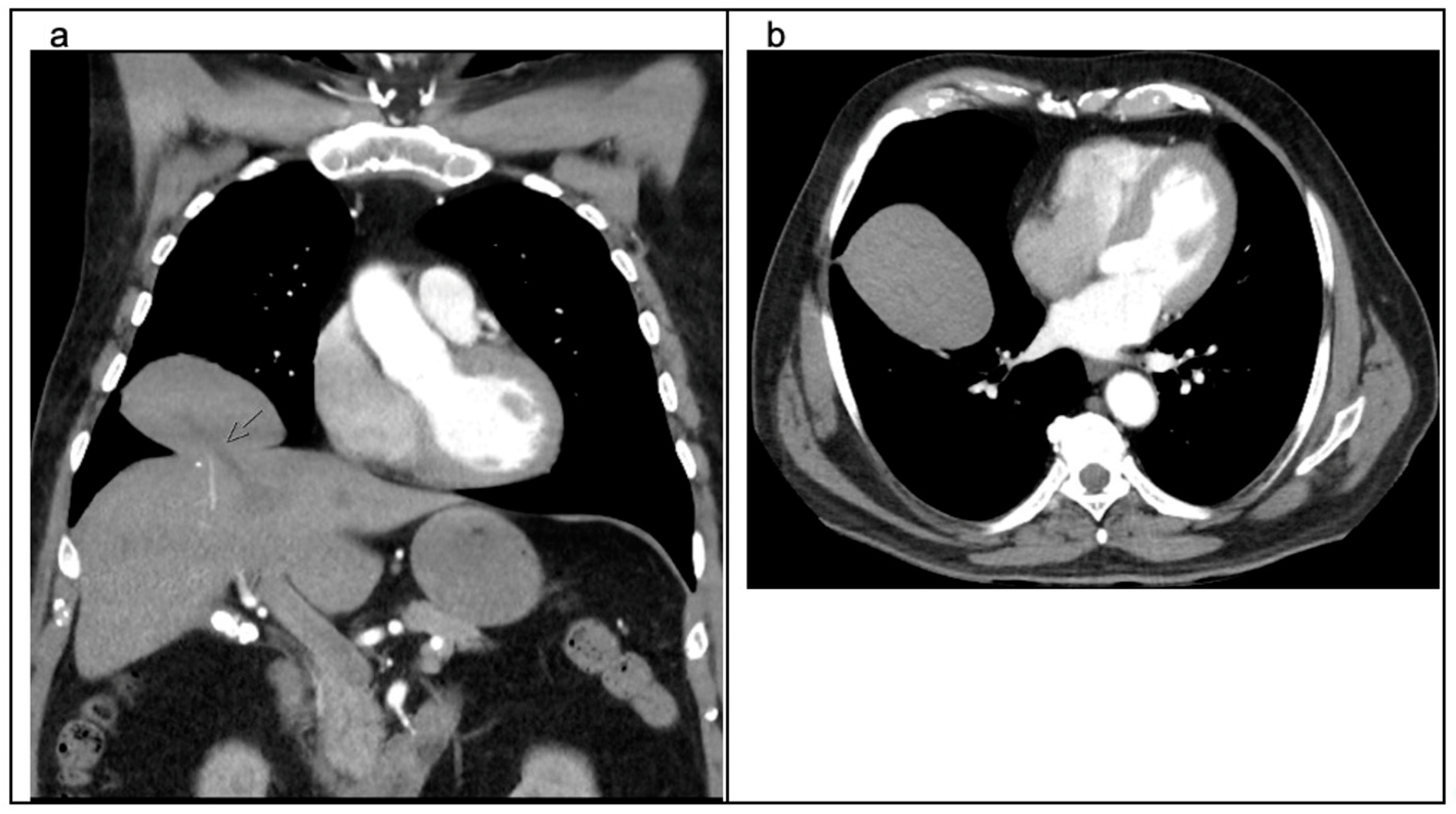
Abstract
:1. Introduction
2. Detailed Case Description
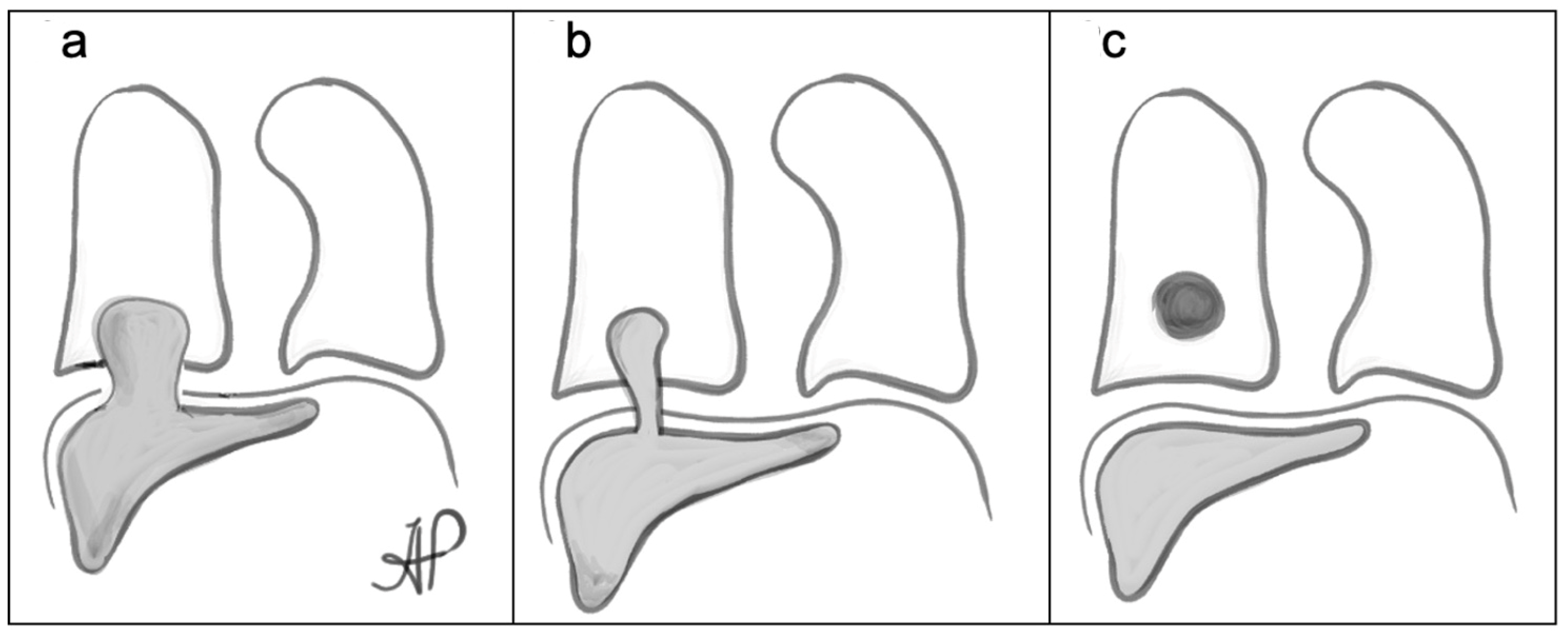
3. Discussion
4. Conclusions
Author Contributions
Funding
Institutional Review Board Statement
Informed Consent Statement
Data Availability Statement
Conflicts of Interest
References
- Negru, D.; Tudorache, C.; Marinescu, M.; Grigoras, M.; Crumpei, F.; Georgescu, S. Torsion and infarction of an accessory liver lobe. Rev. Med. Chir. Soc. Med. Nat. Iasi. 2007, 111, 442–445. [Google Scholar] [PubMed]
- Garba, E.S.; Ameh, E.A. Isolated rupture of an accessory liver from blunt abdominal trauma in childhood. Pediatr. Surg. Int. 2002, 18, 62–63. [Google Scholar] [CrossRef] [PubMed]
- Jambhekar, K.; Pandey, T.; Kaushik, C.; Shah, H.R. Intermittent torsion of accessory hepatic lobe: An unusual cause of recurrent right upper quadrant pain. Indian J. Radiol. Imaging 2010, 20, 135–137. [Google Scholar] [CrossRef] [PubMed]
- Faraj, W.; Dar, F.; Marangoni, G.; Alvarez, F.E.; Howlader, M.; Mukherji, D.; Heaton, N.; Rela, M. Liver transplantation for syndromic biliary atresia with a pedunculated accessory hepatic lobe. Pediatr. Transplant. 2010, 14, E1–E3. [Google Scholar] [CrossRef] [PubMed]
- Pujari, B.D.; Deodhare, S.G. Symptomatic accessory lobe of liver with a review of the literature. Postgrad. Med. J. 1976, 52, 234–236. [Google Scholar] [CrossRef] [PubMed]
- Wang, C.; Cheng, L.; Zhang, Z.; Xie, T.; Ding, H.; Deng, Q.; Yuan, Z. Accessory lobes of the liver: A report of 3 cases and review of the literature. Intractable Rare Dis. Res. 2012, 1, 86–91. [Google Scholar] [CrossRef]
- Kutner, M.A.; Bromberg, A.; Korst, R.J.; Lee, B.E. An unusual case of dysphagia from intrathoracic liver. Ann. Thorac. Surg. 2010, 90, e54–e55. [Google Scholar] [CrossRef]
- Eiserth, P. Beiträge zur Kenntnis der Nebenlebern. Virchows Arch. 1940, 307, 307–313. [Google Scholar] [CrossRef]
- Watanabe, M.; Matsura, T.; Takatori, Y.; Ueki, K.; Kobatake, T.; Hidaka, M.; Hirakawa, H.; Fukukmoto, S.; Shimada, Y. Five cases of ectopic liver and a case of accessory lobe of the liver. Endoscopy 1989, 21, 39–42. [Google Scholar] [CrossRef]
- Sato, S.; Watanabe, M.; Nagasawa, S.; Niigaki, M.; Sakai, S.; Akagi, S. Laparoscopic observations of congenital anomalies of the liver. Gastrointest. Endosc. 1998, 47, 136–140. [Google Scholar] [CrossRef]
- Adin, M.E.; Çetinçakmak, M.G.; Deniz, M.A.; Göya, C. Accessory liver within the thoracic cavity. Surg. Radiol. Anat. 2018, 40, 1085–1091. [Google Scholar] [CrossRef]
- Kurt, A.; Yazıcıoğlu, K.R.; Tosun, Ö.; Coşkun, M. Right sided diaphragmatic hernia in an adult without history of trauma: Unusual CT findings. Eur. J. Gen. Med. 2004, 1, 55–57. [Google Scholar]
- Lemaigre, F.P. Mechanisms of Liver Development: Concepts for Understanding Liver Disorders and Design of Novel Therapies. Gastroenterology 2009, 137, 62–79. [Google Scholar] [CrossRef]
- Ito, F.; Ando, H.; Watanabe, Y.; Seo, T.; Murahashi, O.; Harada, T.; Kaneko, K.; Ishiguro, Y. An accessory lobe of the liver disturbing closure of the umbilical ring. Pediatr. Surg. Int. 1999, 15, 394–396. [Google Scholar] [CrossRef]
- Nora, E.; Carr, C.E. Umbilical accessory liver. Am. J. Obstet. Gynecol. 1946, 52, 330–335. [Google Scholar] [CrossRef]
- Kesavaramanujam, S.; Morell, M.C.; Harigovind, D.; Bhimmanapalli, C.; Cassaro, S. Total thoracic herniation of the liver: A case of delayed right-sided diaphragmatic hernia after blunt trauma. Surg. Case Rep. 2020, 6, 178. [Google Scholar] [CrossRef]
- Elmasalme, F.; Aljudaibi, A.; Matbouly, S.; Hejazi, N.; Zuberi, M.S. Torsion of an accessory lobe of the liver in an infant. J. Pediatr. Surg. 1995, 30, 1348–1350. [Google Scholar] [CrossRef] [PubMed]
- Grunz, J.; Luisiri, A.; Cradock, T. Torsion of a hepatic lobe in the neonate–ultrasound findings. Pediatr. Radiol. 1992, 22, 192–193. [Google Scholar] [CrossRef] [PubMed]
- Sangüesa, C.; Esteban, M.J.; Gomez, J.; Cotina, H. Liver accessory lobe torsion in the infant. Pediatr. Radiol. 1995, 25, 153–154. [Google Scholar] [CrossRef] [PubMed]
- Azmy, A.; Boddy, S.A.; Eckstein, H.B. Torsion of gall bladder, embedded in an accessory lobe of liver in a neonate with Beckwith syndrome. Z. Kinderchir. Grenzgeb. 1980, 30, 277–279. [Google Scholar] [CrossRef] [PubMed]
- Ladurner, R.; Brandacher, G.; Mark, W.; Iannetti, C.; Lottersberger, C.; Steurer, W.; Königsrainer, A.; Margreiter, R. Complete hepatic ischemia due to torsion of a large accessory liver lobe: First case to require transplantation. Transpl. Int. 2005, 18, 467–469. [Google Scholar] [CrossRef]
- Woldeyes, D.H. The occurrence of an additional (accessory) lobe of liver and undescended testis in a single cadaver: A case report. J. Med. Case Rep. 2019, 13, 357. [Google Scholar] [CrossRef]
- Wynn, J.; Yu, L.; Chung, W.K. Genetic causes of congenital diaphragmatic hernia. Semin. Fetal Neonatal Med. 2014, 19, 324–330. [Google Scholar] [CrossRef] [PubMed]
- Silva, C.I.S.; Müller, N.L. Case 158. In The Teaching Files: Chest; Elsevier Enhanced Digital Version; Elsevier Health Sciences: Amsterdam, The Netherlands, 2010. [Google Scholar]
- Spellar, K.; Lotfollahzadeh, S.; Gupta, N. Diaphragmatic Hernia. [Updated 21 August 2023]. In StatPearls [Internet]; StatPearls Publishing: Treasure Island, FL, USA, 2024. [Google Scholar]
- Han, S.; Soylu, L. Accessory liver lobe in the left thoracic cavity. Ann. Thorac. Surg. 2009, 87, 1933–1934. [Google Scholar] [CrossRef] [PubMed]
- Chapman-Fredricks, J.; Birusingh, R.; Ricci, M.; Rodriguez, M. Intracaval liver with cardiac extension. A new developmental anomaly? Fetal Pediatr. Pathol. 2010, 29, 401–406. [Google Scholar] [CrossRef] [PubMed]
- Chen, Y.Y.; Huang, T.W.; Chang, H.; Hsu, H.H.; Lee, S.C. Intrathoracic caudate lobe of the liver: A case report and literature review. World J. Gastroenterol. 2014, 20, 5147–5152. [Google Scholar] [CrossRef] [PubMed]
- Ung, K.A.; Campbell, B.A.; Duplan, D.; Ball, D.; David, S. Impact of the lung oncology multidisciplinary team meetings on the management of patients with cancer. Asia Pac. J. Clin. Oncol. 2016, 12, e298–e304. [Google Scholar] [CrossRef] [PubMed]
- Ost, D.E.; Gould, M.K. Decision Making in Patients with Pulmonary Nodules. Am. J. Respir. Crit. Care Med. 2012, 185, 363–372. [Google Scholar] [CrossRef] [PubMed]
- Hierro, C.R.; Rueda, F.V.; Cruz, V.V.; Betancor, C.L.; Montoro, J.A. Focal nodular hyperplasia on accessory lobe of the liver: Preoperative diagnosis and management. J. Pediatr. Surg. 2013, 48, 251–254. [Google Scholar] [CrossRef] [PubMed]
- Wang, X.; Zhang, Q.; Xu, K. Hepatocellular carcinoma arising from left accessory liver lobe supplied by the branch of left hepatic artery: A case report. Medicine 2019, 98, e16912. [Google Scholar] [CrossRef]
- Huo, L.; Dang, Y.; Feng, R.; Zhuang, H.; Li, F. Hepatocelluar carcinoma in an accessory lobe of the liver revealed by 11C-acetate PET with a negative finding on FDG imaging. Clin. Nucl. Med. 2012, 37, 393–395. [Google Scholar] [CrossRef] [PubMed]
- Arakawa, M.; Kimura, Y.; Sakata, K.; Kubo, Y.; Fukushima, T.; Okuda, K. Propensity of ectopic liver to hepato- carcinogenesis: Case reports and a review of the literature. Hepatology 1999, 29, 57–61. [Google Scholar] [CrossRef] [PubMed]
- Nagavalli, A.; Polanski, S.; Peterson, C.M.; Birkholz, J.H.; Burdette, A.S. The liver twist: A case of accessory liver lobe torsion presenting after mild trauma. Radiol. Case Rep. 2021, 16, 2817–2823. [Google Scholar] [CrossRef] [PubMed]
- Seo, U.H.; Lee, H.J.; Ryu, W.S.; Kwak, J.M.; Shin, B.K.; Kim, W.B.; Lee, S.I.; Park, S.S.; Choi, J.W.; Kim, S.H.; et al. Laparoscopic Resection of a Hepatocellular Carcinoma Arising from an Ectopic Liver. Surg. Laparosc. Endosc. Percutaneous Tech. 2008, 18, 508–510. [Google Scholar] [CrossRef]
- Lasser, A.; Wilson, G.L. Ectopic liver tissue mass in the thoracic cavity. Cancer 1975, 36, 1823–1826. [Google Scholar] [CrossRef]
- Tancredi, A.; Cuttitta, A.; Giorgio, D.; Scaramuzzi, R. Ectopic hepatic tissue misdiagnosed as a tumor of lung. Updates Surg. 2010, 62, 121–123. [Google Scholar] [CrossRef]


Disclaimer/Publisher’s Note: The statements, opinions and data contained in all publications are solely those of the individual author(s) and contributor(s) and not of MDPI and/or the editor(s). MDPI and/or the editor(s) disclaim responsibility for any injury to people or property resulting from any ideas, methods, instructions or products referred to in the content. |
© 2024 by the authors. Licensee MDPI, Basel, Switzerland. This article is an open access article distributed under the terms and conditions of the Creative Commons Attribution (CC BY) license (https://creativecommons.org/licenses/by/4.0/).
Share and Cite
Polikarpova, A.; Bains, H.K.; Thomson, S.; Gao, Y.; Morris, D.L. An Incidental Discovery of the Intrathoracic Accessory Liver Lobe in a 72-Year-Old Man: Case Report and Literature Review. Surgeries 2024, 5, 84-91. https://doi.org/10.3390/surgeries5010010
Polikarpova A, Bains HK, Thomson S, Gao Y, Morris DL. An Incidental Discovery of the Intrathoracic Accessory Liver Lobe in a 72-Year-Old Man: Case Report and Literature Review. Surgeries. 2024; 5(1):84-91. https://doi.org/10.3390/surgeries5010010
Chicago/Turabian StylePolikarpova, Aleksandra, Harinder K. Bains, Samuel Thomson, Yijun Gao, and David L. Morris. 2024. "An Incidental Discovery of the Intrathoracic Accessory Liver Lobe in a 72-Year-Old Man: Case Report and Literature Review" Surgeries 5, no. 1: 84-91. https://doi.org/10.3390/surgeries5010010