Review

Insights into Androgen Receptor Action in Lung Cancer

1
Leiden University Medical Center, 2333 ZA Leiden, The Netherlands
2
Department of Molecular Biology and Endocrinology, “Vinča” Institute of Nuclear Sciences, University of Belgrade, 11000 Belgrade, Serbia
3
Center for Molecular Medicine, University Medical Center Utrecht, 3584 CG Utrecht, The Netherlands
*
Author to whom correspondence should be addressed.
Endocrines 2023, 4(2), 269-280; https://doi.org/10.3390/endocrines4020022
Submission received: 10 January 2023 / Revised: 28 February 2023 / Accepted: 28 March 2023 / Published: 3 April 2023
(This article belongs to the Section Endocrine Oncology)

Abstract

:
Sex hormones and their receptors play a crucial role in human sexual dimorphism and have been traditionally associated with hormone-dependent cancers like breast, prostate, and endometrial cancer. However, recent research has broadened our understanding by revealing connections with other types of cancers, such as lung cancer, where the androgen receptor has been found to be particularly significant. This review aims to explore the molecular mechanisms of androgen action in lung cancer pathogenesis and progression, highlighting the potential of inhibiting the androgen receptor signaling pathway as a therapeutic strategy for lung cancer treatment.

1. Introduction

Lung cancer stands as the foremost cause of cancer-related mortality globally, annually causing nearly 1.8 million fatalities [1], with an estimated 127,000 deaths projected for the year 2023 in the United States by the American Cancer Society [2]. This disease is often asymptomatic, diagnosed at a late stage [3], and characterized by the lowest 5-year relative survival rate out of all cancer types [4]. Importantly, reliable biomarkers for early stages of lung cancer are still missing [5]. Different types of lung cancer exist and can broadly be divided into two major groups: non-small cell lung cancer (NSCLC) and small cell lung cancer (SCLC). The former type, which includes lung squamous cell carcinoma, lung small cell carcinoma, and lung adenocarcinoma, accounts for 80% of all lung cancer cases [4]. The most common subtype of NSCLC is adenocarcinoma, which represents the most prevalent subtype among non-smokers [6]. SCLC is usually diagnosed in patients with smoking history and older than the age of 70, with a major fraction of these patients succumbing to the consequences of the disease rapidly [7].
The main risk factor associated with lung cancer development is tobacco smoke, which contains numerous carcinogens that can cause DNA damage [3]. Smoking-related DNA damage can lead to mutations in tumor suppressor genes, such as p53, which normally help prevent the accumulation of DNA damage and, thus, prevent the development of cancer. In addition to smoking, environmental and/or occupational exposure to pollution, chronic lung disease, lifestyle factors, and genetic factors may also contribute to the development of lung cancer [3]. Mutations in EGFR, KRAS, BRAF, and HER2 have been identified as drivers of lung cancer [8], and the fusion of ALK, ROS1, and RET protein tyrosine kinase oncogenes with multiple partner genes has been linked to the development and progression of non-small cell lung cancer, particularly lung adenocarcinoma [9]. Recent studies have also suggested that sex hormone receptors and their corresponding ligands may play a role in the pathogenesis of lung cancer [10]. Sex hormone receptors, including androgen receptor (AR), estrogen receptors (ER) α and β, as well as progesterone receptor (PR)-A and PR-B, belong to the steroid receptor subgroup of the nuclear receptor (NR) superfamily, which consists of 48 members of transcription factors (TFs) with roles in metabolism, reproduction, inflammation, and development [11]. Dysregulation of sex hormone signaling has been implicated in the development of different types of cancer, including prostate, breast, and lung cancer [10].
It has been reported that females have a higher incidence of lung cancer than males. However, despite frequent diagnosis at advanced stages, females tend to have better 5-year survival outcomes, and, in general, a better prognosis compared to the males [12]. Additionally, there are differences in subtype prevalence, with adenocarcinoma and bronchioloalveolar carcinoma being diagnosed more often in females than males, even after accounting for smoking status as a risk factor [13]. This points to a possible role of sex hormones and their receptors in the development and progression of lung cancer. As the role of ER in lung cancer biology has been extensively reviewed by others [8,14,15,16], the aim of this review is to provide a comprehensive overview of studies investigating the role of androgens and the AR in normal lung development and lung cancer. This includes information on association with survival and disease progression, molecular mechanisms, pathway crosstalk, and potential applications of inhibitors of AR signaling pathway. To that end, a literature analysis was conducted to collect information available concerning the link between the AR, its ligands, and lung cancer. Relevant literature was identified through search engines PubMed and Google Scholar, using the search terms (and their respective synonyms terms) “sex hormone”, “androgen receptor (AR)”, “androgen”, “testosterone”, “dihydrotestosterone”, and “lung cancer” in different combinations. Only studies published in English were included in this review.

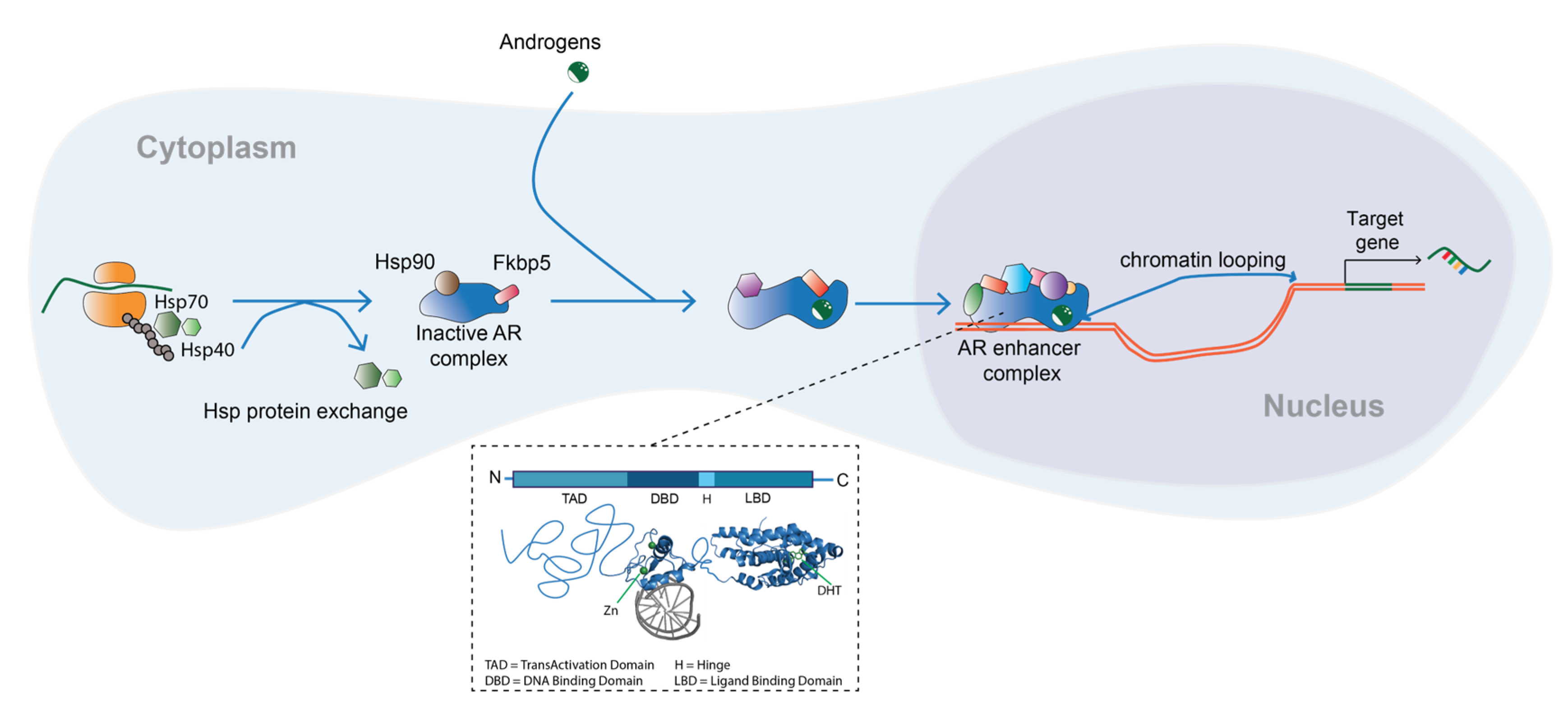
2. Androgens and the Androgen Receptor

Sex hormones are synthesized in the gonads (ovaries and testes), adrenal glands, or locally in other tissues [11]. Cholesterol serves as the precursor to all steroid hormones, including glucocorticoids and mineralocorticoids. Essential for the sex hormone signaling pathway, the enzyme P450-linked side chain cleaving enzyme, converts cholesterol into pregnenolone, which is subsequently used as a precursor for the synthesis of both estrogens and androgens. Pregnenolone is further converted to various, less potent androgens, which ultimately serve as backbones for testosterone biosynthesis. Most of the synthesized testosterone is bound to albumin and sex-binding hormone globulin (SBHG), and only 2–3% circulates free in the serum as biologically active [17,18]. In particular tissues, testosterone itself can be converted to a more potent dihydrotestosterone (DHT) hormone by the enzyme 5-α reductase [19]. Three isoenzymes of 5-α reductase exist: types 1, 2, and 3 [17]. Type 3 is of particular interest as it has been identified in both benign and malignant lung tissue and its overexpression has been detected in lung adenocarcinoma samples [20].
An androgen receptor (AR) is a member of the nuclear receptor (NR) family that shares structural similarities with other members, including estrogen receptors (ER), glucocorticoid receptors (GR), and progesterone receptors (PR). After androgens bind to AR, confirmation of the receptor changes and the protein translocates to the nucleus, where it binds to specific DNA response elements, known as hormone response elements (HREs), mostly found at enhancer regions, distal to AR target genes. After AR engages with HREs, nuclear proteins, such as co-activators and co-repressors, are recruited to ultimately influence transcriptional output of its target genes [6,21] (Figure 1). This is the classical mode of action of AR. Additionally, androgens can also induce rapid activation of other non-classical pathways in cells, including the MAPK pathway, PI3K/Akt pathway, and Src tyrosine kinase pathway [22].
The intricate structure of the AR is characterized by its multi-domain composition, consisting of three distinct functional units (Figure 1). These include an N-terminal transactivation domain (NTD), a DNA-binding domain (DBD), and a ligand-binding domain (LBD) [23]. The NTD houses an activation function (AF)-1 domain that works in tandem with co-regulatory proteins to modulate gene transcription. Meanwhile, the DBD is responsible for binding to specific DNA sequences, known as androgen response elements (AREs), which are strategically located in the promoter regions of target genes. Lastly, the LBD is composed of two critical components, the AF-2 domain and the ligand-binding pocket. The AF-2 domain interacts with co-regulatory proteins, whereas the ligand-binding pocket binds to androgens. Notably, the hinge region connects the DBD and LBD and is vital for AR stability and function [23].
The complexity of the AR is further illustrated by multiple splice variants, including the well-known AR-V7. The constitutively active AR-V7 lacks the ligand-binding domain (LBD) and has been linked to resistance to androgen deprivation therapy in prostate cancer patients [24]. Additionally, other splice variants, such as AR-V1, AR-V2, and AR-V3, could also play a role in the development and progression of prostate cancer [25]. Furthermore, mutations in AR can affect its activity and are associated with various diseases, such as androgen insensitivity syndrome (AIS) and prostate cancer [26]. These mutations can occur in different domains of the receptor, including the DNA-binding domain (DBD) and LBD. Mutations in the LBD can alter the specificity of ligand binding, enabling activation by other hormones, such as progesterone and estrogen [26].
The AR mediates effects of male sex hormones in reproductive and non-reproductive organ systems. Following its discovery in the 1960s, the expression of this receptor was demonstrated in several tissues, including bone, muscle, prostate, and adipose tissue [27]. Over the years, it became evident that the role of AR extends beyond sexual development, with its signaling contributing to various biological processes—for example, brain development, metabolism, and the immune system function [28]. Moreover, researchers demonstrated that AR plays a pivotal role in several pathologies, including androgen insensitivity syndrome, Kennedy’s disease and cancer [29,30]. In particular, in terms of cancer, AR is the critical driver of prostate cancer; nonetheless, its dysregulation has also been proposed to contribute to the development of bladder, kidney, breast, and liver cancer [31,32].

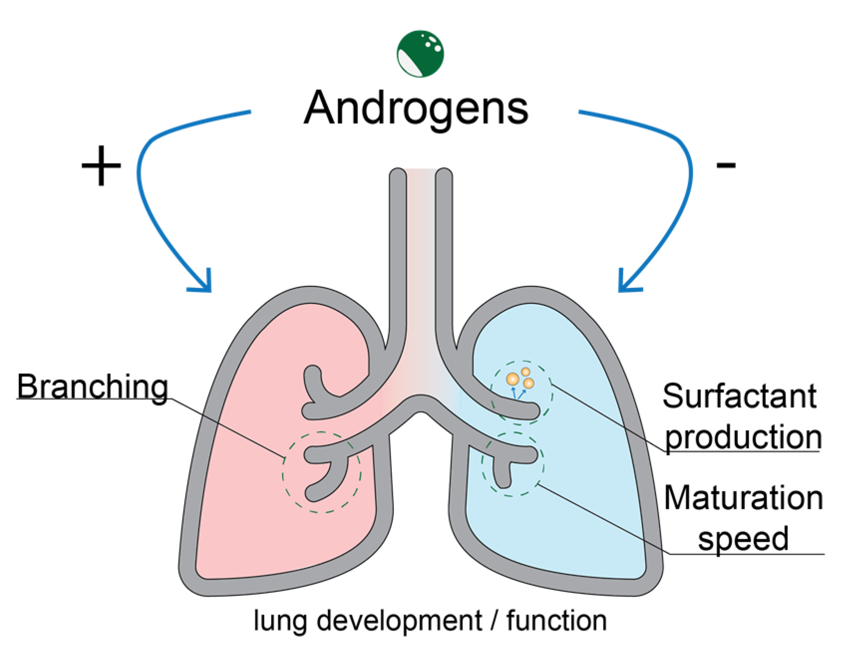
3. Role of the Androgen Receptor in Lung Development

In addition to their role in sexual development, sex hormones play a role in the development of the lung. Mammalian lung development occurs in five consecutive histologically distinct stages: embryonic, pseudoglandular, canalicular, saccular, and alveolar [33,34]. Transitions between stages are under multifunctional molecular control, which involves growth factors, TFs, glucocorticoids, and sex hormones [34]. The fetal lung is exposed to circulating sex hormones produced by C19 steroid precursors in maternal and fetal adrenals. Moreover, in fetal lungs, sex hormones can be inactivated by specific enzymes, adding another layer of regulation [33]. Normal lung development differs between males and females, with maturation and surfactant production occurring earlier in females compared to males [35,36].
Androgens produce distinct effects throughout lung development, depending on the stage. They can act as positive regulators in the process of branching morphogenesis in early development and as negative regulators responsible for delay in male lung maturation in late stages (Figure 2) [33]. Branching morphogenesis occurs at week five of gestation, with all airway branching occurring by week 24 of fetal life; AR is predominantly expressed in epithelial cells found at budding sites during this process [37]. The expression of AR was shown in both murine and human type II pneumocytes and bronchial epithelium [31]. The negative influence of androgens on the lung development was demonstrated in vivo, whereby male animals were found to have less developed lungs compared to their female counterparts [34,36]. Surfactant production in the fetal developing lung is under multifactorial and multihormonal control. Glucocorticoids and estrogens can accelerate the production of surfactant phospholipids and proteins, while androgens can inhibit it [38]. The inhibitory role of testosterone and DHT on pulmonary surfactant production was shown in organ cultures and in vivo mouse and rabbit models, where the effects were attributed to hormone interactions with the AR found in lung fibroblasts and alveolar type II cells [39]. Despite the well-established role of AR in lung development, the precise role of AR in normal lung physiology remains uncertain and warrants further investigation.

4. Androgens and the Androgen Receptor in Lung Cancer

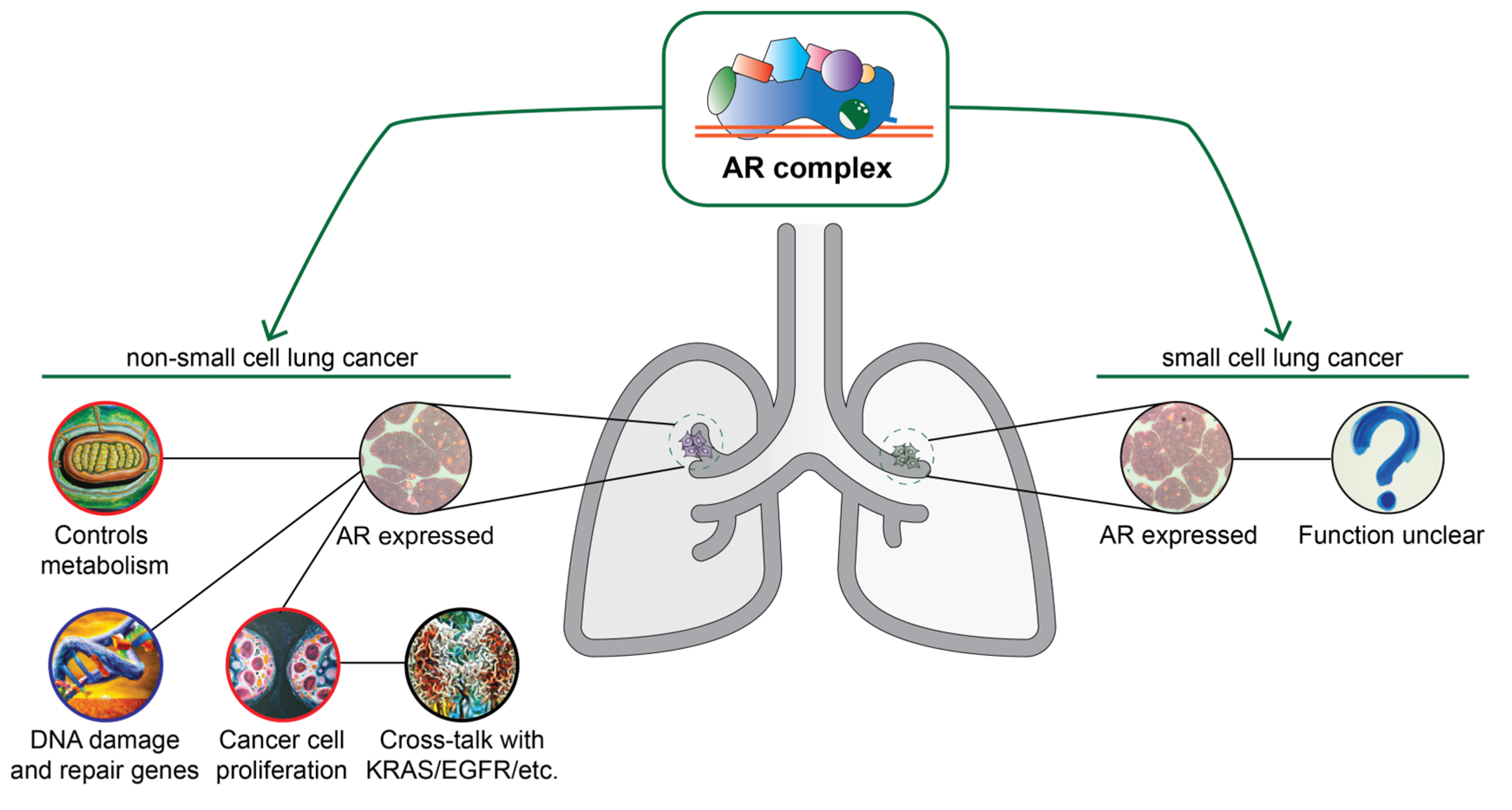
4.1. Androgen Receptor Signaling in Lung Cancer

Early research efforts demonstrated a presence of the functional AR protein in lung cancer, specifically SCLC [40]. In addition to the presence of the receptor and its ability to readily bind the ligand, it was also shown that 5α-reductase is expressed in SCLC models, although at only 10% of the level found in prostate cancer cell lines. Since then, AR expression has also been demonstrated in the more prevalent subtype of lung cancer, NSCLC, both in cell lines and primary lung cancer material [19]. Following these observations, several studies have confirmed the presence of AR in cell line models of lung cancer [32,41]. Moreover, alterations of the AR (i.e., amplifications, deletions, or mutations) have been observed in 5% of lung squamous carcinoma and adenocarcinoma, further suggesting an important role in lung cancer [42].
In addition to the expression of the functional AR protein, activating enzymes that convert steroid precursors to biologically active sex hormones are expressed in lung cancer cell lines [33]. Provost et al. (2000) investigated the intrinsic capacity of A549 to metabolize sex hormones. Cells contained 17β-hydroxysteroid dehydrogenase (HSD) type 5, which catalyzes the transformation of 4-androstenedione into testosterone, and 3α-HSD type 3, which predominantly converts dehydroepiandrosterone (DHEA) to androstenediol, which can also activate the AR. Despite high expression levels of AR, low 3β-HSD activity was detected in an in vitro model, indicative of low capacity of the cell line to produce dihydrotestosterone (DHT) and testosterone themselves [39]. Furthermore, the expression of enzymes that inactivate sex hormones has also been demonstrated in lung cancer cells, as well as in mouse and human fetal lung tissue. For example, low hydroxysteroid 17-β dehydrogenase 6 (17β-HSD B6) expression has an impact on the progression of lung adenocarcinoma [43]. Furthermore, 17β-HSD B6 has both oxidoreductase and epimerase activities and an important function in androgen catabolism. It catalyzes the conversion of 3α-adiol to DHT and the conversion of androsterone to epi-androsterone, and its downregulation has already been linked to poor patient prognosis in hepatocellular carcinoma [44]. It has been demonstrated that 17β-HSD B6 is expressed at low levels in lung adenocarcinoma compared to normal lung tissue. Low expression of 17β-HSD B6 was associated with poor clinicopathological features, including tumor size, advanced tumor stage, low tumor differentiation, and poor patient prognosis [43]. Moreover, it was found that 17β-HSD B6 reduces radio resistance in lung cancer cells and suppresses proliferation, migration, invasion, and epithelial–mesenchymal transition. The research has found evidence that the potential mechanism includes activating PTEN expression and inhibiting AKT phosphorylation [45].
Androgens can affect proliferation of cancer cells. Testosterone is known to have a significant impact on protein synthesis rates, potentially resulting in heightened basal cell metabolism and increased energy [46]. Along the same line, AR activation in murine cells causes upregulation of genes involved in oxygen transport and heme biosynthesis and negative regulation of genes involved in apoptosis, DNA repair, and double-strand break repair [47]. The link between testosterone and hormone-dependent cancers (e.g., prostate cancer) has been well-researched, although with contrasting conclusions about its association to incidence [46].
Recently, studies have investigated the effect of androgens on cellular processes and signaling pathways in NSCLC. In AR-positive murine lung tissue and human NSCLC cell lines, androgen treatment induced profound changes to the transcriptional landscape. Particularly in the A549 cell line model, genes involved in oxygen transport and utilization were upregulated, while the ones involved in DNA repair and DNA recombination were downregulated, consistent with the proposed pro-carcinogenic role of androgens in lung cancer [47]. Wang et al. (2018) treated different NSCLC cell lines with agonists and antagonists of AR and found that the effect was dependent on the KRAS mutational status, proposing that a crosstalk between AR and KRAS exists [42]. This further suggests that AR co-operates with both EGFR (as well as other growth factor signaling pathways) and the Raf/MEK/ERK pathway linked to in various processes, such as differentiation, growth, chemotaxis, and apoptosis [48]. In relation to this, AR knockdown in NSCLC cell lines resulted in a decrease of cyclin D1 expression and subsequently led to inhibition of cell proliferation and anchorage-independent growth in vitro [49,50]. Additionally, suppression of AR expression was hallmarked by a decrease of OCT-4 protein expression, suggesting an interplay between the AR axis and the pathway affecting stemness and self-renewal [49].
Apart from the potential interaction between AR and KRAS, Recchia et al. (2009) shed light on the involvement of EGFR in DHT-induced growth stimulation of A549 lung cancer cells and LNCaP prostate tumor cells [6]. Their study uncovered that DHT treatment led to the up-regulation of CD1 and consequent cell proliferation, both of which could be mitigated by selective inhibitors or knockdown of either AR or EGFR. These findings suggest that the cross-talk between AR and EGFR plays a significant role in the progression of prostate and lung cancer through the activation of the mTOR/CD1 pathway [6].
Compelling evidence for the role of AR in lung cancer comes from the study by Noronha et al. (1983). Female NZR-GD rats treated with a single dose of 20 mg/kg intraperitoneal administration of dimethylnitrosamine (DMN) typically develop lung, kidney, liver, and nasal cavity tumors [51]. It was observed that testosterone treatment before DMN injections resulted in enhanced lung tumor formation, while the same effects could not be observed in the cases of hepatocellular and kidney epithelial tumors [51]. In agreement with the latter are the findings coming from experiments performed in AR knockout (ARKO) mouse models [31]. The ARKO model was used in an NNK-BaP (eight doses of tobacco carcinogens, 4-(methylnitrosamino)-1-(3–pyridyl)-1-butanone (NNK) and benzo[a]pyrene (BaP))-induced mouse model of lung cancer; AR knockout resulted in reduced tumor size compared with control NNK-BaP mice [50].
In summary, the above-discussed research indicates that androgen receptor (AR) expression and activity have been observed in both SCLC and NSCLC. Alterations of the AR gene have been found in lung squamous carcinoma and adenocarcinoma. The expression of the enzymes that convert steroid precursors to biologically active sex hormones and those that inactivate them in lung cancer cells have been shown to impact lung adenocarcinoma progression. Androgens affect cancer cell proliferation and energy consumption, with AR activation leading to upregulation of genes involved in oxygen transport and downregulation of those involved in apoptosis and DNA repair (Figure 3). In vivo studies have revealed that DMN may enhance lung tumor formation, and that AR knockout results in reduced tumor size in mice (Figure 3). These findings suggest that targeting AR signaling may have potential therapeutic implications in lung cancer treatment.

4.2. Androgen Receptor as an Important Factor in Lung Cancer Biology: Evidence from Clinical Studies

The correlation between androgens, intra-tumor AR expression, and patient outcomes has been investigated in numerous studies. Using immunoassays, Hyde et al. (2012) investigated the relationship between total blood T levels and lung cancer risk in a cohort of 3635 community-dwelling elderly men. High T levels were associated with an increase in lung cancer risk, even after accounting for smoking as a double confounder [18]. Using the samples of the same cohort, Chan et al. (2017) investigated the association between plasma T and DHT levels with lung cancer incidence by performing liquid chromatography–mass spectrometry (LC-MS). Comparable to the findings of the first study, higher T and DHT plasma levels were associated with an increased risk of developing lung cancer. One standard deviation (SD) increase in total T, equivalent to 4.87 nmol/L, significantly increased the adjusted risk of lung cancer (HR = 1.30, 95% CI 1.06–1.60; p = 0.012). For every 1 SD (0.73 nmol/l) increase in DHT, the adjusted risk of lung cancer increased by 29% (HR = 1.29, 95% CI 1.08–1.54; p = 0.004). When accounting for current smoking status, higher plasma levels of T did not have an association with lung cancer, however, DHT remained associated with increase in lung cancer risk (HR = 1.26, 95% CI 1.03–1.54; p = 0.026) [41]. In contrast to these reports, Ørsted et al. (2014), who conducted a prospective cohort study (291 male and 193 female participants included, with a median follow up of 22 years), found no association between plasma T levels and the risk of developing lung cancer [46].
In respect to intra-tumor AR expression, Hattori et al. (2015) demonstrated that the AR is expressed only in a minor fraction (18/566 cases) of primary lung cancer tissues (which included squamous cell carcinoma, large neuroendocrine carcinoma, small cell carcinomas, and typical carcinoids) [52]. Irrespective of gender, an immunohistochemistry-based study by Grant et al. (2018) found no association between the AR expression levels and overall survival (OS) or recurrence of NSCLC in 136 tissue samples from Manitoba Tumor Bank (MTB) patients [12]. However, another study by Berardi et al. (2016) investigated the expression of sex hormone receptors in 62 patients with advanced NSCLC and found significant correlation with outcome. They linked the intra-tumor presence of AR, ER-α, and PgR to a significant correlation with outcomes in patients with advanced NSCLC. Patients with nuclear and cytoplasmic AR expression exhibited better survival when compared to those not expressing the protein (49 and 45 months, respectively, vs. 19 months)—although the number of patients demonstrating nuclear AR expression was relatively low (8/62) [53]. In contrast with the latter, Skjefstad et al. (2016), who collected tumor tissues from 335 patients and evaluated the expression of AR in the cytoplasm and nucleus of tumor epithelial and stromal cells, found that both male and female patients had worse outcomes with respect to disease-specific survival compared to the rest of the cohort [54].
Examination of mutation profiles in cell-free DNA found in plasma samples of lung cancer patients revealed that 48.28% of patients exhibited the androgen receptor p.H875Y mutation, which had previously been detected in prostate and breast cancer [55]. Moreover, the researchers found that cancer samples with AR mutation showed a higher total mutational burden compared to the samples without it. Previous reports have shown that mutations of residue 875 of AR expand the ligand repertoire of the receptor, allowing for activation by other steroid hormones [26]. However, the exact functional relevance of this mutation in lung carcinogenesis is yet to be investigated [55].
Furthermore, Rades et al. (2012) investigated the association of AR, ER-α, and PR expression with locoregional control, metastases-free survival, and OS in stage II/III NSCLC tumors of patients who received radiotherapy (RT) [4]. Of importance, while ER-α expression was associated with worse treatment outcomes with regards to locoregional control, metastasis-free survival, and OS in both females and males, no significant association was found between AR or PR expression and these parameters [4]. Regarding OS, a trend towards improved locoregional control was seen for lower N classification and not smoking during RT [4].
The relationship between androgens and lung cancer risk has been studied extensively, with some studies showing that high levels of total testosterone and dihydrotestosterone (DHT) are associated with increased risk of developing lung cancer. However, other studies have found no association between plasma testosterone levels and lung cancer risk. The expression of AR within primary lung cancer tissues is limited, but some studies have shown a correlation between the presence of AR, ER-α, and PgR and better outcomes in patients with late-stage metastatic non-small cell lung cancer (NSCLC). In contrast, other studies have found that patients with AR expression in tumor epithelial and stromal cells had worse outcomes in terms of disease-specific survival. A mutation of the AR gene, p.H875Y, has been found in plasma samples of lung cancer patients and may play a role in lung carcinogenesis, but its functional relevance is yet to be fully understood. In patients with stage II/III NSCLC who received radiotherapy, no association was found between AR expression and treatment outcome, while ER-α expression was associated with worse treatment outcomes.

4.3. Targeting of the Androgen Receptor Pathway

Blocking the androgen receptor signaling pathway can be achieved in several ways, specifically by either targeting AR or the enzymes responsible for the conversion of steroid precursors into T or DHT (as reviewed in detail in [56]). At the level of biosynthesis, CYP17A1 and 5-α reductase inhibitors, such as abiraterone and finasteride, respectively, may be used to block the enzymes responsible for production of T, and ultimately its conversion to DHT. Non-steroidal antiandrogens, such as bicalutamide, nilutamide, flutamide, or the more recently developed enzalutamide, can bind to AR and engage in competitive binding with testosterone and DHT [57]. Steroidal antiandrogens, such as cyproterone acetate, competitively inhibit the action of androgens and the secretion of luteinizing hormone (LH), which leads to decreased testosterone production. Lastly, gonadotropin-releasing hormone (GnRH) agonists, which include buserelin, goserelin, and leuprolide can lead to a downregulation of GnRH receptors and a subsequent decrease in LH and testosterone. [13].
Several studies have investigated the effects of pharmacologically targeting the androgen receptor pathway in lung cancer. In NSLC cell lines, inhibition of AR by enzalutamide has been suggested to cause radiosensitization [42]. Furthermore, expression of miR-224-5p, an exosome-secreted microRNA targeting AR, has been shown in NSCLC cancer specimens. More importantly, lentivirus-mediated inhibition of the expression of miR-224-5p led to decreased proliferative and migratory capacity of A549 and H1299 cells. Using flow cytometry, it was shown that overexpression of miR-224-5p accelerated progression through the cell cycle and inhibited apoptosis. Finally, using a tumor xenograft mouse model, researchers found that injection of A549 cells overexpressing miR-224-5p led to an increase in growth and appearance of necrotic regions in the lungs, along with higher expression of the epithelial-to-mesenchymal transition (EMT) marker N-cadherin [58].
Harlos et al. (2015) investigated the effect of androgen pathway manipulation (APM) on survival in 3018 men diagnosed with lung cancer. Of the total patient population, 339 had used a form of APM, either in the form of an antiandrogen, 5-alpha reductase inhibitor, or GnRH [13]. Patients who received APM, either partial or complete, after their lung cancer diagnosis (HR = 0.36, p = 0.0007) and those who were exposed to APM both before and after their diagnosis (HR = 0.53, p < 0.0001) had a significantly better survival compared to the patients who did not use any APM [13]. Following stratification of the patient sample based on disease stage, researchers found that exposure to APM was significantly associated with longer survival in early-stage disease if treatment occurred after diagnosis. In late-stage disease, a difference was significant in those exposed to APM after diagnosis (HR = 0.41, p = 0.02) and before and after diagnosis (HR = 0.54, p < 0.0001). The effect appeared to diminish in patients who were diagnosed with late-stage lung cancer and with each passing year of receiving therapy after diagnosis [13].
Recently, a proteasome-based system with proteolysis-targeting chimeras (PROTACs) targeting AR has been developed and its effects examined in lung cancer cell lines [1]. PROTACs are heterobifunctional small molecules that harness the ubiquitin-proteasome system to degrade the target protein. It was demonstrated that one of the enzalutamide-based PROTACs with a specific exit vector led to a dose-dependent reduction of viability in the A549 lung cancer cell line model [59].
In light of recent studies, it has become apparent that the androgen receptor and its signaling axis may hold promise as a therapeutic target for NSCLC. Pharmacological targeting of the androgen pathway has shown potential in NSCLC cell lines, with clinical research also demonstrating a correlation between androgen pathway manipulation and improved survival outcomes in lung cancer patients. These findings offer a compelling case for further investigation and development of androgen receptor-targeted therapies for NSCLC treatment.

5. Discussion

A major challenge in understanding the role of androgens in lung cancer is the inconsistent reporting of smoking status in clinical studies. Smoking can confound the effect of androgens on lung cancer as it both increases levels of unbound testosterone in healthy men and independently increases the incidence of lung cancer [18,60]. Furthermore, women who smoke have a higher risk of developing lung cancer compared to non-smoking women [16,61]. To complicate matters further, crosstalk between nuclear receptors exists, and studies on menopausal status and the risk of developing lung cancer have been inconclusive [62,63]. Given these confounding factors, future studies should aim to separate cohorts based on smoking and menopausal status to better understand the effect of androgens on lung cancer.
In addition to these challenges, the role of androgens in SCLC is still not well understood. This is likely due to the lower prevalence of this subtype and the fact that most clinical studies have focused on early rather than late-stage NSCLC, which can have a significant influence on survival analysis.
The collection time of blood or other biological specimens is crucial in studying testosterone levels, as testosterone exhibits diurnal variation with peaks and troughs throughout the day in males [64]. This diurnal variation can confound the results of epidemiological studies if the collection time of samples is not standardized. For instance, samples collected in the morning will likely have higher testosterone levels than those collected in the evening [65]. Therefore, it is important to collect samples at the same time of day to minimize the potential impact of diurnal variation.
Another challenge in assessing the effect of androgens on lung cancer is the measurement of free testosterone levels. Testosterone is mostly found circulating in its bound form to albumin or SBHG, and less than 2% is found in its free, bioactive form [18]. Blood-based association studies should, thus, include measurements of free testosterone, and LC-MS/MS should be used for more precise quantification compared to immunoassays [41]. Additionally, the localization of AR expression in stained tumor tissues may have an influence on the results, yet many immunohistochemistry studies do not specify this localization [53].
While research on the role of androgens in lung cancer has provided valuable insights, there are still many challenges that need to be addressed. Future studies should aim to better account for confounding factors, such as smoking and menopausal status, and use more precise methods for measuring free testosterone levels. These efforts will enable a more accurate understanding of the role of androgens in lung cancer and improve the development of targeted therapies for patients.

6. Conclusions

Given the growing body of research on the role of androgens and the AR in lung cancer, there is increasing interest in exploring their potential as therapeutic targets. Pharmacologically modulating the AR signaling pathway may hold promise as a novel approach to treating patients with lung cancer. To achieve this, a deeper understanding of the exact mechanisms underlying AR action in lung cancer, both at the pathway and genomic levels, is needed. This could involve probing and analyzing high-throughput datasets, such as those on mutation burden and gene expression, to identify new biomarkers and potential therapeutic targets for lung cancer. Additionally, investigations into post-translational modifications of AR could offer insights into the biochemical regulation and cellular function of this protein, providing new avenues for research and potential therapeutic interventions. Overall, further research in this area could have significant clinical implications, potentially leading to the development of novel diagnostic and treatment options for patients with this deadly disease.

Author Contributions

Investigation: D.D., M.J. and S.P.; writing—review and editing: D.D., M.J. and S.P. All authors have read and agreed to the published version of the manuscript.

Funding

This work was supported by the Alpe d’Huzes/KWF Bas Mulder Award. M.J. was supported by the Ministry for Science and Technological Development of the Republic of Serbia (451-03-47/2023-01/200017).

Institutional Review Board Statement

Not applicable.

Informed Consent Statement

Not applicable.

Data Availability Statement

Not applicable.

Acknowledgments

We express our gratitude to our colleagues for their valuable contributions and insightful feedback.

Conflicts of Interest

The authors declare no conflict of interest.

References

  1. Sung, H.; Ferlay, J.; Siegel, R.L.; Laversanne, M.; Soerjomataram, I.; Jemal, A.; Bray, F. Global cancer statistics 2020: GLOBOCAN estimates of incidence and mortality worldwide for 36 cancers in 185 countries. CA A Cancer J. Clin. 2021, 71, 209–249. [Google Scholar] [CrossRef] [PubMed]
  2. Siegel, R.L.; Miller, K.D.; Wagle, N.S.; Jemal, A. Cancer statistics, 2023. CA A Cancer J. Clin. 2023, 73, 17–48. [Google Scholar] [CrossRef] [PubMed]
  3. Bade, B.C.; Dela Cruz, C.S. Lung Cancer 2020: Epidemiology, Etiology, and Prevention. Clin. Chest. Med. 2020, 41, 1–24. [Google Scholar] [CrossRef]
  4. Rades, D.; Setter, C.; Dahl, O.; Schild, S.E.; Noack, F. The prognostic impact of tumor cell expression of estrogen receptor-alpha, progesterone receptor, and androgen receptor in patients irradiated for nonsmall cell lung cancer. Cancer 2012, 118, 157–163. [Google Scholar] [CrossRef]
  5. Rodak, O.; Peris-Díaz, M.D.; Olbromski, M.; Podhorska-Okołów, M.; Dzięgiel, P. Current landscape of non-small cell lung cancer: Epidemiology, histological classification, targeted therapies, and immunotherapy. Cancers 2021, 13, 4705. [Google Scholar] [CrossRef]
  6. Recchia, A.G.; Musti, A.M.; Lanzino, M.; Panno, M.L.; Turano, E.; Zumpano, R.; Belfiore, A.; Ando, S.; Maggiolini, M. A cross-talk between the androgen receptor and the epidermal growth factor receptor leads to p38MAPK-dependent activation of mTOR and cyclinD1 expression in prostate and lung cancer cells. Int. J. Biochem. Cell Biol. 2009, 41, 603–614. [Google Scholar] [CrossRef]
  7. van Meerbeeck, J.P.; Fennell, D.A.; De Ruysscher, D.K.M. Small-cell lung cancer. Lancet 2011, 378, 1741–1755. [Google Scholar] [CrossRef]
  8. Hsu, L.H.; Chu, N.M.; Kao, S.H. Estrogen, Estrogen Receptor and Lung Cancer. Int. J. Mol. Sci. 2017, 18, 1713. [Google Scholar] [CrossRef] [PubMed]
  9. Kohno, T.; Nakaoku, T.; Tsuta, K.; Tsuchihara, K.; Matsumoto, S.; Yoh, K.; Goto, K. Beyond ALK-RET, ROS1 and other oncogene fusions in lung cancer. Transl. Lung Cancer Res. 2015, 4, 156. [Google Scholar]
  10. Dou, M.; Zhu, K.; Fan, Z.; Zhang, Y.; Chen, X.; Zhou, X.; Ding, X.; Li, L.; Gu, Z.; Guo, M.; et al. Reproductive Hormones and Their Receptors May Affect Lung Cancer. Cell Physiol. Biochem. 2017, 44, 1425–1434. [Google Scholar] [CrossRef]
  11. Carey, M.; Card, J.; Voltz, J.; Germolec, D.; Korach, K.; Zeldin, D. The impact of sex and sex hormones on lung physiology and disease: Lessons from animal studies. Am. J. Physiol. Lung Cell Mol. Physiol. 2007, 293, L272–L278. [Google Scholar] [CrossRef] [Green Version]
  12. Grant, L.; Banerji, S.; Murphy, L.; Dawe, D.; Harlos, C.; Myal, Y.; Nugent, Z.; Blanchard, A.; Penner, C.; Qing, G.; et al. Androgen Receptor and Ki67 Expression and Survival Outcomes in Non-small Cell Lung Cancer. Horm. Cancer 2018, 9, 288–294. [Google Scholar] [CrossRef] [PubMed]
  13. Harlos, C.; Musto, G.; Lambert, P.; Ahmed, R.; Pitz, M.W. Androgen pathway manipulation and survival in patients with lung cancer. Horm. Cancer 2015, 6, 120–127. [Google Scholar] [CrossRef] [PubMed]
  14. Hwang, N.M.; Stabile, L.P. Estrogen Receptor ß in Cancer: To ß (e) or not to ß (e)? Endocrinology 2021, 162, bqab162. [Google Scholar] [CrossRef]
  15. Rodriguez-Lara, V.; Avila-Costa, M.R. An overview of lung cancer in women and the impact of estrogen in lung carcinogenesis and lung cancer treatment. Front. Med. 2021, 8, 600121. [Google Scholar] [CrossRef] [PubMed]
  16. Stabile, L.P.; Siegfried, J.M. Estrogen Receptor Pathways in Lung Cancer. Curr. Oncol. Rep. 2004, 6, 259–267. [Google Scholar] [CrossRef]
  17. Azzouni, F.; Godoy, A.; Li, Y.; Mohler, J. The 5 alpha-reductase isozyme family: A review of basic biology and their role in human diseases. Adv. Urol. 2012, 2012, 530121. [Google Scholar] [CrossRef] [Green Version]
  18. Hyde, Z.; Flicker, L.; McCaul, K.A.; Almeida, O.P.; Hankey, G.J.; Chubb, S.A.; Yeap, B.B. Associations between testosterone levels and incident prostate, lung, and colorectal cancer. A population-based study. Cancer Epidemiol Biomark. Prev 2012, 21, 1319–1329. [Google Scholar] [CrossRef] [Green Version]
  19. Chan, Y.X.; Yeap, B.B. Dihydrotestosterone and cancer risk. Curr. Opin. Endocrinol Diabetes Obes. 2018, 25, 209–217. [Google Scholar] [CrossRef]
  20. Godoy, A.; Kawinski, E.; Li, Y.; Oka, D.; Alexiev, B.; Azzouni, F.; Titus, M.A.; Mohler, J.L. 5alpha-reductase type 3 expression in human benign and malignant tissues: A comparative analysis during prostate cancer progression. Prostate 2011, 71, 1033–1046. [Google Scholar] [CrossRef] [Green Version]
  21. Kulik, M.; Bothe, M.; Kibar, G.; Fuchs, A.; Schöne, S.; Prekovic, S.; Mayayo-Peralta, I.; Chung, H.-R.; Zwart, W.; Helsen, C. Androgen and glucocorticoid receptor direct distinct transcriptional programs by receptor-specific and shared DNA binding sites. Nucleic Acids Res. 2021, 49, 3856–3875. [Google Scholar] [CrossRef] [PubMed]
  22. Rahman, F.; Christian, H.C. Non-classical actions of testosterone: An update. Trends Endocrinol. Metab. 2007, 18, 371–378. [Google Scholar] [CrossRef] [PubMed]
  23. Davey, R.A.; Grossmann, M. Androgen receptor structure, function and biology: From bench to bedside. Clin. Biochem. Rev. 2016, 37, 3. [Google Scholar] [PubMed]
  24. Wadosky, K.M.; Koochekpour, S. Androgen receptor splice variants and prostate cancer: From bench to bedside. Oncotarget 2017, 8, 18550. [Google Scholar] [CrossRef] [PubMed] [Green Version]
  25. Wach, S.; Taubert, H.; Cronauer, M. Role of androgen receptor splice variants, their clinical relevance and treatment options. World J. Urol. 2020, 38, 647–656. [Google Scholar] [CrossRef] [PubMed]
  26. Prekovic, S.; Van den Broeck, T.; Moris, L.; Smeets, E.; Claessens, F.; Joniau, S.; Helsen, C.; Attard, G. Treatment-induced changes in the androgen receptor axis: Liquid biopsies as diagnostic/prognostic tools for prostate cancer. Mol. Cell Endocrinol. 2018, 462, 56–63. [Google Scholar] [CrossRef]
  27. Hu, C.; Fang, D.; Xu, H.; Wang, Q.; Xia, H. The androgen receptor expression and association with patient’s survival in different cancers. Genomics 2020, 112, 1926–1940. [Google Scholar] [CrossRef]
  28. Chang, C.; Lee, S.O.; Wang, R.-S.; Yeh, S.; Chang, T.-M. Androgen receptor (AR) physiological roles in male and female reproductive systems: Lessons learned from AR-knockout mice lacking AR in selective cells. Biol. Reprod. 2013, 89, 16–21. [Google Scholar] [CrossRef] [Green Version]
  29. Shukla, G.; Plaga, A.; Shankar, E.; Gupta, S. Androgen receptor-related diseases: What do we know? Andrology 2016, 4, 366–381. [Google Scholar] [CrossRef] [Green Version]
  30. Breza, M.; Koutsis, G. Kennedy’s disease (spinal and bulbar muscular atrophy): A clinically oriented review of a rare disease. J. Neurol. 2019, 266, 565–573. [Google Scholar] [CrossRef]
  31. Chang, C.; Lee, S.O.; Yeh, S.; Chang, T.M. Androgen receptor (AR) differential roles in hormone-related tumors including prostate, bladder, kidney, lung, breast and liver. Oncogene 2014, 33, 3225–3234. [Google Scholar] [CrossRef] [PubMed] [Green Version]
  32. Jeong, Y.; Xie, Y.; Lee, W.; Bookout, A.L.; Girard, L.; Raso, G.; Behrens, C.; Wistuba, I.I.; Gadzar, A.F.; Minna, J.D.; et al. Research resource: Diagnostic and therapeutic potential of nuclear receptor expression in lung cancer. Mol. Endocrinol 2012, 26, 1443–1454. [Google Scholar] [CrossRef] [PubMed] [Green Version]
  33. Seaborn, T.; Simard, M.; Provost, P.R.; Piedboeuf, B.; Tremblay, Y. Sex hormone metabolism in lung development and maturation. Trends Endocrinol. Metab. 2010, 21, 729–738. [Google Scholar] [CrossRef] [PubMed]
  34. Boucher, E.; Provost, P.R.; Plante, J.; Tremblay, Y. Androgen receptor and 17beta-HSD type 2 regulation in neonatal mouse lung development. Mol. Cell Endocrinol. 2009, 311, 109–119. [Google Scholar] [CrossRef] [PubMed]
  35. Patrone, C.; Cassel, T.N.; Pettersson, K.; Piao, Y.S.; Cheng, G.; Ciana, P.; Maggi, A.; Warner, M.; Gustafsson, J.A.; Nord, M. Regulation of postnatal lung development and homeostasis by estrogen receptor beta. Mol. Cell Biol. 2003, 23, 8542–8552. [Google Scholar] [CrossRef] [PubMed] [Green Version]
  36. Nielsen, H. Testosterone Regulation of Sex Differences in Fetal Lung Development. Exp. Biol. Med. 1992, 199, 446–452. [Google Scholar] [CrossRef]
  37. Kimura, Y.; Suzuki, T.; Kaneko, C.; Darnel, A.D.; Akahira, J.; Ebina, M.; Nukiwa, T.; Sasano, H. Expression of androgen receptor and 5α-reductase types 1 and 2 in early gestation fetal lung: A possible correlation with branching morphogenesis. Clin. Sci. 2003, 105, 709–713. [Google Scholar] [CrossRef]
  38. Townsend, E.A.; Miller, V.M.; Prakash, Y.S. Sex Differences and Sex Steroids in Lung Health and Disease. Endocr. Rev. 2012, 33, 1–47. [Google Scholar] [CrossRef] [Green Version]
  39. Provost, P.; Blomquist, C.; Godin, C.; Huang, X.; Flamand, N.; Luu-The, V.; Nadeau, D.; Tremblay, Y. Androgen Formation and Metabolism in the Pulmonary Epithelial Cell Line A549: Expression of 17β-Hydroxysteroid Dehydrogenase Type 5 and 3α—Hydroxysteroid Dehydrogenase Type 3. Endocrinology 2000, 141, 2786–2794. [Google Scholar] [CrossRef]
  40. Maasberg, M.; Rotsch, M.; Jaques, G.; Enderle-Schmidt, U.; Weehle, R.; Havemann, K. Androgen Receptors, Androgen-Dependent Proliferation, And 5α-Reductase Activity of Small-Cell Lung Cancer Cell Lines. Int. J. Cancer 1989, 43, 685–691. [Google Scholar] [CrossRef]
  41. Chan, Y.X.; Alfonso, H.; Chubb, S.A.; Handelsman, D.J.; Fegan, P.G.; Hankey, G.J.; Golledge, J.; Flicker, L.; Yeap, B.B. Higher Dihydrotestosterone Is Associated with the Incidence of Lung Cancer in Older Men. Horm. Cancer 2017, 8, 119–126. [Google Scholar] [CrossRef] [PubMed] [Green Version]
  42. Wang, A.; Beyer, H.; Brennan, S.; Stiles, S.; Wiese, D.; Buehler, D.; Saeed, A.; Baschnagel, A.M.; Iyer, G. Abstract 3946: Androgen receptor drives differential gene expression in KRAS-mediated non-small cell lung cancer. Cancer Res. 2018, 78, 3946. [Google Scholar] [CrossRef]
  43. Tian, T.; Hong, F.; Wang, Z.; Hu, J.; Chen, N.; Lv, L.; Yi, Q. HSD17B6 downregulation predicts poor prognosis and drives tumor progression via activating Akt signaling pathway in lung adenocarcinoma. Cell Death Discov. 2021, 7, 341. [Google Scholar] [CrossRef] [PubMed]
  44. Lv, L.; Zhao, Y.; Wei, Q.; Zhao, Y.; Yi, Q. Downexpression of HSD17B6 correlates with clinical prognosis and tumor immune infiltrates in hepatocellular carcinoma. Cancer Cell Int. 2020, 20, 210. [Google Scholar] [CrossRef]
  45. Auboeuf, D.; Batsché, E.; Dutertre, M.; Muchardt, C.; O’Malley, B.W. Coregulators: Transducing signal from transcription to alternative splicing. Trends Endocrinol. Metab. 2007, 18, 122–129. [Google Scholar] [CrossRef]
  46. Orsted, D.D.; Nordestgaard, B.G.; Bojesen, S.E. Plasma testosterone in the general population, cancer prognosis and cancer risk: A prospective cohort study. Ann. Oncol. 2014, 25, 712–718. [Google Scholar] [CrossRef]
  47. Mikkonen, L.; Pihlajamaa, P.; Sahu, B.; Zhang, F.P.; Janne, O.A. Androgen receptor and androgen-dependent gene expression in lung. Mol. Cell Endocrinol. 2010, 317, 14–24. [Google Scholar] [CrossRef] [Green Version]
  48. Jancik, S.; Drabek, J.; Radzioch, D.; Hajduch, M. Clinical relevance of KRAS in human cancers. J. Biomed Biotechnol. 2010, 2010, 150960. [Google Scholar] [CrossRef] [Green Version]
  49. Lu, H.; Yeh, S.; Chong, Y.; Tsai, Y.; Chang, C.; Wu, C. Abstract 2126: Androgen receptor regulates lung cancer progress through modulation of OCT-4 expression. Cell Mol. Biol. 2011, 71, 963–974. [Google Scholar] [CrossRef]
  50. Yeh, S.-D.; Yang, P.-C.; Lu, H.-H.; Chang, C.; Wu, C.-W. Targeting androgen receptor as a new potential therapeutic approach to battle tobacco carcinogens-induced non-small cell lung cancer. J. Transl. Med. 2012, 10, A8. [Google Scholar] [CrossRef] [Green Version]
  51. Noronha, R.; Goodall, C. Enhancement by testosterone of dimethylnitrosamine carcinogenesis in lung, liver and kidney of inbred NZR/Gd female rats. Carcinogenesis 1989, 4, 613–616. [Google Scholar] [CrossRef] [PubMed]
  52. Hattori, Y.; Yoshida, A.; Yoshida, M.; Takahashi, M.; Tsuta, K. Evaluation of androgen receptor and GATA binding protein 3 as immunohistochemical markers in the diagnosis of metastatic breast carcinoma to the lung. Pathol. Int. 2015, 65, 286–292. [Google Scholar] [CrossRef] [PubMed]
  53. Berardi, R.; Morgese, F.; Santinelli, A.; Onofri, A.; Biscotti, T.; Brunelli, A.; Caramanti, M.; Savini, A.; De Lisa, M.; Ballatore, Z.; et al. Hormonal receptors in lung adenocarcinoma: Expression and difference in outcome by sex. Oncotarget 2016, 7, 82648–82657. [Google Scholar] [CrossRef] [PubMed] [Green Version]
  54. Skjefstad, K.; Grindstad, T.; Khanehkenari, M.R.; Richardsen, E.; Donnem, T.; Kilvaer, T.; Andersen, S.; Bremnes, R.M.; Busund, L.T.; Al-Saad, S. Prognostic relevance of estrogen receptor alpha, beta and aromatase expression in non-small cell lung cancer. Steroids 2016, 113, 5–13. [Google Scholar] [CrossRef]
  55. Tomeva, E.; Switzeny, O.J.; Heitzinger, C.; Hippe, B.; Haslberger, A.G. Comprehensive Approach to Distinguish Patients with Solid Tumors from Healthy Controls by Combining Androgen Receptor Mutation p. H875Y with Cell-Free DNA Methylation and Circulating miRNAs. Cancers 2022, 14, 462. [Google Scholar] [CrossRef]
  56. Helsen, C.; Van den Broeck, T.; Voet, A.; Prekovic, S.; Van Poppel, H.; Joniau, S.; Claessens, F. Androgen receptor antagonists for prostate cancer therapy. Endocr.-Relat. Cancer 2014, 21, T105–T118. [Google Scholar] [CrossRef] [PubMed] [Green Version]
  57. Linder, S.; van der Poel, H.G.; Bergman, A.M.; Zwart, W.; Prekovic, S. Enzalutamide therapy for advanced prostate cancer: Efficacy, resistance and beyond. Endocr.-Relat. Cancer 2019, 26, R31–R52. [Google Scholar] [CrossRef] [PubMed] [Green Version]
  58. Zhou, J.; Wang, H.; Sun, Q.; Liu, X.; Wu, Z.; Wang, X.; Fang, W.; Ma, Z. miR-224-5p-enriched exosomes promote tumorigenesis by directly targeting androgen receptor in non-small cell lung cancer. Mol. Nucleic Acids 2021, 23, 1217–1228. [Google Scholar] [CrossRef]
  59. Gockel, L.M.; Pfeifer, V.; Baltes, F.; Bachmaier, R.D.; Wagner, K.G.; Bendas, G.; Gütschow, M.; Sosič, I.; Steinebach, C. Design, synthesis, and characterization of PROTACs targeting the androgen receptor in prostate and lung cancer models. Arch. Der Pharm. 2022, 355, 2100467. [Google Scholar] [CrossRef]
  60. English, K.; Pugh, P.; Parry, H.; Scutt, N.; Channer, K.; Jones, T. Effect of cigarette smoking on levels of bioavailable testosterone in healthy men. Clin. Sci. 2001, 100, 661–665. [Google Scholar] [CrossRef]
  61. Slowikowski, B.K.; Lianeri, M.; Jagodzinski, P.P. Exploring estrogenic activity in lung cancer. Mol. Biol. Rep. 2017, 44, 35–50. [Google Scholar] [CrossRef] [PubMed] [Green Version]
  62. Schwartz, A.G.; Prysak, G.M.; Murphy, V.; Lonardo, F.; Pass, H.; Schwartz, J.; Brooks, S. Nuclear estrogen receptor beta in lung cancer: Expression and survival differences by sex. Clin. Cancer. Res. 2005, 11, 7280–7287. [Google Scholar] [CrossRef] [Green Version]
  63. Olivo-Marston, S.E.; Mechanic, L.E.; Mollerup, S.; Bowman, E.D.; Remaley, A.T.; Forman, M.R.; Skaug, V.; Zheng, Y.L.; Haugen, A.; Harris, C.C. Serum estrogen and tumor-positive estrogen receptor-alpha are strong prognostic classifiers of non-small-cell lung cancer survival in both men and women. Carcinogenesis 2010, 31, 1778–1786. [Google Scholar] [CrossRef] [PubMed] [Green Version]
  64. Diver, M.J.; Imtiaz, K.E.; Ahmad, A.M.; Vora, J.P.; Fraser, W.D. Diurnal rhythms of serum total, free and bioavailable testosterone and of SHBG in middle-aged men compared with those in young men. Clin. Endocrinol. 2003, 58, 710–717. [Google Scholar] [CrossRef]
  65. Brambilla, D.J.; Matsumoto, A.M.; Araujo, A.B.; McKinlay, J.B. The effect of diurnal variation on clinical measurement of serum testosterone and other sex hormone levels in men. J. Clin. Endocrinol. Metab. 2009, 94, 907–913. [Google Scholar] [CrossRef] [PubMed]
Figure 1. This scheme illustrates the intricate signaling mechanism of the androgen receptor and its corresponding gene/protein structure. Data from Protein Database (https://www.rcsb.org (accessed on 27 February 2023)); IDs 1R4I and 1T5Z.
Endocrines 04 00022 g001
Figure 2. The scheme depicts the impact of androgens on lung development and function.
Endocrines 04 00022 g002
Figure 3. This scheme visually represents how active AR affects lung cancer, with red and blue outlines indicating positive and negative regulation, respectively. All the images were generated with the help of DALL·E 2, a tool developed by OpenAI (https://openai.com/product/dall-e-2 (accessed on 27 February 2023)).
Endocrines 04 00022 g003
Disclaimer/Publisher’s Note: The statements, opinions and data contained in all publications are solely those of the individual author(s) and contributor(s) and not of MDPI and/or the editor(s). MDPI and/or the editor(s) disclaim responsibility for any injury to people or property resulting from any ideas, methods, instructions or products referred to in the content.

