Review

Immunological and microRNA Features of Allergic Rhinitis in the Context of United Airway Disease

by
Kremena Naydenova
1,
Vasil Dimitrov
1 and
Tsvetelina Velikova
2,*
1
Clinical Center of Allergology, University Hospital Alexandrovska, Department of Internal Medicine, Medical University of Sofia, Georgi Sofyiski 1 Str., 1431 Sofia, Bulgaria
2
Clinical Immunology, University Hospital Lozenetz, Sofia University St. Kliment Ohridski, 1407 Sofia, Bulgaria
*
Author to whom correspondence should be addressed.
Sinusitis 2021, 5(1), 45-52; https://doi.org/10.3390/sinusitis5010005
Submission received: 10 December 2020 / Revised: 8 February 2021 / Accepted: 15 February 2021 / Published: 19 February 2021
(This article belongs to the Special Issue Allergic Rhinosinusitis and Airway Diseases)

Abstract

:
Inflammation of the upper respiratory tract in patients with allergic rhinitis (AR) may contribute to lower respiratory airways’ inflammation. T-helper 17 (Th17) cells and related cytokines are also involved in the immunological mechanism of AR along with the classical Th2 cells. It is hypothesized that upon Th2 pressure, the inflammatory response in the lungs may lead to Th17-induced neutrophilic inflammation. However, the findings for interleukin-17 (IL-17) are bidirectional. Furthermore, the role of Th17 cells and their counterpart—T regulatory cells—remains unclear in AR patients. It was also shown that a regulator of inflammation might be the individual circulating specific non-coding microRNAs (miRNAs), which were distinctively expressed in AR and bronchial asthma (BA) patients. However, although several circulating miRNAs have been related to upper and lower respiratory tract diseases, their function and clinical value are far from being clarified. Still, they can serve as noninvasive biomarkers for diagnosing, characterizing, and providing therapeutic targets for anti-inflammatory treatment along with the confirmed contributors to the pathogenesis—Th17 cells and related cytokines. The narrow pathogenetic relationship between the nose and the bronchi, e.g., upper and lower respiratory tracts, confirms the concept of unified airway diseases. Thus, there is no doubt that AR and BA should be diagnosed, managed, and treated in an integrated manner.

1. Introduction

The evidence gathered suggests a link between the upper and lower airways, grouped as “united airway, one disease” [1,2,3,4,5]. The topic is critical, and the interest and involvement of the specialists are continuously increasing.
Several mechanisms explain this connection. It is well known that the nose and sinuses have a significant protective function for the respiratory tract by warming, humidifying, and purifying the inhaled air. Any condition or disease affecting the upper respiratory tract’s mucosal layer impedes this protection. This leads to exposing the lower respiratory tract to the harmful effects of polluted air, irritants, and allergens [6]. Therefore, patients suffering from allergic rhinitis (AR), sinusitis, and/or nasal polyposis may exhibit airway inflammation along with bronchial hyper-reactivity. Nasal and sinus involvement is closely related to bronchial asthma at both pathophysiological and clinical levels [1,2].
This review aims to reveal the recent advances in the “united airway disease pathway,” focusing on allergic rhinitis, rhinosinusitis, and other airway conditions, such as bronchial hyper-reactivity and allergic asthma. Along with the common pathophysiology, the upper and lower airway diseases share common diagnostic and treatment approaches. This is especially valid when multiple conditions present simultaneously.

2. Allergic Rhinitis in the Continuum of Airway Inflammation

One of the most common allergic diseases is AR, affecting up to 40% of the population [7]. The prevalence in the Bulgarian population is 18.2%, as shown in a 2000 study [8]. The disease is IgE-mediated and non-infectious, affecting the nasal mucosa. The condition may involve the conjunctiva in 70–75% of cases after contact with environmental allergens [9]. Although AR is not a life-threatening disease, it significantly impairs patients’ quality of life and the efficiency of daily activities [10].
Approximately 40% of patients have comorbidities such as bronchial asthma (BA), while most patients with BA (85–90%) also have concomitant AR [11]. In patients with BA, the inflammation affecting the nose and sinuses shares common pathological features with that of the lungs [1,10].
It is worth mentioning that in nasal polyposis, the inflammation is predominantly eosinophilic with local production of IgE antibodies [12]. Furthermore, nasal polyps formed by the mucosa’s growth in the paranasal sinuses and that prolapse into the nasal cavity contribute to the nasal obstruction. In clinical practice, the simultaneous presence of BA, polyposis, and aspirin sensitivity is referred to as Samter’s triad, or aspirin-exacerbated respiratory disease [13]. This is another proof of the united airway pathway concept.

3. Upper Airway Cough Syndrome

The relationship between the upper and lower respiratory tracts can be represented with the common symptom of coughing. The latter remains a diagnostic and treatment challenge in clinical practice. Upper respiratory tract infections play a significant role as a risk factor in the development of asthma. Moreover, they are the most common identified cause of chronic cough in adults [14]. Inflammation is common among chronic diseases characterized by coughing. Usually, the inflammatory process is spread from the upper to the lower respiratory tract [15].
In line with this, chronic inflammation that affects the upper and lower respiratory tracts is referred to as sinobronchial syndrome [16]. This syndrome includes chronic rhinosinusitis together with nonspecific inflammation of the lower airways (e.g., chronic bronchitis, bronchiectasis, and diffuse panbronchiolitis). It has been hypothesized that sinusitis occurs first, then the inflammation progresses to bronchial disease. Clinically, markers of bronchial irritation usually correlate with sinusitis severity. This hypothesis assumes that postnasal drip entering the trachea plays an essential role in developing the disease [17,18]. The anatomical relation between the upper and lower airways suggests the possible involvement of nasal discharge to provoke bronchial hyper-reactivity in patients with AR or rhinosinusitis [19]. As postnasal drip is not a diagnosis but a symptom, a broad differential diagnosis should be made, including AR, vasomotor rhinitis, viral or bacterial infections, and nasal polyps [20].
Upper airway cough syndrome (UACS) is the cause of chronic cough in 18.6–67% of cases in China [21]. Cough in patients with UACS is usually secondary due to upper airway diseases (i.e., affecting the nose and sinuses). Pekova et al. reported increased cough sensitivity in patients with AR without cough relative to healthy controls’ sensitivity to cough. The difference between the two groups was predominantly pronounced during the pollen season. Nevertheless, the cough hypersensitivity syndrome observed in allergic patients may be one of the mechanisms leading to cough in patients with UACS [22].
Cough hypersensitivity to capsaicin in patients with allergic asthma increases during the birch pollen season. Increased sensitivity was observed with prolonged pollen exposure in the same patients. This observation suggests that allergic inflammation of the lower and/or the upper respiratory tract stimulates neurogenic mechanisms of significant clinical importance [23]. Explanations include that patients with AR may have an increased number of neurons that release large amounts of neuroinflammatory mediators in the nasal mucosa. Inflammatory mediators not only activate sensory neurons but also sensitize them, lowering their activation threshold.
Kaiser et al. [24] studied neuropeptides’ levels in nasal secretions in patients with and without chronic cough. They found that patients with cough and postnasal drip had significantly higher levels of neuropeptides, such as calcitonin gene-related peptide (CGRP) and substance P (SP), compared to patients without complaints. This study supports and confirms the role of neuropeptides in increased nasopharyngeal discharge and cough hypersensitivity in the context of the united airway disease.

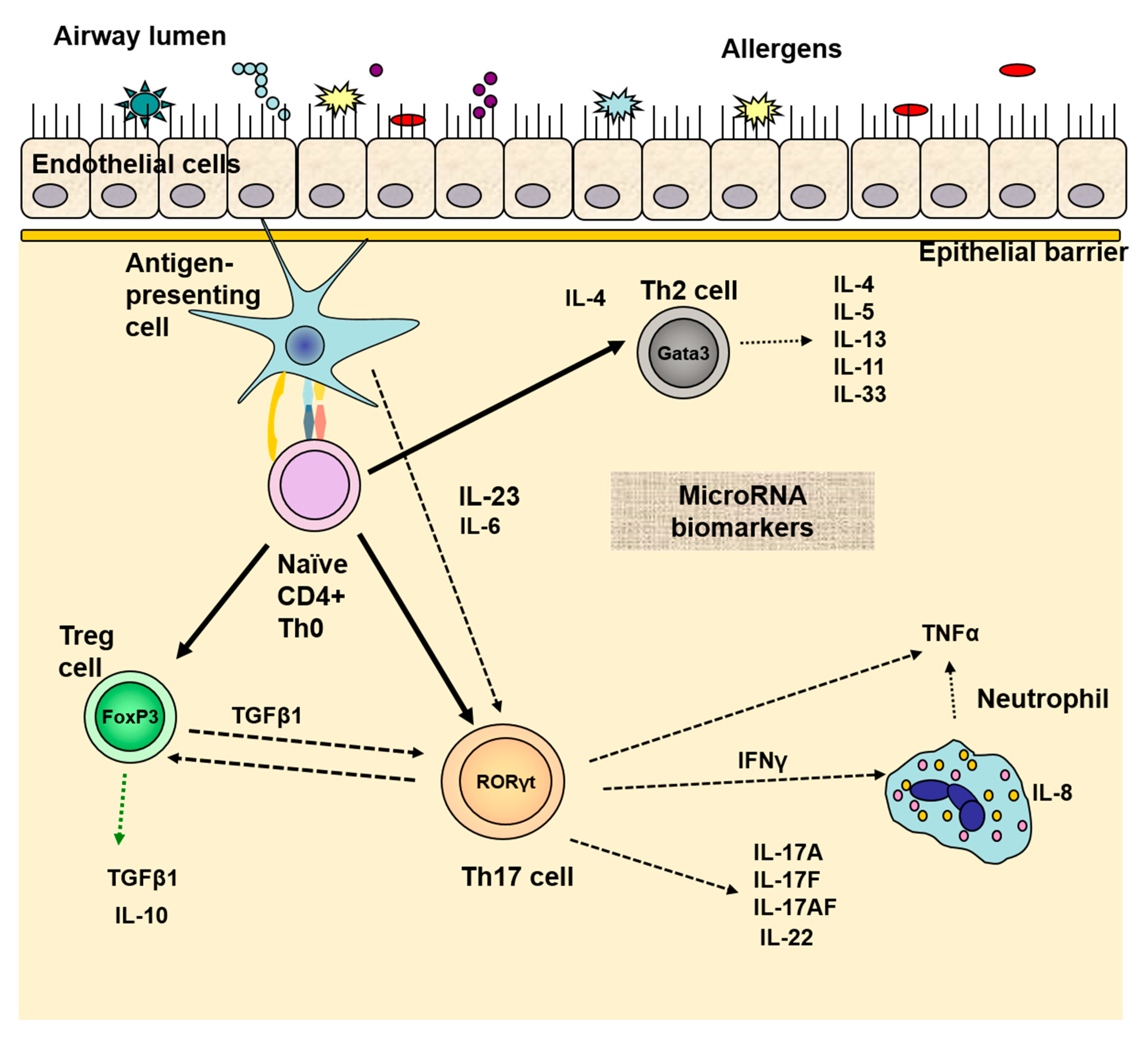
4. Immune Cells Involved in United Airway Inflammation

It was previously established that to maintain immune homeostasis, it is vital to obtain a balance between the regulatory and effectors immune cells, such as T-helper type 1 (Th1) and type 2 (Th2) cells. As the Th1/Th2 balance dysregulates, the released cytokines contribute to the development of chronic inflammation of the mucosa, resulting in autoimmune or allergic diseases [25].
It is assumed that AR is related to the first type of allergic sensitization in Coombs’ classification, where the involvement of IgE antibodies is crucial [26]. Early and late phases of an allergic response are observed.
Briefly, the immunological mechanism can be represented as follows: upon contact with the mucosa, the allergen is taken up by the antigen-presenting cells, which process it and present it to Th2 cells. Activated Th2 lymphocytes release IL-4, IL-6, IL-13, etc., which interact with B lymphocytes. This interaction leads to the activation of B cells and the synthesis of specific IgE antibodies. IgE antibodies bind to their high-affinity receptors on mast cells’ surfaces and, upon contact with the specific allergen, lead to cell degranulation. As a result, several mediators are released, such as histamine, leukotrienes, and prostaglandins. This description represents the early phase of an allergic response observed within the first few minutes after the allergen contact that lasts 2–3 h.
In the late phase, which occurs 4–6 h after antigenic stimulation, cellular infiltration of the mucosa consists mainly of T lymphocytes, eosinophils, and basophils. In the described Th2 response, IgE antibody production requires two main signals to switch B cells to an IgE antibody-producing plasma cell. The first signal is provided by the cytokines IL-4 or IL-13, which interact with B cell receptors. They transmit the signal by activating the tyrosine kinases of the Janus family— Janus kinase 1 (JAK1) and JAK3—which leads to phosphorylation of the STAT6 transcription regulator. The second signal for IgE switching is additional stimulation by contact between the CD40 ligand on the T cell surface and CD40 on the B cell surface [27]. It is worth mentioning that the allergen stimulation of the immune system leads to priming of the entire mucosa of the airways.
Nasal polyps are also rich in inflammatory immune cells, such as eosinophils, Th lymphocytes, plasma cells, and mast cells. Histologically, nasal polyps are characterized by an edematous stroma, eosinophilia, a thickened basement membrane, and a damaged ciliary epithelium. It is not surprising that chronic inflammation in the nasal polyps, allergic or not, resembles the bronchial mucosa inflammation observed in asthma. This once again confirms the united disease pathways of the airways. Nevertheless, in 30–70% of patients with nasal polyposis, accompanying BA is diagnosed [28].
Interestingly, it was shown that AR without polyposis or eosinophilic inflammation may not possess the common airway pathways with asthma, unlike the case where all present together [29]. Taken together, data on the united airway pathway suggest that AR should be seen as predictive risk factor for asthma [30].

5. Th17 Cells Role in the Common Inflammation of the Airways

In recent decades, discovering Th17 cells and regulatory T (Treg) cells has dramatically complicated the established Th1/Th2 paradigm. Involvement of the two counterparts—Th17 cells and Tregs—complicates the understanding of AR’s pathogenesis [31]. The role of Th17 cells in neutrophil infiltration and chronic inflammation in AR and asthma is well established. Moreover, it was confirmed that the balance between Th17/Treg cells matters clinically in allergic and autoimmune diseases.
Speaking of the united airway pathway, the participation of Th17 in the pathogenesis and progression of AR and other diseases is also proven. Moreover, a correlation of IL-17 levels in the inflamed airway mucosa with the severity of the allergic disease was found [32].
Many studies have proven the involvement of Th17 cells and IL-17 in the immunological mechanism of AR. A recent study by Huang et al. [33] examined Th17/Treg cells immunity in AR patients. The results showed that Th17 cells were significantly increased in the peripheral blood of patients with AR, whereas the Treg cell number was decreased. The results suggested that the Th17/Treg imbalance plays a crucial role in AR’s pathogenesis and severity.
Moreover, Milovanovic et al. demonstrated that IL-17 could induce B cell switching to IgE antibody production, endorsing, once again, the involvement of Th17 in allergic diseases [34]. However, Th17 cells produce a large number of mediators, but IL-17A can directly induce IgE production.
A schematic picture of the immune mediators and cells involved in the airway mucosa inflammation is presented in Figure 1.
Another cytokine with a role in the common airway inflammation and allergic diseases is IL-33. It is a member of the IL-1 cytokine family released in response to epithelial cell damage. IL-33 exerts many actions by interacting with the suppressor of tumorigenicity 2 (ST2) receptor. Furthermore, it can induce Th2 cytokine-mediated allergic inflammation [35]. The IL-33/ST2-induced Th2 response has been found to interact with the Th17 immune response in AR pathology [36,37]. IL-33 also induces the production of proinflammatory cytokines and participates in the pathogenesis of diseases other than AR, such as atopic dermatitis, BA, and pollinosis.
The crucial role of IL-33 in the united airway inflammation is based on bridging the innate and acquired immune responses in allergic diseases [38]. Clinically, this connection was evaluated by the reported correlation of IL-33 levels and the AR severity [39].
Other cytokines, such as those with anti-inflammatory properties—IL-10, TGF-β, and IL-35—and related to Th17 and Th22 cells—IL-22 and IL-27—might shape the allergic responses as well. Still, there are few reports on their role [40]. The cytokines related to Th17 and Treg activation while suppressing Th1 cells—IL-17, IL-22, and TGF-β—were found enhanced in AR patients. On the contrary, IL-35, which was shown to inhibit both Th2- and Th17-mediated allergic airway inflammation, was detected low in patients with AR, showing the possible role in the pathogenesis of allergic diseases [40].

6. Diagnostic and Therapeutic Approaches in the Light of the United Airway Pathway

By enhancing the understanding of the inflammation of the upper respiratory tract and the pathogenesis of AR and other allergic diseases, new diagnostic and therapeutic approaches can be established. They can facilitate the management and follow-up of the patients and improve their quality of life.

6.1. microRNAs as a Promising Tool in United Airway Disease Diagnosis

Promising and advanced tools for diagnosing and managing patients with AR and BA are the small non-coding RNAs, known as microRNAs (miRNA or miR). As specific gene expression regulators, miRNAs regulate many biological processes, including cell differentiation, proliferation, and survival [41]. Furthermore, they can serve as noninvasive biomarkers for diagnosis, molecular classification, severity, and relapse prediction [38]. miRNAs tend to participate in the pathogenesis of both AR and BA. It was shown that individual circulating miRNAs were distinctively expressed in AR and BA patients [42].
Moreover, few studies examined their role in clinical settings. Suojalehto et al. found increased levels of miR-143, miR-187, miR-498, miR-874, and miR-886-3p and decreased levels of let-7e, miR-18a, miR-126, miR-155, and miR-224 in BA compared to controls [42]. However, these results were independent of concomitant AR. Thus, no distinction was made between BA and AR based on these expressions.
In the second study, Suojalehto et al. [43] found upregulated expression of miR-155, miR-205, and miR-498 but downregulated expression of let-7e in the nasal mucosa of AR patients and current symptoms in comparison with AR without asthma. However, the cytokine levels (IL-4, IL-5, and IL-13) and miRNA expression profile were comparable in AR with or without AB, suggesting that concomitant asthma might have a minor impact.
In addition, an alternative to the nonsteroidal anti-inflammatory treatment, adjusting and regulating the miRNA network, may be a promising therapy approach.

6.2. Biological Therapy in the Focus of United Airway Disease

The success of all available treatment strategies for united airway disease relies on the combined targeting of all clinically presented diseases. The similar pathological pathways and common mucosal inflammation, along with the parallel incidence of AR and BA, lead to the approach for similar treatment, including biological therapy, as we showed previously [44,45,46,47].
The strategy of using biological agents has been investigated in patients with AR, BA, and other allergic diseases. Biological therapy was considered a beneficial treatment option in patients with severe uncontrolled phenotypes of diseases. Omalizumab, which represents a humanized anti-IgE monoclonal antibody, has been studied extensively for AR and BA. It confirmed its effectiveness in preventing IgE to attach to its high-affinity receptors. Moreover, omalizumab’s clinical outcomes have been linked to reducing nasal and asthma symptoms, decreasing the number of exacerbations by affecting both the upper and lower airways [47]. All of these led to an overall improvement in the quality of life of the patients.
Another monoclonal antibody used for both AR and BA—mepolizumab—acts by blocking the binding of IL-5 to eosinophils. Mepolizumab has also shown efficacy in improving the severity of eosinophilic airway diseases, especially in BA and nasal polyps [48,49].
Nevertheless, we must always have in mind that monoclonal therapy is not without systemic effects. In line with this, in patients with united airway disease due to the effects of therapy on AR improvement, it is hardly possible to design a study to distinguish the improvement in BA alone. In line with this, management of AR and BA must be carried out together to obtain better control of both diseases [50].

7. Conclusions

The concept of unified airway diseases has been the subject of attention in recent years. The pathogenetic relationship of the nose and the bronchi and alveoli, along with the observed common inflammation, provides a niche to create new diagnostic and therapeutic options.
With Th17 cells and other immune cells and mediators, gene alteration and regulation by miRNA complicate the picture of the united airway inflammation. More research here is needed to better understand the associations between the upper and lower airways. However, there is no doubt that AR and BA should be diagnosed, managed, and treated in an integrated manner.

Author Contributions

Conceptualization, K.N. and T.V.; writing—original draft preparation, K.N.; writing—review and editing, T.V.; visualization, T.V.; supervision, V.D. All authors have read and agreed to the published version of the manuscript.

Funding

This research received no external funding.

Institutional Review Board Statement

Not applicable.

Informed Consent Statement

Not applicable.

Data Availability Statement

Not applicable.

Conflicts of Interest

The authors declare no conflict of interest.

References

  1. Caimmi, D.; Marseglia, A.; Pieri, G.; Benzo, S.; Bosa, L.; Caimmi, S. Nose and lungs: One way, one disease. Ital. J. Pediatr. 2012, 38, 60. [Google Scholar] [CrossRef] [PubMed] [Green Version]
  2. Licari, A.; Castagnoli, R.; Denicolò, C.F.; Rossini, L.; Marseglia, A.; Marseglia, G.L. The Nose and the Lung: United Airway Disease? Front. Pediatr. 2017, 5, 44. [Google Scholar] [CrossRef] [Green Version]
  3. Haccuria, A.; van Muylem, A.; Malinovschi, A.; Doan, V.; Michils, A. Small airways dysfunction: The link between allergic rhinitis and allergic asthma. Eur. Respir. J. 2018, 51, 1701749. [Google Scholar] [CrossRef] [Green Version]
  4. Compalati, E.; Ridolo, E.; Passalacqua, G.; Braido, F.; Villa, E.; Canonica, G.W. The link between allergic rhinitis and asthma: The united airways disease. Expert Rev. Clin. Immunol. 2010, 6, 413–423. [Google Scholar] [CrossRef]
  5. Vujnovic, S.D.; Domuz, A. Epidemiological Aspects of Rhinitis and Asthma: Comorbidity or United Airway Disease. In Asthma Diagnosis and Management—Approach Based on Phenotype and Endotype; IntechOpen: London, UK, 2018. [Google Scholar]
  6. Bousquet, J.; Boushey, H.A.; Busse, W.W.; Canonica, G.W.; Durham, S.R.; Irvin, C.G.; Karpel, J.P.; van Cauwenberge, P.; Chen, R.; Iezzoni, D.G.; et al. Characteristics of patients with seasonal allergic rhinitis and concomitant asthma. Clin. Exp. Allergy 2004, 34, 897–903. [Google Scholar] [CrossRef]
  7. Small, P.; Keith, P.K.; Kim, H. Allergic rhinitis. Allergy Asthma Clin. Immunol. 2018, 14, 1–11. [Google Scholar] [CrossRef] [PubMed] [Green Version]
  8. Mileva, Z.; Popov, T.; Staneva, M.; Dimitrov, V.; Mateev, V.; Slavov, S. Frequency and characteristics of allergic diseases in Bulgaria. Allergy Asthma 2000, 1, 3–32. [Google Scholar]
  9. Pawankar, R.; Bunnag, C.; Khaltaev, N.; Bousquet, J. Allergic Rhinitis and Its Impact on Asthma in Asia Pacific and the ARIA Update 2008. World Allergy Organ. J. 2012, 5, S212–S217. [Google Scholar] [CrossRef] [Green Version]
  10. Dykewicz, M.S. 7. Rhinitis and sinusitis. J. Allergy Clin. Immunol. 2003, 111, S520–S529. [Google Scholar] [CrossRef] [PubMed]
  11. Bousquet, P.; Schünemann, H.; Samolinski, B.; Demoly, P.; Baena-Cagnani, C.; Bachert, C.; Bonini, S.; Boulet, L.; Brozek, J.; Canonica, G.; et al. Allergic Rhinitis and its Impact on Asthma (ARIA): Achievements in 10 years and future needs. J. Allergy Clin. Immunol. 2012, 130, 1049–1062. [Google Scholar] [CrossRef] [PubMed] [Green Version]
  12. Bugten, V.; Nordgård, S.; Romundstad, P.; Steinsvåg, S. Chronic rhinosinusitis and nasal polyposis; indicia of heterogeneity. Rhinol. J. 2008, 46, 40–44. [Google Scholar]
  13. Ediger, D.; Sin, B.A.; Heper, A.; Anadolu, Y.; Mitoasitoarlitoagil, Z. Airway inflammation in nasal polyposis: Immunopathological aspects of relation to asthma. Clin. Exp. Allergy 2005, 35, 319–326. [Google Scholar] [CrossRef]
  14. Bousquet, J.; van Cauwenberge, P.; Khaltaev, N. Allergic Rhinitis and Its Impact on Asthma. J. Allergy Clin. Immunol. 2001, 108, S147–S334. [Google Scholar] [CrossRef] [Green Version]
  15. Millqvist, E.; Bende, M. Role of the upper airways in patients with chronic cough. Curr. Opin. Allergy Clin. Immunol. 2006, 6, 7–11. [Google Scholar] [CrossRef] [PubMed]
  16. Kogahara, T.; Kanai, K.-I.; Asano, K.; Suzaki, H. Evidence for passing down of postnasal drip into respiratory organs. Vivo 2009, 23, 297–302. [Google Scholar]
  17. Pratter, M.R. Chronic Upper Airway Cough Syndrome Secondary to Rhinosinus Diseases (Previously Referred to as Postnasal Drip Syndrome). Chest 2006, 129, 63S–71S. [Google Scholar] [CrossRef]
  18. Forer, M.; Ananda, S. The management of postnasal drip. Aust. Fam. Physician 1999, 28, 223–228. [Google Scholar] [PubMed]
  19. Meltzer, E.O.; Szwarcberg, J.; Pill, M.W. Allergic Rhinitis, Asthma, and Rhinosinusitis: Diseases of the Integrated Airway. J. Manag. Care Pharm. 2004, 10, 310–317. [Google Scholar] [CrossRef]
  20. Morice, A.H. The diagnosis and management of chronic cough. Eur. Respir. J. 2004, 24, 481–492. [Google Scholar] [CrossRef] [PubMed]
  21. Lai, K.; Chen, R.; Lin, J.; Huang, K.; Shen, H.; Kong, L.; Zhou, X.; Luo, Z.; Yang, L.; Wen, F.; et al. A Prospective, Multicenter Survey on Causes of Chronic Cough in China. Chest 2013, 143, 613–620. [Google Scholar] [CrossRef]
  22. Pecova, R.; Zucha, J.; Pec, M.; Neuschlova, M.; Hanzel, P.; Tatar, M. Cough reflex sensitivity testing in in seasonal allergic rhinitis patients and healthy volunteers. J. Physiol. Pharmacol. 2008, 59, 557–564. [Google Scholar] [PubMed]
  23. Weinfeld, D.; Ternesten-Hasséus, E.; Löwhagen, O.; Millqvist, E. Capsaicin cough sensitivity in allergic asthmatic patients increases during the birch pollen season. Ann. Allergy Asthma Immunol. 2002, 89, 419–424. [Google Scholar] [CrossRef]
  24. Lim, K.G.; Rank, M.A.; Kita, H.; Patel, A.; Moore, E. Neuropeptide levels in nasal secretions from patients with and without chronic cough. Ann. Allergy Asthma Immunol. 2011, 107, 360–363. [Google Scholar] [CrossRef] [PubMed]
  25. Romagnani, S. T-cell subsets (Th1 versus Th2). Ann. Allergy Asthma Immunol. 2000, 85, 9–21. [Google Scholar] [CrossRef]
  26. Rajan, T. The Gell—Coombs classification of hypersensitivity reactions: A re-interpretation. Trends Immunol. 2003, 24, 376–379. [Google Scholar] [CrossRef]
  27. Janeway, C.A., Jr.; Travers, P.; Walport, M.; Shlomchik, M.J. Immunobiology: The Immune System in Health and Disease, 5th ed.; Garland Science: New York, NY, USA, 2001. [Google Scholar]
  28. Larsen, K. The Clinical Relationship of Nasal Polyps to Asthma. Allergy Asthma Proc. 1996, 17, 243–249. [Google Scholar] [CrossRef]
  29. Muluk, N.B. The united airway disease. Rom. J. Rhinol. 2019, 9, 21–26. [Google Scholar] [CrossRef] [Green Version]
  30. Ketenci, A.; Kalyoncu, A.F.; del Giacco, S. Upper and Lower Airways Interaction: Is the United Airway Disease Concept a Reflection of Reality? How Important Is It? In Challenges in Rhinology; Cingi, C., Muluk, N.B., Scadding, G.K., Mladina, R., Eds.; Springer International Publishing: Cham, Switzerland, 2020; pp. 405–414. [Google Scholar]
  31. Gu, Z.W.; Wang, Y.X.; Cao, Z.W. Neutralization of interleukin-17 suppresses allergic rhinitis symptoms by downregulating Th2 and Th17 responses and upregulating the Treg response. Oncotarget 2017, 8, 22361–22369. [Google Scholar] [CrossRef] [Green Version]
  32. Ciprandi, G.; de Amici, M.; Murdaca, G.; Fenoglio, D.; Ricciardolo, F.L.M.; Marseglia, G.L.; Tosca, M. Serum interleukin-17 levels are related to clinical severity in allergic rhinitis. Allergy 2009, 64, 1375–1378. [Google Scholar] [CrossRef] [PubMed]
  33. Huang, X.; Chen, Y.; Zhang, F.; Yang, Q.; Zhang, G. Peripheral Th17/Treg cell-mediated immunity imbalance in allergic rhinitis patients. Braz. J. Otorhinolaryngol. 2014, 80, 152–155. [Google Scholar] [CrossRef] [PubMed] [Green Version]
  34. Ferretti, E.; di Carlo, E.; Ognio, E.; Guarnotta, C.; Bertoni, F.; Corcione, A.; Prigione, I.; Fraternali-Orcioni, G.; Ribatti, D.; Ravetti, J.L.; et al. Interleukin-17A promotes the growth of human germinal center derived non-Hodgkin B cell lymphoma. OncoImmunology 2015, 4, e1030560. [Google Scholar] [CrossRef]
  35. Haenuki, Y.; Matsushita, K.; Futatsugi-Yumikura, S.; Ishii, K.J.; Kawagoe, T.; Imoto, Y.; Fujieda, S.; Yasuda, M.; Hisa, Y.; Akira, S.; et al. A critical role of IL-33 in experimental allergic rhinitis. J. Allergy Clin. Immunol. 2012, 130, 184–194.e11. [Google Scholar] [CrossRef] [PubMed]
  36. Vocca, L.; di Sano, C.; Uasuf, C.G.; Sala, A.; Riccobono, L.; Gangemi, S.; Albano, G.D.; Bonanno, A.; Gagliardo, R.; Profita, M. IL-33/ST2 axis controls Th2/IL-31 and Th17 immune response in allergic airway diseases. Immunobiology 2015, 220, 954–963. [Google Scholar] [CrossRef]
  37. Ding, W.; Zou, G.-L.; Zhang, W.; Lai, X.-N.; Chen, H.-W.; Xiong, L.-X. Interleukin-33: Its Emerging Role in Allergic Diseases. Molecules 2018, 23, 1665. [Google Scholar] [CrossRef] [Green Version]
  38. Lloyd, C.M. IL-33 family members and asthma—Bridging innate and adaptive immune responses. Curr. Opin. Immunol. 2010, 22, 800–806. [Google Scholar] [CrossRef] [Green Version]
  39. Glück, J.; Rymarczyk, B.; Rogala, B. Serum IL-33 but not ST2 level is elevated in intermittent allergic rhinitis and is a marker of the disease severity. Inflamm. Res. 2012, 61, 547–550. [Google Scholar] [CrossRef] [Green Version]
  40. Degirmenci, P.B.; Aksun, S.; Altin, Z.; Bilgir, F.; Arslan, I.B.; Çolak, H.; Ural, B.; Kahraman, D.S.; Diniz, G.; Ozdemir, B.; et al. Allergic Rhinitis and Its Relationship with IL-10, IL-17, TGF-β, IFN-γ, IL 22, and IL-35. Dis. Markers 2018, 2018, 1–6. [Google Scholar] [CrossRef] [PubMed] [Green Version]
  41. Liu, Z.; Zhang, X.-H.; Callejas-Díaz, B.; Mullol, J. MicroRNA in United Airway Diseases. Int. J. Mol. Sci. 2016, 17, 716. [Google Scholar] [CrossRef] [PubMed] [Green Version]
  42. Panganiban, R.P.; Wang, Y.; Howrylak, J.; Chinchilli, V.M.; Craig, T.J.; August, A.; Ishmael, F.T. Circulating microRNAs as biomarkers in patients with allergic rhinitis and asthma. J. Allergy Clin. Immunol. 2016, 137, 1423–1432. [Google Scholar] [CrossRef] [Green Version]
  43. Suojalehto, H.; Lindström, I.; Majuri, M.-L.; Mitts, C.; Karjalainen, J.; Wolff, H.; Alenius, H. Altered MicroRNA Expression of Nasal Mucosa in Long-Term Asthma and Allergic Rhinitis. Int. Arch. Allergy Immunol. 2014, 163, 168–178. [Google Scholar] [CrossRef]
  44. Naydenova, K.; Velikova, T.; Dimitrov, V. Interactions of allergic rhinitis and bronchial asthma at mucosal immunology level. AIMS Allergy Immunol. 2019, 3, 1–12. [Google Scholar] [CrossRef]
  45. Naydenova, K.; Velikova, T.V.; Dimitrov, V. Chapter 5. Allergic Rhinitis, IL-17 and the Concept of a Common Respiratory Pathway. In Th17 Cells in Health and Disease; Nova Publishing: New York, NY, USA, 2020. [Google Scholar]
  46. Naydenova, K.; Velikova, T.V.; Dimitrov, V. Mucosal Inflammation in Allergic Rhinitis and Bronchial Asthma—Two Sides of a Coin. Clin. Res. Immunol. 2018, 1, 1–2. [Google Scholar]
  47. Gevaert, P.; Calus, L.; van Zele, T.; Blomme, K.; de Ruyck, N.; Bauters, W.; Hellings, P.; Brusselle, G.; de Bacquer, D.; van Cauwenberge, P.; et al. Omalizumab is effective in allergic and nonallergic patients with nasal polyps and asthma. J. Allergy Clin. Immunol. 2013, 131, 110–116.e1. [Google Scholar] [CrossRef] [PubMed]
  48. Pavord, I.D.; Korn, S.; Howarth, P.; Bleecker, E.R.; Buhl, R.; Keene, O.N.; Ortega, H.; Chanez, P. Mepolizumab for severe eosinophilic asthma (DREAM): A multicentre, double-blind, placebo-controlled trial. Lancet 2012, 380, 651–659. [Google Scholar] [CrossRef]
  49. Gevaert, P.; van Bruaene, N.; Cattaert, T.; van Steen, K.; van Zele, T.; Acke, F.; de Ruyck, N.; Blomme, K.; Sousa, A.R.; Marshall, R.P.; et al. Mepolizumab, a humanized anti–IL-5 mAb, as a treatment option for severe nasal polyposis. J. Allergy Clin. Immunol. 2011, 128, 989–995.e8. [Google Scholar] [CrossRef] [Green Version]
  50. Giavina-Bianchi, P.; Aun, M.V.; Takejima, P.; Kalil, J.; Agondi, R.C. United airway disease: Current perspectives. J. Asthma Allergy 2016, 9, 93–100. [Google Scholar] [CrossRef] [PubMed] [Green Version]
Figure 1. Immunological interactions in the inflamed mucosa in the concept of united airway disease. Naïve T cells differentiate either into Th2, Tregs, or Th17 cells. By secreting a distinct array of cytokines, Th17, Th2, and Treg cells connect innate and adaptive immune responses in the airway mucosa, especially during mucosal inflammation. Balance of Th17/Tegs is needed to maintain the immune homeostasis in the mucosa and to resolve the inflammation. Note: allergens are presented as different shaped objects in the airway lumen.
Sinusitis 05 00005 g001
Publisher’s Note: MDPI stays neutral with regard to jurisdictional claims in published maps and institutional affiliations.

