
Prussian Blue and Its Analogs as Novel Nanostructured Antibacterial Materials


Abstract
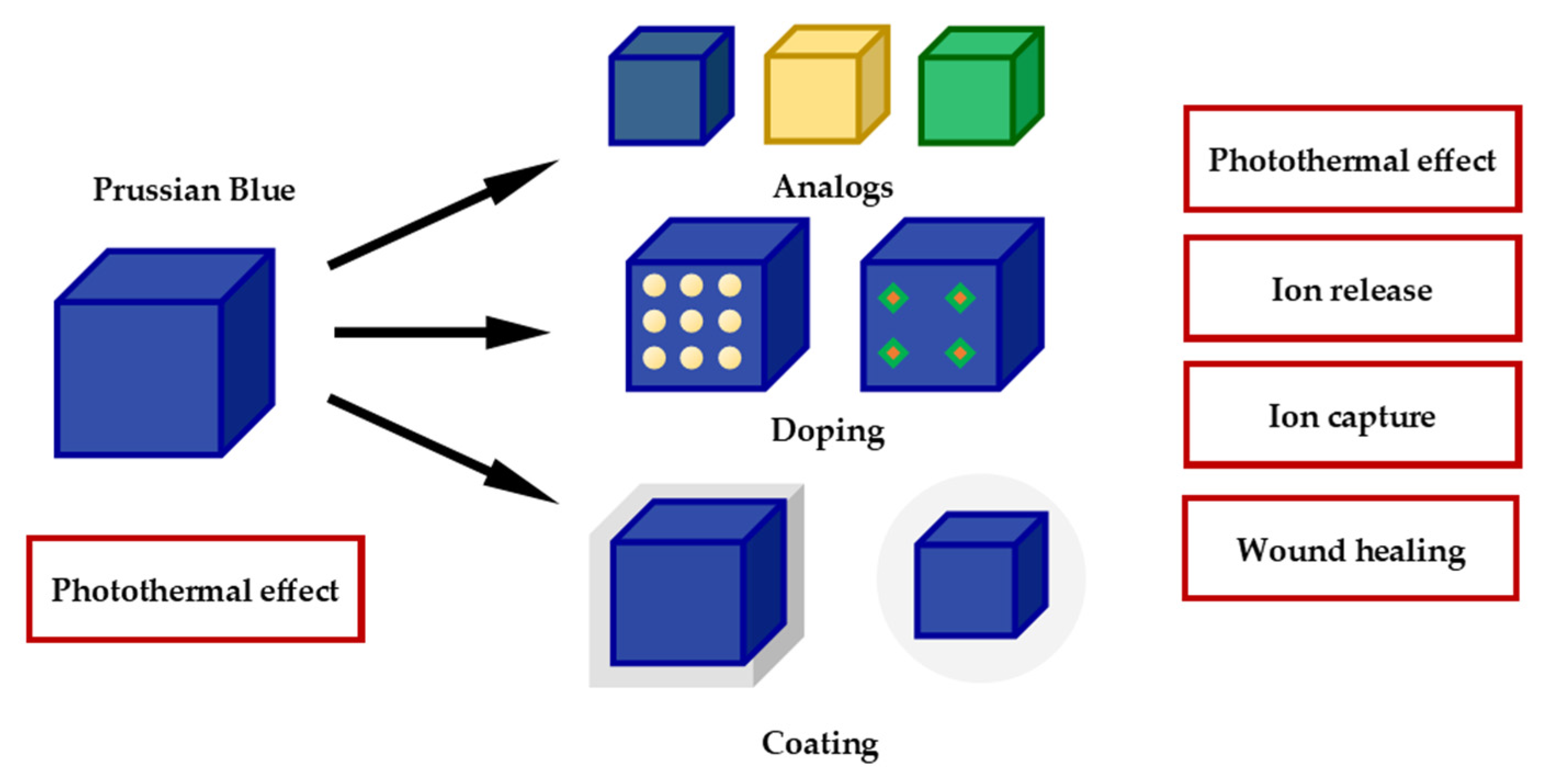
:1. Introduction
2. Synthesis and Coating
3. Antibacterial Activity of Prussian Blue
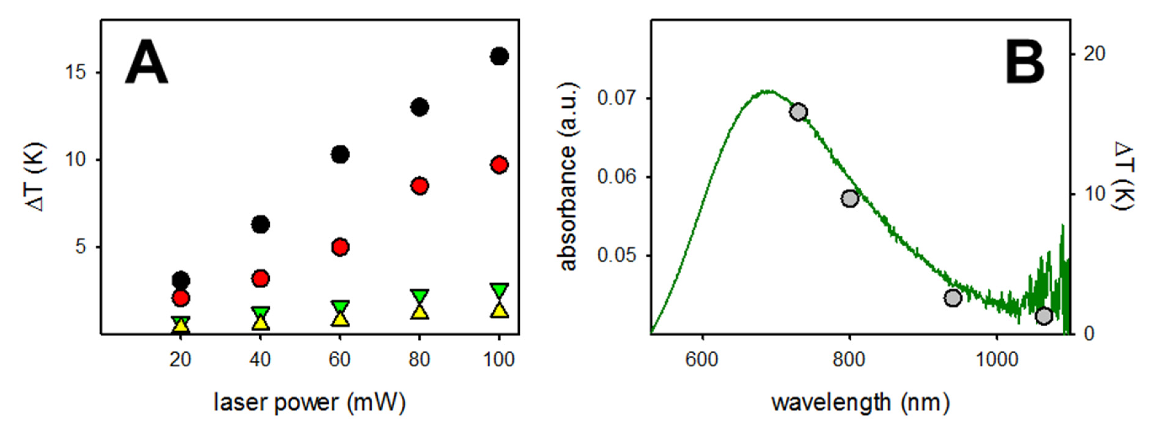
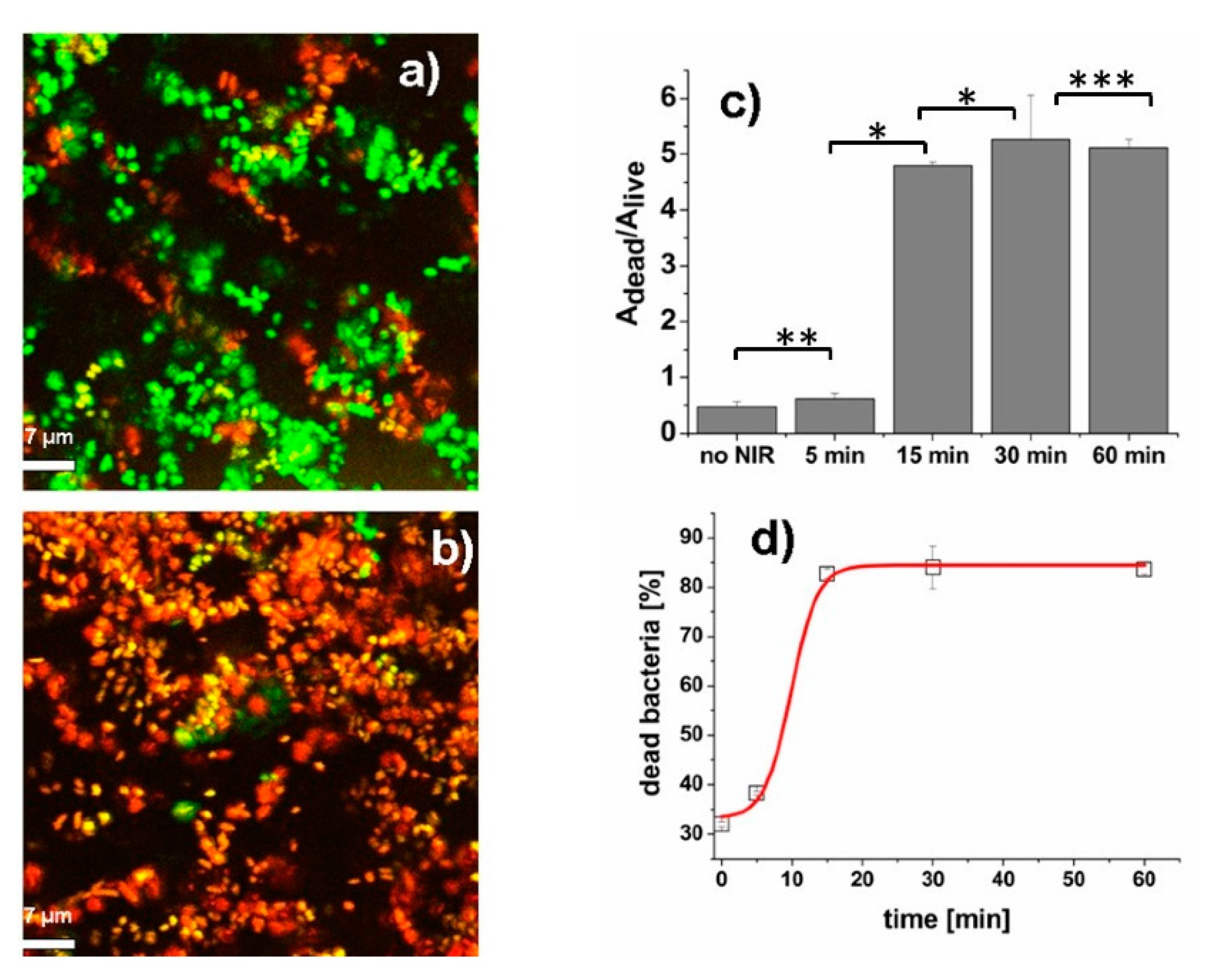
3.1. Photothermal Antibacterial Effect of Prussian Blue Nanoparticles
3.2. Prussian Blue Doped with Traditional Antibiotics
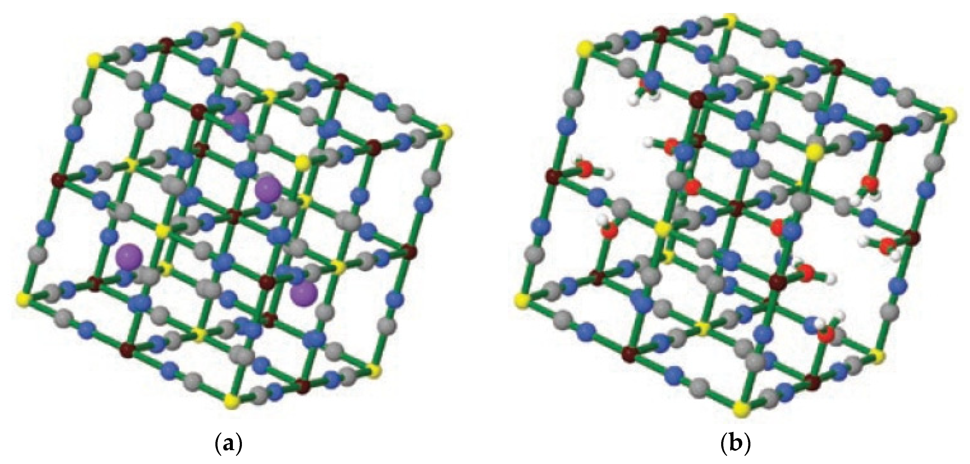
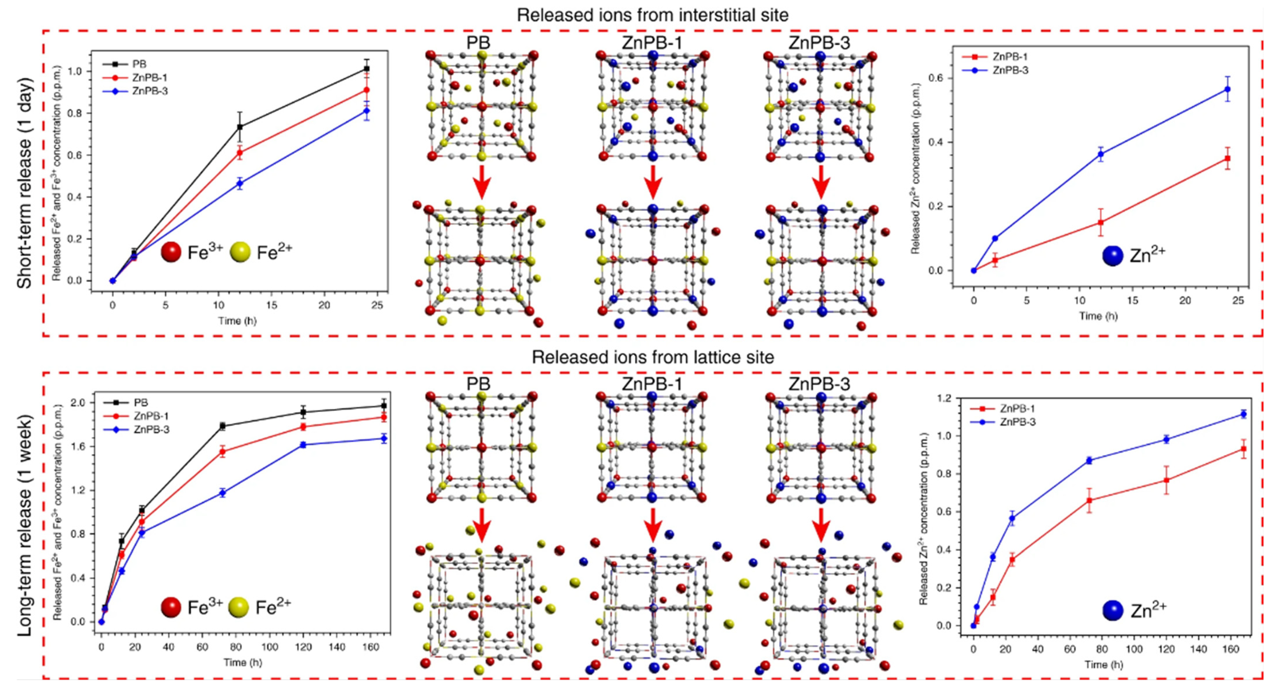
3.3. Prussian Blue Analogs with Antibacterial Effect
3.4. Silver-Containing Prussian Blue
4. Conclusions
Author Contributions
Funding
Institutional Review Board Statement
Informed Consent Statement
Data Availability Statement
Conflicts of Interest
References
- Frisch, J.L. Notitia Caerulei Berolinensis Nuper Inventi. Misc. Berolin. Incrementum Sci. Scr. Soc. Regiae Sci. Exhib. Ed. 1710, 1, 777. [Google Scholar]
- Kraft, A. On the discovery and history of Prussian blue. Bull. Hyst. Chem. 2008, 33, 61–67. [Google Scholar]
- Ludi, A. Prussian Blue, an Inorganic Evergreen. J. Chem. Edu. 1981, 58, 1013. [Google Scholar] [CrossRef]
- Keggin, J.F.; Miles, F.D. Structures and Formulae of the Prussian blues and Related Compounds. Nature 1936, 137, 577–578. [Google Scholar] [CrossRef]
- Robin, M.B. The Color and Electronic Configurations of Prussian Blue. Inorg. Chem. 1962, 1, 337–342. [Google Scholar] [CrossRef]
- Buser, H.J.; Schwarzenbach, D.; Petter, W.; Ludi, A. The Crystal Structure of Prussian Blue: Fe4[Fe(CN)6]3∙xH2O. Inorg. Chem. 1977, 16, 2704–2710. [Google Scholar] [CrossRef]
- Ware, M. Prussian Blue: Artists’ Pigment and Chemists’ Sponge. J. Chem. Educ. 2008, 85, 612–621. [Google Scholar] [CrossRef]
- Whitney, W.R.; Blake, J.C. On Colloidal Gold: Absorption Phenomena and Allotropy. Am. J. Sci. 1903, 16, 381–387. [Google Scholar] [CrossRef] [Green Version]
- Vaucher, S.; Li, M.; Mann, S. Synthesis of Prussian Blue Nanoparticles and Nanocrystal Superlattices in Reverse Microemulsions. Angew. Chem. Int. Ed. 2000, 39, 1793–1796. [Google Scholar] [CrossRef]
- Fda.Gov. Available online: https://www.fda.gov/drugs/bioterrorism-and-drug-preparedness/fda-approves-first-new-drug-application-treatment-radiation-contamination-due-cesium-or-thallium (accessed on 12 March 2021).
- Uemura, T.; Kitagawa, S. Prussian Blue Nanoparticles Protected by Poly(vynilpyrrolidone). J. Am. Chem. Soc. 2003, 125, 7814–7815. [Google Scholar] [CrossRef] [PubMed]
- Yang, Y.; Brownell, C.; Sadrieh, N.; May, J.; Del Grosso, A.; Place, D.; Leutzinger, E.; Duffy, E.; He, R.; Houn, F.; et al. Quantitative Measurement of Cyanide Released from Prussian Blue. Clin. Toxicol. 2007, 45, 776–781. [Google Scholar] [CrossRef]
- Yang, Y.; Faustino, P.J.; Progar, J.J.; Brownell, C.R.; Sadrieh, N.; May, J.C.; Leutzinger, E.; Place, D.A.; Duffy, E.P.; Yu, L.X.; et al. Quantitative Determination of Thallium Binding to Ferric Hexacyanoferrate: Prussian Blue. Int. J. Pharm. 2008, 353, 187–194. [Google Scholar] [CrossRef] [Green Version]
- Mohammad, A.; Faustino, P.J.; Khan, M.A.; Yang, Y. Long-Term Stability Study of Prussian Blue: A Quality Assessment of Water Content and Thallium Binding. Int. J. Pharm. 2014, 477, 122–127. [Google Scholar] [CrossRef]
- Mohammad, A.; Yang, Y.; Khan, M.A.; Faustino, P.J. A Long-Term Stability Study of Prussian Blue: A Quality Assessment of Water Content and Cesium Binding. J. Pharm. Biomed. Anal. 2015, 103, 85–90. [Google Scholar] [CrossRef] [PubMed]
- Long, J.; Guari, Y.; Larionova, J. Prussian Blue type nanoparticles for biomedical applications. Dalton Trans. 2016, 45, 17581. [Google Scholar] [CrossRef] [PubMed]
- Gao, X.; Wang, Q.; Cheng, C.; Lin, S.; Lin, T.; Liu, C.; Han, X. The application of Prussian Blue nanoparticles in Tumor Diagnosis and Treatment. Sensors 2020, 20, 6905. [Google Scholar] [CrossRef] [PubMed]
- Butler, J.S.; Sadler, P.J. Targeted delivery of platinum-based anticancer complexes. Curr. Opin. Chem. Biol. 2013, 17, 175–188. [Google Scholar] [CrossRef] [Green Version]
- Dacarro, G.; Taglietti, A.; Pallavicini, P. Prussian Blue Nanoparticles as Versatile Photothermal Tools. Molecules 2018, 23, 1414. [Google Scholar] [CrossRef] [Green Version]
- De Tacconi, N.R.; Rajeshwar, K. Metal Hexacyanoferrates: Electrosynthesis, in situ Characterization, and Applications. Chem. Mater. 2003, 15, 3046–3062. [Google Scholar] [CrossRef]
- Kale, S.S.; Burga, R.A.; Sweeney, E.E.; Zun, Z.; Sze, R.W.; Tuesca, A.; Subramony, J.A.; Fernandes, R. Composite iron oxide–Prussian blue nanoparticles for magnetically guided T1-weighted magnetic resonance imaging and photothermal therapy of tumors. Int. J. Nanomed. 2017, 12, 6413–6424. [Google Scholar] [CrossRef] [Green Version]
- Zhu, W.; Liu, K.; Sun, X.; Wang, X.; Li, Y.; Cheng, L.; Liu, Z. Mn2+-Doped Prussian Blue Nanocubes for Bimodal Imaging and Photothermal Therapy with Enhanced Performance. ACS Appl. Mater. Interfaces 2015, 7, 11575–11582. [Google Scholar] [CrossRef] [PubMed]
- Xu, M.; Chi, B.; Han, Z.; He, Y.; Tian, F.; Xu, Z.; Li, L.; Wang, J. Controllable synthesis of rare earth (Gd3+,Tm3+) doped Prussian blue for multimode imaging guided synergistic treatment. Dalton Trans. 2020, 49, 12327–12337. [Google Scholar] [CrossRef] [PubMed]
- Forgach, L.; Hegedus, N.; Horvath, I.; Kiss, B.; Kovacs, N.; Varga, Z.; Jakab, G.; Kovacs, T.; Padmanabhan, P.; Szigeti, K.; et al. Fluorescent, Prussian Blue-Based Biocompatible Nanoparticle System for Multimodal Imaging Contrast. Nanomaterials 2020, 10, 1732. [Google Scholar] [CrossRef] [PubMed]
- Karyakin, A.A. Advances of Prussian blue and its analogues in (bio)sensors. Curr. Opin. Electrochem. 2017, 5, 92–98. [Google Scholar] [CrossRef]
- Matos-Peralta, Y.; Antuch, M. Prussian Blue and its analogs as appealing materials for electrochemical sensing and biosensing. J. Electrochem. Soc. 2020, 167, 037510. [Google Scholar] [CrossRef]
- Bu, F.-X.; Du, C.-J.; Zhang, Q.-H.; Jiang, J.-S. One-pot synthesis of Prussian blue superparticles from reverse microemulsion. CrystEngComm 2014, 16, 3113–3120. [Google Scholar] [CrossRef]
- Zakaria, M.B.; Chikyow, T. Recents advances in Prussian Blue analogues: Synthesis and thermal treatments. Coord. Chem. Rev. 2017, 352, 328–345. [Google Scholar] [CrossRef]
- Samain, L.; Grandjean, F.; Long, G.J.; Martinetto, P.; Bordet, P.; Strivay, D. Relationship between the Synthesis of Prussian Blue Pigments, Their Color, Physical Properties, and Their Behavior in Paint Layers. J. Phys. Chem. C 2013, 117, 9693–9712. [Google Scholar] [CrossRef]
- Karyakin, A.A.; Karyakina, E.E. Electroanalytical applications of Prussian Blue and its analogs. Russ. Chem. Bull. 2001, 50, 1811–1817. [Google Scholar] [CrossRef]
- Jia, Z.; Sun, G. Preparation of Prussian Blue nanoparticles with single precursor. Colloids Surf. A 2007, 302, 326–329. [Google Scholar] [CrossRef]
- Qin, Z.; Chen, B.; Mao, Y.; Shi, C.; Li, Y.; Huang, X.; Yang, F.; Gu, N. Achieving Ultrasmall Prussian Blue Nanoparticles as High-Performance Biomedical Agents with Multifunctions. ACS Appl. Mater. Interfaces 2020, 12, 57382–57390. [Google Scholar] [CrossRef]
- Qin, Z.; Li, Y.; Gu, N. Progress in Applications of Prussian Blue Nanoparticles in Biomedicine. Adv. Healthc. Mater. 2018, 7, 1800347. [Google Scholar] [CrossRef]
- Roy, A.K.; Somani, S.P.; Nene, A.; Bipinraj, N.K.; Somani, P.R. Antibacterial Activity of Prussian Blue. J. Green Sci. Technol. 2013, 1, 1–3. [Google Scholar] [CrossRef]
- Pallavicini, P.; Donà, A.; Taglietti, A.; Minzioni, P.; Patrini, M.; Dacarro, G.; Chirico, G.; Sironi, L.; Bloise, N.; Visai, L.; et al. Self-assembled monolayers of gold nanostars: A convenient tool for near-IR photothermal biofilm eradication. Chem. Commun. 2014, 50, 1969–1971. [Google Scholar] [CrossRef] [PubMed] [Green Version]
- Pallavicini, P.; Basile, S.; Chirico, G.; Dacarro, D.; D’Alfonso, L.; Donà, A.; Patrini, M.; Falqui, A.; Sironi, L.; Taglietti, A. Monolayers of gold nanostars with two near-IR LSPRs capable of additive photothermal response. Chem. Commun. 2015, 51, 12928–12930. [Google Scholar] [CrossRef] [Green Version]
- D’Agostino, A.; Taglietti, A.; Grisoli, P.; Dacarro, G.; Cucca, L.; Patrini, M.; Pallavicini, P. Seed mediated growth of silver nanoplates on glass: Exploiting the bimodal antibacterial effect by near IR photo-thermal action and Ag+ release. RSC Adv. 2016, 6, 70414–70423. [Google Scholar] [CrossRef] [Green Version]
- Dacarro, G.; Grisoli, P.; Borzenkov, M.; Milanese, C.; Fratini, E.; Ferraro, G.; Taglietti, A.; Pallavicini, P. Self-assembled monolayers of Prussian blue nanoparticles with photothermal effect. Supramol. Chem. 2017, 823–833. [Google Scholar] [CrossRef]
- Dacarro, G.; Cucca, L.; Grisoli, P.; Pallavicini, P.; Patrini, M.; Taglietti, A. Monolayers of polyethilenimine on flat glass: A versatile platform for cations coordination and nanoparticles grafting in the preparation of antibacterial surfaces. Dalton Trans. 2012, 41, 2456–2463. [Google Scholar] [CrossRef]
- Borzenkov, M.; D’Alfonso, L.; Polissi, A.; Sperandeo, P.; Collini, M.; Dacarro, G.; Taglietti, A.; Chirico, G.; Pallavicini, P. Novel photo-thermally active polyvinyl alcohol-Prussian blue nanoparticles hydrogel films capable of eradicating bacteria and mitigating biofilms. Nanotechnology 2019, 30, 295702. [Google Scholar] [CrossRef] [PubMed]
- Borzenkov, M.; Chirico, G.; Pallavicini, P.; Sperandeo, P.; Polissi, A.; Dacarro, G.; Doveri, L.; Collini, M.; Sironi, L.; Bouzin, M.; et al. Nanocomposite Sprayed Films with Photo-Thermal Properties for Remote Bacteria Eradication. Nanomaterials 2020, 10, 786. [Google Scholar] [CrossRef] [Green Version]
- Jiang, T.; He, J.; Sun, L.; Wang, Y.; Li, Z.; Wang, Q.; Sun, Y.; Wang, W. Highly efficient photothermal sterilization of water mediated by Prussian blue nanocages. Environ. Sci. Nano 2018, 5, 1161. [Google Scholar] [CrossRef]
- Jiang, T.; Wang, Y.; Li, Z.; Aslan, H.; Sun, L.; Sun, Y.; Wang, W.; Yu, M. Prussian blue-encapsulated Fe3O4 nanoparticles for reusable photothermal sterilization of water. J. Coll. Int. Sci. 2019, 540, 354–361. [Google Scholar] [CrossRef]
- Cai, S.; Qian, J.; Yang, S.; Kuang, L.; Hu, D. Acetylcysteine-decorated Prussian blue nanoparticles for strong photothermal sterilization and focal infection treatment. Colloids Surf. B 2019, 181, 31–38. [Google Scholar] [CrossRef]
- Han, D.; Li, Y.; Liu, X.; Li, B.; Han, Y.; Zheng, Y.; Wai, K.; Yeung, K.; Li, C.; Cui, Z.; et al. Rapid bacteria trapping and killing of metal-organic frameworks strengthened photo-responsive hydrogel for rapid tissue repair of bacterial infected wounds. Chem. Eng. J. 2020, 396, 125194. [Google Scholar] [CrossRef]
- Hao, Z.; Lin, X.; Li, J.; Yin, Y.; Gao, X.; Wang, S.; Liu, Y. Multifunctional nanoplatform for dual-mode sensitive detection of pathogenic bacteria and the real-time bacteria inactivation. Biosens. Bioelectron. 2021, 173, 112789. [Google Scholar] [CrossRef] [PubMed]
- Gao, X.; Wu, H.; Hao, Z.; Ji, X.; Lin, X.; Wang, S.; Liu, Y. A multifunctional plasmonic chip for bacteria capture, imaging, detection, and in situ elimination for wound therapy. Nanoscale 2020, 12, 6489–6497. [Google Scholar] [CrossRef] [PubMed]
- Mukherjee, S.; Das, S.; Nuthi, S.; Patra, C.R. Biocompatible nickel-prussian blue@silver nanocomposites show potent antibacterial activities. Future Sci. OA 2017, 3, FSO233. [Google Scholar] [CrossRef]
- Wang, Z.; Yu, B.; Alamri, H.; Yarabarla, S.; Kim, M.-H.; Huang, S.D. KCa(H2O)2[FeIII(CN)6]⋅H2O Nanoparticles as an Antimicrobial Agent against. Staphylococcus aureus. Angew. Chem. Int. Ed. 2018, 57, 2214–2218. [Google Scholar] [CrossRef]
- Li, J.; Liu, X.; Tan, L.; Cui, Z.; Yang, X.; Liang, Y.; Li, Z.; Zhu, S.; Zheng, Y.; Wai, K.; et al. Zinc-doped Prussian blue enhances photothermal clearance of Staphylococcus aureus and promotes tissue repair in infected wounds. Nat. Commun. 2019, 10, 4490. [Google Scholar] [CrossRef]
- Chen, X.; Wua, G.; Tang, J.; Zhou, L.; Wei, S. Ytterbium–Doped Prussian blue: Fabrication, photothermal performance and antibacterial activity. Inorg. Chem. Commun. 2020, 114, 107821. [Google Scholar] [CrossRef]
- Sharma, S.; Chakraborty, N.; Jha, D.; Gautam, H.K.; Roy, I. Robust dual modality antibacterial action using silver-Prussian blue nanoscale coordination polymer. Mater. Sci. Eng. C 2020, 113, 110982. [Google Scholar] [CrossRef] [PubMed]
- Mukherjee, S.; Kotcherlakota, R.; Haque, S.; Das, S.; Nuthi, S.; Bhattacharya, D.; Madhusudana, K.; Chakravarty, S.; Sistla, R.; Patra, C.R. Silver Prussian Blue Analogue Nanoparticles: Rationally Designed Advanced Nanomedicine for Multifunctional Biomedical Applications. ACS Biomater. Sci. Eng. 2020, 6, 690–704. [Google Scholar] [CrossRef] [PubMed]
- Tong, C.; Zhong, X.; Yang, Y.; Liu, X.; Zhong, G.; Xiao, C.; Liu, B.; Wang, W.; Yang, X. PB@PDA@Ag nanosystem for synergistically eradicating MRSA and accelerating diabetic wound healing assisted with laser irradiation. Biomaterials 2020, 243, 119936. [Google Scholar] [CrossRef] [PubMed]


Publisher’s Note: MDPI stays neutral with regard to jurisdictional claims in published maps and institutional affiliations. |
© 2021 by the authors. Licensee MDPI, Basel, Switzerland. This article is an open access article distributed under the terms and conditions of the Creative Commons Attribution (CC BY) license (https://creativecommons.org/licenses/by/4.0/).
Share and Cite
Taglietti, A.; Pallavicini, P.; Dacarro, G. Prussian Blue and Its Analogs as Novel Nanostructured Antibacterial Materials. Appl. Nano 2021, 2, 85-97. https://doi.org/10.3390/applnano2020008
Taglietti A, Pallavicini P, Dacarro G. Prussian Blue and Its Analogs as Novel Nanostructured Antibacterial Materials. Applied Nano. 2021; 2(2):85-97. https://doi.org/10.3390/applnano2020008
Chicago/Turabian StyleTaglietti, Angelo, Piersandro Pallavicini, and Giacomo Dacarro. 2021. "Prussian Blue and Its Analogs as Novel Nanostructured Antibacterial Materials" Applied Nano 2, no. 2: 85-97. https://doi.org/10.3390/applnano2020008