Article

Influence of Placement of Ultrashort Implant at Sub-Crestal, Crestal and Supra-Crestal Level with Titanium or Polyetheretherketone Hybrid Abutment: 3D Finite Element Analysis

by
Roberto Lo Giudice
1,
Pablo Soares Machado
2,
Amanda Maria de Oliveira Dal Piva
3 and
João Paulo Mendes Tribst
4,*
1
Department of Human Pathology in Adulthood and Childhood “G. Barresi”, University Hospital “G. Martino” of Messina, Via Consolare Valeria 1, 98123 Messina, Italy
2
Post-Graduate Program in Oral Sciences (Prosthodontics Units), Faculty of Odontology, Federal University of Santa Maria (UFSM), Santa Maria 97105-340, Brazil
3
Department of Dental Materials, Academic Centre for Dentistry Amsterdam (ACTA), University of Amsterdam and Vrije Universiteit Amsterdam, 1081 LA Amsterdam, The Netherlands
4
Department of Reconstructive Oral Care, Academic Centre for Dentistry Amsterdam (ACTA), University of Amsterdam and Vrije Universiteit Amsterdam, 1081 LA Amsterdam, The Netherlands
*
Author to whom correspondence should be addressed.
Prosthesis 2023, 5(3), 721-732; https://doi.org/10.3390/prosthesis5030051
Submission received: 20 June 2023 / Revised: 20 July 2023 / Accepted: 24 July 2023 / Published: 2 August 2023

Abstract

:
The aim of this study was to evaluate and compare the stress concentration of short dental implants supporting different conical abutments using 3D finite element analysis (3D-FEA). A tridimensional model of single-unit short dental implants (5.2 mm × 5 mm) was designed using the computer-aided design (CAD) software based on the manufacturer’s stereolithography. The short implants were positioned in a bone model to support titanium or ceramic-reinforced PEEK conical abutments considering different bone levels (supra-crestal, crestal or sub-crestal). With the aid of a computer-aided engineering (CAE) software, the finite element model was created and an axial load of 500 N was applied. Observing the mechanical response of the implant, abutment and screw, both evaluated materials resulted in homogeneous stress and could be indicated for implant-supported restorations with short fixtures. However, aiming to decrease the strain in the bone tissue, placing the implant in the sub-crestal position is a preferable option; while the supra-crestal placement decreases the stress at the screw and implant.

1. Introduction

The fixed rehabilitation of an edentulous area can be achieved using dental implants, which are widely employed nowadays [1]. However, the long-term success of these rehabilitations requires the presence of adequate bone quantity and quality. Therefore, it is crucial to evaluate these factors before the surgery using digital devices and imaging exams [1,2]. In some cases, the edentulous region exhibits poor bone quality and large narrow spaces. Additionally, there may be a reduction in both vertical and horizontal bone volumes due to severe atrophy, increased sinus pneumatization and potential iatrogenic factors associated with the prosthesis [2,3].
In extreme cases like these, the usual options include surgical sinus lift procedures, the use of zygomatic implants or even the use of tilted distal implants to avoid anatomical boundaries and to allow better bone insertion [4,5]. However, these treatments pose clinical challenges and are associated with higher morbidity rates, as well as an increased risk of intra and postoperative complications [1,2,3]. As a result, some authors propose that the utilization of short or ultra-short implants can serve as a minimally invasive alternative for treatment in such situations [3,6].
According to the literature, implants with a length of ≤8 mm are classified as short implants, while those longer than 8 mm are considered standard implants. Ultrashort implants, on the other hand, have a length of less than 6 mm. A literature review reported an overall cumulative success rate of 97.1% for short implants and 95.1% for ultrashort implants [2,3]. However, it is important to note that the study did not specifically discuss the individual impact of abutment material on the success rates.
According to a previous study, it was found that the increase in crown height on short implants has a negative impact on peri-implant tissue. However, ultrashort implants with a Morse taper connection demonstrated improved stress distribution, with loads concentrated in the prosthetic connection and a better dissipation of axial and angular loads in the implant body compared to internal hexagon implants [6]. FEA, or finite element analysis, is a widely applied method for solving complex numerical problems and mathematical models. It involves dividing a large system into smaller, simpler parts known as finite elements [6,7,8,9,10,11]. FEA analysis is considered a non-invasive and effective tool to study the impact of mechanical forces on biological systems [7,8,9]. This approach allows the simulation and evaluation of applied forces in terms of their location, magnitude and direction. Additionally, FEA is repeatable and does not require the involvement of humans or animal subjects, as it does not affect the physical properties of the materials being analyzed [6,10,11]. In the context of implantology, FEA is extensively employed to assess the stress patterns between implants and bone, taking into account various parameters such as implant number, diameter, length, thread profile, mechanical properties of implant components and the quality and quantity of the surrounding bone [4,5,6,7,10,11,12,13,14].
There is a consensus in the literature that mechanical stresses play a crucial role in maintaining the quality and quantity of peri-implant bone, while occlusal overloads have been identified as a risk factor for peri-implant bone loss [15,16]. Histological changes that occur around overloaded implants provide additional evidence for the clinically significant loss of osseointegration. These findings underscore the importance of the biomechanical conditions at the implant–bone interface, as well as of the insertion of the implant in the crestal bone, for the long-term success of the implants [15,16,17,18]. It is worth noting that, while there is substantial research on the impact of mechanical stresses on conventional implants, there is a lack in the literature regarding ultrashort implants and their load distribution characteristics. In addition, there is not enough information about the influence of alternative and softer abutment materials than titanium, such as polyetheretherketone (PEEK), on the stress concentration when such short implants are used. Further investigation in this area is warranted to better understand the biomechanical behavior and outcomes of ultrashort implants. A widely applied PEEK material in dentistry is Bio-HPP (High-Performance Polymer), a semi-crystalline linear polycyclic thermoplastic material used for various applications such as superstructures, implant abutments and implant bodies. It offers several clinical advantages over titanium alloys, including lower hypersensitivity and allergic reactions, radiolucency, reduced artifacts on magnetic resonance imaging and non-metallic color [13]. Bio-HPP, approved as a Class II medical device, is a semi-crystalline and pigmented thermoplastic material that contains 20% of homogeneous ceramic filler with grain size between 0.3 to 0.5 µm. These properties have contributed to the increasing popularity of Bio-HPP in implant-supported restorations [13].
Therefore, the purpose of this study was to evaluate, by the finite element method, the stress distribution in ultrashort dental implants placed in different bone levels with two abutment designs (titanium or BIO-HPP hybrid abutment). The null hypotheses were that neither (1) the abutment material nor (2) the bone level of the short implant would affect the stress distribution in the restorative set.

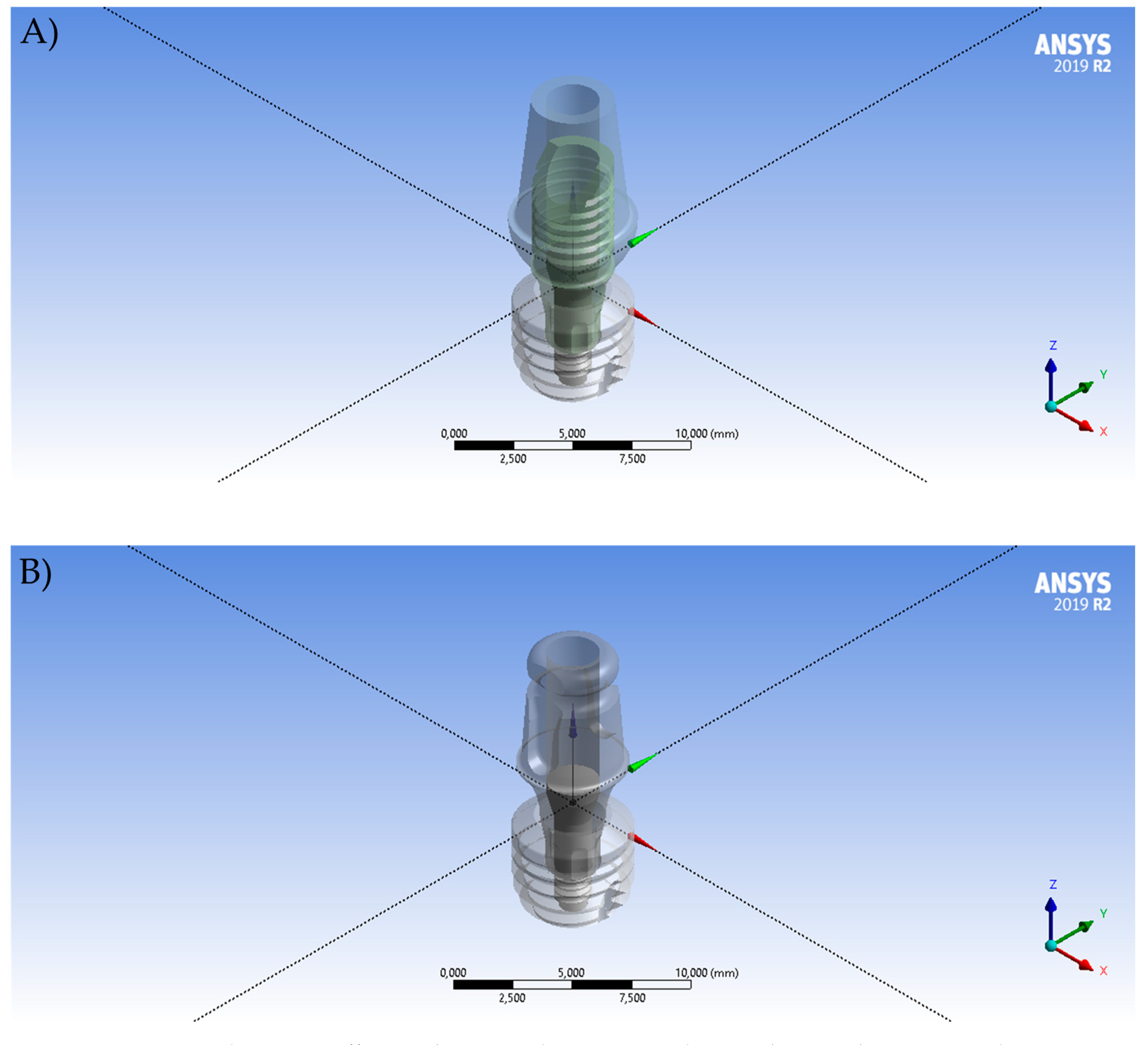
2. Materials and Methods

Ultrashort dental implants (copaSKY) STL files were obtained from the manufacturer (Bredent GmbH & Co. KG, Senden, Germany). The 3D file of the implant (5.2 mm × 5 mm), conical titanium abutment (copaSKY exso), titanium base and screw were converted into solids in the modelling software (Rhinoceros version 5.0 SR8, 2013, McNeel North America, Seattle, WA, USA) using the reverse engineering tool. For the BIO-HPP abutment (BioHPP copaSKY elegance) model, a cement layer (Panavia F 2.0, Kuraray Noritake Dental Inc., Okayama, Japan) was created between the titanium base and the mesostructure with 0.1 mm thickness (Figure 1).
The set of each model (titanium or BIO-HPP) was replicated in three conditions according to the bone level: sub-crestal, crestal and supra-crestal level (Figure 2). The difference between each model was standardized in 1.0 mm from the implant platform. The solid bone consisted of a simplified model (between cortical and cancellous bone tissue) with a cylinder shape (diameter and length: 15 mm and 20 mm). To guarantee correct connection at the bone–implant contact (BIC,) a Boolean operation was used, by subtracting the implant from the bone volume. In total, six 3D models were created and composed of bone tissue, dental implant, prosthetic screw and abutment.
The solid models were exported to the computer-aided engineering (CAE) software (ANSYS 19.0, 2018, ANSYS Inc., Houston, TX, USA) and a 10% mesh control convergence test was applied determining the total number of nodes and tetrahedral elements (Figure 3). The mechanical properties are summarized in Table 1 [12,13]. An axial load of 500 N was applied to simulate the occlusal force, at the upper surface of the abutment, based upon a coordinate system that combined vectors on different axes of orientation [12].
In the present study, microstrains, caused by the loading forces, were analyzed for the peri-implant tissue while von Mises stresses were analyzed for the implant, abutment and screw.

3. Results

The stress peaks and microstrain values obtained from the finite element analysis are summarized in Table 2, providing a comprehensive overview of the mechanical behavior of the system. These values serve as valuable indicators of the stress concentrations and deformation patterns within the implant and surrounding tissues. Additionally, Figure 4 presents a visual representation of the von Mises stress distribution across the entire evaluated set. This stress map provides a clear depiction of the areas experiencing the highest stress levels, allowing for a more detailed understanding of the load-bearing characteristics and potential areas of concern.
The influence of the abutment material on stress distribution was found to be minimal in the evaluated components, with the exception of the abutment itself. In models wherein the implant was placed in crestal and supra-crestal positions, it was observed that BIO-HPP, as the abutment material, exhibited higher stress levels. This finding suggests that the choice of abutment material may have implications for the mechanical performance of the implant restoration, particularly when considering the positioning of the implant. The results, depicted in Figure 5, highlight the importance of considering the mechanical properties of the abutment material and its potential impact on stress distribution, aiding clinicians and researchers in making informed decisions regarding implant restorations.
The positioning of the implant in the sub-crestal position had a notable effect on stress concentration within the peri-implant tissue. In this position, the stress distribution was found to be more homogenous and lower in the surrounding bone compared to other placement positions. This finding, as illustrated in Figure 6, suggests that sub-crestal implant placement can contribute to a more favorable biomechanical environment around the implant, potentially reducing the risk of complications and promoting better long-term stability.
On the other hand, when evaluating the stress distribution in the implant and screw components, it was observed that both experienced lower stress concentrations when the implant was placed in the supra-crestal position. This finding, depicted in Figure 7 and Figure 8, suggests that the supra-crestal placement of the implant can contribute to a more favorable load distribution and stress dissipation within these components. By reducing the stress concentration, the risk of mechanical failure and component fatigue may be minimized, thereby enhancing the overall durability and longevity of the implant-supported restoration.

4. Discussion

The present study aimed to evaluate, by the finite element method, the stress distribution in ultrashort dental implants placed in different bone levels with two abutment designs (titanium or BIO-HPP hybrid abutment) with a load of 500 N. The results show that both factors were able to affect the mechanical behavior of each evaluated structure in a different way. Therefore, both null hypotheses have been rejected.
To ensure the enduring success of a dental implant, a comprehensive understanding of how chewing forces are transmitted from the implant prosthetic components to the surrounding bone tissue is paramount. The distribution of stress is influenced by various critical factors, including the type of load applied, the interface between the bone and the implant, the shape and materials of the fixture as well as the quality and quantity of the bone. These factors collectively play a fundamental role in guaranteeing implant stability and evaluating the bone tissue’s ability to remodel and adapt to its biomechanical environment [19]. Such insights are crucial in optimizing the long-term performance and functionality of dental implants. The bone remodeling algorithm and FEA analysis are reported in the literature as reliable methods to evaluate the most appropriate implant prosthetic rehabilitation and prevent possible implant failures for each patient [19].
Short implants offer a viable solution for implant-supported restorations in cases wherein the edentulous area exhibits compromised bone quality and quantity. They serve as a more straightforward and less invasive alternative to procedures such as sinus lifts and bone grafts. By utilizing short implants, clinicians can minimize the need for extensive surgical interventions and achieve successful outcomes in situations wherein conventional implants may not be feasible. This approach provides a valuable option for patients with limited bone availability, offering a simplified and efficient treatment pathway while maintaining a high level of clinical success [3,6].
Indeed, the placement of implants in relation to the bone level is a crucial consideration for clinicians. It directly influences the preservation of peri-implant tissues, including gingival margins and the formation of interdental papilla. Optimal implant positioning plays a vital role in achieving favorable esthetic outcomes and ensuring the long-term stability of the soft tissues surrounding the implant [19]. Moreover, the stress concentration is usually located in the bone/implant contact area [20]. According to the findings of the present study, the sub-crestal placement decreased the microstrain in the bone, probably due to the higher contact area between the bone and the implant, thus generating a more homogeneous and lower stress concentration. The findings of previous studies align with the results that sub-crestal implant placement offers benefits for the peri-implant tissue when compared to crestal-level placement [20,21,22]. These studies have demonstrated that placing implants in a sub-crestal position can promote the improved preservation of the surrounding bone and enhance the stability of the soft tissues, including the gingival margins and interdental papilla as the peri-implant soft tissues play a pivotal role in dental implant success, impacting not only on the esthetic appearance but also on the maintenance and long-term stability of the implants [23,24]. Furthermore, evidence suggests that sub-crestal implant placement facilitates better osseointegration around the implant platform [25], further supporting its positive impact on the long-term success of implant-supported restorations.
The material used for the implant/abutment is another factor that may affect the peri-implant tissues’ maintenance [26]. Although it is still considered the gold-standard material for abutments, titanium is much stiffer than cortical and trabecular bone, since it presents high elastic modulus [27]. This fact was previously associated with the occurrence of the stress shielding effect, characterized by atrophy and bone reabsorption [28]. To reduce such phenomenon, softer materials emerged as options for abutment materials, like hybrid polyetheretherketone (PEEK) abutments, which consist of a high-performance synthetic polymer that presents biocompatibility, chemical stability and mechanical performance [29,30]. Moreover, PEEK presents an elastic modulus similar to the bone (around 3.6 GPa) [31]. When considering the stress in the abutment, the PEEK model depicted higher values of stress when compared to the titanium model for crestal and supra-crestal implant placement. This may be explained by the aforementioned properties of both materials, since in the hybrid abutment (PEEK/titanium) the polymer underwent greater deformation and concentrated less stress on itself, generating a peak of stress in the adjacent titanium component, while the solid titanium abutment presented a homogenous and lower stress concentration. These findings are corroborated by previous studies that evaluated the mechanical behavior of PEEK when used as abutment material through FEA [30,32,33].
These findings should be considered along with consideration related to the different all-ceramic crown designs as stress patterns have been reported to be different when comparing conventional bilayer zirconia covered with porcelain, a monolithic full-contour zirconia crown and the cutback modified zirconia crown with porcelain-veneered buccal face [34].
Despite the difference in elastic modulus between the abutment materials, the present study showed that the stress distribution in the bone, screw and implant components was not affected by such factor. This may be explained by the used test geometry, wherein the load application was positioned over the top surface of the abutment, which is far from the set bone/implant. Moreover, the implant placement and bone anchorage seem to be predominant for the biomechanical behavior of the models [35], where the higher support of the implant–bone contact in the sub-crestal model overcame the effect of the abutment elastic modulus, thus generating similar stress concentration even within the BIO-HPP and titanium abutments. Hence, both materials appear as suitable options for clinical applications.
The failure of the screw (loose or fracture) can compromise the entire implant-supported restorations, since it may lead to the fracture of the implant, instability and other complications [36]. In this sense, researchers and clinicians search for alternatives that reduce the stress concentration and consequently the risk of failures in the screw region [37,38]. The findings of the present study show that the supra-crestal implant placement decreased the amount of stress when compared to the sub-crestal and crestal positioning, thus reducing the risk of screw loosening and implant fracture. This may also be due to the lower contact implant/bone area in the crestal and supra-crestal models, which increased the lever arm and concentrated more stress on the bone [20], while generating less stress on the screw and the implant itself.
Despite the relevance of the present study, its limitations must also be considered. As an in silico study, it is not possible to consider all factors that are present in a clinical scenario, such as different loads and cyclic mechanical stimuli that are applied over the restorations. Even so, the use of FEA is important since it allows the isolated evaluation of a factor, which is essential to determine the performance of complex oral rehabilitations. Moreover, the present study reports important findings to predict the biomechanics of ultrashort implants according to their placement and abutment material. Through the finite element analysis, it was clear that ultrashort implants behaved adequately during the load application, as well as promoted homogenous stress distribution on the adjacent bone, mainly when placed in a sub-crestal position. In addition, new studies considering the use of short-implant are encouraged, including different materials and surgery protocols as well as their possible effects on the bone quality around the implant [39,40,41,42,43]. Also, FEA studies evaluating the presence of crowns, cantilever bridges and full-arch prostheses over ultrashort implants are needed, as well as the distinction between different bone types, fatigue and alternative implant designs [44].

5. Conclusions

The optimal implant placement plays a crucial role in achieving a more homogenous stress distribution within the peri-implant tissues. The sub-crestal implant placement has been found to offer mechanical benefits, particularly in terms of strain levels within the bone. This positioning helps reduce the risk of failure in both the bone and the abutment. Moreover, the results indicate that BIO-HPP serves as a suitable material for hybrid abutments, as it exhibits stress concentration patterns partially similar to titanium. Considering these factors, careful consideration of implant placement and the choice of abutment material can contribute to improved outcomes in implant-supported restorations, enhancing long-term stability and success.

Author Contributions

Conceptualization R.L.G., A.M.d.O.D.P. and J.P.M.T.; methodology, R.L.G., A.M.d.O.D.P. and J.P.M.T.; formal analysis, R.L.G., P.S.M. and J.P.M.T.; investigation, R.L.G., P.S.M. and J.P.M.T.; resources, R.L.G. and J.P.M.T.; data curation, R.L.G. and J.P.M.T.; writing—original draft preparation, R.L.G. and P.S.M.; writing—review and editing, A.M.d.O.D.P. and J.P.M.T.; supervision, J.P.M.T.; project administration, R.L.G. and J.P.M.T. All authors have read and agreed to the published version of the manuscript.

Funding

This research received no external funding.

Institutional Review Board Statement

Not applicable.

Informed Consent Statement

Not applicable.

Data Availability Statement

Not applicable.

Acknowledgments

The authors would like to thank Bredent Medical® for providing the STL files required for the simulation.

Conflicts of Interest

The authors declare no conflict of interest.

References

  1. Morand, M.; Irinakis, T. The Challenge of Implant Therapy in the Posterior Maxilla: Providing a Rationale for the Use of Short Implants. J. Oral Implant. 2007, 33, 257–266. [Google Scholar] [CrossRef]
  2. Lombardo, G.; Pighi, J.; Marincola, M.; Corrocher, G.; Simancas-Pallares, M.; Nocini, P.F. Cumulative Success Rate of Short and Ultrashort Implants Supporting Single Crowns in the Posterior Maxilla: A 3-Year Retrospective Study. Int. J. Dent. 2017, 2017, 8434281. [Google Scholar] [CrossRef] [Green Version]
  3. Magdy, M.; Msc, M.A.A.; Alloush, S.; El-Sayed, K.M.F.; Nawwar, A.A.; Shoeib, M.; ElNahass, H.; Msc, M.M.; Msc, S.A. Ultra-short versus standard-length dental implants in conjunction with osteotome-mediated sinus floor elevation: A randomized controlled clinical trial. Clin. Implant. Dent. Relat. Res. 2021, 23, 520–529. [Google Scholar] [CrossRef] [PubMed]
  4. Heboyan, A.; Giudice, R.L.; Kalman, L.; Zafar, M.S.; Tribst, J.P.M. Stress Distribution Pattern in Zygomatic Implants Supporting Different Superstructure Materials. Materials 2022, 15, 4953. [Google Scholar] [CrossRef]
  5. Pera, F.; Pesce, P.; Menini, M.; Fanelli, F.; Kim, B.-C.; Zhurakivska, K.; Mayer, Y.; Isola, G.; Cianciotta, G.; Crupi, A.; et al. Immediate loading full-arch rehabilitation using transmucosal tissue-level implants with different variables associated: A one-year observational study. Minerva Dent. Oral Sci. 2023. [Google Scholar] [CrossRef]
  6. García-Braz, S.H.; Prados-Privado, M.; Zanatta, L.C.S.; Calvo-Guirado, J.L.; Prados-Frutos, J.C.; Gehrke, S.A. A Finite Element Analysis to Compare Stress Distribution on Extra-Short Implants with Two Different Internal Connections. J. Clin. Med. 2019, 8, 1103. [Google Scholar] [CrossRef] [Green Version]
  7. Fiorillo, L.; Milone, D.; D’andrea, D.; Santonocito, D.; Risitano, G.; Cervino, G.; Cicciù, M. Finite Element Analysis of Zirconia Dental Implant. Prosthesis 2022, 4, 490–499. [Google Scholar] [CrossRef]
  8. Emam, M.; Arafa, A.M. Stress Distribution and Fracture Resistance of Green Reprocessed Polyetheretherketone (PEEK) Single Implant Crown Restorations Compared to Unreprocessed PEEK and Zirconia: An in-Vitro Study. BMC Oral Health 2023, 23, 275. [Google Scholar] [CrossRef]
  9. Cicciù, M.; Cervino, G.; Terranova, A.; Risitano, G.; Raffaele, M.; Cucinotta, F.; Santonocito, D.; Fiorillo, L. Prosthetic and Mechanical Parameters of the Facial Bone under the Load of Different Dental Implant Shapes: A Parametric Study. Prosthesis 2019, 1, 41–53. [Google Scholar] [CrossRef] [Green Version]
  10. De Andrade, G.S.; Kalman, L.; Giudice, R.L.; Adolfi, D.; Feilzer, A.J.; Tribst, J.P.M. Biomechanics of implant-supported restorations. Braz. Dent. Sci. 2023, 26, 1–15. [Google Scholar] [CrossRef]
  11. Tribst, J.P.M.; Dal Piva, A.M.d.O.; Lo Giudice, R.; Borges, A.L.S.; Bottino, M.A.; Epifania, E.; Ausiello, P. The Influence of Custom-Milled Framework Design for an Implant-Supported Full-Arch Fixed Dental Prosthesis: 3D-FEA Study. Int. J. Environ. Res. Public Health 2020, 17, 4040. [Google Scholar] [CrossRef]
  12. Ausiello, P.; Tribst, J.P.M.; Ventre, M.; Salvati, E.; di Lauro, A.E.; Martorelli, M.; Lanzotti, A.; Watts, D.C. The role of cortical zone level and prosthetic platform angle in dental implant mechanical response: A 3D finite element analysis. Dent. Mater. 2021, 37, 1688–1697. [Google Scholar] [CrossRef]
  13. Giudice, R.L.; Sindoni, A.; Tribst, J.P.M.; Piva, A.M.D.O.D.; Giudice, G.L.; Bellezza, U.; Giudice, G.L.; Famà, F. Evaluation of Zirconia and High Performance Polymer Abutment Surface Roughness and Stress Concentration for Implant-Supported Fixed Dental Prostheses. Coatings 2022, 12, 238. [Google Scholar] [CrossRef]
  14. Paracchini, L.; Barbieri, C.; Redaelli, M.; Di Croce, D.; Vincenzi, C.; Guarnieri, R. Finite Element Analysis of a New Dental Implant Design Optimized for the Desirable Stress Distribution in the Surrounding Bone Region. Prosthesis 2020, 2, 225–236. [Google Scholar] [CrossRef]
  15. Fiore, D.; Montagner, A.; Sivolella, M.; Stellini, S.; Yilmaz, E.; Brunello, B. Peri-Implant Bone Loss and Overload: A Systematic Review Focusing on Occlusal Analysis through Digital and Analogic Methods. J. Clin. Med. 2022, 11, 4812. [Google Scholar]
  16. Oliveros-López, L.-G.; Castillo-De-Oyagüe, R.; Serrera-Figallo, M.; Martínez-González, J.; Pérez-Velasco, A.; Torres-Lagares, D.; Gutiérrez-Pérez, J.-L. Bone Loss in Bruxist Patients Wearing Dental Implant Prostheses: A Finite Element Analysis. Metals 2020, 10, 1132. [Google Scholar] [CrossRef]
  17. D’amico, C.; Bocchieri, S.; Sambataro, S.; Surace, G.; Stumpo, C.; Fiorillo, L. Occlusal Load Considerations in Implant-Supported Fixed Restorations. Prosthesis 2020, 2, 252–265. [Google Scholar] [CrossRef]
  18. Bambini, F.; Orilisi, G.; Quaranta, A.; Memè, L. Biological Oriented Immediate Loading: A New Mathematical Implant Vertical Insertion Protocol, Five-Year Follow-Up Study. Materials 2021, 14, 387. [Google Scholar] [CrossRef]
  19. Santonocito, D.; Nicita, F.; Risitano, G. A Parametric Study on a Dental Implant Geometry Influence on Bone Remodelling through a Numerical Algorithm. Prosthesis 2021, 3, 157–172. [Google Scholar] [CrossRef]
  20. Ellendula, Y.; Sekar, A.C.; Nalla, S.; Basany, R.B.; Sailasri, K.; Thandu, A. Biomechanical Evaluation of Stress Distribution in Equicrestal and Sub-crestally Placed, Platform-Switched Morse Taper Dental Implants in D3 Bone: Finite Element Analysis. Cureus 2022, 2914, e24591. [Google Scholar] [CrossRef]
  21. Baggi, L.; Cappelloni, I.; Di Girolamo, M.; Maceri, F.; Vairo, G. The influence of implant diameter and length on stress distribution of osseointegrated implants related to crestal bone geometry: A three-dimensional finite element analysis. J. Prosthet. Dent. 2008, 100, 422–431. [Google Scholar] [CrossRef] [PubMed] [Green Version]
  22. Chou, H.-Y.; Müftü, S.; Bozkaya, D. Combined effects of implant insertion depth and alveolar bone quality on periimplant bone strain induced by a wide-diameter, short implant and a narrow-diameter, long implant. J. Prosthet. Dent. 2010, 104, 293–300. [Google Scholar] [CrossRef]
  23. Del Amo, F.S.L.; Yu, S.-H.; Sammartino, G.; Sculean, A.; Zucchelli, G.; Rasperini, G.; Felice, P.; Pagni, G.; Iorio-Siciliano, V.; Grusovin, M.G.; et al. Peri-implant Soft Tissue Management: Cairo Opinion Consensus Conference. Int. J. Environ. Res. Public Heal. 2020, 17, 2281. [Google Scholar] [CrossRef] [Green Version]
  24. Felice, P.; Bonifazi, L.; Pistilli, R.; Ferri, A.; Gasparro, R.; Barausse, C. Influence of Keratinized Tissue on Short Dental Implants: A Parallel Cohort Retrospective Study on 217 Implants with a Mean Follow-up of 4.1 Years. Int. J. Oral Maxillofac. Implant. 2023, 38, 462–467. [Google Scholar] [CrossRef]
  25. Aimetti, M.; Ferrarotti, F.; Mariani, G.; Ghelardoni, C.; Romano, F. Soft Tissue and Crestal Bone Changes Around Implants with Platform-Switched Abutments Placed Nonsubmerged at Subcrestal Position: A 2-Year Clinical and Radiographic Evaluation. Int. J. Oral Maxillofac. Implant. 2015, 30, 1369–1377. [Google Scholar] [CrossRef] [Green Version]
  26. Chan, Y.-H.; Lew, W.-Z.; Lu, E.; Loretz, T.; Lu, L.; Lin, C.-T.; Feng, S.-W. An evaluation of the biocompatibility and osseointegration of novel glass fiber reinforced composite implants: In vitro and in vivo studies. Dent. Mater. 2018, 34, 470–485. [Google Scholar] [CrossRef] [PubMed]
  27. Çaglar, A.; Bal, B.T.; Karakoca, S.; Aydın, C.; Yılmaz, H.; Sarısoy, S. Three-dimensional finite element analysis of titanium and yttrium-stabilized zirconium dioxide abutments and implants. Int. J. Oral Maxillofac. Implant. 2011, 26, 961–969. [Google Scholar]
  28. Lee, W.-T.; Koak, J.-Y.; Lim, Y.-J.; Kim, S.-K.; Kwon, H.-B.; Kim, M.-J. Stress shielding and fatigue limits of poly-ether-ether-ketone dental implants. J. Biomed. Mater. Res. Part B Appl. Biomater. 2012, 100B, 1044–1052. [Google Scholar] [CrossRef]
  29. Najeeb, S.; Zafar, M.S.; Khurshid, Z.; Siddiqui, F. Applications of polyetheretherketone (PEEK) in oral implantology and prosthodontics. J. Prosthodont. Res. 2016, 60, 12–19. [Google Scholar] [CrossRef] [PubMed]
  30. Barbosa-Júnior, S.A.; Pereira, G.K.R.; DaPieve, K.S.; Machado, P.S.; Valandro, L.F.; Schuh, C.; Consani, R.L.X.; Bacchi, A. Mechanical Fatigue Analysis of PEEK as Alternative to Zirconia for Definitive Hybrid Abutments Supporting All-Ceramic Crowns. Int. J. Oral Maxillofac. Implant. 2020, 35, 1209–1217. [Google Scholar] [CrossRef]
  31. Mishra, S.; Chowdhary, R. PEEK materials as an alternative to titanium in dental implants: A systematic review. Clin. Implant. Dent. Relat. Res. 2018, 21, 208–222. [Google Scholar] [CrossRef] [Green Version]
  32. Soares, P.M.; Cadore-Rodrigues, A.C.; Borges, A.L.S.; Valandro, L.F.; Pereira, G.K.R.; Rippe, M.P. Load-bearing capacity under fatigue and FEA analysis of simplified ceramic restorations supported by Peek or zirconia polycrystals as foundation substrate for implant purposes. J. Mech. Behav. Biomed. Mater. 2021, 123, 104760. [Google Scholar] [CrossRef]
  33. Tretto, P.H.W.; Dos Santos, M.B.F.; Spazzin, A.O.; Pereira, G.K.R.; Bacchi, A. Assessment of stress/strain in dental implants and abutments of alternative materials compared to conventional titanium alloy-3D non-linear finite element analysis. Comput. Methods Biomech. Biomed. Eng. 2020, 23, 372–383. [Google Scholar] [CrossRef] [PubMed]
  34. Ramos, N.d.C.; Ramos, G.F.; Penteado, M.M.; de Melo, R.M.; Borges, A.L.S.; Bottino, M.A.; Tribst, J.P.M. Comparative Stress Evaluation between Bilayer, Monolithic and Cutback All-Ceramic Crown Designs: 3D Finite Element Study. Prosthesis 2021, 3, 173–180. [Google Scholar] [CrossRef]
  35. Brånemark, P.-I.; Breine, U.; Adell, R.; Hansson, B.O.; Lindström, J.; Ohlsson, Å. Intra-Osseous Anchorage of Dental Prostheses:I. Experimental Studies. Scand. J. Plast. Reconstr. Surg. 1969, 3, 81–100. [Google Scholar] [CrossRef]
  36. Geckili, E.; Geckili, O.; Bilhan, H.; Kutay, O.; Bilgin, T. Clinical comparison of screw-retained and screwless Morse taper im-plant-abutment connections: One-year postloading results. Int. J. Oral Maxillofac. Implant. 2017, 32, 1123–1131. [Google Scholar] [CrossRef] [Green Version]
  37. Fiorillo, L.; Meto, A.; Cicciù, M. Bioengineering Applied to Oral Implantology, a New Protocol: “Digital Guided Surgery”. Prosthesis 2023, 5, 234–250. [Google Scholar] [CrossRef]
  38. Choi, S.; Kang, Y.S.; Yeo, I.-S.L. Influence of Implant–Abutment Connection Biomechanics on Biological Response: A Literature Review on Interfaces between Implants and Abutments of Titanium and Zirconia. Prosthesis 2023, 5, 527–538. [Google Scholar] [CrossRef]
  39. Menini, M.; Pesce, P.; Delucchi, F.; Ambrogio, G.; Canepa, C.; Carossa, M.; Pera, F. One-stage versus two-stage technique using two splinted extra-short implants: A multicentric split-mouth study with a one-year follow-up. Clin. Implant. Dent. Relat. Res. 2022, 24, 602–610. [Google Scholar] [CrossRef]
  40. El-Shabrawy, M.M.; Zaki, A.A.; Elguindy, J.; El Nahass, H.; Bakr, K.A. Patient Satisfaction and Pink Esthetics of PEEK Versus Zirconium Abutments in the Esthetic Zone (A Randomized Controlled Trial). Braz. Dent. Sci. 2021, 24, 1–16. [Google Scholar] [CrossRef]
  41. Giordano, M.; Ausiello, P.; Martorelli, M. Accuracy evaluation of surgical guides in implant dentistry by non-contact reverse engineering techniques. Dent. Mater. 2012, 28, e178–e185. [Google Scholar] [CrossRef]
  42. de Kok, P.; Kleverlaan, C.J.; de Jager, N.; Kuijs, R.; Feilzer, A.J. Mechanical performance of implant-supported posterior crowns. J. Prosthet. Dent. 2015, 114, 59–66. [Google Scholar] [CrossRef] [PubMed]
  43. Van Oers, R.F.M.; Feilzer, A.J. Abutment-to-fixture load transfer and peri-implant bone stress. Am. J. Dent. 2015, 28, 247–250. [Google Scholar] [PubMed]
  44. Tribst, J.P.M.; Piva, A.M.d.O.D.; Blom, E.J.; Kleverlaan, C.J.; Feilzer, A.J. Dental biomechanics of root-analog implants in different bone types. J. Prosthet. Dent. 2022, 116, 104370. [Google Scholar] [CrossRef] [PubMed]
Figure 1. Different abutment designs according to the prosthetic material. (A) Ceramic-reinforced PEEK abutment with titanium base and (B) conventional titanium abutment.
Prosthesis 05 00051 g001
Figure 2. Different bone levels positioning simulated in the present study. (A) sub-crestal, (B) crestal and (C) supra-crestal.
Prosthesis 05 00051 g002
Figure 3. Numerical model and boundary conditions. (A) Model after meshing process, (B) loading application and (C) fixed support.
Prosthesis 05 00051 g003
Figure 4. Von Mises stress distribution along the entire set according to the implant placement (supra-crestal, crestal and sub-crestal) and abutment material (BIO-HPP and titanium).
Prosthesis 05 00051 g004
Figure 5. Stress distribution along the abutment through von Mises stress test, according to the implant placement (supra-crestal, crestal and sub-crestal) and abutment material (BIO-HPP and titanium).
Prosthesis 05 00051 g005
Figure 6. Microstrain distribution along the peri-implant tissue (bone) through strain analysis, according to the implant placement (supra-crestal, crestal and sub-crestal) and abutment material (BIO-HPP and titanium).
Prosthesis 05 00051 g006
Figure 7. Stress distribution along the implant through von Mises stress test, according to the implant placement (supra-crestal, crestal and sub-crestal) and abutment material (BIO-HPP and titanium).
Prosthesis 05 00051 g007
Figure 8. Stress distribution along the screw through von Mises stress test, according to the implant placement (supra-crestal, crestal and sub-crestal) and abutment material (BIO-HPP and titanium).
Prosthesis 05 00051 g008
Table 1. Mechanical properties of the structures adopted in the present study.
StructuresElastic Modulus (GPa)Poisson Ratio
Titanium *1100.3
BIO-HPP30.3
Cement80.3
Bone tissue5.50.3
* Both screw, ultrashort implant and the correspondent abutment were made of titanium.
Table 2. Equivalent strains in the bone tissue and stress per region for each evaluated model.
PlacementAbutment DesignMicrostrain (µ/µ)Abutment
Stress (MPa)
Implant
Stress (MPa)
Screw
Stress (MPa)
Sub-crestalBIO-HPP1.08 × 10−2222.67338.6371.10
Titanium1.07 × 10−2220.53342.0575.05
CrestalBIO-HPP1.17 × 10−2248.08287.3471.19
Titanium1.17 × 10−2219.88291.9475.61
Supra-crestalBIO-HPP1.23 × 10−2248.08274.2563.74
Titanium1.20 × 10−2221.77276.9361.33
Disclaimer/Publisher’s Note: The statements, opinions and data contained in all publications are solely those of the individual author(s) and contributor(s) and not of MDPI and/or the editor(s). MDPI and/or the editor(s) disclaim responsibility for any injury to people or property resulting from any ideas, methods, instructions or products referred to in the content.

