Article

Effectiveness of Different Irrigation Techniques on Post Space Smear Layer Removal: SEM Evaluation

1
Department of Medicine and Surgery, School of Dentistry, University of Salerno, 84081 Salerno, Italy
2
Department of Endodontics, Faculty of Dentistry, University of Alexandria, Alexandria 21531, Egypt
3
Department of Biomedical and Neuromotor Sciences, School of Dentistry, University of Bologna, 40125 Bologna, Italy
*
Author to whom correspondence should be addressed.
Prosthesis 2023, 5(2), 539-549; https://doi.org/10.3390/prosthesis5020037
Submission received: 12 April 2023 / Revised: 31 May 2023 / Accepted: 1 June 2023 / Published: 2 June 2023
(This article belongs to the Special Issue Modern Management of Endodontically Treated Teeth)

Abstract

:
Background: Effective debris and smear layer removal affects post-cementation and bond strength. Aim: The aim was to compare the effectiveness of debris and smear layer removal using standard irrigation and activated irrigation with heated EDTA. Moreover, an irrigant activation technique was chosen in the current research, employing the ultrasonic activation of an EDTA solution after being heated directly inside the post space preparation. Materials and Methods: 30 single-rooted human mandibular premolar teeth were used in the current study to test the proposed techniques. First, the teeth were cut to have standardised roots 18 mm in length and prepared endodontically, and then the post space was carried out on all the samples. Then the specimens were randomly distributed into three study groups according to the post space irrigation technique. In detail, the groups were group 1, where a 3D cleaning technique was used; group 2, where the traditional irrigation with EDTA was applied; and group 3, where only saline as an irrigant was used. The third group was considered the control group. Next, the teeth were segmented and analysed via scanning electron microscopy (SEM). The magnification was used to evaluate and score the smear layer and debris. Statistical analysis was undertaken using the classic statistical software package (SPSS, version 28.0; SPSS IBM, Armonk, NY, USA). Then, the data were interpreted with a non-parametric analysis of variance (Kruskal–Wallis ANOVA) among the experiment groups. The significance level was decided as p < 0.05. In addition, statistically significant (p < 0.05) lower mean smear layer and debris scores were found in both the examination groups compared to the control group. Results: group 1 demonstrated better results compared to group 2 in terms of cleaning the dentinal walls. Conclusions: The current research concluded that the EDTA 3D cleaning technique is an effective irrigation technique for clearing debris and smear layers in the post space. Future research, such as on push-out bond strength, should be used to verify this research’s conclusions. Key findings: The use of ultrasonically activated heated EDTA improved the smear layer removal. The use of the 3D cleaning technique resulted in better post space cleaning.

1. Introduction

Undoubtedly, the quality of coronal restoration is a fundamental requirement for the long-term success of endodontic treatment as it prevents microbial infiltration into the treated canal [1]. A post should be used when the remaining coronal tooth structure is insufficient to support the final restoration [2]. Teeth that need endodontic treatment, most of the time, have often lost a significant amount of coronal tooth structure, and the remaining one is usually much compromised. This condition requires restoration with a crown that can be supported by a post inserted into the root canal space [3]. Fibre post systems are more commonly used today because they have similar mechanical characteristics to dentine [4].
Furthermore, their advantages include biocompatibility, corrosion resistance, improved light transmission, diffraction, and reflection effects in aesthetic restorations, and improved restoration retention [5]. For adequate retention of the composite core on the post and tooth, as well as increased fracture resistance of endodontically treated teeth, fibre posts are cemented with adhesives and composite resin luting cement for dentin [6]. Generally, the adhesion between resin and dentin is considered a weak point in cementing a fibre post [7].
As a rule, the canal space for the post is created using a parallel-sided preparation [8]. Moreover, approximately 3–5 mm of root-canal-filling material below the post space is left in the apical third region [9].
Chiefly, the depth of the post space preparation varies depending on the required post length and root morphology, bearing in mind that post retention is higher with longer posts [3].
Posts can be rigid or flexible; rigid posts are constructed from a metal alloy, while flexible posts can be made from various types of ceramic or fibre materials [10]. The literature describes two timings for preparing the post space: immediate and delayed. However, the two approaches have been shown to influence endodontic treatment outcomes with controversial results [11].
The post space preparation phase requires the use of burs to remove the filling material from the root canal; in particular, Gates-Glidden burs and pin burs with parallel sides are used. Additionally, specific drills are used for core removal in endodontic cases treated with obturation systems with plastic core carriers.
For correct and effective cementation, the post space walls must be made of dentin to create an adequate hybrid zone between the resin cement and the dentinal tubules [12]. Enlarging the canal beyond the size of the initial preparation reduces the amount of obturating material residue on the canal walls [10]. It is advisable to minimise the removal of root dentin and support preserving the fracture toughness of the tooth; thus, the post drill should match the size of the prepared canal [13].
Once the mechanical preparation phase of the post space is complete, the dentine walls are covered with a layer of dentinal debris, sealant, and gutta-percha residues which can hinder adhesion [14].
Methods of restoring endodontically treated teeth still generate discussions in dentistry, especially when it comes to difficulties in obtaining good adhesion between a fibre post and root dentin. Difficulties in removing the smear layer, drying excess moisture from the root canal, as well as complete polymerization of the adhesive system and resin cement are inherent challenges in achieving high adhesion of the fibre post to the root dentin [15]. To achieve sufficient adhesion, removal of the smear layer, which contains inorganic components, sealer residues, and gutta-percha, is required, so proper irrigation of the post space is mandatory to remove this debris. With traditional irrigation, it is difficult to obtain adequate cleaning of the post space, which is why it is essential to look for new techniques and protocols for the complete removal of the smear layer [15].
The irrigation phase is still a highly debated topic today. The use of sodium hypochlorite is recommended by some manufacturers, while others advise otherwise [16]. New irrigation protocols with alternative chemical solutions have been proposed; in particular, calcium hypochlorite has been tested [17]. A preliminary study showed no significant differences between the tissue-dissolving properties of 5% calcium hypochlorite or 10% CH and 4.65% sodium hypochlorite (Chlorax solution) or 1.36% solutions after 60 min [18]. In a study conducted by Görduysus et al. [19], it was shown that neither sodium hypochlorite nor calcium hypochlorite was effective in removing the smear layer and dentinal debris.
Recent studies have analysed the use of EDTA (ethylenediaminetetraacetic acid) in smear layer removal with satisfactory results [20]. In one study [21], the effectiveness of pre-heated EDTA was evaluated, obtaining good results in terms of cleaning. However, it has been demonstrated that irrigants pre-heated and inserted into root canals stabilise at body temperature after a few seconds [21]. For this reason, a new technique has been proposed, which involves heating the irrigant directly inside the post space. Due to controversies in the literature regarding the post space irrigation phase, the following work aims to evaluate the effectiveness of a new cleansing protocol. This study involves evaluating the effect of internal heating on the EDTA followed by the use of ultrasonic activation. Therefore, the study’s null hypothesis was that the new cleansing protocol should not increase the degree of cleansing of the post space.

2. Materials and Methods

This research was performed according to the policies of the Declaration of Helsinki and permitted by the Board of the Institutional Review (University of Naples Federico II, Naples, Italy).
Extracted single-rooted mandibular premolars (n = 30) were picked for this study.
The sample size was calculated utilising G Power Software employing an effect size of 0.4 and α = 0.05 [5]. A sum of 30 samples was required, 10 per group, to set the power at 80%.
These teeth were extracted after collecting informed consents, unrelated to our work, and were extracted for orthodontic treatment plans (patients’ ages from 18 to 25 years).
Teeth with any signs of cracks, fractures, resorption, periapical lesions, previously endodontically treated teeth, history of trauma, immature apices, or calcification were excluded from this study. On the other hand, teeth with mature apices, normal anatomy, single canal, and canal curvatures less than 10° according to the Schneider system (by buccolingual and mesiodistal radiographic analysis) [22] were included in this study.
Once the teeth were extracted, a curette was used to remove debris and small pieces of external periodontal tissues. For applying the exclusion criteria, digital radiography and a stereomicroscope (Karl Kaps Optik-Feinmechanik-Gerätebau GmbH & Co. KG.Europastraße, Aßlar, Germany) under ×20 magnification were used to evaluate each specimen.
Next, the samples were conserved in physiological saline in separated vials at +4 °C till the beginning of the experimentation, after five days [23].
The study groups were created before testing by dividing the roots randomly into 3 groups of 10 samples per group.

Root Canal Preparation

All the samples were cut using a diamond disc (Kerr Italy SRL, Via Passanti 332, 84018 Scafati, Italy) to have 18 mm length roots in order to standardize the length and have the same post space area. Next, to establish the working length, a size ten k-file (Coltene/Whaldedent Inc., Cuyahoga Falls, OH, USA) was used in the canal until it was visible from the apex, and then 0.5 mm was deducted from the file length measurement.
The apices were then sealed by applying wax to avoid irrigant extrusion during the experiments to simulate a clinical situation. All the canals were next mechanically prepared using nickel–titanium rotating files (Hyflex EDM, Coltene/Whaldedent Inc., Cuyahoga Falls, USA). Specifically, files 10/0.05 and 20/0.05 Hyflex EDM were selected to prepare the canals for the entire working length. This step was accomplished deliberately to provide minimal shaping for the canals.
The chemical canal cleaning was conducted using 3% NaOCl for three minutes (Canal pro, Coltene/Whaldedent Inc., Cuyahoga Falls, OH, USA) through a side-vented 30 G needle syringe (Coltene/Whaldedent Inc., Cuyahoga Falls, OH, USA). A 5 mL NaOCl solution was used per tooth throughout the preparation, and a new solution was revived every 60 s.
At the end of the shaping phase, the root canals were irrigated with sterile saline, and then the following cleaning protocol was used for all the samples:
First, 3 mL of 17% EDTA (Canal pro, Coltene/Whaldedent Inc., Cuyahoga Falls, OH, USA) was left in the canal for 1 min. Second, the canals were again flushed with 3 mL of sterile saline. The irrigant was inserted in the canal using an endodontic needle of a 30 G open-vented (Coltene/Whaldedent Inc., Cuyahoga Falls, OH, USA) at 2 mm away from the working length. The chemo-mechanical phase was concluded with 2 mL of distilled water used as the final flush, and the canals were dried using paper points (Coltene/Whaldedent Inc., Cuyahoga Falls, OH, USA) of appropriate size.
Then, all the root canals were obturated using a continuous wave of condensation (gutta-percha cone and bioseal, Coltene/Whaldedent Inc., Cuyahoga Falls, OH, USA). All the samples used the same quantity of cement. The bioseal syringe tip was inserted 6 mm away from the working length, and for each tooth, 2 mm of biosealer was injected.
After 30 min from the obturation phase, after having reached the hardening of the endodontic cement (12 min), the post spaces were prepared using Gates-Glidden drills 1, 2, 3 and 4 (Kerr Italy SRL, Via Passanti 332, 84018 Scafati, Italy) for 10 mm inside the root canal. Each Gates-Glidden was used for 20 s (total: 80 s).
At the end of the root canal treatment, different protocols of post space cleaning were used for all the samples. Roots (n = 30) were randomly allocated (www.random.org, accessed on 10 January 2023) into 3 groups (n = 10):
Group (1): 3 mL of 17% EDTA was utilised, followed by 2 mL of saline solution; both solutions were added using a 30 G endodontic needle.
Ultimately, the 3D cleaning procedure was conducted (Figure 1 and Figure 2):
An amount of 17% EDTA was inserted, placing the same type of needle as in previous steps in contact with the previous filling material. The activation was done using a system-B heat tip (Analytic Endodontics, Orange, CA, USA) set at 180 °C with an X-fine tip (30/04). During the process, the tip was not in contact with dentinal walls and was activated for 6 s. At this temperature, for 6 s, the heat carrier transmits about 80 °C into the canal (21).
This was followed by 20 s of ultrasonic activation utilising the Ultra Smart AI (Coxo, Fushan, China). The tip used for ultrasonic activation was 25.00. This activation procedure was repeated thrice, and 17% EDTA was refreshed with a new solution at each cycle. A total of 3 mL of 17% EDTA was used. The duration time of EDTA irrigation was 90 s. 2 mL of distilled water was applied as the final flush for 30 s, and then the post space was dried using paper points.
Group (2): An endodontic 30 G needle was used to deliver the irrigants in contact with the previous filling material. Finally, traditional irrigation was performed. The post space was filled with 3 mL of 17% concentration of EDTA (Canal pro, Coltene/Whaldedent Inc., Cuyahoga Falls, OH, USA) and was left inside for 1 min. Following this, a flush of 2 mL of sterile saline was applied for 30 s as the final rinse, and paper points were used to dry the post space.
Group (3), the saline solution (control): an endodontic needle reached the previous filling material, and 5 mL of saline was used. Finally, 2 mL of distilled water was utilised as the final flush, and the post space was dried with paper points.
After completion of the experiments, two longitudinal grooves were prepared on each root’s palatal/lingual and buccal surfaces with a diamond bur (Kerr Italy SRL, Via Passanti 332, 84018 Scafati, Italy) used with a high-speed water-cooled handpiece to facilitate vertical splitting. Each sample was dipped in liquid nitrogen immediately after canal preparation and split longitudinally into two halves with a stainless-steel chisel. The sections were then prepared for SEM analysis: They were allowed to be air-dried overnight in a desiccator at room temperature. Then the specimens were sputter-coated using gold to be ready for SEM analysis (EVO MA 10 Carl Zeiss SMT AG, Oberkochen, Germany).
SEM images were obtained at a magnification of ×1000 (Figure 3), and 30 photomicrographs were taken in three different areas (10 for the area; coronal, middle, and apical thirds of the post space). For the assessment of residual debris and smear layer, each specimen’s coronal, middle, and apical areas were chosen randomly. The areas were selected at low magnification by the SEM operator, who was blinded to the study’s aims and needed to be informed about the respective preparation technique. The images were captured at 5 kV.
In order to score the presence or absence of debris and smear layer, three trained operators blindly assessed them on the surface of the post space preparation at each tooth’s coronal, middle, and apical portions. The rating system applied in the current study was proposed by Hulsmann et al. [24], and the criteria for the scoring were reported as follows [25]:
Debris Scores
Score 1: Clean post space with only a few small debris particles.
Score 2: Few small accumulations of debris.
Score 3: Multiple accumulations of debris that covered <50% of the post space.
Score 4: >50% of the post space was covered by debris.
Score 5: All or almost all post space walls were covered by debris.
Scores of the smear layer
Score 1: No smear layer was detected, and the orifices of dentinal tubules were open.
Score 2: Small quantity of smear layer and some dentinal tubules were open.
Score 3: A homogenous smear layer covered the post space with just a few open dentinal tubules.
Score 4: A homogenous smear layer covered the entire post space wall, and no dentinal tubules were open.
Score 5: The heavy, homogenous smear layer covers the whole post space walls.
Statistical analysis
Statistical analysis was performed with a standard statistical software package (SPSS, version 28.0; SPSS IBM, Armonk, NY, USA). The scores of smear layer and debris were individually registered. Explicatory statistics for ordinal data were calculated per group, including the median and 25th and 75th percentiles. Data were analysed with a non-parametric analysis of variance (Kruskal–Wallis) among the groups tested and among the thirds of the post space. The significance level was determined at p < 0.05.

3. Results

The means, medians, and 25th and 75th percentiles of each group are shown in Table 1 and Table 2. The means and the medians of the debris score were inferior in group 1 (EDTA 3D cleaning approach) compared to group 2 (traditional irrigation method). The Kruskall–Wallis non-parametric test demonstrated that the differences in the medians are statistically significant (p < 0.05). Similar results were gained regarding the smear layer’s residual quantity on the dentinal wall’s surface with the medians of group 2, which were higher than those in group 1 (p < 0.05). Therefore, the null hypothesis of this work should be rejected. When comparing the three thirds of the post space, the test showed no statistically significant differences between the third smear layer groups (p > 0.05). Conversely, in the debris groups, the apical thirds revealed higher values (p < 0.05).

4. Discussion

The present research found that the 3D cleaning protocol had the highest smear layer removal compared to traditional irrigation. Thus, the null hypothesis was rejected.
Through the SEM analysis, it is possible to highlight how the 3D cleaning protocol has obtained excellent results in removing the smear layer at all three levels, coronal, middle, and apical, of the post space. On the other hand, the traditional irrigation protocol was able to remove only a small amount of smear layer in the coronal third, while removing the smear layer in the middle and apical third of the post space was impossible. The smear layer was abundant in the control group at all three levels.
In summary, the heated EDTA with subsequent ultrasonic activation, the 3D cleaning protocol, was able to optimally expose the dentinal tubules at all levels of the post space.
The first step in endodontic treatment is achieving the correct diagnosis and organising an adequate treatment plan, after which it is possible to proceed with the subsequent phases. Moreover, the treated tooth is isolated with a rubber dam. Next, the access cavity is made under magnification and illumination. Only after identifying all the root canals is it possible to proceed with the shaping, using modern rotating files, the cleaning, and, finally, the obturation of the complex endodontic space [21].
After endodontic therapy, the treated tooth’s final restoration may or may not include inserting a fibre post and resin restoration, which is a standard clinical approach.
The mechanical preparation of a post space indicates removing the obturating material’s coronal portion. The persisting filling materials propose the apical seal between the post and coronal restoration and the periapical tissues. When the remaining root filling length is compromised, it may offer a more vulnerable barrier to coronal leakage. Therefore, it is crucial to utilise materials and approaches to sufficiently seal the apical root canal third when a post is mandated. Furthermore, using an operating microscope during the mechanical phase of post space preparation has been documented to enhance the bond strength of a glass fibre post to the dentin [26]. At the end of the mechanical phase, it is critical to confirm that there is no remaining gutta-percha.
Furthermore, magnification has become increasingly standard in dental procedures and there is awareness of the earlier onset of presbyopia about the age of 40 [26]. Optical aids such as loops or operating microscopes are recommended to be employed early to compensate for the operator’s visual impairment or development [26]. The widespread use of loupes and microscopes is due to the confidence of the utilising professionals that these instruments have benefits and enhance their work’s quality and ergonomics.
Indeed, restorative materials’ adherence to the dentine is created by micro-mechanical retentions formed after demineralising the surface and by constructing a hybrid layer and resin tags [27]. Consequently, post space cleansing is important and can influence the strength of the bond of the post to the dentin [28].
The adhesive systems presently available in the dental market are numerous. Some researchers argue that the self-etching technique has advantages compared to the total-etch system. This conclusion is mainly based on the fact that the self-etch does not mandate a moist dentine substrate that is hard to control in the root canal [29].
Moreover, the self-etch adhesive strength is not influenced by the profundity of the post space preparation [10]. Nevertheless, a thick layer, the smear layer, is formed on the walls after mechanical preparation, and applying the self-etch method becomes more complicated [7].
Self-adhesive resin cement has a primary bonding approach created by micromechanical retention and a chemical interaction between monomeric acid groups and dentine hydroxyapatite [3]. The multifunctional monomers with phosphoric acid groups concurrently demineralise and penetrate the enamel and dentin. Furthermore, self-adhesive luting types of cement are incapable of removing the smear layer [10]. Accordingly, they lack the ability to etch through the smear layer created from the post space mechanical preparation. Henceforth, removing the smear layer is required during biomechanical post space preparation to permit better collagen infiltration, improving the contact surface of the cement with the dentin and acquiring sufficiently clean dentinal walls [11,12].
For these reasons, the total-etch methods are regarded as better for the adhesion procedure of the fibre post owing to the etching effect of orthophosphoric acid that can aid in removing the smear layer [7]. Considerable studies have proposed that EDTA is capable of removing the smear layer effectively from the dentinal tubules along the complete post space [20,30]. Nevertheless, obtaining a clean dentinal surface in more profound zones of the post space is more challenging. Many researchers demonstrated that when EDTA is used in combination with sodium hypochlorite [31,32,33,34,35] it can cleanse the canal walls of the post space.
In a systematic review by Bohrer TC, post space treatments that improve the post’s capability to resist adhesive bonds comprised solutions like ethanol, sodium hypochlorite, ethylene diamine tetraacetic acid (NaOCl + EDTA), NaOCl + EDTA + ultrasonic activation, erbium-doped yttrium aluminium garnet laser (Er: YAG laser), diode laser, and neodymium-doped yttrium aluminium garnet laser (Nd: YAG laser) [36]. Another important factor to consider is the operational timing (immediate or delayed) of the post space preparation. A systematic review and meta-analysis undertaken by Dos Reis-Prado AH et al. [37] showed that delayed preparation of the post space adversely affects the apical sealing. For this reason, it is recommended to prepare the post space immediately after the endodontic treatment. However, NaOCl induces oxygen release, impeding the polymerisation of resinous materials [16]. Hence, the acid etching of dentine is an indispensable step for obtaining cleansed canal walls and a higher adhesion power of the post bond strength. Correspondingly, this result can be achieved by activating the heated EDTA ultrasonically during the cleansing of the post space, as shown in this study leading to a more satisfactory dentine substrate for adhesive post cementation.
Limitations of this research are the sole use of SEM for smear layer and debris evaluation. For example, a micro-ct test should have helped to better analyse all the post space’s dentinal walls.
Future research is needed to validate the 3D cleaning technique, such as push-out bond strength tests to verify the impact of the ultrasonically activated heated EDTA on post retention and adhesion.

5. Conclusions

The new technique of activating irrigants in post space cleansing showed promising results compared to traditional protocols.
Using the ultrasonic activation of the heated EDTA, dentinal walls with open dentinal tubules are obtained without the presence of smear layers.
In summary, after mechanical preparation of the post space, the clinician should:
Use EDTA within the post space. Heat it, using a heat carrier at 180° for 6 s, and then activate it ultrasonically using a thin, smooth tip for 20 s.
Remove the EDTA, insert fresh EDTA, and repeat the cycle of heat and ultrasound at least two more times. Finally, dry with saline solution and paper cones to proceed to the fibre post adhesion protocols.

Author Contributions

Methodology, A.I., A.A., A.B. and M.C.; software, A.I., A.A. and F.G.; validation, A.I.; formal analysis, M.P. and G.S.; investigation, A.I. and D.A.; resources, A.A.; data curation, M.P., A.B. and M.C.; writing—original draft, A.I.; writing—review and editing, D.A.; visualization, F.G. and G.S.; project administration, A.I. All authors have read and agreed to the published version of the manuscript.

Funding

This research received no external funding.

Institutional Review Board Statement

Not applicable.

Informed Consent Statement

Not applicable.

Data Availability Statement

Not applicable.

Conflicts of Interest

The authors declare no conflict of interest.

References

  1. Gillen, B.M.; Looney, S.W.; Gu, L.-S.; Loushine, B.A.; Weller, R.N.; Loushine, R.J.; Pashley, D.H.; Tay, F.R. Impact of the quality of coronal restoration versus the quality of root canal fillings on success of root canal treatment: A systematic review and meta-analysis. J. Endod. 2011, 37, 895–902. [Google Scholar] [CrossRef] [PubMed] [Green Version]
  2. Schroeder, A.A.; Ford, N.L.; Coil, J.M. Micro-computed tomography analysis of post space preparation in root canals filled with carrier-based thermoplasticized gutta-percha. Int. Endod. J. 2016, 50, 293–302. [Google Scholar] [CrossRef] [PubMed]
  3. Özkurt-Kayahan, Z.; Barut, G.; Ulusoy, Z.; Oruçoğlu, H.; Kayahan, M.B.; Kazazoğlu, E.; Haznedaroğlu, F. Influence of post space preparation on the apical leakage of calamus, single-cone and cold lateral condensation obturation techniques: A computerized fluid filtration study. J. Prosthodont. 2019, 28, 587–591. [Google Scholar] [CrossRef] [PubMed]
  4. Asmussen, E.; Peutzfeldt, A.; Heitmann, T. Stiffness, elastic limit, and strength of newer types of endodontic posts. J. Dent. 1999, 27, 275–278. [Google Scholar] [CrossRef] [PubMed]
  5. Perdigão, J.; Geraldeli, S.; Lee, I.K. Push-out bond strengths of tooth-colored posts bonded with different adhesive systems. Am. J. Dent. 2004, 17, 422–426. [Google Scholar] [PubMed]
  6. Hayashi, M.; Takahashi, Y.; Hirai, M.; Iwami, Y.; Imazato, S.; Ebisu, S. Effect of endodontic irrigation on bonding of resin cement to radicular dentin. Eur. J. Oral Sci. 2005, 113, 70–76. [Google Scholar] [CrossRef]
  7. Zhang, L.; Huang, L.; Xiong, Y.; Fang, M.; Chen, J.H.; Ferrari, M. Effect of post-space treatment on retention of fiber posts in differen root regions using two self-etching systems. Eur. J. Oral Sci. 2008, 116, 280–286. [Google Scholar] [CrossRef]
  8. Caputo, A.A.; Standlee, J.P. Pins and posts—Why, when and how. Dent. Clin. N. Am. 1976, 20, 299–311. [Google Scholar] [CrossRef]
  9. Kremeier, K.; Fasen, L.; Klaiber, B.; Hofmann, N. Influence of endodontic post type (glass fiber, quartz fiber or gold) and luting material on push-out bond strength to dentin in vitro. Dent. Mater. 2008, 24, 660–666. [Google Scholar] [CrossRef]
  10. Akgungor, G.; Akkayan, B. Influence of dentin bonding agents and polymerization modes on the bond strength between translucent fiber posts and three dentin regions within a post space. J. Prosthet. Dent. 2006, 95, 368–378. [Google Scholar] [CrossRef]
  11. Aksornmuang, J.; Nakajima, M.; Foxton, R.M.; Panyayong, W.; Tagami, J. Regional bond strengths and failure analysis of fiber posts bonded to root canal dentin. Oper. Dent. 2008, 33, 636–643. [Google Scholar] [CrossRef] [PubMed] [Green Version]
  12. Teoh, Y.Y.; Walsh, L.J. Residual Endodontic Filling Material after Post Space Preparation: A Confocal Microscopic Study. Materials 2017, 10, 1333. [Google Scholar] [CrossRef] [PubMed] [Green Version]
  13. Mannocli, F.; Innocenti, M.; Ferrari, M.; Watson, T.F. Confocal and scanning electron microscopic study of teeth restored with fiber posts, metal posts, and composite resins. J. Endod. 1999, 25, 789–794. [Google Scholar] [CrossRef] [PubMed]
  14. Perdigão, J.; Gomes, G.; Augusto, V. The effect of dowel space on the bond strengths of fiber posts. J. Prosthodont. 2007, 16, 154–164. [Google Scholar] [CrossRef] [PubMed]
  15. Seballos, V.G.; Barreto, M.S.; Rosa, R.A.D.; Machado, E.; Valandro, L.F.; Kaizer, O.B. Effect of Post-Space Irrigation with NaOCl and CaOCl at Different Concentrations on the Bond Strength of Posts Cemented with a Self-Adhesive Resin Cement. Braz. Dent. J. 2018, 29, 446–451. [Google Scholar] [CrossRef] [PubMed]
  16. Morris, M.D.; Lee, K.W.; Agee, K.A.; Bouillaguet, S.; Pashely, D.H. Effects of sodium hypoclorite and RC-prep on bond strengths of resin cement to endodontic surfaces. J. Endod. 2001, 27, 753–757. [Google Scholar] [CrossRef]
  17. Ferreira, M.B.d.C.; Carlini Júnior, B.; Galafassi, D.; Gobbi, D.L. Calcium hypochlorite as a dentin deproteinization agent: Microleakage, scanning electron microscopy and elemental analysis. Microsc. Res. Tech. 2015, 78, 676–681. [Google Scholar] [CrossRef]
  18. Dutta, A.; Saunders, W.P. Comparative evaluation of calcium hypochlorite and sodium hypochlorite on soft-tissue dissolution. J. Endod. 2012, 38, 1395–1398. [Google Scholar] [CrossRef]
  19. Görduysus, M.; Küçükkaya, S.; Bayramgil, N.P.; Görduysus, M.Ö. Evaluation of the effects of two novel irrigants on intraradicular dentine erosion, debris and smear layer removal. Restor. Dent. Endod. 2015, 40, 216–222. [Google Scholar] [CrossRef]
  20. Macário, T.R.L.; de Carvalho, R.F.; Domingues, P.B.A.; Maia, B.d.G.O.; Marinho, C.C.; Barcellos, A.S.d.P.; Junqueira, R.B. Evaluation of the cleaning protocol of post space on bond strength of glass fiber posts. Int. J. Odontostomatol. 2021, 15, 427–433. [Google Scholar] [CrossRef]
  21. Iandolo, A.; Simeone, M.; Orefice, S.; Rengo, S. 3D cleaning, a perfected technique: Thermal profile assessment of heated NaOCl. G. Ital. Di Endod. 2017, 31, 58–61. [Google Scholar] [CrossRef]
  22. Schneider, S.W. A Comparison of Canal Preparations in Straight and Curved Root Canals. Oral Surg. Oral Med. Oral Pathol. 1971, 32, 271–275. [Google Scholar] [CrossRef] [PubMed]
  23. Rajamanickam, K.; Teja, K.V.; Ramesh, S.; AbuMelha, A.S.; Alkahtany, M.F.; Almadi, K.H.; Bahammam, S.A.; Janani, K.; Choudhari, S.; Jose, J.; et al. Comparative Study Assessing the Canal Cleanliness Using Automated Device and Conventional Syringe Needle for Root Canal Irrigation-An Ex-Vivo Study. Materials 2022, 15, 6184. [Google Scholar] [CrossRef] [PubMed]
  24. Metzger, Z.; Teperovich, E.; Cohen, R.; Zary, R.; Paqué, F.; Hülsmann, M. The self-adjusting file (SAF). Part 3: Removal of debris and smear layer—A scanning electron microscope study. J. Endod. 2010, 36, 697–702. [Google Scholar] [CrossRef] [PubMed]
  25. Rödig, T.; Hülsmann, M.; Kahlmeier, C. Comparison of root canal preparation with two rotary NiTi instruments: ProFile. 04 and GT Rotary. Int. Endod. J. 2007, 40, 553–562. [Google Scholar] [CrossRef]
  26. Silva, N.R.D.; Rodrigues, M.D.P.; Bicalho, A.A.; De Deus, R.A.; Soares, P.B.F.; Soares, C.J. Effect of Magnification during Post Space Preparation on Root Cleanness and Fiber Post Bond Strength. Braz. Dent. J. 2019, 30, 491–497. [Google Scholar] [CrossRef]
  27. Van Meerbeek, B.; De Munck, J.; Yoshida, Y.; Inoue, S.; Vargas, M.; Vijay, P.; Van Landuyt, K.; Lambrechts, P.; Vanherle, G. Buonocore memorial lecture. Adhesion to enamel and dentin: Current status and future challenges. Oper. Dent. 2003, 28, 215–235. [Google Scholar]
  28. Boone, K.J.; Murchison, D.F.; Schindler, W.G.; Walker, W.A. Post retention: The effect of sequence of post-space preparation, cementation time, and different sealers. J. Endod. 2001, 27, 768–771. [Google Scholar] [CrossRef]
  29. Nakajima, M.; Kanemura, N.; Pereira, P.N.; Tagami, J.; Pashley, D.H. Comparative microtensile bond strenght and SEM analysis of bonding to wet and dry dentin. Am. J. Dent. 2000, 13, 324–328. [Google Scholar]
  30. Serper, A.; Çalt, S. The demineralizing effects of EDTA at different concentrations and pH. J. Endod. 2002, 28, 501–502. [Google Scholar] [CrossRef]
  31. Şen, B.H.; Wesselink, P.R.; Türkün, M. The smear layer: A phenomenon in root canal therapy. Int. Endod. J. 1995, 28, 141–148. [Google Scholar] [CrossRef] [PubMed]
  32. Iandolo, A.; Amato, A.; Martina, S.; Latif, D.A.; Pantaleo, G. Management of severe curvatures in root canal treatment with the new generation of rotating files using a safe and predictable protocol. Open Dent. J. 2020, 14, 421–425. [Google Scholar] [CrossRef]
  33. Martina, S.; Pisano, M.; Amato, A.; Abdellatif, D.; Iandolo, A. Modern rotary files in minimally invasive endodontics: A case report. Front. Biosci. 2021, 13, 299–304. [Google Scholar]
  34. Abdellatif, D.; Amato, A.; Calapaj, M.; Pisano, M.; Iandolo, A. A novel modified obturation technique using biosealers: An ex vivo study. J. Conserv. Dent. 2021, 24, 369–373. [Google Scholar]
  35. Simeone, M.; Valletta, A.; Giudice, A.; Di Lorenzo, P.; Iandolo, A. The activation of irrigation solutions in Endodontics: A perfected technique. G. Ital. Di Endod. 2015, 29, 65–69. [Google Scholar] [CrossRef] [Green Version]
  36. Bohrer, T.C.; Fontana, P.E.; Rocha, R.O.; Kaizer, O.B. Post-Space Treatment Influences the Bond Strength in Endodontically Treated Teeth: A Systematic Review and Meta-Analysis of In Vitro Studies. Oper. Dent. 2021, 46, E132–E157. [Google Scholar] [CrossRef]
  37. Dos Reis-Prado, A.H.; Abreu, L.G.; Tavares, W.L.F.; Peixoto, I.F.D.C.; Viana, A.C.D.; de Oliveira, E.M.C.; Bastos, J.V.; Ribeiro-Sobrinho, A.P.; Benetti, F. Comparison between immediate and delayed post space preparations: A systematic review and meta-analysis. Clin. Oral Investig. 2021, 25, 417–440. [Google Scholar] [CrossRef]
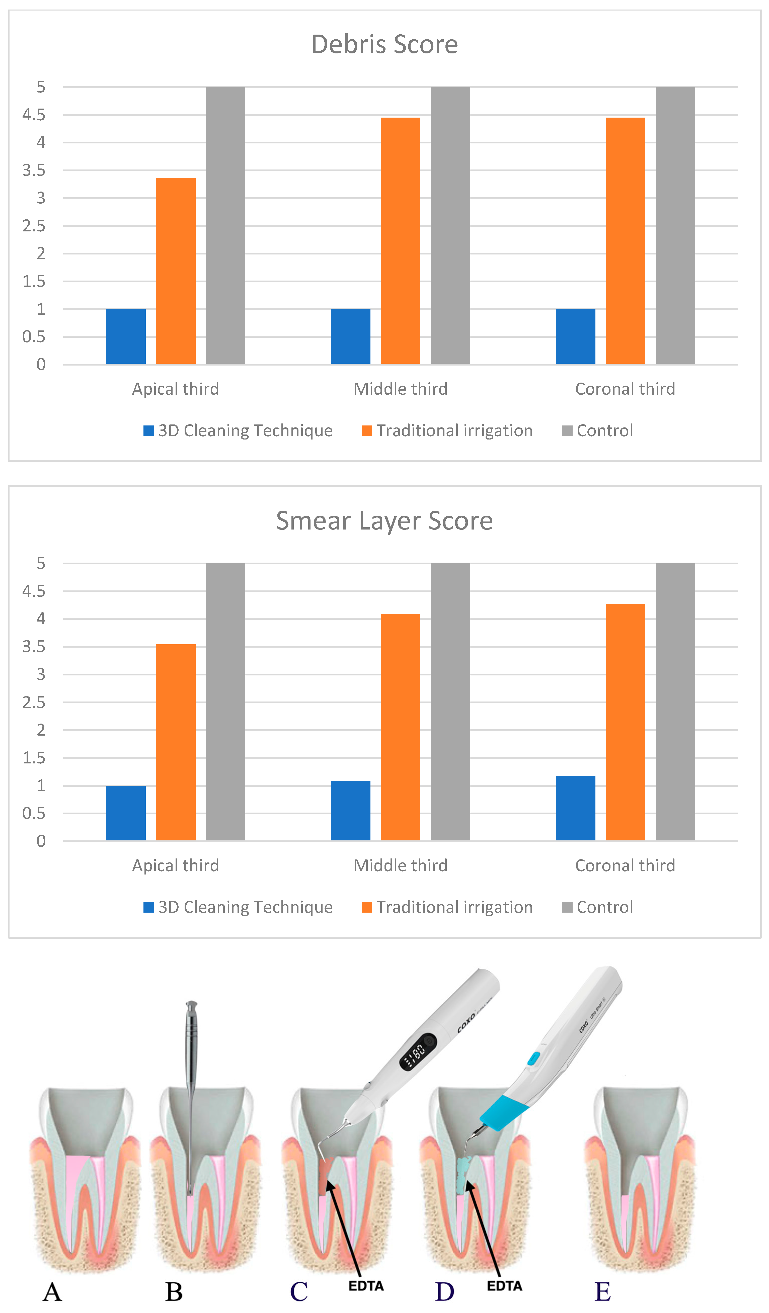
Figure 1. Showing 3D cleaning procedure for post space cleaning. (A) Root canal filled with gutta-percha, (B) Post space prepared using Gates-Glidden, (C) Internal Heating of EDTA, (D) Ultrasonic activation of heated EDTA, (E) Post space cleaned.
Prosthesis 05 00037 g001
Figure 2. Second maxillary premolar after obturation phase. (A,B) The root canals are filled with obturating materials (sealer and gutta-percha). (C) Post space after 3D cleaning protocol; it is possible to see clear walls without macroscopic debris. The pictures were taken using an operative microscope at 15×.
Prosthesis 05 00037 g002
Figure 3. Showing samplings of SEM images of the coronal, middle, and apical thirds of the post space after irrigation of group 1 (3D cleaning), group 2 (traditional irrigation) and the control group (1000× magnification).
Prosthesis 05 00037 g003
Table 1. Means, medians, minimum and maximum scores per group.
GroupD CoronalD MiddleD ApicalD Total
1Mean1111.06 ± 0.24
N11111133
Median
25 Percentile
75 Percentile
1
1
1
1
1
1
1
1
1
1
1
1
2Mean3.36 ± 0.674.45 ± 0.694.45 ± 0.694.06 ± 0.79
N11111133
Median
25 Percentile
75 Percentile
3
3
4
5
4
5
5
4
5
4
4
5
3Mean5555
N11111133
Median
25 Percentile
75 Percentile
5
5
5
5
5
5
5
5
5
5
5
5
TotalMean3.12 ± 1.713.45 ± 1.83.54 ± 1.77
N333333
Median
25 Percentile
75 Percentile
3
1
5
4
1
5
5
1
5
Table 2. Means with standard deviations, medians, minimum and maximum scores per group.
GroupSL CoronalSL MiddleSL ApicalSL Total
1Mean11.09 ± 0.31.18 ± 0.41.09 ± 0.29
N11111133
Median
25 Percentile
75 Percentile
1
1
1
1
1
1
1
1
1
1
1
1
2Mean3.54 ± 0.824.09 ± 0.74.27 ± 0.93.96 ± 0.85
N11111133
Median
25 Percentile
75 Percentile
4
3
4
4
4
5
4
4
5
4
3.5
5
3Mean5555
N11111133
Median
25 Percentile
75 Percentile
5
5
5
5
5
5
5
5
5
5
5
5
TotalMean3.18 ± 1.743.39 ± 1.753.48 ± 1.77
N333333
Median
25 Percentile
75 Percentile
4
1
5
4
1
5
4
1
5
Disclaimer/Publisher’s Note: The statements, opinions and data contained in all publications are solely those of the individual author(s) and contributor(s) and not of MDPI and/or the editor(s). MDPI and/or the editor(s) disclaim responsibility for any injury to people or property resulting from any ideas, methods, instructions or products referred to in the content.

