Article

Clinical Evaluation of Cement-Retained Implant-Supported CAD/CAM Monolithic Zirconia Single Crowns in Posterior Areas: Results of a 6-Year Prospective Clinical Study

Department of Neurosciences, Reproductive and Odontostomatological Sciences, Division of Prosthodontics and Digital Dentistry, University “Federico II” of Naples, 80131 Naples, Italy
*
Author to whom correspondence should be addressed.
Prosthesis 2022, 4(3), 383-393; https://doi.org/10.3390/prosthesis4030031
Submission received: 3 July 2022 / Revised: 24 July 2022 / Accepted: 25 July 2022 / Published: 27 July 2022
(This article belongs to the Special Issue Novel Zirconia Materials Applied in Dental Prostheses)

Abstract

:
Background: Different technologies and materials can be used for implant-supported restorations in posterior areas. Our study aimed to evaluate the clinical performance of CAD/CAM implant-supported monolithic zirconia single crowns (ZrSCs) cemented onto titanium abutments with a dual-curing resin cement after 6 years of use. Methods: Fifty patients in need of one single crown in posterior regions were enrolled. The patients were recalled for a follow-up after 6 months and then yearly for a total observation period of 6 years. The biological and technical complications were examined using criteria set by the United States Public Health Service (USPHS), whereas visual analogue scales (VASs) were used to evaluate the patients’ aesthetic and functional satisfaction. Results: No patient was lost at follow-up; both the survival and success rates were 100%. No fractures, decementation, peri-implantitis, or soft tissue inflammation were recorded. Forty-six restorations were rated Alpha in each evaluated parameter of the USPHS’s criteria. According to the patients’ VASs assessments, the overall function of the ZrSCs was 8.4 (±2.1), whereas the overall aesthetic was 8.7 (±0.7). Conclusions: Monolithic zirconia single crowns cemented with dual-curing resin cements onto titanium abutments can be considered an effective clinical option in posterior regions after 6 years of function.

1. Introduction

To date, dental implants represent a predictable treatment for full or partially edentulous patients, with high success rates for both function and aesthetics [1,2]. In the last decades, the application of dental implants has become more and more widespread, and, at the same time, many different types of fixtures and restorative materials have been introduced for use in different clinical situations [3].
To date, there is no univocal consensus in the scientific literature regarding the ideal retention system: some authors suggest the use of cement-retained implant restorations, while others prefer screw-retained implant prostheses. Both typologies exhibit advantages and drawbacks [3,4,5,6].
The main advantages of cement-retained restorations are good biomechanical stability, passive fit, absence of screw-access holes, better occlusal design, and adaptation for implant prosthetic malposition. Moreover, the cement layer can compensate for dimensional discrepancies between the abutment and restoration and work as a shock absorber, transferring occlusal loads to implant–restoration–bone complex [4,6,7].
The downside is the difficulty of removing cement excess, which has always been considered the major issue [8] since it may increase the risk of peri-implant mucositis [9,10], peri-implantitis [9], and marginal bone loss [11,12].
Introduced in the early years of implantology as an integral part of the “Toronto” full-arch bridge concept, screw-retained prostheses, for their part, offer easier retrievability, particularly advantageous in the case of long-span and cantilevered restorations [6,13]. Moreover, the use of screw-retained prosthetics is preferred in cases of limited interocclusal space, when the height of the abutment should be ≤4 mm, not offering sufficient retention. Another indication for screw-retained restorations is the use of temporary prostheses aimed at soft tissue conditioning and the customization of the emergence profile [6].
Compared with cemented restorations, screw-retained systems are reported to be more frequently prone to technical complications, such as a screw loosening and components/restoration fractures [14,15,16]. Moreover, the presence of an occlusal hole can impair the occlusal design, especially in the case of implant malposition. Finally, screw-retained prostheses are more expensive due to the higher cost of the components [6].
Cemented prostheses can ease the restoration design when the fixture location is not ideal and when a screw-retained solution would face the problem of a screw hole emerging in a critical position (e.g., incisal margin, buccal surface in an anterior site, and cusp tip in a posterior site). In these cases, a proper cement-retained restoration design and the correct selection of the implant abutments are paramount since they allow for customization and compensate the emergence profile, limiting micromovements and consequent bacterial contamination at the implant–prosthetic microgap [17,18].
Titanium abutments are reported to show significantly higher fracture strength than zirconia abutments, reducing the incidence of clinical complications; consequently, they represent the first choice in posterior areas subjected to heavy occlusal forces [19,20]. Very high survival rates were reported in clinical investigations for titanium abutments that showed reliable clinical behavior and can be safely used in daily practice [21,22].
Prosthetic crowns made from different restorative materials are currently available as clinical options. Conventional metal–ceramic restorations and bilayered all-ceramic prostheses showed high rates of technical complications mainly due to cohesive fractures of veneering ceramics (i.e., chipping) [23,24,25,26,27]; consequently, all-ceramic crowns in the monolithic configuration were recently proposed in posterior sites to reduce mechanical drawbacks, thanks to their excellent mechanical resistance to fractures and their biocompatibility [28].
CAD/CAM monolithic zirconia was developed to limit the incidence of mechanical complications due to the chipping of veneering ceramics, reduce production times, and improve cost-effectiveness [23,29]. However, different CAD/CAM zirconia milling procedures may produce discrepancies in the cementation space, which eventually influence final crown retention [30]. In addition to providing retention of prostheses, cementation techniques and mechanical properties of luting agents can affect the fracture strength and leakage of all-ceramic crowns [31]. Some investigations reported that zirconia should be submitted to specific mechanical and chemical surface treatments to develop durable bond strength to resin cements, like mild sandblasting and the application of primers containing 10-Methacryloyloxydecyl dihydrogen phosphate (MDP) [32,33].
The primary aim of our prospective clinical study was to evaluate the survival and success rates of CAD/CAM monolithic zirconia single crowns (ZrSCs) cemented onto titanium implant abutments with a dual-curing resin cement containing MDP. The secondary aim was to assess possible biological and technical complications during use and to assess the patients’ aesthetic and functional satisfaction.

2. Materials and Methods

2.1. Participants

Fifty consecutive patients (18 women and 32 men) in need of 1 single crown in the posterior regions of either maxilla or mandible in order to replace a missing premolar or molar (Table 1) were enrolled as participants in our prospective clinical study. The age of the patients ranged between 21 and 70, with a mean age of 45.6 (±14.3 y). All patients presented at the prosthodontics department of the University “Federico II” of Naples (Italy) from October 2015 to March 2016 (baseline).
The patients received comprehensive explanations regarding the clinical protocol and signed written informed consent forms. Each patient was provided with only 1 ZrSC.
The patients were recruited according to the following inclusion criteria:
  • Age ≥ 18 years;
  • Good general health;
  • ASA I (healthy) or ASA II (mild systemic disease), according to the American Society of Anesthesiologists (ASA);
  • Good oral hygiene;
  • Angle class I occlusal relationship;
  • No evident signs of occlusal parafunction and/or temporomandibular disorders;
  • No pregnancy or lactation;
  • Smoking ≤ 10 cigarettes/day;
  • Pocket probing depth ≤ 4 mm, no bleeding on probing, and plaque index ≤ 20%;
  • Single missing tooth being a premolar or a molar in the maxilla or mandible, with a minimum post-extraction healing of 3 months;
  • Absence of infection at the implant site;
  • Adequate bone volume to place an implant (length 8.5–10 mm, diameter 4.1 mm, and class I to III bone quality according to Lekholm and Zarb) [34];
  • Adequate prosthetic space to receive an anatomic restoration.
Conversely, patients fulfilling at least one of the following exclusion criteria were excluded from the study:
  • General and medical contraindication for surgical procedures;
  • Poor oral hygiene;
  • Reduced prosthetic space at the edentulous site (≤5 mm);
  • Severe wear facets, clenching, and bruxism;
  • Heavy smokers (>10 cigarettes/day);
  • Severe or not controlled periodontal disease;
  • Poor compliance.

2.2. Surgical Procedures

The surgical and prosthetic treatments were carried out at the implant surgery and prosthodontics departments of the University “Federico II” of Naples. One experienced oral surgeon performed the surgical procedures, and 2 skilled prosthodontists carried out the restorative treatments.
The patients presented with an edentulous site with a single missing tooth for more than 3 months, showing sufficient bone volume to achieve implant primary stability. One hour before surgery, antibiotic prophylaxis (i.e., 1 gr amoxicillin or, if allergic to penicillin, 600 mg clindamycin) was given, and a 0.2% chlorhexidine mouthwash for oral disinfection (twice a day for 10 days) was prescribed.
In the surgical session, a crestal incision was made under local anesthesia, followed by the preparation of a mucoperiosteal flap to expose the alveolar bone. Each patient received 1 endosseous dental implant (Outlink2, Sweden&Martina; Padova, Italy) in the posterior region. The implant diameter was 4.1 mm, and the length varied from 8.5 to 10 mm, dependent on the available bone height at the implant site. A 2-stage surgical technique with submerged fixtures and no additional soft or hard tissue grafts was planned for all the patients. One week after implant placement, the patients were recalled for a follow-up to remove the sutures and control the healing process.
Three months after implant placement, the osseointegration was verified clinically and radiographically; then, surgical re-entry was performed, and a trans-mucosal healing abutment (Healing Abutment, Sweden&Martina; Padova, Italy) was screwed onto the fixture. This second surgical stage was performed by the same surgeon that placed the implants.

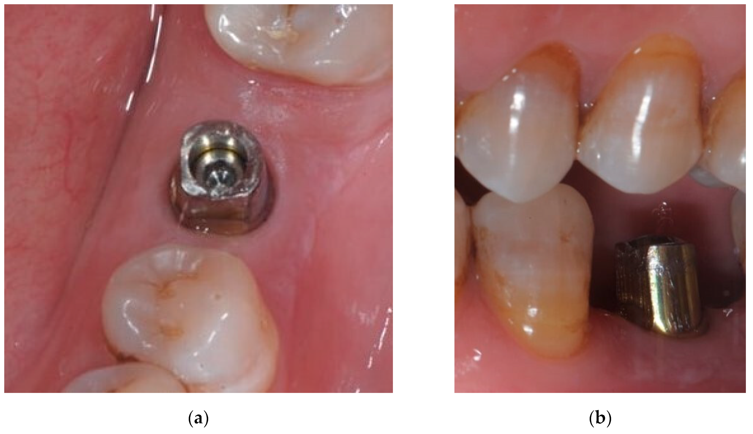
2.3. Prosthetic and Laboratory Procedures

Two weeks after surgical re-entry, a precision pick-up impression was made with the “open tray—pick-up technique” [35,36,37] using polyether materials (Impregum, 3M ESPE; Seefeld, Germany). The color of the final restorations was determined using a conventional shade scale (VITA Linearguide 3D-MASTER, VITA Zahnfabrik; Bad Sackingen, Germany).
All the prostheses were fabricated in a single dental laboratory. Master casts with implant analogs were poured and then digitized with a laboratory scanner (DScan 3, EGS S.R.L.; Bologna, Italy).
CAD/CAM titanium abutments (Titanium Abutment, Sweden&Martina; Padova, Italy) (Figure 1) were selected and customized strictly following the manufacturer’s instructions. Each titanium abutment was scanned, and 50 monolithic ZrSCs were designed using a dedicated CAD software (Exocad DentalCAD, Exocad GmbH; Darmstadt, Germany). The zirconia blocks (Katana Zirconia HT, Kuraray Noritake; Aichi, Japan) were then milled with a 5-axis milling system (Plus Mill S5, Dental Plus; Gyeonggi, Korea). Finally, each crown was polished manually without any veneering or a glazing layer.

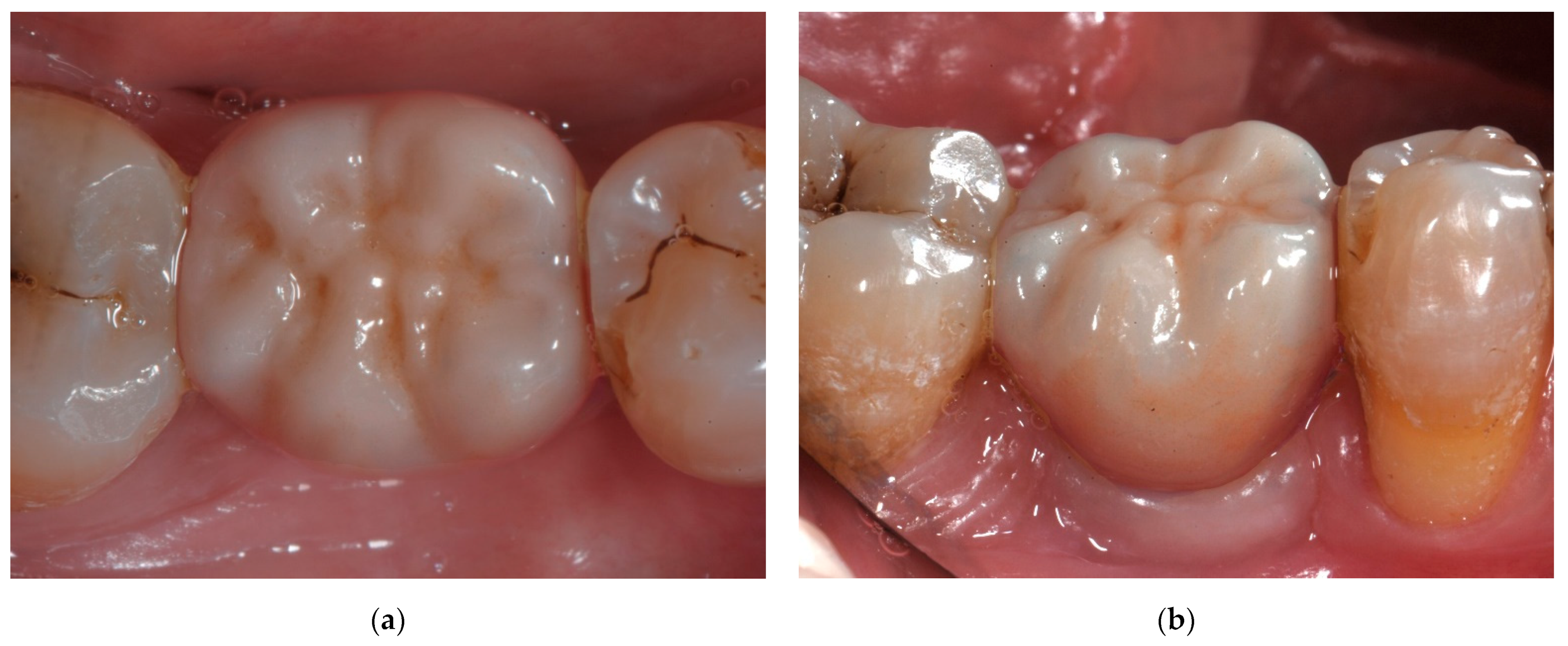
2.4. Delivery of Restorations

After a total healing time of 4 months, the titanium abutments were screwed onto the implants and torqued at 35 N/cm with a dynamometric wrench, according to the manufacturer’s recommendations; the screwing channels were protected and filled with polytetrafluoroethylene (PTFE) tape.
The internal surface of each ZrSC was sandblasted with 50 μm aluminum oxide (Al2O3) powder at 1 bar; then, the ZrSCs were cleaned with steam for 60 s. The monolithic ZrSCs were luted onto titanium abutments with a dual-curing resin cement (Panavia V5, Kuraray Noritake; Okayama, Japan); an MDP-containing ceramic primer (Ceramic Primer Plus, Kuraray Noritake; Okayama, Japan) was applied with a microbrush onto the intaglio’s surface of each ZrSC for 20 s and dried. The cement was dispensed into each restoration with an automating tip, and the crowns were initially seated onto the titanium abutments with finger pressure; the excess cement was tack-cured with a curing light (Elipar™ S10, 3M ESPE; St. Paul, MN, USA) for 5 s to reach a gel stage and then cleaned away with plastic tips and dental floss; the patients were asked to clench onto a wooden stick for 5 more minutes to ensure the complete seating of the restorations. Finally, each surface was further light-cured for 10 s, according to the manufacturer’s instructions. Postoperative radiographs were taken to detect possible cement remnants.
The occlusion was checked with 8 μm thick articulating foil (Shimstock foil, Bausch; Köln, Germany) to allow correct occlusal contacts, avoiding occlusal precontacts and interferences on the restorations. When necessary, occlusal adjustments were made using fine-grit diamond burs; then, the adjusted surfaces were meticulously polished with a polishing system dedicated to zirconia (Komet nos. 9425, 9426, and 9547; Brasseler; Savannah, GA, USA). Finally, scrupulous oral hygiene instructions were provided to all patients.

2.5. Baseline Evaluations

Two experienced calibrated and trained clinicians made the baseline evaluations, which were recorded 7 days after the cementation of the ZrSCs.
Furthermore, the static and dynamic occlusal contacts were carefully checked and adjusted if necessary, as previously described.
Standardized peri-apical radiographs and clinical photographs of the restorations were taken.
The evaluation of technical complications was performed following the modified USPHS’s criteria [38,39,40], classified on the basis of the clinical serviceability of the restorations (Table 2). The following parameters were evaluated: fracture behavior, decementation, anatomical form, and marginal adaptation.
The biological parameters were evaluated by referring to the modified plaque index (mPI), modified Sulcus Bleeding Index (mSBI), hypertrophy/hyperplasia of soft tissue, and peri-implantitis.
The mPI was scored from 0 to 3, as follows:
  • 0 = no plaque and no inflammation;
  • 1 = mild inflammation and a film of plaque adhering to the free soft tissues margin that cannot be seen with the naked eye but only with probes;
  • 2 = moderate inflammation with moderate glazing, redness, bleeding on probing, and moderate accumulation of deposits within the soft tissue pocket and on the margin, which can be seen with the naked eye;
  • 3 = abundance of soft matter within the soft tissue pocket, on the margin, and/or on the restoration; severe inflammation with redness, hypertrophy, and tendency to spontaneous bleeding [41,42,43,44].
The mSBI was scored from 0 to 3, as follows:
  • 0 = no bleeding when a periodontal probe is passed along the peri-implant soft tissue;
  • 1 = isolated bleeding spots visible;
  • 2 = blood forms a confluent red line on the margin;
  • 3 = heavy or profuse bleeding [41,44,45].
VASs were used to allow patients to rate the overall functional and aesthetic results of the restorations (0 = scarce, 10 = excellent).

2.6. Follow-Up Recalls

All the patients were recalled at follow-up after 6 months from the baseline and then yearly, for a total prospective observational period of 6 years. The baseline assessments were repeated, and their results were recorded, as previously described.
Furthermore, standardized peri-apical radiographs were made to evaluate possible marginal bone resorption at implant sites; moreover, clinical photographs of the restorations were taken to record the peri-implant soft tissue morphology and conditions.

3. Results

After 6 years of clinical service, no patient was lost at follow-up or censored; consequently, all the restorations were available for follow-up examinations.
No technical problems such as fractures or decementations were observed (Figure 2); as a result, both the survival and success rates were 100%, considering zirconia–ceramic fractures and/or loss of retention as events.
During the entire follow-up period, neither radiographic evidence nor signs and symptoms of peri-implant pathology nor marginal bone resorption were noticed.
The technical evaluation using the USPHS’s criteria showed very good clinical performances of the ZrSCs cemented onto titanium abutments. In terms of fracture behavior and decementation, all the ZrSCs were rated Alpha. For anatomical form, 46 ZrSCs were rated Alpha, and 4 ZrSCs were rated Bravo; differently, for the marginal adaptation, 47 ZrSCs were rated Alpha, and 3 ZrSCs were rated Bravo.
According to the patients’ VASs assessments, the overall function of the ZrSCs reported a mean value of 8.4 (±2.1), whereas the overall aesthetics rated an average value of 8.7 (±0.7). The statistical results of the VASs for gender, age, and jaw are reported in Table 3.
For mPI and mSBI, 32 and 31 ZrSCs, respectively, were rated 0; 16 and 17 ZrSCs, respectively, were rated 1; 2 and 0 ZrSCs, respectively, were rated 2; 0 and 2 ZrSCs, respectively, were rated 3.
No peri-implantitis or hypertrophy/hyperplasia of soft tissues was detected.

4. Discussion

Among the ideal retention systems, some authors prefer the use of cement-retained prostheses, while others suggest screw-retained restorations [3,4,5,6]. On one hand, the main advantages of cement-retained prostheses are a passive fit, good biomechanical stability, the absence of screw-access holes, better occlusal design, and adaptation for implant prosthetic malposition. Moreover, the cement layer can compensate for dimensional discrepancies between the restoration and abutment and work as a shock absorber [4,6,7]. Nevertheless, a cement excess that is not removed may increase the risk of peri-implantitis [9], peri-implant mucositis [9,10], and marginal bone loss [11,12]. On the other hand, screw-retained prostheses allow for easier retrievability [6,13] and are preferable for limited interocclusal space, but they are prone to technical complications such as components/restoration fractures and screws loosening [14,15,16].
In our study, the clinical performance of cement-retained implant-supported restorations was analyzed. Such restorative solutions offer the passive fit of crowns, absence of screw-access holes, easier control of occlusion, and compensation of possible prosthetic implant malposition [4]. The restorative system made up of monolithic ZrSCs cemented onto titanium abutments with a dual-curing resin cement reported survival and success rates of 100% at 6 years, considering the loss of retention and/or fractures of zirconia as events for the cumulative survival, and excellent scores according to the USPHS’s criteria. Particularly, the parameters “fracture behavior” and “decementation” scored Alpha in 100% of restorations; this means that, in the medium-term of clinical service, neither fractures of the ZrSCs nor decementation between zirconia crowns and titanium abutments were observed. According to such evidence, it was concluded that the use of dual-curing MDP-containing resin cements is a reliable choice for the cementation of zirconia monolithic crowns onto titanium abutments as well as a viable clinical option to strengthen the restorative complex. This statement is in accordance with a previous investigation where an MDP monomer showed the ability to increase the bond strength between zirconia and resin-based luting agents [33].
Moreover, for a whole observation period of 6 years, no hypertrophy/hyperplasia of soft tissue or peri-implantitis was detected, indicating a complete absence of inflammation in the peri-implant tissues surrounding the ZrSCs; such occurrence confirmed the outstanding biocompatibility of zirconia, particularly in its monolithic configuration, and the optimal mechanical coupling and biological integration of the zirconia–titanium restorative complex.
For the “anatomical form” parameter, 46 ZrSCs were rated Alpha, meaning that the ideal anatomical shape and proximal contacts were achieved. Differently, four ZrSCs were rated Bravo because of the modifications of the buccal peri-implant soft tissues over time, resulting in slightly over- or under-contoured restorations; this occurrence could be due to a less than ideal position of fixtures in the buccal–lingual direction and/or thin peri-implant biotype [46]. Furthermore, Bravo scores for "anatomical form" were also reported because of weak proximal contacts; such a phenomenon is not uncommon in posterior implant-supported prostheses and should be considered a minor complication related to different associated factors, such as the anatomical position of restorations, lower alveolar bone support level, and time since the delivery of the prosthesis [47,48].
For “marginal adaptation”, 47 ZrSCs were rated Alpha, indicating excellent marginal precision, while the remaining 3 ZrSCs were rated Bravo with a slight probe catch but no gap between the crown and abutment. Considering the optimal mechanical coupling between the implant necks and milled restorations, thanks to the well-known reliability of CAD/CAM technologies, it could be speculated that such an occurrence was due either to the patients’ relaxation during the cementation clenching or to a premature cement-excess cleaning before complete gelification, resulting in voids at the prosthetic margin.
Regarding the periodontal parameters mPI and mSBI, there were reported 32 and 31, respectively, for the score 0; 16 and 17, respectively, for the score 1; 2 and 0, respectively, for the score 2; and 0 and 2, respectively, for the score 3, showing extremely satisfactory biological integration of the restorations. Particularly, these results indicated the absence of bleeding, plaque, and inflammation for at least 31 restorations, whereas a single discreet bleeding point or mild inflammation was reported for at least 16 ZrSCs. Furthermore, moderate inflammation was observed in two restorations, and in two more, the interdental spaces were filled with blood shortly after probing; this evidence was noticed in smokers and patients with less satisfactory compliance to the supportive periodontal care and oral hygiene prescriptions. The findings of our investigation are in agreement with those of other studies and confirm the excellent biological response of peri-implant soft tissues to zirconia restorations and the outstanding biocompatibility of this material, particularly in its polished monolithic configuration [40,49,50,51,52,53].
According to the VASs’ scores of the overall function (8.4 ± 2.1) and aesthetics (8.7 ± 0.7), ZrSCs were considered fully satisfactory for restoring proper chewing activity and for achieving a natural tooth-like appearance of the restorations.
The results of our prospective clinical study are very promising, likely due to an extremely careful selection of the patients. In any case, the investigation presented some limitations, namely, an observational period limited to the medium-term and no randomization of participants. Moreover, the antagonist tooth and its restorative material were not variables taken into consideration in our study. In addition, the 100% success result might be explained by the limited sample size (n = 50), the favorable implant diameter used (4.1 mm), and the retentive abutment height (> 2mm). Further RCTs with a wider sample population and longer observational periods are essential to validate long-term serviceability, biomechanical effectiveness, and biological and aesthetical outcomes of the restorative system evaluated in our clinical investigation.

5. Conclusions

Within the limitations of our prospective clinical study, the restorative system based on the use of titanium abutments and monolithic zirconia single crowns and cemented with an MDP-based cement proved to be a viable clinical option for restoring posterior missing teeth in the medium term. The following conclusions may be drawn:
  • The tested restorative system was highly effective and reliable for restoring occlusal function, showing 100% survival and success rates.
  • Neither fracture nor loss of retention were noticed.
  • The most-frequent technical complications were minor marginal misfit and weak proximal contacts, but none of them impaired function.
  • The tested restorative system was highly biocompatible, as shown by the stability and optimal health status of the surrounding peri-implant tissues.
  • Patients reported being very satisfied by the overall function and aesthetics.

Author Contributions

Conceptualization, F.Z. and R.S.; design, E.T. and G.R.; data analysis, G.R., F.Z., and R.L.; data acquisition, E.T. and G.R.; data interpretation, R.S. and E.T.; writing—original draft preparation, G.R. and R.L.; writing—review and editing, F.Z. and R.S.; article approval, F.Z., R.S., G.R., and E.T.; data collection, E.T, G.R., and R.L. All authors have read and agreed to the published version of the manuscript.

Funding

This research received no external funding.

Institutional Review Board Statement

The study was conducted according to the guidelines of the Declaration of Helsinki and approved by the Institutional Review Board of the University “Federico II” of Naples (protocol code: NCT04374201; date of approval: 11 June 2015).

Informed Consent Statement

Informed consent was obtained from all subjects involved in the study.

Data Availability Statement

The datasets used and/or analyzed during the current study are available from the corresponding author on reasonable request.

Acknowledgments

The authors would like to thank the following industries for providing the needed materials: Kuraray Noritake (PANAVIA V5 and Katana Zirconia HT); Sweden & Martina (Outlink2 endosseous implant, healing abutment, and titanium abutment).

Conflicts of Interest

The authors declare no conflict of interest.

References

  1. Pjetursson, B.E.; Thoma, D.; Jung, R.; Zwahlen, M.; Zembic, A. A systematic review of the survival and complication rates of implant-supported fixed dental prostheses (FDPs) after a mean observation period of at least 5 years. Clin. Oral Implant. Res. 2012, 23, 22–38. [Google Scholar] [CrossRef] [PubMed]
  2. Schwarz, S.; Schroder, C.; Corcodel, N.; Hassel, A.J.; Rammelsberg, P. Retrospective comparison of semipermanent and permanent cementation of implant-supported single crowns and FDPs with regard to the incidence of survival and complications. Clin. Implant. Dent. Relat. Res. 2012, 14, e151–e158. [Google Scholar] [CrossRef] [PubMed]
  3. Lee, A.; Okayasu, K.; Wang, H.L. Screw- versus cement-retained implant restorations: Current concepts. Implant. Dent. 2010, 19, 8–15. [Google Scholar] [CrossRef] [PubMed]
  4. Nissan, J.; Narobai, D.; Gross, O.; Ghelfan, O.; Chaushu, G. Long-term outcome of cemented versus screw-retained implant-supported partial restorations. Int. J. Oral Maxillofac Implant. 2011, 26, 1102–1107. [Google Scholar]
  5. Kraus, R.D.; Epprecht, A.; Hämmerle, C.H.F.; Sailer, I.; Thoma, D.S. Cemented vs screw-retained zirconia-based single implant reconstructions: A 3-year prospective randomized controlled clinical trial. Clin. Implant. Dent. Relat. Res. 2019, 21, 578–585. [Google Scholar] [CrossRef]
  6. Wittneben, J.G.; Joda, T.; Weber, H.P.; Brägger, U. Screw retained vs. cement retained implant-supported fixed dental prosthesis. Periodontol 2000 2017, 73, 141–151. [Google Scholar] [CrossRef]
  7. Zarone, F.; Sorrentino, R.; Traini, T.; Di lorio, D.; Caputi, S. Fracture resistance of implant-supported screw- versus cement-retained porcelain fused to metal single crowns: SEM fractographic analysis. Dent. Mater. 2007, 23, 296–301. [Google Scholar] [CrossRef]
  8. Linkevicius, T.; Vindasiute, E.; Puisys, A.; Linkeviciene, L.; Maslova, N.; Puriene, A. The influence of the cementation margin position on the amount of undetected cement. A prospective clinical study. Clin. Oral Implant. Res. 2013, 24, 71–76. [Google Scholar] [CrossRef]
  9. Gapski, R.; Neugeboren, N.; Pomeranz, A.Z.; Reissner, M.W. Endosseous implant failure influenced by crown cementation: A clinical case report. Int. J. Oral Maxillofac. Implant. 2008, 23, 943–946. [Google Scholar]
  10. Wasiluk, G.; Chomik, E.; Gehrke, P.; Pietruska, M.; Skurska, A.; Pietruski, J. Incidence of undetected cement on CAD/CAM monolithic zirconia crowns and customized CAD/CAM implant abutments. A prospective case series. Clin. Oral Implant. Res. 2017, 28, 774–778. [Google Scholar] [CrossRef]
  11. Wittneben, J.G.; Millen, C.; Bragger, U. Clinical performance of screw-versus cement-retained fixed implant-supported reconstructions—A systematic review. Int. J. Oral Maxillofac. Implant. 2014, 29, 84–98. [Google Scholar] [CrossRef] [Green Version]
  12. Hamed, M.T.; Abdullah, M.H.; Khalid, A.S.; Hossam, H.A.B.; Hussein, N.G. A Systematic Review of Screw versus Cement-Retained Fixed Implant Supported Reconstructions. Clin. Cosmet. Investig. Dent. 2020, 12, 9–16. [Google Scholar] [CrossRef] [PubMed] [Green Version]
  13. Shadid, R.; Sadaqa, N. A comparison between screw- and cement-retained implant prostheses. A literature review. J. Oral Implant. 2012, 38, 298–307. [Google Scholar] [CrossRef] [PubMed] [Green Version]
  14. Sailer, I.; Mühlemann, S.; Zwahlen, M.; Hämmerle, C.H.; Schneider, D. Cemented and screw-retained implant reconstructions: A systematic review of the survival and complication rates. Clin. Oral Implant. Res. 2012, 23, 163–201. [Google Scholar] [CrossRef] [PubMed]
  15. Chee, W.; Jivraj, S. Screw versus cemented implant supported restorations. Br. Dent. J. 2006, 201, 501–507. [Google Scholar] [CrossRef] [PubMed] [Green Version]
  16. Michalakis, K.X.; Hirayama, H.; Garefis, P.D. Cement-retained versus screw-retained implant restorations: A critical review. Int. J. Oral Maxillofac. Implant. 2003, 18, 719–728. [Google Scholar]
  17. Steinebrunner, L.; Wolfart, S.; Bössmann, K.; Kern, M. In vitro evaluation of bacterial leakage along the implant-abutment interface of different implant systems. Int. J. Oral Maxillofac. Implant. 2005, 20, 875–881. [Google Scholar]
  18. Kurbad, A.; Kurbad, S. CAD/CAM-based implant abutments. Int. J. Comput. Dent. 2013, 16, 125–141. [Google Scholar]
  19. Apicella, D.; Veltri, M.; Balleri, P.; Apicella, A.; Ferrari, M. Influence of abutment material on the fracture strength and failure modes of abutment-fixture assemblies when loaded in a bio-faithful simulation. Clin. Oral Implant. Res. 2011, 22, 182–188. [Google Scholar] [CrossRef]
  20. Fabbri, G.; Fradeani, M.; Dellificorelli, G.; De Lorenzi, M.; Zarone, F.; Sorrentino, R. Clinical Evaluation of the Influence of Connection Type and Restoration Height on the Reliability of Zirconia Abutments: A Retrospective Study on 965 Abutments with a Mean 6-Year Follow-Up. Int. J. Periodontics Restor. Dent. 2017, 37, 19–31. [Google Scholar] [CrossRef] [Green Version]
  21. Pjetursson, B.E.; Zarauz, C.; Strasding, M.; Sailer, I.; Zwahlen, M.; Zembic, A. A systematic review of the influence of the implant-abutment connection on the clinical outcomes of ceramic and metal implant abutments supporting fixed implant reconstructions. Clin. Oral Implant. Res. 2018, 29, 160–183. [Google Scholar] [CrossRef] [Green Version]
  22. Ferrari, M.; Tricarico, M.G.; Cagidiaco, M.C.; Vichi, A.; Gherlone, E.F.; Zarone, F.; Sorrentino, R. 3-Year Randomized Controlled Prospective Clinical Trial on Different CAD-CAM Implant Abutments. Clin. Implant. Dent. Relat. Res. 2016, 18, 1134–1141. [Google Scholar] [CrossRef] [Green Version]
  23. Zarone, F.; Russo, S.; Sorrentino, R. From porcelain-fused-to-metal to zirconia: Clinical and experimental considerations. Dent. Mater. 2011, 27, 83–96. [Google Scholar] [CrossRef]
  24. Alsarani, M.; Souza, G.; Rizkalla, A.; El-Mowafy, O. Influence of crown design and material on chipping-resistance of all-ceramic molar crowns: An in vitro study. Dent. Med. Probl. 2018, 55, 35–42. [Google Scholar] [CrossRef] [PubMed] [Green Version]
  25. Poggio, C.E.; Ercoli, C.; Rispoli, L.; Maiorana, C.; Esposito, M. Metal-free materials for fixed prosthodontic restorations. Cochrane Database Syst. Rev. 2017, 12, CD009606. [Google Scholar] [CrossRef] [PubMed]
  26. Anusavice, K.J. Standardizing failure, success, and survival decisions in clinical studies of ceramic and metal-ceramic fixed dental prostheses. Dent. Mater. 2012, 28, 102–111. [Google Scholar] [CrossRef] [PubMed] [Green Version]
  27. Sadowsky, S.J. Has zirconia made a material difference in implant prosthodontics? A review. Dent. Mater. 2020, 36, 1–8. [Google Scholar] [CrossRef]
  28. Sorrentino, R.; Triulzio, C.; Tricarico, M.G.; Bonadeo, G.; Gherlone, E.F.; Ferrari, M. In vitro analysis of the fracture resistance of CAD-CAM monolithic zirconia molar crowns with different occlusal thickness. J. Mech. Behav. Biomed. Mater. 2016, 61, 328–333. [Google Scholar] [CrossRef]
  29. Seydler, B.; Schmitter, M. Clinical performance of two different CAD/CAM- fabricated ceramic crowns: 2-year results. J. Prosthet. Dent. 2015, 114, 212–216. [Google Scholar] [CrossRef] [PubMed]
  30. Patzelt, S.B.; Spies, B.C.; Kohal, R.J. CAD/CAM-fabricated implant-supported restorations: A systematic review. Clin. Oral Implant. Res. 2015, 26, 77–85. [Google Scholar] [CrossRef] [PubMed]
  31. Blatz, M.B.; Alvarez, M.; Sawyer, K.; Brindis, M. How to Bond Zirconia. The APC Concept. Compend. Contin. Educ. Dent. 2016, 37, 611–618. [Google Scholar] [PubMed]
  32. Qeblawi, D.M.; Muñoz, C.A.; Brewer, J.D.; Monaco, E.A.J. The effect of zirconia surface treatment on flexural strength and shear bond strength to a resin cement. J. Prosthet. Dent. 2010, 103, 210–220. [Google Scholar] [CrossRef]
  33. Koizumi, H.; Nakayama, D.; Komine, F.; Blatz, M.B.; Matsumura, H. Bonding of resin-based luting cements to zirconia with and without the use of ceramic priming agents. J. Adhes. Dent. 2012, 14, 385–392. [Google Scholar] [PubMed]
  34. Lekholm, U.; Zarb, G.A. Patient selection and preparation. In Tissue Integrated Prostheses: Osseointegration in Clinical Dentistry, 1st ed.; Branemark, P.I., Zarb, G.A., Albrektsson, T., Eds.; Quintessence Publishing Company: Chicago, IL, USA, 1985; pp. 199–209. [Google Scholar]
  35. Sorrentino, R.; Gherlone, E.F.; Calesini, G.; Zarone, F. Effect of implant angulation, connection length, and impression material on the dimensional accuracy of implant impressions: An in vitro comparative study. Clin. Implant. Dent. Relat. Res. 2010, 12, e63–e76. [Google Scholar] [CrossRef] [PubMed]
  36. Moreira, A.H.; Rodrigues, N.F.; Pinho, A.C.; Fonseca, J.C.; Vilaca, J.L. Accuracy Comparison of Implant Impression Techniques: A Systematic Review. Clin. Implant. Dent. Relat. Res. 2015, 17, e751–e764. [Google Scholar] [CrossRef] [Green Version]
  37. Calesini, G.; Zarone, F.; Sorrentino, R.; Micarelli, C.; Fabianelli, A.; Papacchini, F.; Gherlone, E. Effect of 2 impression techniques on the dimensional accuracy of working implant prosthesis models: An in vitro study. J. Craniofac. Surg. 2014, 25, 822–827. [Google Scholar] [CrossRef]
  38. Bayne, S.C.; Schmalz, G. Reprinting the classic article on USPHS evaluation methods for measuring the clinical research performance of restorative materials. Clin. Oral Investig. 2005, 9, 209–214. [Google Scholar] [CrossRef]
  39. Pol, C.W.P.; Raghoebar, G.M.; Maragkou, Z.; Cune, M.S.; Meijer, H.J.A. Full-zirconia single-tooth molar implant-supported restorations with angulated screw channel abutments: A 1-year prospective case series study. Clin. Implant. Dent. Relat. Res. 2020, 22, 138–144. [Google Scholar] [CrossRef] [PubMed] [Green Version]
  40. Sorrentino, R.; De Simone, G.; Tetè, S.; Russo, S.; Zarone, F. Five-year prospective clinical study of posterior three-unit zirconia-based fixed dental prostheses. Clin. Oral Investig. 2012, 16, 977–985. [Google Scholar] [CrossRef] [PubMed]
  41. Mombelli, A.; van Oosten, M.A.; Schurch, E.J.; Land, N.P. The microbiota associated with successful or failing osseointegrated titanium implants. Oral Microbiol. Immunol. 1987, 2, 145–151. [Google Scholar] [CrossRef] [PubMed]
  42. Bradley, H.; Owen, B.; Keys, W. Zirconia implants: A promising alternative to titanium? Evid. Based Dent. 2021, 22, 102–103. [Google Scholar] [CrossRef]
  43. Curiel-Aguilera, F.P.; Griffiths, G.R.; Rossmann, J.A.; Gonzalez, J.A. Titanium versus zirconia complete-arch implant-supported fixed prostheses: A comparison of plaque accumulation. J. Prosthet. Dent. 2021. [Google Scholar] [CrossRef]
  44. Farrag, K.M.; Khamis, M.M. Effect of anodized titanium abutment collars on peri-implant soft tissue: A split-mouth clinical study. J. Prosthet. Dent. 2021. [Google Scholar] [CrossRef] [PubMed]
  45. Newbrun, E. Indices to measure gingival bleeding. J. Periodontol. 1996, 67, 555–561. [Google Scholar] [CrossRef] [PubMed]
  46. Ferrari, M.; Cagidiaco, M.C.; Garcia-Godoy, F.; Goracci, C.; Cairo, F. Effect of different prosthetic abutments on peri-implant soft tissue. A randomized controlled clinical trial. Am. J. Dent. 2015, 28, 85–89. [Google Scholar] [PubMed]
  47. Wong, A.T.; Wat, P.Y.; Pow, E.H.; Leung, K.C. Proximal contact loss between implant-supported prostheses and adjacent natural teeth: A retrospective study. Clin. Oral Implant. Res. 2015, 26, e68–e71. [Google Scholar] [CrossRef] [PubMed]
  48. Pang, N.S.; Suh, C.S.; Kim, K.D.; Park, W.; Jung, B.Y. Prevalence of proximal contact loss between implant-supported fixed prostheses and adjacent natural teeth and its associated factors: A 7-year prospective study. Clin. Oral Implant. Res. 2017, 28, 1501–1508. [Google Scholar] [CrossRef]
  49. Sailer, I.; Gottnerb, J.; Kanelb, S.; Hammerle, C.H. Randomized controlled clinical trial of zirconia-ceramic and metal-ceramic posterior fixed dental prostheses: A 3-year follow-up. Int. J. Prosthodont. 2009, 22, 553–560. [Google Scholar]
  50. Sorrentino, R.; Galasso, L.; Tetè, S.; De Simone, G.; Zarone, F. Clinical evaluation of 209 all-ceramic single crowns cemented on natural and implant-supported abutments with different luting agents: A 6-year retrospective study. Clin. Implant. Dent. Relat. Res. 2012, 14, 184–197. [Google Scholar] [CrossRef]
  51. Zarone, F.; Di Mauro, M.I.; Ausiello, P.; Ruggiero, G.; Sorrentino, R. Current status on lithium disilicate and zirconia: A narrative review. BMC Oral Health 2019, 19, 134. [Google Scholar] [CrossRef] [PubMed] [Green Version]
  52. da Cruz, M.B.; Marques, J.F.; Fernandes, B.F.; Madeira, S.; Carvalho, Ó.; Silva, F.S.; da Mata, A.D.S.P.; Caramês, J.M.M. Peri-implant cell response on groove and pore-textured zirconia surfaces. J. Oral Biosci. 2022, 64, 100–107. [Google Scholar] [CrossRef] [PubMed]
  53. Zarone, F.; Ruggiero, G.; Leone, R.; Breschi, L.; Leuci, S.; Sorrentino, R. Zirconia-reinforced lithium silicate (ZLS) mechanical and biological properties: A literature review. J. Dent. 2021, 109, 103661. [Google Scholar] [CrossRef] [PubMed]
Figure 1. Titanium implant abutment on the mandibular first molar area: (a) occlusal view; (b) buccal view.
Prosthesis 04 00031 g001
Figure 2. Six-year recall evaluation. Healthy condition of monolithic zirconia single crown cemented onto titanium implant abutment: (a) occlusal view; (b) buccal view.
Prosthesis 04 00031 g002
Table 1. Anatomical distribution of monolithic zirconia single crowns.
Maxilla (n = 10)Mandible (n = 40)
n%n%
1st premolar12%918%
2nd premolar48%1224%
1st molar510%1938%
n = number of implant-supported restorations.
Table 2. United States Public Health Service’s modified criteria for the evaluation of restorations at the 6-year follow-up.
USPHS CriteriaAlphaBravoCharlieDelta
Fracture behaviorNo fracture of zirconiaFracture but polishing possibleFracture but polishing not possibleNew restoration needed
DecementationNo decementation between crown and abutment--Decementation between crown and abutment
Anatomical formIdeal anatomical shape, good proximal contactsSlightly over- or under-contoured; weak proximal contactsHighly over- or under-contoured; open proximal contactsNew restoration needed
Marginal adaptationNo probe catchesSlight probe catches but no gapGap with abutment exposureNew restoration needed
Table 3. Statistical results for gender, age, and jaw with VASs.
GroupsVAS Aesthetics Score (mean)VAS Function Score (mean)
Men (n = 32)8.98.7
Women (n = 18)8.27.9
21–40 years old (n = 17)8.68.4
41–70 years old (n = 33)8.88.4
Maxilla (n = 10)8.38.0
Mandible (n = 40)8.88.5
n = number of implant-supported restorations.
Publisher’s Note: MDPI stays neutral with regard to jurisdictional claims in published maps and institutional affiliations.

