Case Report

Tissue Recession around a Dental Implant in Anterior Maxilla: How to Manage Soft Tissue When Things Go Wrong?

1
Independent Researcher, 03100 Frosinone, Italy
2
Independent Researcher, 33100 Udine, Italy
3
Independent Researcher, 00100 Rome, Italy
4
School of Dentistry, Medical, Surgical and Experimental Science, University of Sassari, 07100 Sassari, Italy
5
Biomedical, Dental and Morphological and Functional Images Department, School of Dentistry, University of Messina, 98100 Messina, Italy
*
Author to whom correspondence should be addressed.
Prosthesis 2021, 3(3), 209-220; https://doi.org/10.3390/prosthesis3030021
Submission received: 5 August 2021 / Revised: 19 August 2021 / Accepted: 20 August 2021 / Published: 23 August 2021

Abstract

:

Abstract

Dental implants represent the gold standard for the treatment of single edentulism, even in anterior areas. Today, the basic criteria for implant success has changed from mobility, pain, radiolucency, and peri-implant bone loss (>1.5 mm) to prosthetic level success, aesthetics, soft tissue parameters, as well as patient satisfaction. This case report documents a combination of surgical and prosthetic procedures for the treatment of gingival recessions in the anterior maxilla, appearing after tooth extraction, socket preservation, and staged guided implant placement. Prosthetic management of the temporary restoration, orthodontic treatment, and a connective tissue graft were performed. The decision-making process and step-by-step execution of the treatments are presented to describe the entire clinical and surgical management of the reported case. Finally, good aesthetic outcomes, patient satisfaction, and recovery of the soft tissue recession were observed with the combination of these techniques.

Key Clinical Message

Implant placement is not without complications. Aesthetically, the results can be dramatic. An interdisciplinary approach is often mandatory in order to resolve these types of complications.

1. Introduction

Tooth loss causes inevitable remodeling processes of the alveolar bone in the area of the extraction. These changes usually result in bone volume loss, especially against the buccal side, and this can lead to functional and aesthetic problems. Although the bone resorption is highly variable, alveolar ridge resorption takes place mainly during the first three months after tooth extraction, resulting in the loss of about 50% of the buccal wall [1,2,3]. Even if bone volume loss is a global problem, it is mostly stressed in the anterior area because of the high aesthetic requirements [4,5,6]. Immediate implant placement and loading seem to be the gold standard in the anterior area, but mid-facial mucosa recession could compromise the final aesthetic outcomes [7]. Moreover, a higher risk of implant failure can be expected when compared with immediate or early loading performed in healed ridges [8,9]. In order to overcome these drawbacks, several techniques and materials intended to minimize the alveolar bone resorption, including different procedures to cover the grafted material during the healing phase, have been introduced [3,4,5,6]. Leaving out the differences between various socket preservation techniques, the common point is that the material introduced into the residual socket should act as space maker, avoiding the complete collapse of the alveolar bone. Socket preservation procedures have been introduced to preserve not only the hard tissue but also the soft tissue contour after a tooth extraction, representing an opportunity to avoid more invasive bone augmentation techniques at a later stage [10]. Despite the progress that has been achieved, post-extractive implants in the anterior area remain a challenge because of possible unacceptable aesthetics, and consequently poor patient satisfaction, as a result of soft tissue recessions, unfavorable color, and visible crown margins [1,2,3]. The present case describes the management of an aesthetic complication occurring after the rehabilitation of a central incisor extracted due to a horizontal fracture, with a dental implant.

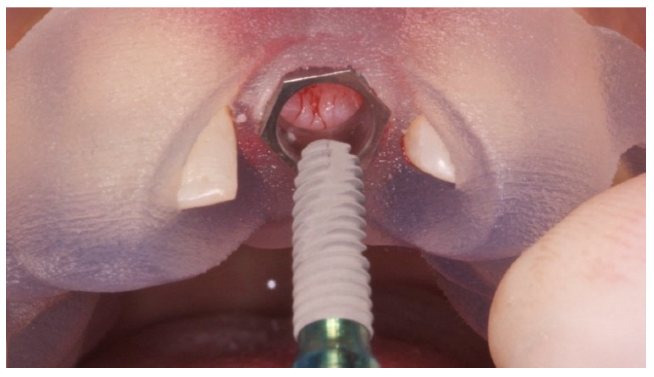
2. Case Presentation

A 10-year-old boy presented with a composed horizontal fracture (ski trauma) of a previously treated maxillary left incisor, involving full root thickness, and splitting the tooth into two parts. Initial pictures and radiographs were taken for evaluation (Figure 1 and Figure 2). Due to the very young age of the patient, endodontic retreatment was performed and the fracture rhyme was sealed with MTA, after its isolation through an ultrasonic device. After composite reconstruction, two single composite veneer crowns were delivered on teeth 11 and 22. At the follow-up examinations, planned at 4 month intervals, the site of the trauma showed a palatal probing up to 5 mm.
After 12 years, as a result of a second trauma (elbow strike while playing football) the tooth fractured completely. Then, bone sounding with periodontal probing revealed a buccal probing depth of approximately 6 mm (Figure 3) and pathological mobility. Clinical signs of inflammation were visible, particularly at the palatal side (Figure 4 and Figure 5). Overall, the clinical and periodontal conditions of the tooth made the extraction unavoidable. Due to the patient’s high aesthetic and functional demands, the proposed treatment plan included initial periodontal therapy, a minimally invasive tooth extraction with socket preservation/seal, and a Maryland bridge for at least 4 months. Then, computer-guided, template-assisted implant placement with conventional loading was implemented. Finally, a cement-retained, implant-supported single crown was installed on the left incisor in addition to a veneer on the right incisor.
After the patient gave written consent for the proposed treatment, and permission to write up the case, all the clinical and surgical procedures began. The patient was prescribed amoxicillin 2 g, 2 times per day, for 2 days before and 5 days after surgery. The extraction was performed flapless, as atraumatically as possible, using a periotome and atraumatic elevators (PT1 and EPTSMS, Hu-Friedy Italy, Milan, Italy). A carefully curettage of the socket was performed with alveolar curettes, and then the area was washed with sterile saline. After that, the socket (Figure 6) was grafted with deproteinized bovine bone (Bio-Oss, Geistlich Pharma AG, Wolhusen, Switzerland), and sealed using a connective tissue graft (CTG) harvested from the palate. Finally, the wound was sutured with non-absorbable surgical suture (Vicryl, Ethicon J&J International, St. Stevens, Woluwe, Belgium). A temporary Maryland bridge was cemented to the neighboring teeth, without compressing the extraction site. The first clinic check, one week later, showed a perfect healing of the wound.
Four months later, the patient underwent a cone beam computed tomography (CBCT) scan (Cranex 3Dx, Soredex, Tuusula, Finland), using a wax bite to separate dental arches. Then, the patient received an intraoral digital impression taken using the 3M True Definition Scanner (3M Italia, Pioltello, Milano). The digital data (STL, Surface Tessellation Language) were imported into 3D design software (Exocad DentalCAD, Exocad GmbH, Darmstadt, Germany) to realize a virtual wax-up according to the functional and aesthetic requirements. Then, the STL and DICOM (Digital Imaging and Communications in Medicine) data were imported in a 3D software planning program (3Diagnosys ver. 4.2, 3DIEMME srl, Cantù, Italy). Afterwards, prosthetic-driven implant position was planned and a surgical template was ordered [3,6,11,12,13,14,15,16,17,18,19,20,21,22,23,24]. Before implant placement, the patient underwent professional oral hygiene, prophylactic antiseptic with 0.2% chlorhexidine (Curasept, Curaden Healthcare, Saronno, Italy) for one minute, and prophylactic antibiotic therapy (2 g of amoxicillin). Local anesthesia (articaine with adrenaline 1:100,000) was administered 20 min before surgery. Immediately before implant placement, the fit of the surgical template was tested in the patient’s mouth to achieve a stable fit (Fit Checker, GC—Tokyo, Japan). The surgical templates were stabilized on the residual teeth and fixed with two preplanned anchor pins. The surgical template was temporarily removed to elevate a minimally invasive flap without relation incisors. Then, planned implant (Osstem TSIII, Osstem, Seoul, South Korea) was placed at 35 Ncm using dedicated drills (OsstemGuide Kit (Taper), Osstem Seoul, Korea) in combination with reduction tools, within the surgical templates containing metallic sleeves (Figure 7, Figure 8 and Figure 9). Finally, the wound was closed with single-stitch sutures using 4.0 resorbable suture material (Vicryl, Ethicon J&J International) and the previous temporary prosthesis was cemented.
Two weeks later, after suture removal, clinical examination revealed papillary recession at the mesial area of the left maxillary lateral incisor (Figure 10). It was classified as Class III based on Nordland and Tarnow’s classification [25], which means mesio-distal crestal bone loss. Different non-surgical and surgical techniques, such as guided bone regeneration or connective tissue graft, were widely discussed with the patient to increase the amount of available bone and/or gingival tissue. After all the benefits and limitations of these procedures were evaluated, the patient was scheduled for a fixed orthodontic appliance in order to level the isolated infrabone defect and reposition of the gingival margin, increasing the amount of attached bone and soft tissue. The orthodontic therapy (Figure 11) was carried out for four months after non-surgical periodontal therapy, which included scaling and root planing, and in combination with odontoplasty to allow the extrusion of the tooth in the arch [26].
After debonding (five months after implant placement), an incision using microsurgial blade and avoiding perpendicular incisions was performed, allowing for more tissue adaptation. After that, a screw-retained temporary restoration, designed with an ideal emergence profile, was delivered (Figure 12 and Figure 13). Temporary restoration was shaped with a concavity in the distal part avoiding any compression. This design was chosen in preparation for connective tissue grafting, also allowing for soft tissue creeping.
One month later, local anesthesia was administered in the facial and palatal regions using articaine with adrenaline 1:100,000. Intrasulcular incisions without vertical releasing incisions were made on either side of the papilla to raise a full-thickness flap. After achieving sufficient anesthesia in the maxillary tuberosity region, epithelialized connective tissue was harvested using two parallel split-thickness incisions close to the bone tuberosity on the buccal and palatal aspects. A fibrine sponge was sutured on the site in order to protect the wound during healing (by second intention). The CTG was de-epithelialized, trimmed, and placed over the recipient site (Figure 14), and finally stabilized with resorbable 6.0 suture (Vicryl, Ethicon J&J International). Flaps were coronally advanced and sutured along with single stitches using Supramid non-resorbable, synthetic, multi-filament 5-0 sutures (Braun Milano S.p.A., Milan, Italy) (Figure 15). Nine months after soft tissue maturation (Figure 16, Figure 17 and Figure 18), the patient received three cemented single crowns, made in lithium disilicate. A zirconia abutment was delivered on the implant (Figure 19). The patient was enrolled in a hygiene maintenance program with visits every 6 months.

3. Discussion

The present case describes the management of an aesthetic complication occurring after the implant rehabilitation of a central incisor extracted due to a fracture, with loss of the interdental papilla. The treatment of aesthetic defects around teeth and implants (e.g., marginal tissue recession, deficient ridges, ridge collapse) presents some of the most challenging and most unpredictable problems, and hence is a real challenge in modern dentistry. The loss of papilla can lead to aesthetic, phonetic (space allows passage for air or saliva), and functional (lateral food impaction) problems. Reconstruction of lost interdental papilla can be performed by means of surgical and/or non-surgical techniques. Nevertheless, non-surgical approaches such as restorative intervention can only mask the loss of the tissues. In the present case, the loss of interdental papilla could have been caused by flap necrosis and/or a pre-existing periodontal defect. The clinical approach was a combination of non-surgical (repeated curettage of the papilla during maintenance therapy, soft tissue development using a screw-retained restoration, and orthodontic extrusion) and surgical procedures (de-epithelialized CTG harvested from the maxillary tuberosity), in order to restore the interdental papilla after a significant loss of soft tissue between the implant and the lateral incisor [27,28,29]. Orthodontic extrusion techniques have been described in the literature since the 1940s as tools to restore the interdental bone peak with the idea that attached hard and soft tissues follow tooth movement coronally, increasing the height of the alveolar crest [30]. Orthodontic extrusion can be achieved with slow forces (1 mm/month) to allow periodontal structures to follow the tooth’s eruption, or higher forces in cases where elongation of the clinical crown needs to be obtained (fast eruption, 1 mm/week). These last cases are associated with fibrotomy [31]. According to Tarnow et al., the presence of the interproximal dental papilla depends on the distance between the bone crest and the contact point [32]. Following this principle, the left lateral incisor was forced to erupt along its axis, allowing the bone to follow its movement and relocating the crestal bone within 5 mm of the contact point for papilla support. This is particularly useful in cases where there is an implant adjacent to a tooth with crestal bone apical to the cementoenamel junction of the natural tooth [33].
According to a recent randomized controlled trial that compared the use of CTG with guided bone reconstruction using resorbable membrane to re-establish the convexity at the buccal aspect of single implants, both procedures seem to be effective without significant differences [34]. Nevertheless, all the cases presented with a horizontal defect, without loss of vertical bone around the adjacent teeth. In the present case report, in order to reconstruct the interproximal papilla next to the implant, a CTG harvested from the tuberosity was used. Connective tissue graft is considered the gold standard in the treatment of soft tissue defects. There is proof in the literature that connective tissue graft harvested from the maxillary tuberosity presents several advantages compared to traditional palatal graft. Connective tissue graft harvested from the tuberosity is full of connective fibers and poor in glandular components. Moreover, it is represented by its peculiar tendency to a hyperplastic response over time, which makes it a better choice to increase soft tissue thickness compared to the same graft harvested from other donor sites. Finally, CTG from the tuberosity seems to be associated with lower patient morbidity.
The present case report shows a possible strategy to manage the loss of interdental papilla next to a dental implant in aesthetically important areas using a CTG harvesting the graft from maxillary tuberosity followed by a guided orthodontic tooth extrusion to induce the remodeling of the bone architecture in order to restore the periodontal peak.
Although both the authors and the patient agreed upon the most conservative treatment with low morbidity for the patient as well as high patient satisfaction, this approach presented some limitations, including the long treatment time, the need for orthodontics, a surgical procedure, and adjunctive veneer on the lateral incisor that became necessary following the orthodontic treatment.

4. Conclusions

The presented case report describes a successful combination of non-surgical and surgical procedures for the management of an aesthetic complication occurring after implant placement in the anterior maxilla. Slow orthodontic extrusion in combination with a CTG harvested from the maxillary tuberosity, and correct management of the prosthetic profiles, proved effective in the present clinical case. An integrated team approach involving clinicians with different expertise may be considered the gold standard for the management of aesthetic complications.

Author Contributions

Interventions—surgical and prosthetic treatments, U.U., A.F. and M.T.; writing—original draft preparation, S.L. and A.I.L.; review and supervision, S.M.M. and M.C. All authors have read and agreed to the published version of the manuscript.

Funding

This research received not external funding.

Institutional Review Board Statement

Ethical committee of the Aldent University approved the publication of the present clinical case (protocol N° 6/2020).

Informed Consent Statement

Informed consent was obtained from the subject involved in this study.

Data Availability Statement

Original radiographs and adjunctive pictures are available from the corresponding author.

Conflicts of Interest

The authors declare no conflict of interest.

References

  1. Canullo, L.; Caneva, M.; Tallarico, M. Ten-year hard and soft tissue results of a pilot double-blinded randomized controlled trial on immediately loaded post-extractive implants using platform-switching concept. Clin. Oral Implant. Res. 2017, 10, 1195–1203. [Google Scholar] [CrossRef] [PubMed]
  2. Esposito, M.; Tallarico, M.; Trullenque-Eriksson, A.; Gianserra, R. Endodontic retreatment vs dental implants of teeth with an uncertain endodontic prognosis: 1-year results from a randomised controlled trial. Eur. J. Oral Implantol. 2017, 10, 293–308. [Google Scholar] [PubMed]
  3. Meloni, S.M.; Tallarico, M.; Lolli, F.M.; Deledda, A.; Pisano, M.; Jovanovic, S.A. Post-extraction socket preservation using epithelial connective tissue graft versus porcine collagen matrix. One-year results of a randomised controlled trial. Eur. J. Oral Implantol. 2015, 8, 39–48. [Google Scholar] [PubMed]
  4. Canullo, L.; Wiel Marin, G.; Tallarico, M.; Canciani, E.; Musto, F.; Dellavia, C. Histological and Histomorphometrical Evaluation of Postextractive Sites Grafted with Mg-Enriched Nano-Hydroxyapatite: A Randomized Controlled Trial Comparing 4 Versus 12 Months of Healing. Clin. Implant. Dent. Relat. Res. 2016, 18, 973–983. [Google Scholar] [CrossRef] [PubMed]
  5. Pozzi, A.; Tallarico, M.; Moy, P.K. Immediate loading with a novel implant featured by variable threaded geometry, internal conical connection and platform shifting: Three-year results from a prospective cohort study. Eur. J. Oral Implantol. 2015, 8, 51–63. [Google Scholar] [PubMed]
  6. Tallarico, M.; Meloni, S.M.; Canullo, L.; Caneva, M.; Polizzi, G. Five-Year Results of a Randomized Controlled Trial Comparing Patients Rehabilitated with Immediately Loaded Maxillary Cross-Arch Fixed Dental Prosthesis Supported by Four or Six Implants Placed Using Guided Surgery. Clin. Implant. Dent. Relat. Res. 2016, 18, 965–972. [Google Scholar] [CrossRef] [PubMed]
  7. Kan, J.Y.K.; Rungcharassaeng, K.; Lozada, J.L.; Zimmerman, G. Facial gingival tissue stability following immediate placement and provisionalization of maxillary anterior single implants: A 2 to 8-year follow-up. Int. J. Oral Maxillofac. Implant. 2011, 26, 179–187. [Google Scholar]
  8. Atieh, M.A.; Payne, A.G.T.; Duncan, W.J.; Cullinan, M.P. Immediate restoration/loading of immediately placed single implants: Is it an effective bimodal approach? Clin. Oral Implant. Res. 2009, 20, 645–659. [Google Scholar] [CrossRef] [PubMed]
  9. Atieh, M.A.; Payne, A.G.T.; Duncan, W.J.; de Silva, R.K.; Cullinan, M.P. Immediate placement or immediate restoration/loading of single implants for molar tooth replacement: A systematic review and meta-analysis. Int. J. Oral Maxillofac. Implant. 2010, 25, 401–415. [Google Scholar]
  10. Jung, R.E.; Zembic, A.; Pjetursson, B.E.; Zwahlen, M.; Thoma, D.S. Systematic review of the survival rate and the incidence of biological, technical, and aesthetic complications of single crowns on implants reported in longitudinal studies with a mean follow-up of 5 years. Clin. Oral Implant. Res. 2012, 23, 2–21. [Google Scholar] [CrossRef]
  11. Meloni, S.M.; De Riu, G.; Pisano, M.; Dell’Aversana Orabona, G.; Piombino, P.; Salzano, G.; Quarato, D.; Riccardi, E.; Belli, E.; Ungari, C. Computer-assisted implant surgery and immediate loading in edentulous ridges with dental fresh extraction sockets. Two years results of a prospective case series study. Eur. Rev. Med. Pharmacol. Sci. 2013, 17, 2968–2973. [Google Scholar]
  12. Meloni, S.M.; De Riu, G.; Pisano, M.; Lolli, F.M.; Deledda, A.; Campus, G.; Tullio, A. Implant Restoration of Edentulous Jaws with 3D Software Planning, Guided Surgery, Immediate Loading, and CAD-CAM Full Arch Frameworks. Int. J. Dent. 2013, 2013, 683423. [Google Scholar] [CrossRef]
  13. Meloni, S.M.; De Riu, G.; Pisano, M.; Tullio, A. Full arch restoration with computer-assisted implant surgery and immediate loading in edentulous ridges with dental fresh extraction sockets. One year results of 10 consecutively treated patients: Guided implant surgery and extraction sockets. J. Maxillofac. Oral Surg. 2013, 12, 321–325. [Google Scholar] [CrossRef] [Green Version]
  14. Meloni, S.M.; Tallarico, M.; De Riu, G.; Pisano, M.; Deledda, A.; Lolli, F.M.; Massarelli, O.; Tullio, A. Guided implant surgery after free-flap reconstruction: Four-year results from a prospective clinical trial. J. Craniomaxillofac. Surg. 2015, 43, 1348–1355. [Google Scholar] [CrossRef]
  15. Meloni, S.M.; Tallarico, M.; Pisano, M.; Xhanari, E.; Canullo, L. Immediate Loading of Fixed Complete Denture Prosthesis Supported by 4-8 Implants Placed Using Guided Surgery: A 5-Year Prospective Study on 66 Patients with 356 Implants. Clin. Implant. Dent. Relat. Res. 2017, 19, 195–206. [Google Scholar] [CrossRef]
  16. Meloni, S.M.; Jovanovic, S.A.; Pisano, M.; De Riu, G.; Baldoni, E.; Tallarico, M. One-stage horizontal guided bone regeneration with autologous bone, anorganic bovine bone and collagen membranes: Follow-up of a prospective study 30 months after loading. Eur. J. Oral Implantol. 2018, 11, 89–95. [Google Scholar]
  17. Meloni, S.M.; Spano, G.; Mattia Ceruso, F.; Gargari, M.; Lumbau, A.; Baldoni, E.; Massarelli, G.; Pisano, M.; Tallarico, M. Upper jaw implant restoration on six Implants with flapless guided template surgery and immediate loading: 5 years results of prospective case series. Oral. Implantol. 2019, 12, 151–160. [Google Scholar] [CrossRef] [Green Version]
  18. Meloni, S.M.; Jovanovic, S.A.; Urban, I.; Baldoni, E.; Pisano, M.; Tallarico, M. Horizontal ridge augmentation using GBR with a native collagen membrane and 1:1 ratio of particulate xenograft and autologous bone: A 3-year after final loading prospective clinical study. Clin. Implant Dent. Relat. Res. 2019, 21, 669–677. [Google Scholar] [CrossRef]
  19. Tallarico, M.; Meloni, S.M. Retrospective Analysis on Survival Rate, Template-Related Complications, and Prevalence of Peri-implantitis of 694 Anodized Implants Placed Using Computer-Guided Surgery: Results Between 1 and 10 Years of Follow-Up. Int. J. Oral Maxillofac. Implant. 2017, 32, 1162–1171. [Google Scholar] [CrossRef]
  20. Tallarico, M.; Esposito, M.; Xhanari, E.; Caneva, M.; Meloni, S.M. Computer-guided vs freehand placement of immediately loaded dental implants: 5-year postloading results of a randomised controlled trial. Eur. J. Oral Implantol. 2018, 11, 203–213. [Google Scholar]
  21. Tallarico, M.; Kim, Y.J.; Cocchi, F.; Martinolli, M.; Meloni, S.M. Accuracy of newly developed sleeve-designed templates for insertion of dental implants: A prospective multicenters clinical trial. Clin. Implant Dent. Relat. Res. 2019, 21, 108–113. [Google Scholar] [CrossRef] [Green Version]
  22. Tallarico, M.; Xhanari, E.; Pisano, M.; De Riu, G.; Tullio, A.; Meloni, S.M. Single post-extractive ultrawide 7 mm-diameter implants versus implants placed in molar healed sites after socket preservation for molar replacement: 6-month post-loading results from a randomised controlled trial. Eur. J. Oral Implantol. 2016, 9, 263–275. [Google Scholar]
  23. Tallarico, M.; Martinolli, M.; Kim, Y.; Cocchi, F.; Meloni, S.M.; Alushi, A.; Xhanari, E. Accuracy of Computer-Assisted Template-Based Implant Placement Using Two Different Surgical Templates Designed with or without Metallic Sleeves: A Randomized Controlled Trial. Dent. J. 2019, 7, 41. [Google Scholar] [CrossRef] [Green Version]
  24. Tallarico, M.; Xhanari, E.; Kim, Y.J.; Cocchi, F.; Martinolli, M.; Alushi, A.; Baldoni, E.E.; Meloni, S.M. Accuracy of computer-assisted template-based implant placement using conventional impression and scan model or intraoral digital impression: A randomised controlled trial with 1 year of followup. Int. J. Oral Implantol. 2019, 12, 197–206. [Google Scholar]
  25. Nordland, W.P.; Tarnow, D.P. A classification system for loss of papillary height. J. Periodontol. 1998, 69, 1124–1126. [Google Scholar] [CrossRef] [PubMed] [Green Version]
  26. de Molon, R.S.; de Avila, D.; de Souza, J.A.; Nogueira, A.V.; Cirelli, C.C.; Cirelli, J.A. Combination of orthodontic movement and periodontal therapy for full root coverage in a Miller class III recession: A case report with 12 years of follow-up. Braz. Dent. J. 2012, 23, 758–763. [Google Scholar] [CrossRef] [PubMed]
  27. Han, T.J.; Takei, H.H. Progress in gingival papilla reconstruction. Periodontol. 2000 1996, 11, 65–68. [Google Scholar] [CrossRef]
  28. Kokich, V.G. Esthetics: The orthodontic-periodontic restorative connection. Semin. Orthod. 1996, 2, 21–30. [Google Scholar] [CrossRef]
  29. Shapiro, A. Regeneration of interdental papillae using periodic curettage. Int. J. Periodontics Restor. Dent. 1985, 5, 27–33. [Google Scholar]
  30. Oppenheim, A. Artificial elongation of teeth. Am. J. Orthod. Oral Surg. 1940, 26, 931. [Google Scholar] [CrossRef]
  31. Buskin, R.; Castellon, P.; Hochstedler, J.L. Orthodontic extrusion and orthodontic extraction in preprosthetic treatment using implant therapy. Pract. Periodontics Aesthet. Dent. 2000, 12, 213–219. [Google Scholar]
  32. Tarnow, D.P.; Magner, A.W.; Fletcher, P. The effect of the distance from the contact point to the crest of bone on the presence or absence of the interproximal dental papilla. J. Periodontol. 1992, 63, 995–996. [Google Scholar] [CrossRef] [Green Version]
  33. Salama, H.; Salama, M. The role of orthodontic extrusive remodeling in the enhancement of soft and hard tissue profiles prior to implant placement: A systematic approach to the management of extraction site defects. Int. J. Periodontics Restor. Dent. 1993, 13, 312. [Google Scholar]
  34. De Bruyckere, T.; Eeckhout, C.; Eghbali, A.; Younes, F.; Vandekerckhove, P.; Cleymaet, R.; Cosyn, J. A randomized controlled study comparing guided bone regeneration with connective tissue graft to re- establish convexity at the buccal aspect of single implants: A one-year CBCT analysis. J. Clin. Periodontol. 2018, 45, 1375–1387. [Google Scholar] [CrossRef]
Figure 1. Initial situation at the first trauma, frontal view.
Prosthesis 03 00021 g001
Figure 2. Initial situation at the first trauma, periapical radiograph.
Prosthesis 03 00021 g002
Figure 3. Preclinical intraoral picture, after the second trauma, frontal view.
Prosthesis 03 00021 g003
Figure 4. Preclinical intraoral picture after the second trauma, palatal view.
Prosthesis 03 00021 g004
Figure 5. Preclinical radiograph after the second trauma.
Prosthesis 03 00021 g005
Figure 6. Tooth extraction and socket preservation with deproteinized bovine bone and CTG.
Prosthesis 03 00021 g006
Figure 7. Guided implant placement four months after the socket preservation.
Prosthesis 03 00021 g007
Figure 8. Implant position after guided implant placement, with minimally invasive flap elevation, and submerged healing.
Prosthesis 03 00021 g008
Figure 9. Periapical radiograph after guided implant placement.
Prosthesis 03 00021 g009
Figure 10. Papilla recession at the mesial area of the left maxillary lateral incisor two weeks after implant placement.
Prosthesis 03 00021 g010
Figure 11. Orthodontic therapy.
Prosthesis 03 00021 g011
Figure 12. Five months after implant placement, management of soft tissue with screw-retained temporary restoration.
Prosthesis 03 00021 g012
Figure 13. Five months after implant placement, management of soft tissue with screw-retained temporary restoration (without crown).
Prosthesis 03 00021 g013
Figure 14. Six months after implant placement, CTG placed over the recipient site.
Prosthesis 03 00021 g014
Figure 15. Flaps coronally advanced and sutured along with single stitches using a non-resorbable suture.
Prosthesis 03 00021 g015
Figure 16. Soft tissue maturation 10 days after CTG.
Prosthesis 03 00021 g016
Figure 17. Soft tissue maturation and definitive restorations 9 months after CTG.
Prosthesis 03 00021 g017
Figure 18. Soft tissue maturation and definitive restorations 1 year after CTG.
Prosthesis 03 00021 g018
Figure 19. Radiographic evaluation 1 year after CTG.
Prosthesis 03 00021 g019
Publisher’s Note: MDPI stays neutral with regard to jurisdictional claims in published maps and institutional affiliations.

