1. Introduction
Bovine mastitis, also known as an intra-mammary infection, is a mammary gland inflammation brought on by bacterial invasion and infection of the gland’s secretory tissue [
1,
2,
3]. The disease is considered one of the most prevalent, expensive, and widely applicable diseases in the dairy sector worldwide [
4,
5,
6]. Each year, 30–50% of cows suffer from bovine mastitis worldwide [
7,
8,
9]. Mastitis is a significant problem impacting animal welfare, productivity, and food safety and is thought to be the primary cause of dairy cow culling, in addition to the monetary losses brought on by decreased milk quality and yield, higher personnel costs, veterinary care, and medication [
10]. Generally, cows with mastitis exhibit a wide range of symptoms, including lethargy, anorexia, increased body temperature, udder swelling, heat and pain, and milk with unusual appearance [
10]. According to the severity of the inflammation, bovine mastitis can be divided into three categories: subclinical, chronic, and clinical [
4]. Many risk factors have been linked to the incidence of clinical mastitis; in studies examining these risk factors, the sampling unit can range from the quarter level to the herd level [
9,
10]. The variance in the incidence of clinical mastitis in various quarters of an identical animal can be attributed to risk factors that are specific to that quarter [
9,
10]. The infection is mainly brought on by
S. aureus.
S. aureus is one of the most significant zoonotic bacterial diseases which affects both humans and a variety of animals, particularly dairy cows [
10]. Indeed,
S. aureus has a smart pathogenesis, including facultative intracellular parasitism, increasingly serious antimicrobial resistance, and biofilm formation, making it challenging to treat using conventional therapy [
4]. Additionally,
S. aureus does not respond well to antibiotic therapy and can cause long-lasting infections that can persist through the lactation and into subsequent lactations [
11,
12,
13]. As a result, it is important to find and employ innovative medications that have specific targets. Modern medicine is in danger due to the spread of microorganisms that are resistant to antibiotics, as well as from the overuse and abuse of antibiotics. To address this, the Medicines for Malaria Venture (MMV), a public–private partnership, recently developed the Malaria Box, a set of substances split into different compounds that are drug-like and different compounds that are probe-like [
6,
14,
15]. The compounds with physicochemical characteristics that are appropriate for oral absorption are known as drug-like compounds and were subjected to substructure filters [
16,
17,
18], as well as REOS (Rapid Elimination Of Swill) and the PAINS (Pan Assay Interference Compounds) filters [
19], to remove known toxicophores. The hits classified as probe-like were those that did not pass through any of these filters [
19,
20]. This collection was selected from 19,000 structurally unique molecules that had previously shown activity against the erythrocytic stage of
Plasmodium (P.) falciparum in three large phenotypic high-throughput screening (HTS) campaigns reported by GlaxoSmithKline (GSK) [
21], St. Jude Children’s Research Hospital [
22], and the Genomics Institute of the Novartis Research Foundation (GNF) [
23]. The Malaria Box is designed to be a starting point for drug discovery and the identification of drug targets and pathways for
P. falciparum and other medically important pathogens [
20,
24]. All of these compounds are commercially available, which adds further merit to this huge library [
20,
25]. Most previous studies evaluated different candidates against laboratory
S. aureus strains. There is paucity in the studies evaluating the selected drugs in terms of bacteria isolated from field samples. Using a broth microdilution experiment on a microtiter plate, it was possible to analyze the impact of bacterial strains on the efficacy of the tested medicine and assess MIC. The powerful hit against the bacterium strain would have the greatest value. Therefore, in the current investigation, we assessed the Malaria Box compounds’ in vitro growth-inhibiting ability against
S. aureus isolated from cows with clinical mastitis as a preliminary step within the drug discovery process. This study opens the way for the completion other phases of drug discovery procedures, including pre-clinical phases, clinical phases, and regulatory approval.
2. Materials and Methods
2.1. Chemical Reagents
Amoxicillin/clavulanate (Augmentin 625
®) is a broad-spectrum antibacterial drug that was used as a control drug. Augmentin was prepared as a 100 Mm stock solution and, prior to use, MMV compounds were made as a 100 mM stock solution and kept at −30 °C. The Malaria Box compounds were sent frozen in 20 µL of a 10 mM DMSO (Sigma-Aldrich, Tokyo, Japan) solution and were provided in V-shaped 96-well plates. As supporting information for the Malaria Box, plate mapping and complete data with the original GSK/St Jude/Novartis compound number, ID, set, molecular weight, canonical SMILES, source, and biological data are provided as supporting information for Malaria Box [
26,
27,
28]. Additionally available [
28,
29,
30] is a list of vendors that provided the compounds for the Malaria Box, along with the number of compounds from each vendor and their website address.
2.2. Sampling and Isolation
A total of 200 milk samples were collected from cows affected by clinical mastitis in three different dairy farms in Dakahlia Governorate, Egypt. Every sample was obtained in an aseptic manner and placed in sterile falcon tubes before being sent for analysis to the Laboratory of Bacteriology, Immunology, and Mycology, Faculty of Veterinary Medicine, Mansoura University, Egypt. Adequate biosecurity and biosafety measures were taken. Hands were washed before and after specimen collection. In line with standard precautions, appropriate personal protective equipment was worn when collecting or handling specimens. Specimens were collected in sterile containers with close-fitting lids to avoid contamination and spoilage.
The isolation of
S. aureus strains was carried out as follows: 100 μL of phosphate buffered saline (PBS)-diluted milk samples was spread onto Baird-Parker agar (Becton Dickinson and Co., Sparks, MD, USA) plates and incubated at 37 °C for 48 h. Resulting colonies were then subjected to Gram staining and standard biochemical tests, including catalase, oxidase, DNase, and coagulase tests [
24,
25]. For catalase and coagulase tests, 3% hydrogen peroxide and fresh rabbit plasma were used, respectively. Presumptive
S. aureus isolates were further scrutinized on Mannitol Salt Agar (Acumedia Manufacturers Inc., Lansing, MI, USA) plates. All isolates were stored in 30% glycerol solution at −20 °C for further investigation.
2.3. Molecular Characterization of S. aureus
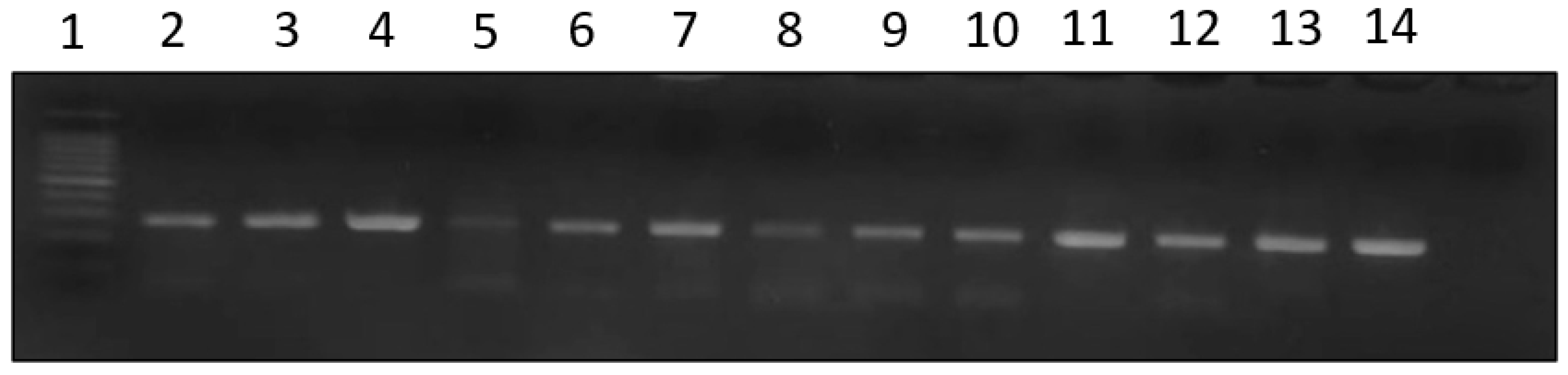
The uniplex PCR test for
S. aureus was carried out by amplification of the
nuc gene using oligonucleotide primers (Metabion, Planegg, Germany)
nuc-F (5GTGCTGGCATATGTATGGCAATTG3) and
nuc-R (5CTGAATCAGCGTTGTCTTCGCTCCAA3) [
26,
27]. A thermocycler (Biometra, Göttingen, Germany) was used to perform PCR amplifications. The reaction mixture (25 µL) contained 3 µL of genomic DNA, 12.5 µL of 2X PCR Master Mix (Takara code number RR310A), and 1 µL of each of the two primers, with the remaining volume comprising nuclease-free water. The cycling conditions were initial denaturation at 95 °C for 5 min followed by 30 cycles of denaturation at 95 °C for 30 s, annealing at 60 °C for 1 min, extension at 72 °C for 1 min, and final elongation at 72 °C for 7 min. The amplified PCR products were separated by electrophoresis in 1.5% agarose gel (Lonza, Walkersville, MD, USA) at 100 V for 30 min, stained with ethidium bromide, and then viewed and recorded using a UV trans-illuminator (Biometra, Göttingen, Germany).
2.4. Antimicrobial Susceptibility Assay
The broth microdilution assay was performed in accordance with Clinical and Laboratory Standards Institute (CLSI) standards for evolution MMV hits against S. aureus in vitro. The assay was performed for the MVV compounds provided by the MMV. After 24 h incubation of the bacterial cells with different dilutions of Malaria Box compounds at 37 °C, the lowest dilution columns with no bacterial growth were reported as the minimum inhibitory concentration (MIC) value. All assays were performed at least twice in triplicate. Using the broth microdilution assay, the 80 compounds (40 drug-like and 40 probe-like) from the Malaria Box were found to have inhibitory effects on S. aureus growth. S. aureus was isolated in pure cultures and the final inoculum size for broth dilution was 5 × 10⁵ colony-forming units (CFU).
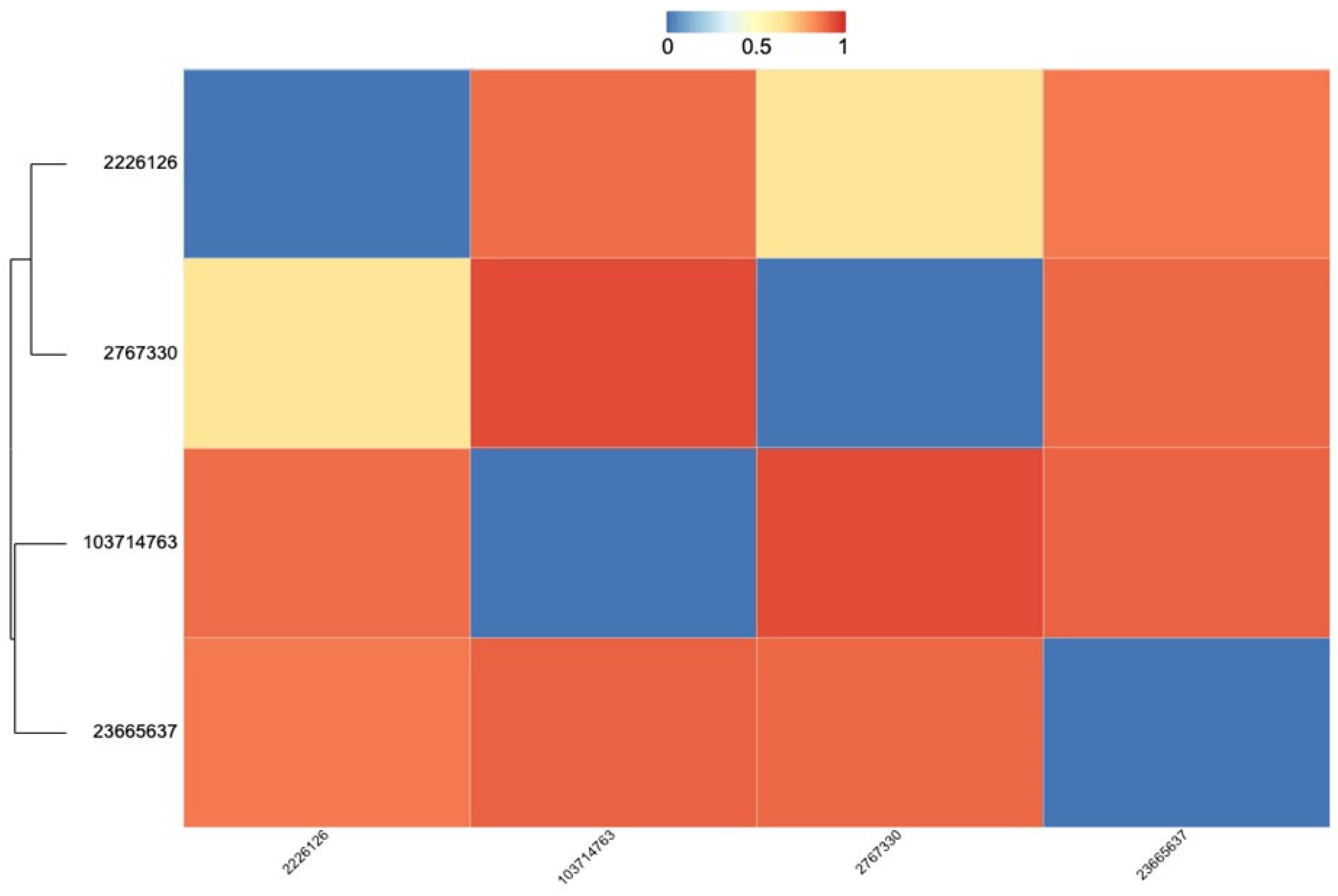
2.5. Structural Similarity Measurements
The degree of structural similarity was determined between the drug-like MMV compounds and Augmentin 625
®, a medication that is frequently used to treat
S. aureus. The chemical ID acquired from PubChem for each compound was used to calculate the atom pair fingerprint (APfp). The CIDs were then used to calculate the APfp of all compounds using ChemMine Tools software [
13,
14]. ChemmineR software was used to perform hierarchical clustering analysis (HCA) on the APfp [
15].
2.6. Microdilution Assays and Minimum Inhibitory Concentration (MIC)
Augmentin 625
® was prepared as 0.016989 gm dissolved 100 µL DMSO. Then, 20 µL of liquid drug was diluted by 180 µL of sterile broth to obtain a final concentration of 1 mM of the liquid drug. The 96-well plates (Nunc, Roskilde, Denmark) were prepared by dispensing 25 µL of sterile broth into each well. In order to determine the MIC values of the positive controls, an experiment was performed every time. The MIC was ascertained using the broth microdilution method in accordance with the Clinical and Laboratory Standards Institute’s (CLSI, Clinical and Laboratory Standards Institute, 2018) recommendations. For the preparation of bacterial inoculum, 1 mL of 24 h culture was suspended in 9 mL of sterile broth. The bacterial suspension was further diluted through five-fold broth dilution, and the last one was used in the experiment, with 25 µL added to each well of the micro-titer plate and prepared freshly every time [
30]. A total of 25 µL of the liquid drug and 25 µL of the sterile broth were added and mixed in the first well, which contained the highest concentration of drug. Then, 25 µL of the solution was used for downstream serial dilutions in ten consecutive wells, each well already containing 25 µL of sterile broth. The last 25 µL was discarded. Then, 25 µL of diluted bacteria was added to each well so the final volume in each well was 50 µL. The microplates were incubated in an incubator for 24 h.
The Malaria Box compounds were first screened at a concentration of 1 mM. For every experiment, there was a negative control (50 µL of sterile Tryptone Soya Broth) and positive control (25 µL of sterile Tryptone Soya Broth, 25 µL diluted bacteria).
In order to calculate the MIC values of the positive controls for each experiment, 96-well plates (U shapes) were generally created by dispensing 25 µL of sterile broth into each well. The microplates spent 24 h in an incubator. The MIC values for the trials were calculated in triplicate, just like they were for the positive controls.
2.7. In Vitro Drug Combination Test
Combination therapy involving the MMV compound that exhibited the highest MIC value, MMV665941, and the traditionally used anti-S. aureus drug at a concentration of 1mM was used. Every time an experiment began, 12.5 µL of the liquid drug, 12.5 µL of Augmentin, and 25 µL of sterile broth were added and mixed in the first well, which had the highest concentration of the drug.
2.8. Determination of Fractional Inhibitory Concentration (FIC)
The effect of the combination consisting of MMV66594 and the traditionally used anti-
S. aureus drug was investigated by determining the ∑ FIC. The European Committee for Antimicrobial Susceptibility Testing (EUCAST) of the European Society of Clinical Microbiology and Infectious Diseases (ESCMID)
65 described the method for quantifying MIC results in terms of the FIC index, which is defined as the sum of the FIC values of two drugs in combination [
16]. An example of the method used to calculate the
SFIC is as follows:
For two anti-bacterial substances (A and B) both alone and in combination:
2.9. Statistical Analysis
A two-way ANOVA test was used to identify the significant differences between the analyzed groups using GraphPad Prism (version 5.0 for Windows; GraphPad Software, Inc., San Diego, CA, USA). Statistical significance was defined as a p-value of less than 0.05.
4. Discussion
The Malaria Box was created to find new medications to fight different apicomplexan parasites and is a rich source of commercially accessible compounds that represent families of structures found in phenotypic screenings of pharmaceutical and academic libraries against
P. falciparum [
8,
28]. The 400-compound collection known as the Malaria Box has been broken down into 200 distinct drug-like compounds and 200 distinct probe-like compounds; toxophores are included in the drug-like compounds category. Additional hits fall under the category of probe-like hits. A comprehensive dataset encompassing sixteen protozoa, seven helminths, nine bacterial and mycobacterial species, and the NCI60 human cancer cell line was released along with the in vitro inhibitory effects of Malaria Box compounds [
8]. We were motivated to conduct a large-scale in vitro screening of MMV compounds (n = 80) against S. aureus in the current study due to the encouraging candidates found from the extensive screening of Malaria Box compounds and the zoonotic significance of
S. aureus. In this study, we have screened the antibacterial activity of the Malaria Box compounds against
S. aureus using broth microdilution assays in microtiter plates in which we put 25 µL of broth in each well and then 25 µL of the drug via serial dilution, with 25 µL of bacterial inoculum then added in each well. The final inoculum size for broth dilution was 5 × 10
5 colony-forming units (cfu), which was then incubated for 24 h.
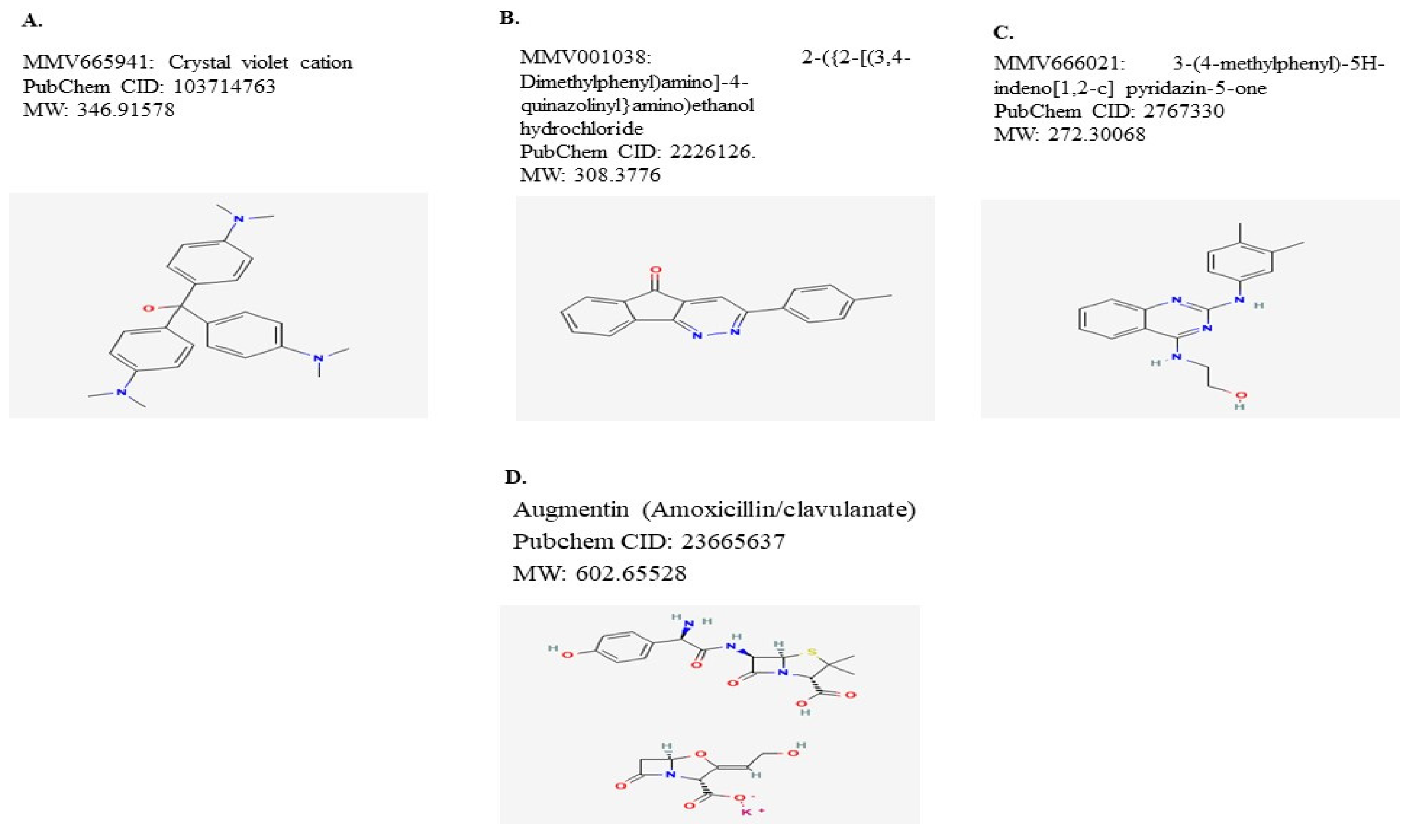
Amoxicillin/clavulanate (Augmentin 625®) was included as the internal control in every microtiter plate, which showed activity against S. aureus with an MIC value of 0.25 mg/L. On the other hand, three compounds (MMV665941, MMV666021, and MMV001038) showed excellent activity against S. aureus with MIC values of 0.0078 mg/L, 0.125 mg/L, and 0.125 mg/L. Combination therapy was employed to combine the most potent anti-S. aureus drug, the MMV665941 compound, with a traditional anti-S. aureus drug, and according to fractional inhibitory concentration (FIC), there is antagonism between both of them.
Methylrosaniline, tris [4-(dimethylamino)phenyl]methanol, and the crystal violet carbinol base are among the names for MMV665941, a triarylmethane dye with a variety of medicinal applications, including antibacterial, antifungal, antiseptic, antihelminthic, anti-trypanosomal, anti-angiogenic, and anti-tumor applications [
28]. A significant protozoal illness in oysters, Perkinsus marinus, was also found to be susceptible to MMV665941′s effectiveness; however, not all strains of the parasite showed the same level of resistance [
31]. It has been demonstrated that
Brugia malayi and
B. pahangi, Cryptosporidium, Tritrichomonas fetus trophozoites, and
Trypanosoma cruzi are all vulnerable to this chemical [
32,
33,
34,
35]. MMV665941 and gentian violet share structural similarities. Gentian violet is used as a mold inhibitor in feed components and has also been used in human medicine to treat a range of bacterial infections [
36]. However, it is strictly forbidden to use gentian violet in any food animal in the United States [
37]. A previous study [
28] demonstrated the CC
50 of MMV665941 to be equal to 353.55 nM when evaluated in a culture against human fibroblast (MRC-5) cells. Of note, there have been few grounds given for gentian violet prohibition, other than the FDA’s statement that the impact of drug residues on human health has not been sufficiently studied [
37].
Typically, the LD
50 of the drug under test in the chosen animal model is used to calculate the administrative dose in an in vivo study [
38,
39]. Even the same medication, administered via various routes, showed varying LD
50 values in the same experimental animal model [
38]. Among the identified potent MMV hits in the present study, namely MMV665941, the only available information for its administrative safe dose is by the oral route in mice [
40]. However, future studies need to identify the best administration method (intramuscular, intravenous, subcutaneous, or intramammary) of the identified potent anti-
S.aureus MMV compounds and determine the withdrawal period for each compound from the treated animals.
The decreasing quantity of viable antimicrobial medications poses a challenge to modern medicine, as does the emergence of drug-resistant bacteria and the abuse and overuse of antibiotics. In light of the way that pathogen-eliminating medicines exert strong selective pressure on the development of antimicrobial resistance, it is imperative to explore novel therapeutic approaches that can decelerate the formation of resistance. MMV665941, MMV666021, and MMV001038 showed excellent activity against S. aureus in the present study. It is known that the drug discovery process involves many different stages and series of actions. Typically, it can be divided into four main stages: early drug discovery, the pre-clinical phase, clinical phases, and regulatory approval. Identifying and characterizing molecules with the potential to safely modulate disease with the goal of bringing medicines that can improve the lives of patients represents the main aim of the drug discovery process. In the present study, the earlier phases of this process were achieved. Further experiments into antibiofilm activity and intracellular killing tests for the identified potent MMV compound are required to be performed in the future. Completing other phases in an experimental animal model in the future may suggest the usage of MMV665941, MMV666021, and MMV001038 in the treatment of previously untreatable S. aureus.
This study is considered the starting point for the evaluation of antimalarial compounds against the growth of S. aureus. Other studies are required to complete the screening of other plates in the Malaria Box against S. aureus. Furthermore, given that this work is thought to be the first to compare MMV compounds against bacteria collected from field samples, future studies are required to evaluate the identified potent compounds in an experimental animal model involving animals infected with S. aureus.


