
Dyes Are the Rainbow of Our Health
 , , and
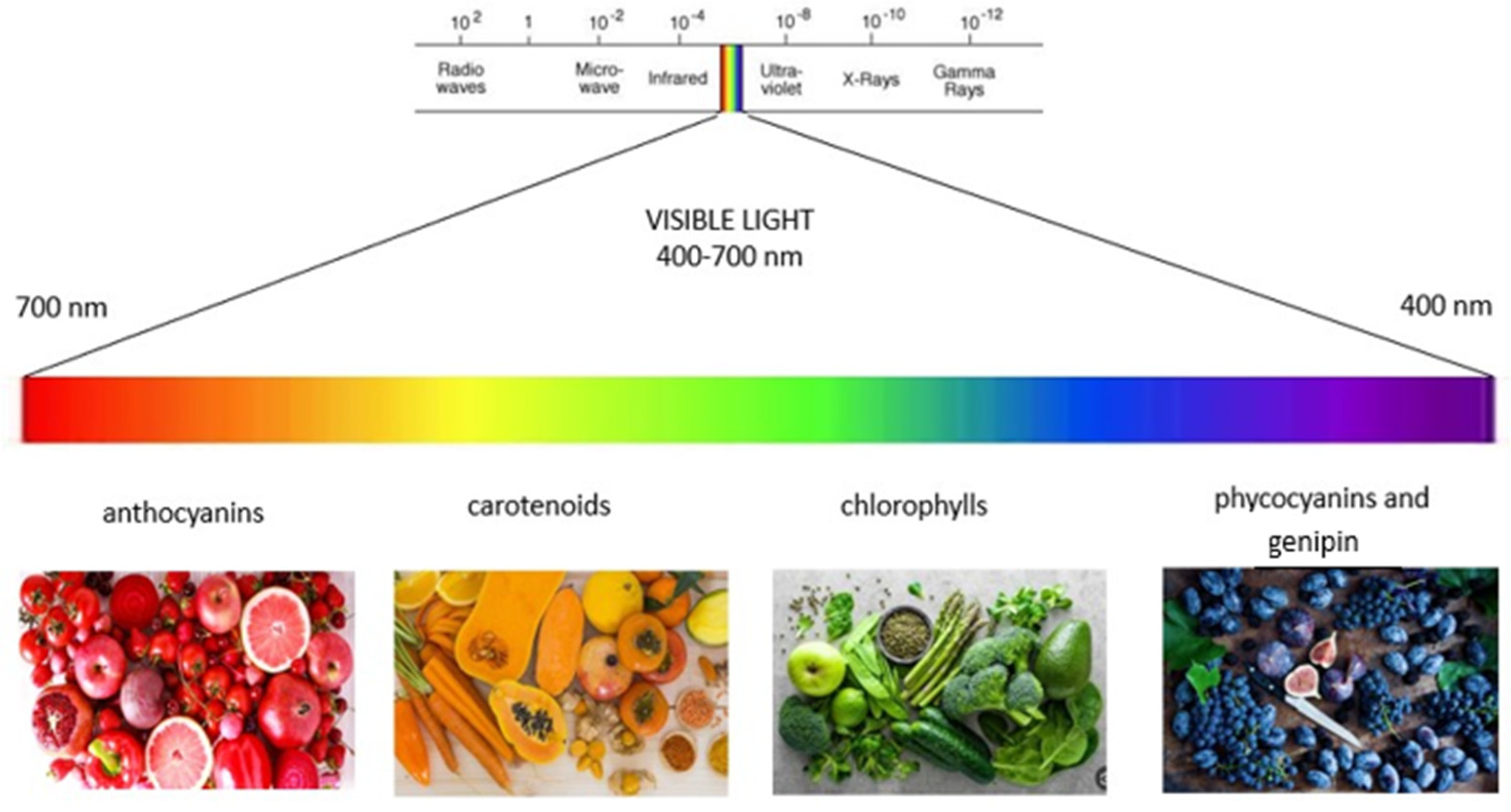
Abstract
:1. Introduction
2. Color Categories
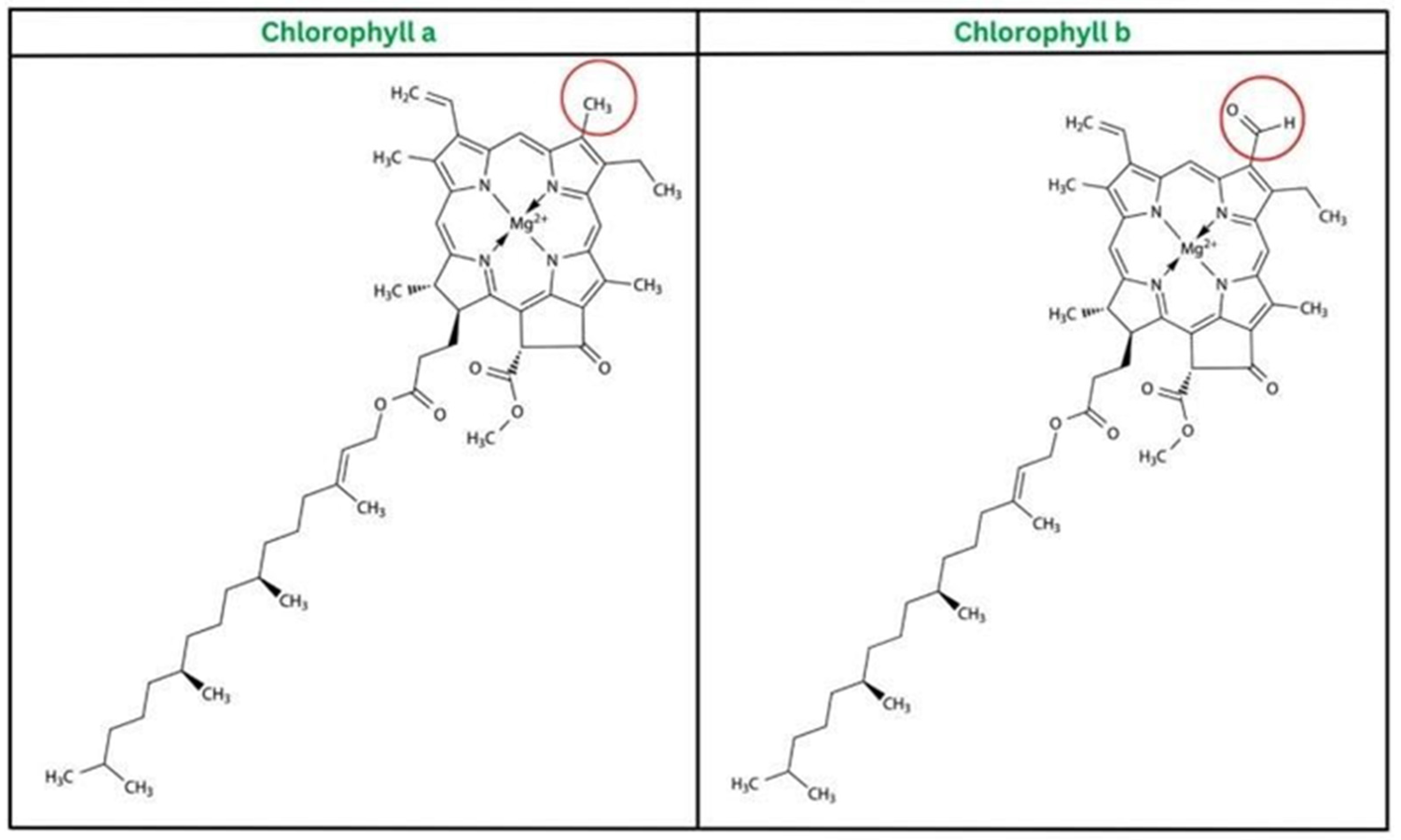
2.1. Green Color (Chlorophyll)
2.1.1. Chlorophyll as Antimicrobial Agent
2.1.2. Chlorophyll as Antioxidant Agent
2.1.3. Chlorophyll as Anticancer Agent
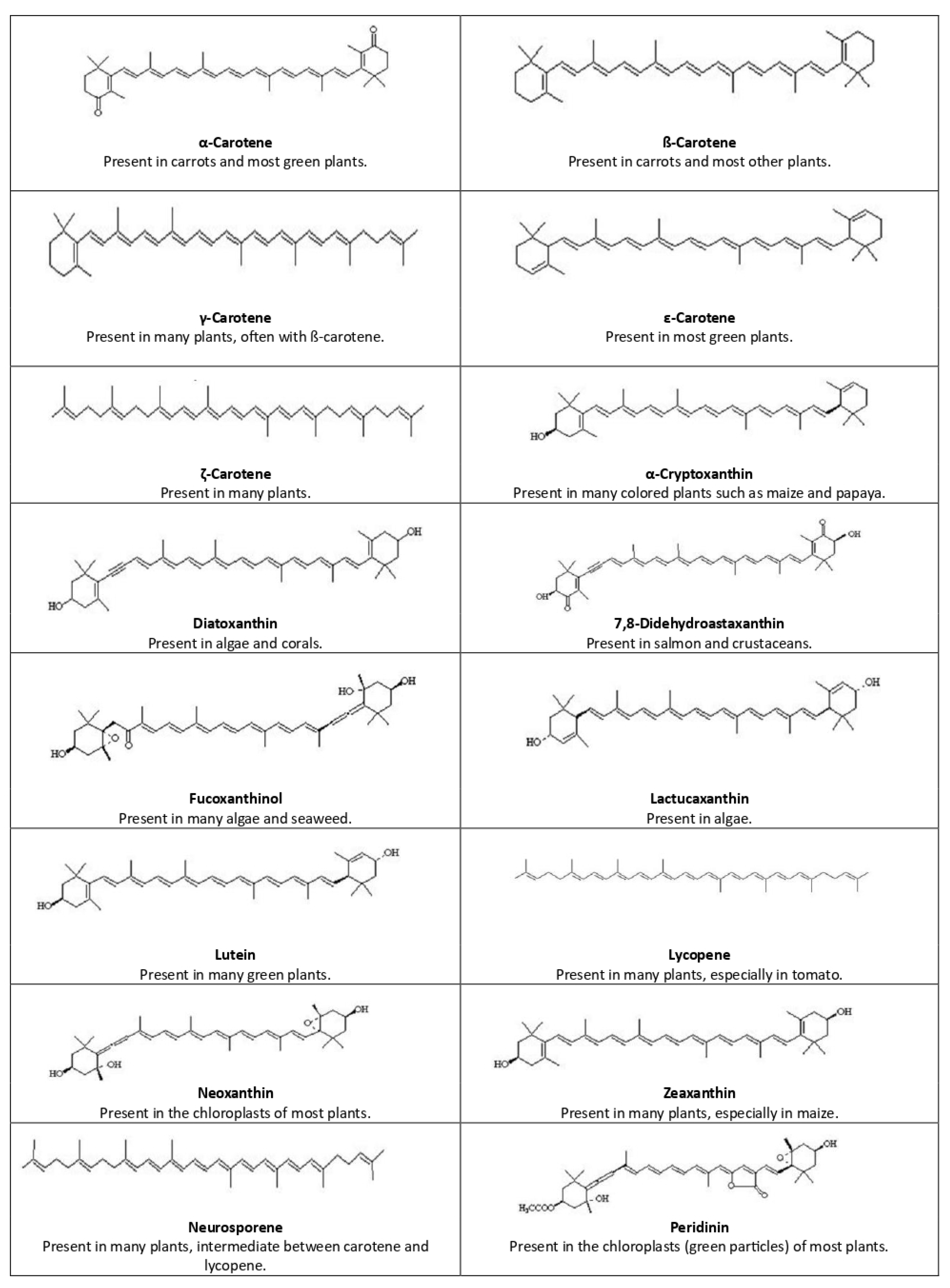
2.2. Orange Color (Carotenoids)
2.2.1. Carotenoids as Antimicrobial Agents

2.2.2. Carotenoids as Antioxidant Agents
2.2.3. Carotenoids as Antidiabetic Agents
2.2.4. Carotenoids as Anticancer Agents
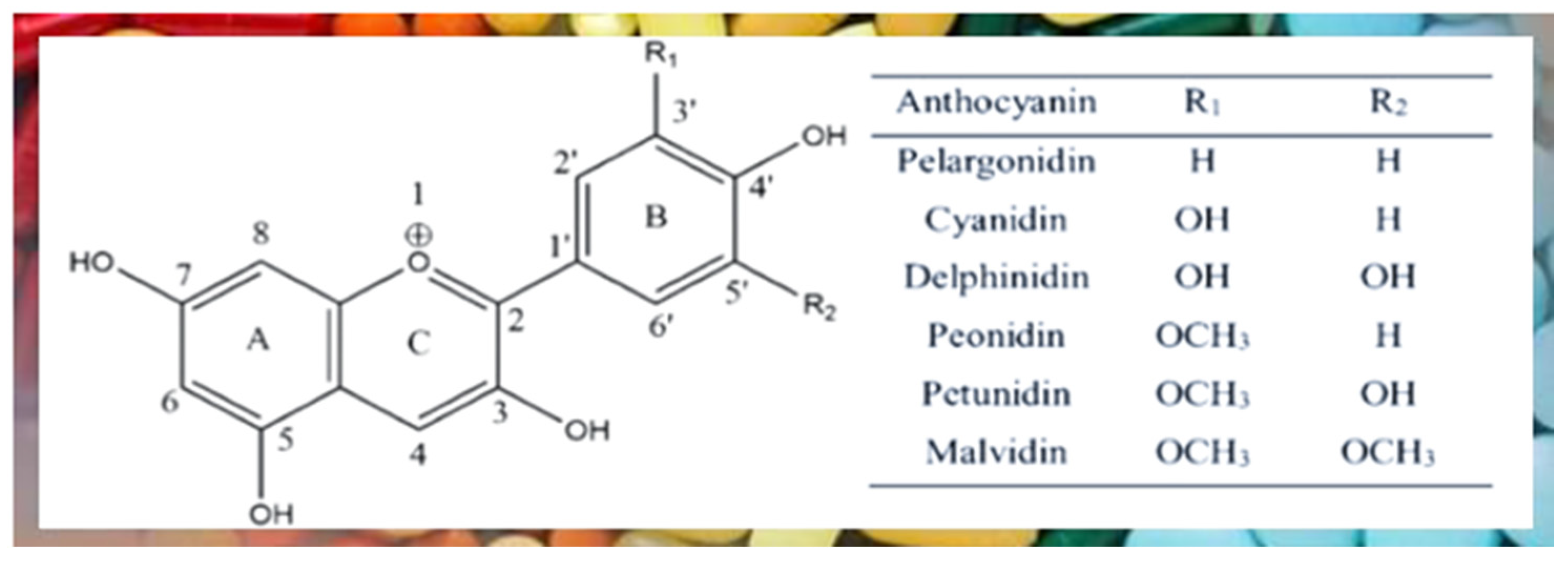
2.3. Red Color (Anthocyanin)
2.3.1. Anthocyanin as Antimicrobial Agent
2.3.2. Anthocyanin as Antioxidant Agent
2.3.3. Anthocyanin as Anticancer Agent
2.3.4. Anthocyanin as Antidiabetic Agent
2.4. Blue Color (Phycocyanins and Genipin)
2.4.1. Phycocyanin Health Benefits
2.4.2. Genipin Health Benefits
3. Conclusions
Author Contributions
Funding
Data Availability Statement
Conflicts of Interest
References
- Kadolph, S.J. Natural Dyes: A Traditional Craft Experiencing New Attention. Delta Kappa Gamma Bull. 2008, 75, 14–17. [Google Scholar]
- Bhat, S.V.; Nagasampagi, B.A.; Sivakumar, M. Chemistry of Natural Products. ChemBioChem 2005, 6, 1127–1128. [Google Scholar]
- Hernández-Ceruelos, A.; Madrigal-Bujaidar, E.; De La Cruz, C. Inhibitory effect of chamomile essential oil on the sister chromatid exchanges induced by daunorubicin and methyl methanesulfonate in mouse bone marrow. Toxicol. Lett. 2002, 135, 103–110. [Google Scholar] [CrossRef] [PubMed]
- Rahayu, M.S.; Wahyuni, S.; Fitriani, I.; Agung, H.B. Effect of tartrazine on blood urea nitrogen, creatinine levels, and renal tubular necrosis in adult male Wistar rats (Rattus norvegicus): An experimental study. Bali Med. J. 2022, 11, 1755–1759. [Google Scholar] [CrossRef]
- Chengaiah, B.; Rao, K.M.; Kumar, K.; Alagusundaram, M.; Chetty, C. Medicinal importance of natural dyes—A review. Int. J. Pharmtech. Res. 2010, 2, 144–154. [Google Scholar]
- Młodzin’ska, E. Survey of plant pigments: Molecular and environmental determinants of plant colors. Acta Biol. Crac. Ser. Bot. 2009, 51, 7–16. [Google Scholar]
- Yusuf, M.; Shabbir, M.; Mohammad, F. Natural Colorants: Historical, Processing and Sustainable Prospects. Nat. Prod. Bioprospect. 2017, 7, 123–145. [Google Scholar] [CrossRef]
- Ebrahimi, P.; Shokramraji, Z.; Tavakkoli, S.; Mihaylova, D.; Lante, A. Chlorophylls as Natural Bioactive Compounds Existing in Food By-Products: A Critical Review. Plants 2023, 12, 1533. [Google Scholar] [CrossRef]
- Ulatowska-Jarza, A. Antimicrobial PDT with chlorophyll-derived photosensitizer and semiconductor laser. Med. Laser Appl. 2006, 21, 177–183. [Google Scholar] [CrossRef]
- Vedhanarayanan, P.; Vaithiyanathan, T.; Sundaramoorthy, P. Antimicrobial activity of chlorophyll and methanol extract of Lennea coromandelica Merr. Int. Lett. Nat. Sci. 2015, 45, 67–74. [Google Scholar] [CrossRef]
- Oktavia, L.; Mulyani, I.; Suendo, V. Investigation of Chlorophyl-a Derived Compounds as Photosensitizer for Photodynamic Inactivation. Bull. Chem. React. Eng. Catal. 2021, 16, 161–169. [Google Scholar] [CrossRef]
- Kozo, T.; Yoshiyuki, F.; Shunji, I.; Atsushi, T. Anticancer Agent Decomposition Method and Anticancer Agent Decomposition Device. JP Patent PCT/JP2014/066199; Filed 18 June 2014, and Issued 31 December 2014, Available online: https://patents.google.com/patent/WO2014208428A1/en (accessed on 3 September 2023).
- Mitsuo, K.; Kaiichiro, O.; Toru, I. Copper or Iron Chlorophyllin Sodium, Pulp, Aqueous Pigment Solution, Paper Yarn and Process for Producing the Same. JP Patent PCT/JP2001/006997, 13 August 2001. Available online: https://patents.google.com/patent/WO2002016366A1/en (accessed on 6 September 2023).
- Fujiura, Y.; Kuriyama, S.; Ando, N.; Toda, Y.; Imai, T. Antimicrobial Agent in Bag, and Sheet-Form Antimicrobial Agent. JP Patent 2014/068093, 15 January 2015. Available online: https://patents.google.com/patent/WO2015005295A1/en (accessed on 6 September 2023).
- Kuniko, K.; Yuko, T.; Otori, M. Production Method of Chlorophyll Solute. JP Patent 4465675 B2 2010.5.19, 19 May 2010. Available online: https://patents.google.com/patent/JP4465675B2/en (accessed on 6 September 2023).
- Hsiao, C.J.; Lin, J.F.; Wen, H.Y.; Lin, Y.M.; Yang, C.H.; Huang, K.S.; Shaw, J.F. Enhancement of the stability of chlorophyll using chlorophyll-encapsulated polycaprolactone microparticles based on droplet microfluidics. Food Chem. 2020, 306, 125300. [Google Scholar] [CrossRef] [PubMed]
- Wareham, D.W.; Wilson, P. Chloramphenicol in the 21st century. Hosp. Med. 2002, 63, 157–161. [Google Scholar] [CrossRef] [PubMed]
- Romodin, L.A. Chlorophyllin Inhibits Chemiluminescence That Accompanies a Quasi-Hypoxygenase Reaction Catalyzed by the Cytochrome c-Cardiolipin Complex. Izvestiya of Saratov University. Ecology 2020, 20, 427–432. [Google Scholar] [CrossRef]
- Cuong, D.X. Antioxidant chlorophyll purification from maize leaves by liquid-to-liquid extraction method. J. Drug Deliv. Ther. 2020, 10, 152–158. [Google Scholar] [CrossRef]
- Georgiopoulou, I.; Tzima, S.; Louli, V.; Magoulas, K. Supercritical CO2 Extraction of High-Added Value Compounds from Chlorella vulgaris: Experimental Design, Modelling and Optimization. Molecules 2022, 27, 5884. [Google Scholar] [CrossRef] [PubMed]
- Suwa, K.; Kimura, T.; Schaap, A. Reactivity of singlet molecular oxygen with cholesterol in a phospholipid membrane matrix. A model for oxidative damage of membranes. Biochem. Biophys. Res. Commun. 1977, 75, 785–792. [Google Scholar] [CrossRef]
- Christiana, R.; Kristopo, H.; Limantara, L. Photodegradation and antioxidant activity of chlorophyll a from spirulina (Spirulina sp.) powder. Indones. J. Chem. 2010, 8, 236–241. [Google Scholar] [CrossRef]
- Usatiuk, S.; Pelekhova, L. Chlorophyll content and antioxidant activity of sunflower oil with aromatic raw materials. Food Environ. Saf. J. 2014, 13, 317–320. [Google Scholar]
- Niu, X.D.; Li, G.R.; Kang, Z.H.; Huang, J.L.; Wang, G.X. Photosynthetic characteristics and antioxidant enzyme system in high-chlorophyll rice Gc mutant. Russ. J. Plant Physiol. 2012, 59, 691–695. [Google Scholar] [CrossRef]
- Federica, B.; Federica, F.; Gimenez, A.; Canaparo, R.; Durando, G.; Andreana, I.; Barge, A.; Peira, E.; Arpicco, S.; Serpe, L.; et al. Exploiting Lipid and Polymer Nanocarriers to Improve the Anticancer. Sonodynamic Activity of Chlorophyll. Pharmaceutics 2020, 12, 605. [Google Scholar]
- Han-Zhi, L.; Lei, X.; Han, R. Anti-invasion activity of several plant-originated anticancer drugs with different mechanism of action. Acta Pharm. Sin. 1998, 33, 18–21. [Google Scholar]
- Butt, M.S.; Ahmad, R.S.; Sultan, M.T.; Qayyum, M.M.N.; Naz, A. Green Tea and Anticancer Perspectives: Updates from Last Decade. Crit. Rev. Food Sci. Nutr. (Taylor Fr.) 2015, 55, 792–805. [Google Scholar] [CrossRef]
- Pinelli, A.; Ferrario, P.; Trivulzio, S. Preliminary Observation on the Cytotoxic Activity of New Chlorophyllin Derivative RCD on Human Tumour Cell Lines In Vitro. Anticancer Res. 2019, 39, 1807–1812. [Google Scholar] [CrossRef] [PubMed]
- Vaňková, K.; Marková, I.; Jašprová, J.; Dvořák, A.; Subhanová, I.; Zelenka, J.; Novosádová, I.; Rasl, J.; Vomastek, T.; Sobotka, R.; et al. Chlorophyll-Mediated Changes in the Redox Status of Pancreatic Cancer Cells Are Associated with Its Anticancer Effects. Oxidative Med. Cell. Longev. 2018, 2018, 4069167. [Google Scholar] [CrossRef] [PubMed]
- Iqbal, J.; Abbasi, B.A.; Mahmood, T.; Kanwal, S.; Ali, B.; Shah, S.A.; Khalil, A.T. Plant-derived anticancer agents: A green anticancer approach. Asian Pac. J. Trop. Biomed. 2017, 7, 1129–1150. [Google Scholar] [CrossRef]
- Kobayashi, Y.; Maniki, M.; Nakamura, K. Cationic chlorophyl derivatives with SOD mimicking activity suppress the proliferation of human ovarian cancer cells. Cancer Biother. Radiopharm. 1996, 11, 197–202. [Google Scholar] [CrossRef] [PubMed]
- Tsuchiya, A.; Kanno, T.; Shimizu, T.; Nakao, S.; Tanaka, A.; Tabata, C.; Nakano, T.; Nishizaki, T. A novel PP2A enhancer induces caspase-independent apoptosis of MKN28 gastric cancer cells with high MEK activity. Cancer Lett. 2014, 347, 123–128. [Google Scholar] [CrossRef]
- Young, A.J.; Phillip, D.; Ruban, A.V.; Horton, P.; Frank, H.A. The xanthophyll cycle and carotenoid-mediated dissipation of excess excitation energy in photosynthesis. Pure Appl. Chem. 1997, 69, 2125–2130. [Google Scholar] [CrossRef]
- Jouni, Z.; Makhoul, Z. Carotenoid-Containing Compositions and Methods. US Patent US13/486,760, 20 September 2012. Available online: https://patents.google.com/patent/US9072314 (accessed on 3 September 2023).
- Vargas-Sinisterra, A.F.; Ramírez-Castrillón, M. Yeast carotenoids: Production and activity as antimicrobial biomolecule. Arch. Microbiol. 2021, 203, 873–888. [Google Scholar] [CrossRef]
- Mostofian, B.; Johnson, Q.R.; Smith, J.C.; Cheng, X. Carotenoids promote lateral packing and condensation of lipid membranes. Phys. Chem. Chem. Phys. 2020, 22, 12281–12293. [Google Scholar] [CrossRef] [PubMed]
- Johnson, J.D. Do carotenoids serve as transmembrane radical channels? Free Radic. Biol. Med. 2009, 47, 321–323. [Google Scholar] [CrossRef] [PubMed]
- Krinsky, N.I. Antioxidant functions of carotenoids. Free Radic. Biol. Med. 1989, 7, 90143–90146. [Google Scholar] [CrossRef]
- Eldahshan, O.A.; Singab, A.N.B. Carotenoids. J. Pharmacogn. Phytochem. 2013, 2, 225–234. [Google Scholar]
- González-Peña, M.A.; Lozada-Ramírez, J.D.; Ortega-Regules, A.E. Antioxidant activities of spray-dried carotenoids using maltodextrin-Arabic gum as wall materials. Bull. Natl. Res. Cent. 2021, 45, 58. [Google Scholar] [CrossRef]
- Srivastava, R. Physicochemical, antioxidant properties of carotenoids and its optoelectronic and interaction studies with chlorophyll pigments. Sci. Rep. 2021, 11, 1–14. [Google Scholar] [CrossRef] [PubMed]
- Dewanjee, S.; Bhattacharjee, N.; Chakraborty, P.; Bhattacharjee, S. Carotenoids as Antioxidants. In Proceedings of the Carotenoids: Structure and Function in the Human Body; Zia-Ul-Haq, M., Dewanjee, S., Riaz, M., Eds.; Springer: Cham, Switzerland, 2021; pp. 447–473. [Google Scholar]
- Shi, G.; Gu, L.; Jung, H.; Chung, W.J.; Koo, S. Apocarotenals of Phenolic Carotenoids for Superior Antioxidant Activities. ACS Omega 2021, 6, 25096–25108. [Google Scholar] [CrossRef] [PubMed]
- Ribeiro, D.; Freitas, M.; Silva, A.M.S.; Carvalho, F.; Fernandes, E. Antioxidant and pro-oxidant activities of carotenoids and their oxidation products. Food Chem. Toxicol. 2018, 120, 681–699. [Google Scholar] [CrossRef]
- Saini, R.K.; Song, M.H.; Yu, J.W.; Lee, J.H.; Ahn, H.Y.; Keum, Y.S.; Lee, J.H. Profiling of Nutritionally Vital Bioactive Compounds in Emerging Green Leafy Vegetables: A Comparative Study. Foods 2022, 11, 3867. [Google Scholar] [CrossRef]
- Nishida, Y.; Nawaz, A.; Hecht, K.; Tobe, K. Astaxanthin as a novel mitochondrial regulator: A new aspect of carotenoids, beyond antioxidants. Nutrients 2021, 14, 107. [Google Scholar] [CrossRef]
- Kręcisz, M.; Kolniak-Ostek, J.; Stępień, B.; Łyczko, J.; Pasławska, M.; Musiałowska, J. Influence of Drying Methods and Vacuum Impregnation on Selected Quality Factors of Dried Sweet Potato. Agriculture 2021, 11, 858. [Google Scholar] [CrossRef]
- Sayahi, M.; Shirali, S. The Antidiabetic and Antioxidant Effects of Carotenoids: A Review. Asian J. Pharm. Res. Health Care 2017, 9, 186–191. [Google Scholar] [CrossRef] [PubMed]
- Gutiérrez-Grijalva, E.P.; Contreras-Angulo, L.A.; Emus-Medina, A.; Heredia, J.B. Plant Alkaloids with Antidiabetic Potential. In Proceedings of the Structure and Health Effects of Natural Products on Diabetes Mellitus; Chen, H., Zhang, M., Eds.; Springer: Singapore, 2021; pp. 251–266. [Google Scholar]
- Fischer, J.; Fodor, T.; Lévai, S.; Balló, I.; Petényi, E. Antidiabetic Thiazolidinediones and Their Preparation. HU Patent HUP9904634, 21 June 2001. Available online: https://patents.google.com/patent/WO2001044240A1/en (accessed on 5 September 2023).
- Mynatt, R.L. Carnitine and type 2 diabetes. Ann. N. Y. Acad. Sci. 2004, 1033, 1–11. [Google Scholar] [CrossRef]
- Hegazy, G.E.; Abu-Serie, M.M.; Abo-Elela, G.M.; Ghozlan, H.; Sabry, S.A.; Soliman, N.A.; Abdel-Fattah, Y.R. In vitro dual (anticancer and antiviral) activity of the carotenoids produced by haloalkaliphilic archaeon Natrialba sp. M6. Sci. Rep. 2020, 10, 5986. [Google Scholar] [CrossRef] [PubMed]
- Avila-Roman, J.; Garda-Gil, S.; Rodriguez-Luna, A.; Motilva, V.; Talero, E. Anti-Inflammatory and Anticancer Effects of Microalgal Carotenoids. Mar. Drugs 2021, 19, 531. [Google Scholar] [CrossRef] [PubMed]
- Rahman, N.; Khan, H.; Zia, A.; Khan, A.; Fakhri, S.; Aschner, M.; Gul, K.; Saso, L. Bcl-2 modulation in p53 signaling pathway by flavonoids: A potential strategy towards the treatment of cancer. Int. J. Mol. Sci. 2021, 22, 11315. [Google Scholar] [CrossRef] [PubMed]
- da Costa Pereira Soares, N.; Teodoro, A.J.; Lotsch, P.F.; Granjeiro, J.M.; Borojevic, R. Anticancer properties of carotenoids in prostate cancer. A review. Histol. Histopathol. 2015, 30, 1143–1154. [Google Scholar] [CrossRef]
- Hou, J.; Cui, H.L. In vitro antioxidant, antihemolytic, and anticancer activity of the carotenoids from halophilic archaea. Curr. Microbiol. 2018, 75, 266–271. [Google Scholar] [CrossRef] [PubMed]
- Huang, C.H.; Huang, C.S.; Hu, M.L.; Chuang, C.H. Multi-Carotenoids at Physiological Levels Inhibit VEGF-Induced Tube Formation of Endothelial Cells and the Possible Mechanisms of Action Both In Vitro and Ex Vivo. Nutr. Cancer 2018, 70, 116–124. [Google Scholar] [CrossRef] [PubMed]
- Yang, C.; Zhang, L.; Tsao, R. Chemistry and biochemistry of dietary carotenoids: Bioaccessibility, bioavailability and bioactivities. J. Food Bioact. 2020, 10, 32–46. [Google Scholar] [CrossRef]
- Khoo, H.E.; Azlan, A.; Tang, S.T.; Lim, S.M. Anthocyanidins and anthocyanins: Colored pigments as food, pharmaceutical ingredients, and the potential health benefits. Food Nutr. Res. 2017, 61, 1361779. [Google Scholar] [CrossRef] [PubMed]
- Laleh, G.H.; Frydoonfar, H.; Heidary, R.; Jameei, R.; Zare, S. The effect of light, temperature, pH and species on stability of anthocyanin pigments in four Berberis species. Pak. J. Nutr. 2006, 5, 90–92. [Google Scholar] [CrossRef]
- Cahyana, Y.; Mills, C.E.; Huda, S.; Gordon, M.H. Factors affecting cellular uptake of anthocyanins: The role of pH, glucose and anthocyanin structure. Nutrients 2022, 14, 4807. [Google Scholar] [CrossRef] [PubMed]
- Nassour, R.; Ayash, A.; Al-tameemi, K. Anthocyanin pigments: Structure and biological importance. J. Chem. Pharm. Sci. 2020, 13, 45–57. [Google Scholar]
- Martín, J.; Navas, M.J.; Jiménez-Moreno, A.M.; Asuero, A.G. Anthocyanin Pigments: Importance, Sample Preparation and Extraction. In Proceedings of the Phenolic Compounds-Natural Sources, Importance and Applications; Soto-Hernandez, M., Palma-Tenango, M., del Rosario, M., Eds.; IntechOpen: London, UK, 2017. [Google Scholar] [CrossRef]
- Martín, J.; Kuskoski, E.M.; Navas, M.J.; Asuero, A.G. Antioxidant Capacity of Anthocyanin Pigments. In Proceedings of the Flavonoids-From Biosynthesis to Human Health; Justino, G.C., Ed.; IntechOpen: London, UK, 2017; Volume 3, pp. 202–255. [Google Scholar] [CrossRef]
- Bassler, B.L.; Semmelhack, M.F.; Drescher, K.; Siryaporn, A.; Miller, L.C.; O’Loughlin, C.T. Molecules and Compositions That Inhibit Gram Negative Bacteria and Their Uses. US Patent US15/023,279, 26 March 2015. Available online: https://patents.google.com/patent/WO2015042363A1/en (accessed on 10 September 2023).
- Silva, S.; Costa, E.M.; Mendes, M.; Morais, R.M.; Calhau, C.; Pintado, M.M. Antimicrobial, antiadhesive and antibiofilm activity of an ethanolic, anthocyanin-rich blueberry extract purified by solid phase extraction. J. Appl. Microbiol. 2016, 121, 693–703. [Google Scholar] [CrossRef] [PubMed]
- Yoon, B.I.; Bae, W.J.; Choi, Y.S.; Kim, S.J.; Ha, U.S.; Hong, S.H.; Sohn, D.W.; Kim, S.W. Anti-inflammatory and Antimicrobial Effects of Anthocyanin Extracted from Black Soybean on Chronic Bacterial Prostatitis Rat Model. Chin. J. Integr. Med. 2018, 24, 621–626. [Google Scholar] [CrossRef] [PubMed]
- Mahmad, N.; Taha, R.M.; Othman, R.; Abdullah, S.; Anuar, N.; Elias, H.; Rawi, N. Anthocyanin as potential source for antimicrobial activity in Clitoria ternatea L. and Dioscorea alata L. Pigment. Resin Technol. 2018, 47, 490–495. [Google Scholar] [CrossRef]
- Filimonova, N.; Gliebova, K.; Shakun, O.; Tishchenko, I.; Bosenko, O.; Domarev, A.; Krichkovskaya, L.; Gorbach, T. Antimicrobial and antioxidant activity of anthocyanin complexes of some berries’ species of ukraine. Ionori’Ihi H:Lykh 2018, 6, 304–309. [Google Scholar] [CrossRef]
- Coelho, M.; Silva, S.; Costa, E.; Pereira, R.N.; Rodrigues, A.S.; Teixeira, J.A.; Pintado, M. Anthocyanin Recovery from Grape by-Products by Combining Ohmic Heating with Food-Grade Solvents: Phenolic Composition, Antioxidant, and Antimicrobial Properties. Molecules 2021, 26, 3838. [Google Scholar] [CrossRef]
- Chatterjee, N.S.; Dara, P.K.; Raman, S.P.; Vijayan, D.K.; Sadasivam, J.; Mathew, S.; Ravishankar, C.N.; Anandan, R. Nanoencapsulation in low-molecular-weight chitosan improves in vivo antioxidant potential of black carrot anthocyanin. J. Sci. Food Agric. 2021, 101, 5264–5271. [Google Scholar] [CrossRef]
- Meng, X.; Li, Y.; Lu, C.; Zhao, M.; Li, M.; Wang, S.; Zhao, C.; Lin, B.; Shang, L.; Chu, Z.; et al. Purification and antioxidant capacity analysis of anthocyanin glucoside cinnamic ester isomers from Solanum nigrum fruits. J. Sep. Sci. 2020, 43, 2311–2320. [Google Scholar] [CrossRef]
- Farmani, F.; Moein, M.; Khoshnood, M.; Sabahi, Z. Anthocyanin Isolation from Berberis integerrima Bunge Fruits and Determination of their Antioxidant Activity. Free Radic. Antioxid. 2018, 8, 1–5. [Google Scholar]
- Tena, N.; Asuero, A.G. Antioxidant capacity of anthocyanins and other vegetal pigments. Antioxidants 2020, 9, 665. [Google Scholar] [CrossRef] [PubMed]
- Cappellini, F.; Marinelli, A.; Toccaceli, M.; Tonelli, C.; Petroni, K. Anthocyanins: From mechanisms of regulation in plants to health benefits in foods. Front. Plant Sci. 2021, 12, 748049. [Google Scholar] [CrossRef] [PubMed]
- Lin, B.W.; Gong, C.C.; Song, H.F.; Cui, Y.Y. Effects of anthocyanins on the prevention and treatment of cancer. Br. J. Pharmacol. 2017, 174, 1226–1243. [Google Scholar] [CrossRef] [PubMed]
- Bontempo, P.; De Masi, L.; Carafa, V.; Rigano, D.; Scisciola, L.; Iside, C.; Grassi, R.; Molinari, A.M.; Aversano, R.; Nebbioso, A.; et al. Anticancer activities of anthocyanin extract from genotyped Solanum tuberosum L. “Vitelotte”. J. Funct. Foods 2015, 19, 584–593. [Google Scholar] [CrossRef]
- Hazafa, A.; Rehman, K.-U.; Jahan, N.; Jabeen, Z. The Role of Polyphenol (Flavonoids) Compounds in the Treatment of Cancer Cells. Nutr. Cancer 2020, 72, 386–397. [Google Scholar] [CrossRef] [PubMed]
- Huang, P.C.; Kuo, W.W.; Shen, C.Y.; Chen, Y.F.; Lin, Y.M.; Ho, T.J.; Padma, V.V.; Lo, J.F.; Huang, C.Y.; Huang, C.Y. Anthocyanin Attenuates Doxorubicin-Induced Cardiomyotoxicity via Estrogen Receptor-α/β and Stabilizes HSF1 to Inhibit the IGF-IIR Apoptotic Pathway. Int. J. Mol. Sci. 2016, 17, 1588. [Google Scholar] [CrossRef] [PubMed]
- Veeramuthu, D.; Raja, W.R.T.; Al-Dhabi, N.A.; Savarimuthu, I. Flavonoids: Anticancer Properties. In Proceedings of the Flavonoids from Biosynthesis to Human Health; Justino, G.C., Ed.; InTech: London, UK, August 2017; Volume 287. [Google Scholar] [CrossRef]
- Kopustinskiene, D.M.; Jakstas, V.; Savickas, A.; Bernatoniene, J. Flavonoids as anticancer agents. Nutrients 2020, 12, 457. [Google Scholar] [CrossRef]
- Ding, W.; Liu, H.; Qin, Z.; Liu, M.; Zheng, M.; Cai, D.; Liu, J. Dietary Antioxidant Anthocyanins Mitigate Type II Diabetes through Improving the Disorder of Glycometabolism and Insulin Resistance. J. Agric. Food Chem. 2021, 69, 13350–13363. [Google Scholar] [CrossRef]
- Sivamaruthi, B.S.; Kesika, P.; Kumaresan, S.; Chaiyasut, C. Beneficial effects of anthocyanins against diabetes mellitus associated consequences-A mini review. Asian Pac. J. Trop. Biomed. 2018, 8, 471–477. [Google Scholar] [CrossRef]
- Gowd, P.V.; Jia, Z.; Chen, W. Anthocyanins as promising molecules and dietary bioactive components against diabetes-A review of recent advances. Trends Food Sci. Technol. 2017, 68, 1–13. [Google Scholar] [CrossRef]
- Arathy, R.; Murugan, K.; Babu, K.D.; Manoj, G. Assessment of In Vitro Antidiabetic Potential of Purified Anthocyanin Extract from Floral Petals of Wild Balsam Species. J. Drug Deliv. Ther. 2020, 10, 31–35. [Google Scholar] [CrossRef]
- Les, F.; Cásedas, G.; Gómez, C.; Moliner, C.; Valero, M.S.; López, V. The role of anthocyanins as antidiabetic agents: From molecular mechanisms to in vivo and human studies. J. Physiol. Biochem. 2021, 77, 109–131. [Google Scholar] [CrossRef] [PubMed]
- Krępska, M.; Lasoń-Rydel, M.; Jagiełło, J. Charakterystyka, właściwości, perspektywy i trudności stosowania niebieskich barwników naturalnych do barwienia produktów spożywczych. Technol. I Jakos´C´ Wyr. 2016, 61, 63–68. [Google Scholar]
- Olas, B.; Białecki, J.; Urban´ska, K.; Brys´, M. The Effects of Natural and Synthetic Blue Dyes on Human Health: A Review of Current Knowledge and Therapeutic Perspectives. Adv. Nutr. 2021, 12, 2301–2311. [Google Scholar] [CrossRef] [PubMed]
- Szalontai, B.; Gombos, Z.; Csizmadia, V.; Bagyinka, C.; Lutd, M. Structure and Interactions of Phycocyanobilin Chromophores in Phycocyanin and Allophycocyanin from an Analysis of Their Resonance Raman Spectra. Biochemistry 1994, 33, 11823–11832. [Google Scholar] [CrossRef] [PubMed]
- Manickam, B.; Sreedharan, R.; Elumalai, M. ‘Genipin’–The Natural Water Soluble Cross-linking Agent and Its Importance in the Modified Drug Delivery Systems: An Overview. Curr. Drug Deliv. 2014, 11, 139–145. [Google Scholar] [CrossRef]
- Agrawal, M.; Bansal, S.; Chopra, K. Evaluation of the in vitro and in vivo antioxidant potentials of food grade Phycocyanin. J. Food Sci. Technol. 2021, 58, 4382–4390. [Google Scholar] [CrossRef]
- Renugadevi, K.; Nachiyar, C.V.; Sowmiya, P.; Sunkar, S. Antioxidant activity of phycocyanin pigment extracted from marine filamentous cyanobacteria Geitlerinema sp TRV57. Biocatal. Agric. Biotechnol. 2018, 16, 237–242. [Google Scholar] [CrossRef]
- Nilamsari, A.M.; Yunanda, A.; Hadiyanto, H. Thermal degradation kinetics of phycocyanin encapsulation as an antioxidant agent. Int. Symp. Food Agro-Biodivers. 2018, 102, 12055. [Google Scholar] [CrossRef]
- Chentir, I.; Nasri, M. One structure, multiple features: The phycocyanin in biotechnology. Nutr. St. 2020, 9, 28–42. [Google Scholar] [CrossRef]
- de Amarante, M.C.A.; Braga, A.R.C.; Sala, L.; Kalil, S.J. Colour stability and antioxidant activity of C-phycocyanin-added ice creams after in vitro digestion. Food Res. Int. 2020, 137, 109602. [Google Scholar] [CrossRef] [PubMed]
- Hadiyanto, H.; Harjanto, G.D.; Huzain, M.L.; Aji, R.W. Production of antioxidant C-phycocyanin using extraction process of Spirulina platensis in large scale industry. IOP Conf. Ser. Mater. Sci. Eng. 2019, 633, 12025. [Google Scholar]
- Safari, R.; Amiri, R.Z.; Kenari, R.E. Antioxidant and antibacterial activities of C-phycocyanin from common name Spirulina platensis. Iran. J. Fish. Sci. 2020, 19, 1911–1927. [Google Scholar] [CrossRef]
- Sonani, R.R.; Patel, S.; Bhastana, B.; Jakharia, K.; Chaubey, M.G.; Singh, N.K.; Madamwar, D. Purification and antioxidant activity of phycocyanin from Synechococcus sp. R42DM isolated from industrially polluted site. Bioresour. Technol. 2017, 245, 325–331. [Google Scholar] [CrossRef] [PubMed]
- Farajzadeh, N.; Özdemir, S.; Gonca, S.; Atmaca, G.Y.; Erdoğmuş, A.; Bayır, Z.A.; Koçak, M.B. Photophysicochemical and Biological Properties of New Phthalocyanines Bearing 4-(trifluoromethoxy) phenoxy and 2-(4-methylthiazol-5-yl) ethoxy Groups on Peripheral Positions. Photochem. Photobiol. 2022, 98, 894–906. [Google Scholar] [CrossRef] [PubMed]
- Sethi, S.; Joshi, A.; Arora, B.; Bhowmik, A.; Sharma, R.; Kumar, P.; Sethi, S.; Joshi, A.; Arora, B.; Bhowmik, A.; et al. Significance of FRAP, DPPH, and CUPRAC assays for antioxidant activity determination in apple fruit extracts. Eur. Food Res. Technol. 2020, 246, 591–598. [Google Scholar] [CrossRef]
- Liang, Y.; Kaczmarek, M.B.; Kasprzak, A.K.; Tang, J.; Shah, M.M.R.; Jin, P.; Klepacz-Smółka, A.; Cheng, J.J.; Ledakowicz, S.; Daroch, M. Thermosynechococcaceae as a source of thermostable C-phycocyanins: Properties and molecular insights. Algal Res. 2018, 35, 223–235. [Google Scholar] [CrossRef]
- Arulselvan, P.; Fard, M.T.; Tan, W.S.; Gothai, S.; Fakurazi, S.; Norhaizan, M.E.; and Kumar, S. Role of Antioxidants and Natural Products in Inflammation. Oxidative Med. Cell. Longev. 2016, 2016, 5276130. [Google Scholar] [CrossRef]
- Hao, S.; Li, S.; Wang, J.; Zhao, L.; Zhang, C.; Huang, W.; Wang, C.; Hao, S.; Li, S.; Wang, J.; et al. Phycocyanin Reduces Proliferation of Melanoma Cells through Downregulating GRB2/ERK Signaling. J. Agric. Food Chem. 2018, 66, 10921–10929. [Google Scholar] [CrossRef] [PubMed]
- Jiang, L.; Wang, Y.; Yin, Q.; Liu, G.; Liu, H.; Huang, Y.; Li, B. Phycocyanin: A Potential Drug for Cancer Treatment. J. Cancer 2017, 8, 3416. [Google Scholar] [CrossRef] [PubMed]
- Yong, W.; Feng, Q.; Kaixian, Q.; Qiang, D. Anticancer activity of phycocyanin. J. Zhejiang Univ. (Eng. Sci.) 2001, 35, 672–675. [Google Scholar]
- Morcos, N.C. Anatomical Imaging with Phycocyanins. US Patent US08/631,670, 15 September 1998. Available online: https://patents.google.com/patent/US5807536A/en?q=(Anatomical+imaging+phycocyanins)&oq=Anatomical+imaging+with+phycocyanins (accessed on 13 September 2023).
- Cimmino, A.; Andolfi, A.; Evidente, A. Phenazine as an anticancer agent. In Proceedings of the Microbial Phenazines; Chincholkar, S., Thomashow, L., Eds.; Springer: Berlin/Heidelberg, Germany, December 2013; pp. 217–243. [Google Scholar]
- Gruda, I.; Pagé, M.; Bolduc, F.; Laliberté, S.; Noel, C. Merocyanines: Potential new anticancer agents. Anticancer Res 1987, 7, 1124–1127. [Google Scholar] [PubMed]
- Scoglio, S.; Baffone, W.; Campana, R.; Colombo, E. The Use of Phycocyanins, Purified or as Present in Cyanobacterial Microalgae or Extracts Thereof, as Prebiotics, to Enhance the Viability, Gastrointestinal Survival, Pathogen-Fighting Ability, and the Overall Health-Enhancing Properties of Probiotic Cultures and Products. US Patent US16/098,618, 9 November 2017. Available online: https://patents.google.com/patent/WO2017191078A1/en (accessed on 13 September 2023).
- Qiu, Y.T.; Wang, B.J.; Weng, Y.M. Preparation and characterization of genipin cross-linked and lysozyme incorporated antimicrobial sodium caseinate edible films. Food Packag. Shelf Life 2020, 26, 100601. [Google Scholar] [CrossRef]
- Chu, X.; Wu, F.; Sun, B.; Zhang, M.; Song, S.; Zhang, P.; Wang, Y.; Zhang, Q.; Zhou, N.; Shen, J. Genipin cross-linked carbon dots for antimicrobial, bioimaging and bacterial discrimination. Colloids Surf. B Biointerfaces 2020, 190, 110930. [Google Scholar] [CrossRef] [PubMed]
- Ma, W.; Tang, C.H.; Yin, S.W.; Yang, X.Q.; Qi, J.R. Genipin-crosslinked gelatin films as controlled releasing carriers of lysozyme. Food Res. Int. 2013, 51, 321–324. [Google Scholar] [CrossRef]
- Khan, A.; Salmieri, S.; Fraschini, C.; Bouchard, J.; Riedl, B.; Lacroix, M. Genipin cross-linked nanocomposite films for the immobilization of antimicrobial agent. ACS Appl. Mater. Interfaces 2014, 6, 15232–15242. [Google Scholar] [CrossRef]
- Kim, T.H.; Yoon, S.J.; Lee, S.M. Genipin attenuates sepsis by inhibiting Toll-like receptor signaling. Mol. Med. 2012, 18, 455–465. [Google Scholar] [CrossRef]
- Liu, Q.; Li, Y.; Xing, S.; Wang, L.; Yang, X.; Hao, F.; Liu, M. Genipin-crosslinked amphiphilic chitosan films for the preservation of strawberry. Int. J. Biol. Macromol. 2022, 213, 804–813. [Google Scholar] [CrossRef]
- Luo, N.; Chen, G.; Zhang, T.; Zhao, J.; Fu, J.; Lu, N.; Ma, T. Genipin Attenuates Sepsis-Induced Splenocyte Apoptosis via the Inhibition of Endoplasmic Reticulum Stress. Biol. Pharm. Bull. 2023, 46, 187–193. [Google Scholar] [CrossRef] [PubMed]
- Shanmugam, M.K.; Shen, H.; Tang, F.R.; Arfuso, F.; Rajesh, M.; Wang, L.; Kumar, A.P.; Bian, J.; Goh, B.C.; Bishayee, A.; et al. Potential role of genipin in cancer therapy. Pharm. Res. 2018, 133, 195–200. [Google Scholar] [CrossRef] [PubMed]
- Kim, B.R.; Jeong, Y.A.; Kim, D.Y.; Kim, J.L.; Jeong, S.; Na, Y.J.; Yun, H.K.; Park, S.H.; Jo, M.J.; Ashktorab, H.; et al. Genipin increases oxaliplatin-induced cell death through autophagy in gastric cancer. J. Cancer 2020, 11, 460–467. [Google Scholar] [CrossRef] [PubMed]
- Cho, Y.S. Genipin, an Inhibitor of UCP2 as a Promising New Anticancer Agent: A Review of the Literature. Int. J. Mol. Sci. 2022, 23, 5637. [Google Scholar] [CrossRef] [PubMed]
- Lee, S.; Kim, H.J.; Oh, S.C.; Lee, D.H. Genipin inhibits the invasion and migration of colon cancer cells by the suppression of HIF-1α accumulation and VEGF expression. Food Chem. Toxicol. 2018, 116, 70–76. [Google Scholar] [CrossRef] [PubMed]
- Shin, Y.S.; Cho, M.; Park, G.H.; Cho, H.; Kang, H. Antitumor Effects of Genipin: New and Emerging Insights from Recent Studies. J. Bacteriol. Virol. 2016, 46, 108–113. [Google Scholar] [CrossRef]
- Ahani, N.; Sangtarash, M.H.; Houshmand, M.; Eskandani, M.A. Genipin induces cell death via intrinsic apoptosis pathways in human glioblastoma cells. J. Cell. Biochem. 2019, 120, 2047–2057. [Google Scholar] [CrossRef] [PubMed]
- Ye, J.; Li, J.; Wang, X.; Li, L. Medicinal supplement genipin induces p53 and Bax-dependent apoptosis in colon cancer cells. Oncol. Lett. 2018, 16, 2957–2964. [Google Scholar] [CrossRef]
- Li, Z.; Ma, T.; Zhang, W.; Shang, Y.; Zhang, Y.; Ma, Y. Genipin attenuates dextran sulfate sodium-induced colitis via suppressing inflammatory and oxidative responses. Inflammopharmacology 2020, 28, 333–339. [Google Scholar] [CrossRef]
- Jo, M.J.; Jeong, S.; Yun, H.K.; Kim, D.Y.; Kim, B.R.; Kim, J.L.; Na, Y.J.; Park, S.H.; Jeong, Y.A.; Kim, B.G. Genipin induces mitochondrial dysfunction and apoptosis via downregulation of Stat3/mcl-1 pathway in gastric cancer. BMC Cancer 2019, 19, 1–12. 763. [Google Scholar] [CrossRef]
- Zuo, T.; Xu, W.; Li, H.; Song, H.; Zhu, M. Geniposide and geniposidic acid, modified forms of genipin, attenuate genipin-induced mitochondrial apoptosis without altering the anti-inflammatory ability in KGN cell line. Med. Chem. Res. 2017, 26, 499–508. [Google Scholar] [CrossRef]
- Cho, H.I.; Kim, S.J.; Choi, J.W.; Lee, S.M. Genipin alleviates sepsis-induced liver injury by restoring autophagy. Br. J. Pharmacol. 2016, 173, 980–991. [Google Scholar] [CrossRef]
- Wang, Z.; Liu, H.; Luo, W.; Cai, T.; Li, Z.; Liu, Y.; Gao, W.; Wan, Q.; Wang, X.; Wang, J.; et al. Regeneration of skeletal system with genipin crosslinked biomaterials. J. Tissue Eng. 2020, 11. [Google Scholar] [CrossRef]
- Koo, B.S.; Kwon, T.S.; Kim, C.H. Salviae miltiorrhizae Radix Inhibits Superoxide Generation by Activated Rat Microglias and Mimics the Action of Amphetamine on in vitro Rat Striatal Dopamine Release. Neurochem. Res. 2004, 29. [Google Scholar] [CrossRef]


Disclaimer/Publisher’s Note: The statements, opinions and data contained in all publications are solely those of the individual author(s) and contributor(s) and not of MDPI and/or the editor(s). MDPI and/or the editor(s) disclaim responsibility for any injury to people or property resulting from any ideas, methods, instructions or products referred to in the content. |
© 2023 by the authors. Licensee MDPI, Basel, Switzerland. This article is an open access article distributed under the terms and conditions of the Creative Commons Attribution (CC BY) license (https://creativecommons.org/licenses/by/4.0/).
Share and Cite
Shammout, M.-J.A.; Alsaleh, M.M.; Natsheh, I.Y.; Albadawi, D.K.; Alkhawaldeh, A.K. Dyes Are the Rainbow of Our Health. Chemistry 2023, 5, 2229-2245. https://doi.org/10.3390/chemistry5040149
Shammout M-JA, Alsaleh MM, Natsheh IY, Albadawi DK, Alkhawaldeh AK. Dyes Are the Rainbow of Our Health. Chemistry. 2023; 5(4):2229-2245. https://doi.org/10.3390/chemistry5040149
Chicago/Turabian StyleShammout, Mohammad-Jamal A., Majd M. Alsaleh, Iyad Y. Natsheh, Duaa K. Albadawi, and Ahmad K. Alkhawaldeh. 2023. "Dyes Are the Rainbow of Our Health" Chemistry 5, no. 4: 2229-2245. https://doi.org/10.3390/chemistry5040149