Review

Green Synthesis of Gold Nanoparticles: An Eco-Friendly Approach

by
Poornima Budime Santhosh
*,
Julia Genova
* and
Hassan Chamati
*
Institute of Solid State Physics, Bulgarian Academy of Sciences, Tzarigradsko Chaussée 72, 1784 Sofia, Bulgaria
*
Authors to whom correspondence should be addressed.
Chemistry 2022, 4(2), 345-369; https://doi.org/10.3390/chemistry4020026
Submission received: 16 March 2022 / Revised: 12 April 2022 / Accepted: 23 April 2022 / Published: 25 April 2022
(This article belongs to the Section Chemistry at the Nanoscale)

Abstract

:
By virtue of their unique physicochemical properties, gold nanoparticles (AuNPs) have gained significant interest in a broad range of biomedical applications such as sensors, diagnosis, and therapy. AuNPs are generally synthesized via different conventional physical and chemical methods, which often use harmful chemicals that induce health hazards and pollute the environment. To overcome these issues, green synthesis techniques have evolved as alternative and eco-friendly approaches to the synthesis of environmentally safe and less-expensive nanoparticles using naturally available metabolites from plants and microorganisms such as bacteria, fungi, and algae. This review provides an overview of the advances in the synthesis of AuNPs using different biological resources with examples, and their profound applications in biomedicine. A special focus on the biosynthesis of AuNPs using different medicinal plants and their multifunctional applications in antibacterial, anti-inflammatory, and immune responses are featured. Additionally, the applications of AuNPs in cancer theranostics, including contrast imaging, drug delivery, hyperthermia, and cancer therapeutics, are comprehensively discussed. Moreover, this review will shed light on the importance of the green synthesis approach, and discuss the advantages, challenges, and prospects in this field.

1. Introduction

Nanotechnology is a promising field that integrates the various disciplines of science, engineering, and technology. The rapid scientific advances in this field have led to the development of different types of functional nanoparticles (NPs), with at least one dimension in the typical size range of 1 to 100 nm. Among the various metallic NPs, gold nanoparticles (AuNPs) have attracted huge attention due to their unique surface plasmon resonance properties, facile synthesis, tunable sizes, and multifunctional abilities with well-characterized properties [1,2]. They are versatile materials, relatively inert, biocompatible, and generally stable. Due to their well-defined surface chemistry, AuNPs can be easily conjugated with different molecules such as proteins, dyes, drugs, antibodies, enzymes, and nucleic acids [3,4,5]. AuNPs functionalized with different targeting moieties have enormous scope in various biomedical applications such as diagnosis, targeting, drug/nucleic acid delivery, imaging, and therapy (Figure 1). Furthermore, by employing the surface-enhanced Raman scattering technique, AuNPs are used as sensitive probes in Raman scattering and imaging applications [6]. The potential of AuNPs in biomedical fields has been tremendously increased by virtue of their applications in photothermal therapy, radiation therapy, computed tomography, biosensors, etc. [7]. Due to their intrinsic electrical and optical properties, as well as their ability to conjugate with different biomolecules, AuNPs-based biosensors with high sensitivity and selectivity are being developed [8,9]. In the last decade, AuNPs-based biosensors have attracted great attention in the diagnosis of various types of diseases. Recently, Antonio et al. [10] highlighted the various AuNPs-based biological assays for the detection and quantification of analytes in urinary samples, with a focus on protein analysis. Such assays using AuNPs are useful in the diagnosis of several illnesses such as kidney disorders, cancer, and heart diseases [4,11].
Gold nanomaterials can be synthesized via different techniques in various internal structure, sizes, and shapes structures, including nanospheres, nanorods, nanocubes, nanoshells, nanowires, nanocages, nanoflowers, etc. [12]. They exhibit exceptional properties such as fluorescence, attenuation of X-rays, etc., and act as excellent contrast agents for optical, fluorescence, X-ray, and photoacoustic imaging [13,14]. Their intrinsic features (optics, electronics, and physicochemical characteristics) can be altered by adjusting their size and shape. To improve their compatibility and stability in a biological environment, AuNPs are coated with different biomolecules such as phospholipids, proteins, and polymers such as polyethylene glycol [15,16]. By fine-tuning the aspect ratio (length/width of the particle), gold nanorods can be manipulated to absorb light very strongly in the near-infrared region, convert it into heat energy, and transmit it to the surrounding environment. This process, called photohyperthermia, is widely used to attenuate cancer cells, where gold nanorods are administered near the tumor region to destroy the cancer cells without causing much damage to the healthy neighboring cells [17,18]. These properties have made AuNPs a widely used nanomaterial for global academic research and in the production of various industrial products and medical devices.

2. General Methods for Synthesis of AuNPs

2.1. Physicochemical Methods

NPs are typically synthesized via two basic methods: “top-down” or “bottom-up” (Figure 2). In the top-down approach, the constituent bulk materials are initially broken down to powder form and subsequently reduced to fine nanoparticles using various techniques such as etching, grinding, sputtering, thermal/laser ablation, etc. On the other hand, the bottom-up method involves the self-assembly of atoms to form nuclei, which then transform into particles of nanoscale range. The bottom-up method is widely used to obtain NPs with uniform morphology and chemical composition. The Turkevich method is a conventional chemical synthesis method commonly used to produce spherical small AuNPs around 10 to 30 nm in diameter [19]. However, it was observed that for the synthesis of AuNPs above 30 nm size, the results were less reproducible and resulted in a broader size distribution of particles [20]. The major limitation of this method is the strict process control that needs to be followed and the precise maintenance of temperature, pH, and salt concentration to obtain monodisperse NPs in a specific size range. Alternatively, the Brust method and seed-mediated growth method are widely used for the synthesis of AuNPs [3].
The different methods of AuNPs synthesis, such as physical (ultrasonication, irradiation, electrochemical, etc.), chemical (vapor deposition, sol–gel process, etc.), and biological methods (using plants and microbial sources), are shown in Figure 3. However, the various physical and chemical methods used for the synthesis of NPs are expensive and hazardous to human health due to the use of toxic components and the production of byproducts, either during the synthesis or during the capping/stabilization process of NPs. The use of chemicals raises serious environmental and toxicity concerns when administered to living organisms. For instance, the surfactant cetyltrimethylammonium bromide (CTAB), which acts as a stabilizer and template for the growth of gold nanorods in the seed-mediated method, is reported to be highly toxic to different types of cells [21]. Therefore, gold nanorods stabilized with CTAB are not suitable for biomedical applications and need to be properly surface-modified with biocompatible materials such as phospholipids or polyethylene glycol [22]. Hence, there is a growing requirement to search for reliable, non-expensive, biocompatible, and environmentally friendly methods for the synthesis of NPs.

2.2. Biosynthetic Mechanism of AuNPs

The mechanism of AuNPs biosynthesis is a simple two-step process and does not require a dramatic increase in temperature and pressure. In the first step, the biological extract (e.g., plant, bacterial, or fungal extract) is mixed with the HAuCl 4 salt solution, which causes the reduction of gold (Au 3 + ) ions to gold atoms (Au 0 ). In the second step, growth and stabilization result in the AuNPs formation (Figure 4). Finally, the color change of the resulting solution indicates the formation of AuNPs [23,24]. The chemical reactions involved in the reduction of Au 3 + to Au 0 in the presence of H 2 O molecules are expressed in the below reactions:
Dissociation: HAuCl 4 ( Chloroauric acid ) H 2 O H + + Au 3 + + 4Cl ,
Oxidation: 4Cl ⟶ 2Cl 2 + 4e ,
Reduction: Au 3 + + 4e ⟶ Au 0 + e .
Figure 4. Schematic representation of AuNPs biosynthesis mechanism.
Chemistry 04 00026 g004
A variety of biocompounds (enzymes, phenols, sugars, etc.) can participate both in the reduction and stabilization of different types of particles, including AuNPs [25]. Figure 5a,b show the biosynthesis mechanism of bacterial microorganisms, which can act as a “factory” for the production of AuNPs. The biosynthesis mechanism of the microorganisms can be either extracellular or intracellular based on the location of AuNPs production [26]. Extracellular biosynthesis occurs outside the bacterial cell by trapping and reducing metal ions in the presence of enzymes. On the contrary, in the intracellular method, metal ions are transported into the microbial cell to form NPs in the presence of enzymes [27].

3. Green Synthesis of AuNPs

3.1. Importance of Green Synthesis of AuNPs

As the applications of AuNPs are increasing day by day, the demand for their synthesis also increases simultaneously. It has aroused interest among researchers worldwide to develop novel interdisciplinary routes for the synthesis of highly stable, monodisperse, and safe AuNPs for various applications. To overcome the challenges in the conventional chemical synthesis method, an alternative approach to synthesize biocompatible NPs, termed “green synthesis”, has evolved. It is an emerging branch of nanotechnology and has attracted huge attention among researchers and industries, as well as people concerned about environmental pollution and health hazards. Green synthesis techniques are important as they are an eco-friendly approach that involves the use of natural bioresources and avoids toxic chemicals to synthesize different types of NPs [28]. For instance, green synthesis of cobalt ferrite nanoparticles using extracts of grape peel, pulp, and honey-mediated synthesis of cobalt-zinc ferrite NPs were reported earlier [29,30].
A variety of plants, microorganisms, and biomolecules derived from them are used as a source for the synthesis of various types of NPs. Extracts from different parts of the plant such as leaves, roots, seeds, flowers, fruits, bark, etc., and microbes, including bacteria, fungi, and algae, are widely used to synthesize NPs with varying sizes and shapes using interdisciplinary routes [19,31]. The precursor gold salt solution is treated either with the microbial culture or plant extracts, which are then bioreduced to form AuNPs. Different metabolites and biomolecules such as sugars, fatty acids, proteins, enzymes, and phenols play a key role in the synthesis of the AuNPs [32,33]. Further, this biological approach involves the use of (i) an environmentally acceptable solvent medium for NPs synthesis, (ii) natural reducing agents, and (iii) nontoxic capping agents, mostly polyphenols and other secondary metabolites. Green synthesis of AuNPs using different bioagents and their applications is shown in Table 1.

3.2. Advantages of Green Synthesis of AuNPs

The green synthesis method offers the following advantages over the chemical methods: (i) safety: this method avoids the exposure of chemicals or their toxic byproducts, either during the NPs synthesis step or during their stabilization process; (ii) cost-effective: no external stabilizing agent is required. On the contrary, chemical methods use expensive and hazardous chemicals and stabilizing agents; (iii) simplicity: biosynthesis of NPs from plant extract is a simple process; (iv) renewable feedstock: e.g., algal biomass; (v) easy availability of source materials; (vi) biocompatibility: since natural sources are used to synthesize and stabilize the particles, AuNPs synthesized via green synthesis methods are biocompatible to different cell types; (vii) the whole green synthesis process is dynamic, reproducible, and energy-efficient; (viii) suitable for large scale production of NPs for commercial applications; (ix) AuNPs synthesized via green synthesis method are also reported to exhibit antibacterial, antifungal, anticancer, and anti-inflammatory properties, and antioxidant and catalytic activity due to the presence of phytochemicals from the bioextract [69,70]. All these factors have rendered the green synthesis approach more rewarding than conventional methods.

4. Biosynthesis of AuNPs from Plant Sources

Biosynthesis of AuNPs from plant sources is facile and involves a single-step process in the one-pot method. To synthesize AuNPs from plant sources, different parts of the plant (leaves, fruits, bark, flower, peels, seed, rhizome root, etc.) are washed with distilled water, dried, ground into powder or chopped into small pieces, and boiled in distilled water to a specific temperature to obtain the extract. Then, filtration or centrifugation techniques are used to purify the extract, which is then simply mixed with various concentrations of gold salt solution (based on the plant parts and their species). The gold salt solution is reduced into AuNPs and the reaction completes in minutes to a few hours. The reaction mixture is further incubated to reduce the gold salt completely, and is visually monitored by color change. Finally, the synthesized AuNPs are purified by centrifugation and washed thoroughly in water for further use. The whole process is simple, eco-friendly, and can be scaled up easily.
Plants are rich in alkaloids, flavonoids, saponins, steroids, tannins, and other natural compounds [71]. The plant extract contains various secondary metabolites, which act as both reducing and stabilizing agents for the biogenesis of NPs [72]. Numerous reports have shown the successful synthesis of different types of NPs, such as silver, copper, gold, cobalt, palladium, magnetite, and zinc oxide [73,74]. Although various parts of plants have been reported in the biosynthesis of AuNPs, leaves are widely used. Variations in the level of metabolites content from different plant parts, and even variation among plants, play a crucial role in shaping the morphology of NPs.
Islam et al. [75] reported a reproducible green synthetic method to produce highly stable AuNPs, using the leaves extract of the plant Salix alba L. (syn: white willow), which belongs to the family Salicaceae. The leaves and bark of this plant are rich in phenolic contents such as salicin, which acts as a precursor in the development of aspirin. Hence, they are traditionally used for musculoskeletal pain relief and treatment of different ailments, owing to their antipyretic and anti-inflammatory properties. When the aqueous gold ions were treated with Salix alba L. leaves extract, they were reduced, leading to the synthesis of AuNPs. The UV–Vis absorption spectra data revealed that the synthesized AuNPs were found to be colloidally stable at different pH and salt concentrations. The AuNPs functionalized with the phytochemicals of leaf extracts exhibited good antifungal activity, pain-relieving, and muscle relaxant effect, which enhances their potential for various biomedical and pharmaceutical applications. Narayan et al. [76] reported the extracellular synthesis of AuNPs using coriander leaf extract as the reducing agent. Transmission electron microscopy (TEM) images have shown the formation of stable AuNPs in the size range of 6.75–57.91 nm (Figure 6) with varying shapes, such as spherical, triangle, truncated triangles, and decahedral morphologies.
Similarly, Boruah et al. [77] reported a simple, faster, low-cost, eco-friendly technique to biosynthesize AuNPs using fresh young leaves and leaf buds of Camellia Sinensis (widely used to prepare tea). When chloroauric acid was treated with tea extract prepared from young leaves and leaf buds at room temperature, they were reduced (Au 3 + Au 0 ) by polyphenols present in the extract, leading to the formation of AuNPs. Chen et al. [78] reported that AuNPs (∼8–25 nm) synthesized from the aqueous leaf extract of Curcumae Kwangsiensis Folium exhibited excellent, dose-dependent anti-human ovarian cancer potential due to their antioxidant properties. Biogenic AuNPs synthesized using different plant extracts exhibit remarkable cytotoxic antibacterial properties. For instance, Chahardoli et al. [79] studied the reduction of gold ions into spherical AuNPs (3–37 nm) using Nigella arvensis leaf extract in the one-step green synthesis method. They showed the cytotoxicity effects against H1299 and MCF-7 cancer cell lines with an IC50 value of 10 and 25 μ g/mL, respectively. Fourier transform infrared spectroscopy (FTIR) analysis confirmed the presence of phytocompounds involved in the reduction and stabilization of NPs.
Apart from leaves, other parts of the plants, such as barks, flowers, fruits, seeds, and even peels of fruits, are used in the synthesis of NPs. Bahram et al. [80] used the bark extract from the S. alba (willow tree) to synthesize AuNPs which exhibited high potential as colorimetric sensors for selective recognition and monitoring of cysteine, among other amino acids. Leon et al. [81] synthesized AuNPs through a one-pot synthesis method using Mimosa tenuiflora (Mt) bark extract, which is rich in different types of polyphenols. The synthesized AuNPs were characterized by a series of analytical techniques including FTIR and X-ray photoelectron spectrometry for functional group determination. The results indicated that AuMt (colloids formed by AuNPs and molecules of Mt) NPs interact mainly with carbonyl groups (ketones), in addition to hydroxyl groups of Mimosa tannins, saponins, and other molecules that participate in the reduction of Au 3 + to Au 0 and stabilization of nanomaterials. Due to the fluorescence property at low excitation power and a high cellular uptake, AuMtNPs synthesized with Mt bark extracts are good candidates for implementation as drug nanocarriers and fluorescent probes in cells. Elmiitwalli et al. [82] reported the green synthesis of AuNPs using cinnamon bark extract, which acts both as reducing agent and stabilizer.
Wang et al. [83] synthesized stable AuNPs in the size range 10–30 nm, utilizing lignin NPs at room temperature without the addition of chemicals. Lignin–AuNP composites exhibited enhanced stability in suspension for more than 7 days. Parida et al. [84] reported the synthesis of cost-effective and environment-friendly AuNPs using onion (Allium cepa) extract as reducing agent. Lee et al. [85] reported the synthesis of AuNPs (32.96 ± 5.25 nm) by the reduction of aqueous gold metal ions in contact with the aqueous peel extract of the plant Garcinia mangostana (G. mangostana). FTIR results revealed the presence of phenols, flavonoids, benzophenones, and anthocyanins, which suggests that they may act as reducing agent.
Reports [86,87] have revealed that the temperature and pH of the reaction mixture play an important role in determining the final size and shape of the synthesized AuNPs. For example, Bogireddy et al. [86] described the formation of size-tunable crystalline AuNPs using sundried Coffea arabica seed (CAS) extract at room temperature. The influence of pH on the size of AuNPs was investigated by manipulating the pH of the reaction mixture (pH 5, 7, 9, and 11). The size, shape, and crystallinity of the NPs were analyzed using different techniques, including TEM and X-ray diffraction (XRD). The results showed the formation of larger NPs (∼69 nm) at lower pH value (∼5), which was probably due to the limited availability of capping agents (OH functional groups), whereas smaller, quasi-spherical NPs (∼13 nm) were formed at higher pH values (>10). Thus, the obtained results stipulate the possibility to manipulate the size and shape anisotropy of NPs by controlling the pH of the reaction mixture. FTIR results revealed that the phenolic groups present in the CAS extract helped to reduce Au 3 + to Au 0 and stabilize the synthesized AuNPs. Similarly, Oueslati et al. [87] reported the synthesis of ultra-small and large AuNPs using polyphenol extracted from the Salvia officinalis plant. In both alkaline (pH∼11) and acidic media (pH∼5), polyphenols induced rapid reduction of the Au (III) salt and led to the formation of highly monodisperse, ultra-small (∼6 nm), and larger (∼27 nm) spherical AuNPs, respectively. FTIR results revealed that different polyphenols were capped onto the surface of NPs favoring high colloidal stability.
Anbu et al. [88] synthesized spherical-shaped AuNPs with an average size of 15 nm using Platycodon grandiflorum (balloon flower plant) extracts and evaluated their antibacterial potential against Escherichia coli and Bacillus subtilis. The synthesized AuNPs significantly inhibited bacterial growth and demonstrated their antibacterial applications. Sett et al. [89] reported a novel method of AuNPs synthesis using aqueous fruit extract of Dillenia indica. The high phenolic content of the aqueous core extract of D. indica with a strong antioxidant property helped in the reduction of gold ions to AuNPs. The phytochemicals present in the fruit extract act as an effective reducing and capping agent to synthesize AuNPs. TEM images of AuNPs revealed an average size range of 5–50 nm, which is very promising for most biological applications. The synthesized AuNPs did not show any form of cytotoxicity in the normal fibroblast cell line L929, thus proving their compatibility.
Elia et al. [90] compared the biocompatibility and stability of AuNPs synthesized using the extracts of the following four different plants: Salvia officinalis, Lippia citriodora, Pelargonium graveolens, and Punica granatum. When chloroauric ions were treated with the extract of different plants, the gold ions were reduced to gold atoms, which then aggregated to form AuNPs. TEM images have shown the formation of smaller spherical/triangular NPs starting from about 10 nm in size, whereas larger particles (∼150 nm) were also formed with different geometrical shapes, such as triangles, pentagons, and hexagons. The cytotoxicity studies of all the synthesized AuNPs on L-cells (a murine fibroblast cell line) did not show deleterious effects and expressed biocompatibility as well as high stability for over 3 weeks. Therefore, the synthesized NPs have the potential to be applied in biomedical applications. Literature data have shown the formation of AuNPs of varying geometrical shapes when extracts from different plants were used [91,92].
Rao et al. [93] reported the green synthesis of AuNPs (20–30 nm) by reducing chloroauric acid with flower and leaf extracts of Ocimum tenuiflorum, leaves of Azadirachta indica and Mentha spicata, and peel of Citrus sinensis plants. The synthesized AuNPs were tested on pathogenic Staphylococcus aureus (Gram-positive) and Pseudomonas aeruginosa (Gram-negative) bacteria, which are dangerous to humans and other living organisms. The phytochemicals from the plant extract formed in situ capping on the NPs surface and exhibited antibacterial properties up to 99%. The toxicity study inferred that the phytochemicals-capped AuNPs ruptured the bacterial cell wall and affected the normal metabolic process of pathogenic bacteria. Interestingly, the phytochemicals-capped AuNPs produced via green synthesis exhibited higher antibacterial activities than the other metallic NPs produced by chemical methods. This proves the benefits of NPs synthesized through green routes compared to chemical methods and their superior features for biomedical applications.

5. Biosynthesis of AuNPs Using Microorganisms

Green synthesis of AuNPs using different types of microorganisms such as bacteria, fungi, algae, actinomycetes, etc., has triggered great interest in industrial microbiology owing to the numerous benefits they offer. Easy handling and processing, low-cost medium for their growth, and ability to adsorb and reduce various metal ions into NPs are some of the attractive reasons [19,94,95]. Large-scale cultivation of microbes in bulk fermenters will enable the surplus extraction of enzymes and various secondary metabolites in a less economical way. Various fungal strains can be cultivated on different substrates such as cellulosic wastes, coir-pith, and agricultural wastes, thereby enabling the usage of less-expensive raw materials for their growth, helping in waste recycling and reducing environmental pollution [96].
Microorganisms such as bacteria, filamentous fungi, yeast, algae, and actinomycetes have huge scope in the bioremediation process and have the potential to degrade contaminants, such as heavy metals, dyes, and toxic chemicals, that pose environmental and human risks [97,98]. In other words, microbes can be effectively utilized to biologically degrade harmful pollutants into nontoxic substances.

5.1. Fungi and Algae

Fungi are excellent candidates for large-scale production of NPs because of the simplicity, high scalability, downstream processing, easy handling, and cost-efficiency of fungal growth on both the laboratory and the industrial scale. The filamentous fungi are well known for their high metal tolerance and bioaccumulation properties. Fungal cell walls possess different functional groups such as amine, carboxyl, sulfhydryl, hydroxyl, and phosphate groups, that act as ligands and help to chelate metal ions [99]. Further, they secrete a wide array of proteins and enzymes such as ATPase, 3-glucanase, hemicellulose, glyceraldehyde-3-phosphate dehydrogenase, cell wall lytic enzyme β -1, etc., which play an important role in the feasible, large-scale synthesis of metallic NPs. Fungi such as Penicillium chrysogenum, Fusarium oxysporum, and Verticillium sp. are reported in the biosynthesis of metallic NPs such as platinum, silver, silicon, and titanium [100].
Numerous reports have elaborated the biogenesis of AuNPs using unicellular and multicellular fungi [56,101]. Extracellular or intracellular extracts of different fungi such as Candida albicans, Aspergillus niger, Aspergillus clavatus, and Penicillium sp. are widely used for the synthesis of AuNPs [102,103]. Priyadarshini et al. [104] reported an ecofriendly, ambient temperature protocol for size-controlled synthesis of AuNPs, using the fungus Aspergillus terreus IF0. AuNPs were formed immediately by adding chloroauric acid to the aqueous fungal culture extract. TEM results have revealed that the particles were found to be in the size range of 10–19 nm. FTIR analysis has indicated the presence of carboxyl, amino, and thiol functional groups from the fungal extracts, which were responsible for both bioreduction and stabilization of NPs. The synthesized AuNPs demonstrated excellent antibacterial activity against the Gram-negative bacteria, Escherichia coli, and have exciting scope in clinical applications.
Quite recently, Nguyen et al. [105] demonstrated the green-synthesis of silver and gold NPs using Ganoderma lucidum, mushroom extract, as reducing and capping agents. The synthesized NPs showed excellent catalytic, antibacterial activity, and colorimetric detection of Fe 3 + ions in real water systems and exhibited their outstanding properties in environmental and biotechnological applications. Sastry et al. [106] observed the intracellular and extracellular production of AuNPs using two different genera of fungi, Verticillium sp. and Fusarium oxysporum. When the aqueous gold and silver ions were exposed to Verticillium sp., the metal ions were reduced intracellularly to form gold and silver NPs in the size range 2–20 nm. On the other hand, the same aqueous gold and silver ions were reduced extracellularly in the case of F. oxysporum, leading to the formation of gold and silver NPs around 2–50 nm in size. The active biomolecules produced by the fungi, the concentration of precursor gold salt solution, and optimization of the experimental conditions play an important role in controlling the size distribution, shape, and biochemical composition of the synthesized NPs. For instance, Dhanasekar et al. [107] explained a simple and eco-friendly approach to synthesize AuNPs of different sizes (7–93 nm) and shapes by exposing the cell-free filtrate of filamentous fungus Alternaria sp. to three different concentrations (0.3, 0.5, and 1 mM) of chloroauric solution. In all cases, the Au 3 + ions were reduced to Au 0 , leading to the formation of stable AuNPs. TEM analysis has revealed the presence of spherical, square, rod, pentagonal, and hexagonal morphologies for 1 mM chloroauric solution and quasi-spherical and spherical NPs for lower concentrations (0.3 and 0.5 mM) of chloroauric solution. FTIR analysis has revealed the presence of aromatic primary amines, amino acids such as tryptophan/tyrosine, or phenylalanine as the capping and stabilizing agents on the surface of AuNPs.
Different approaches have been used for the biosynthesis of AuNPs from fungal extracts. However, there is no clear knowledge about the limitations of all these methods. To gain better understanding, Molnar et al. [108] investigated 29 different thermophilic filamentous fungal strains to compare the AuNPs formed using either the extracellular fraction, the autolysate of fungi, or the intracellular fraction of fungi. The results have shown that AuNPs of varying sizes (6–40 nm) with high standard deviations ranging between 30% and 70% were formed based on the difference in the fungal strain and environmental conditions. Mishra et al. [59] reported the fungus-mediated synthesis of AuNPs using an industrially important fungus Penicillium rugulosum. TEM results revealed that the size of synthesized NPs was in the range of 20–80 nm. The AuNPs were then conjugated with isolated genomic DNA of bacteria Escherichia coli and Staphylococcus aureus. Stability analysis results have shown that DNA-conjugated AuNPs were highly stable and monodispersed, which infers that the presence of genomic DNA on the surface of NPs prevents them from aggregation due to their negatively charged phosphate backbone. Such surface modification of AuNPs will improve their shelf life during in vivo applications and enhance their scope in biomedicine.
A variety of algae, such as Turbinaria conoides, Spirulina platensis, Galaxaura elongate, and Shewanella algae, are used as bionanofactories for the synthesis of AuNPs [67,68]. Singh et al. [109] reported the synthesis of AuNPs using aqueous extract of Dunaliella salina, a unicellular, halotolerant microalga. The synthesis, characterization, and in vitro anticancer activity of the biosynthesized AuNPs is shown in Figure 7. The anticancer potential of AuNPs was tested against the breast cancer cell line (MCF7) and normal breast epithelial cell line (MCF 10A), and commercial anticancer drug cisplatin was used as a positive control. The cell viability results (Figure 8A–D) have indicated that AuNPs synthesized using D. salina selectively attenuated cancer cells and were not detrimental to the normal cell line, whereas cisplatin affected normal cells as well at 48 h exposure. Chellapandian et al. [110] demonstrated a facile one-pot synthesis of AuNPs using an aqueous solution of the marine red seaweed, Gracilaria verrucosa. The biocompatibility of the synthesized AuNPs was assessed using human embryonic kidney (HEK-293) cells. The fluorescence microscopy images using Trypan blue exclusion and AO/EB staining have shown (Figure 9) that the cells treated with biosynthesized AuNPs (100 μ g/mL) appear similar to control cells, indicating the cell viability.

5.2. Bacteria

Different types of bacterial species have been reported to play an important role in several biotechnological applications such as food processing, bioremediation, biofuels, genetic engineering, and biomining [111]. They are actively involved in the production of inorganic NPs such as silver, gold, and selenium [112]. Bacteria are good candidates in NPs synthesis due to their abundance in the environment, rapid growth, and ability to survive in extreme conditions. Prokaryotic bacteria and actinomycetes have been extensively used to synthesize AuNPs, either intracellularly or extracellularly. Various enzymes, fatty acids, and sugars present in the bacterial cell can reduce metal ions to their respective NPs. Reports have shown the biosynthesis of AuNPs using different bacteria such as Bacillus subtilis, Escherichia coli, Rhodopseudomonas capsulate, Lactobacillus, Pseudomonas aeruginosa, Bacillus megaterium, and Desulfovibrio desulfuricans [113,114,115].
Kumari et al. [116] demonstrated the formation of AuNPs of various sizes (2–500 nm) and shapes (spheres, triangles, pentagons, hexagons, and nanosheets) by modulating different physical parameters using Trichoderma viride filtrate. The synthesized NPs were characterized by different techniques, including dynamic light scattering, UV–visible spectroscopy, FTIR, TEM, and X-ray diffraction. Experimental studies have indicated that various parameters such as pH, temperature, time, and culture filtrate concentration play a major role in altering the morphology of NPs.
He et al. [117] reported the extracellular biosynthesis of stable AuNPs using the bacterial Rhodopseudomonas capsulate, which secretes cofactor NADH- and NADH-dependent enzymes that induce bioreduction of Au 3 + to Au 0 and the subsequent formation of AuNPs. TEM results have demonstrated the formation of spherical AuNPs in the range of 10–20 nm at pH 7. On the contrary, few gold nanoplate structures were formed when the pH was reduced to 4. Interestingly, the results indicate that the pH plays an important role in determining the size and shape of NPs. At low pH, the functional groups (amino, carboxyl, sulfhydryl, etc.) possess more positive charge, and the reducing power of the biomass is weak. This leads to a very slow reaction rate and strong Au-biomass biosorbent, which would possibly result in the formation of nanoplate structures. On the other hand, an increase in the pH increases the reducing power and reaction rate and contributes to the formation of spherical-shaped NPs, which are thermodynamically favorable. Similarly, Sathiyanarayanan et al. [118] reported the extracellular synthesis of AuNPs using the Bacillus megaterium MSBN04. The exopolysaccharide (EPS) produced from this bacterium acts as both reducing and stabilizing agents. TEM and XRD analysis have confirmed the spherical crystalline nature of AuNPs (5–20 nm), which were capped with an EPS layer. Nadaf et al. [119] demonstrated a facile bacteriogenic route for the extracellular synthesis of AuNPs from Bacillus marisflavi YCIS MN 5, which showed their potential in catalytic dye degradation of congo red and methylene blue in the presence of sodium borohydride.
Few bacterial species can survive at toxic metal ion concentrations and extreme temperatures. They have unique defense mechanisms to tolerate such high stress and toxicity of metal ions. Ahmad et al. [120] reported the intracellular synthesis of AuNPs using a novel alkalotolerant actinomycete (Rhodococcus species). When the cells were exposed to chloroauric acid, gold ions were rapidly reduced. TEM analysis of thin sections of actinomycete cells has shown that highly monodispersed NPs in the size range of 5–15 nm were formed on the cytoplasmic membrane. The synthesized AuNPs were not toxic to the cells and the cells continued to grow even after the biogenesis of AuNPs. Llanten et al. [121] reported the biosynthesis of AuNPs using a thermophilic bacterium belonging to the genus Geobacillus, strain ID17, isolated from Antarctica. When exposed to Au 3 + ions, the bacterial cells turned from colorless into a dark purple color. This bioconversion process is enzymatically mediated by the reductase enzyme and NADH as cofactors. TEM results have shown intracellular accumulation of quasi-hexagonal-shaped AuNPs with sizes ranging from 5–50 nm. Sharma et al. [94] exploited a novel strain of Marinobacter pelagius, which belongs to marine bacteria and can tolerate high salt concentration and can evade toxicity of different metal ions for the production of AuNPs. TEM images have shown the formation of stable and monodisperse AuNPs around 10 nm size upon exposure of the chloroauric acid solution to whole cells. The result indicated that this bacterial strain can synthesize stable, quick, and monodisperse AuNPs around 2–6 nm in size.
Several bacterial strains have the potential of adsorbing/binding metal ions and reducing them into NPs by enzymes produced during metabolic processes in cells, and this property helps to enhance their applications in bioremediation and bioleaching. Nangia et al. [52] identified a new bacterial strain, Stenotrophomonas malophilia (AuRed02), and isolated it from gold-enriched soil samples, which can synthesize well-dispersed AuNPs of size about 40 nm. FTIR results showed that the synthesized AuNPs were capped with negatively charged phosphate groups, which improves their stability in the aqueous medium. Kunoh et al. [122] reported that the cells of Leptothrix (iron-oxidizing bacteria) released extracellular RNA which has the ability to reduce Au (III) to form spherical AuNPs when treated with an aqueous chloroauric acid solution under ambient conditions.
There are some drawbacks in the usage of bacteria for the synthesis of AuNPs. First, maintaining the bacterial culture is a tedious process. Second, safety measures have to be strictly followed in a clean environment to prevent them from mass contamination. Third, the reduction process is slow and takes time from hours to days. Hence, they are not a preferable choice for the commercial synthesis of AuNPs. Nevertheless, few recent reports have highlighted that the AuNPs synthesized from various bacterial strains exhibited superior properties when compared to the NPs prepared by chemical methods. For instance, Li et al. [123] reported that the AuNPs synthesized from Deinococcus radiodurans showed significant antibacterial activity against both Gram-positive and Gram-negative bacteria. This opens up additional scope for the NPs as an antibacterial agent. Similarly, Shabani et al. [124] reported the enzymatic synthesis of AuNPs (∼10 nm) using Escherichia coli. The synthesized NPs exhibited strong antifungal properties against various human pathogenic fungi and nontoxicity for Vero and Hep-2 cell lines in vitro at concentrations ranging from 0.31 to 10%.

6. Green Synthesis of Different Types of Nanoparticles

Apart from AuNPs, the green synthesis technique is commonly used to synthesize different types of nanoparticles. For instance, Vinodhini et al. [125] demonstrated the green synthesis of silver nanoparticles (AgNPs) in the size range of 40–57 nm from the leaf extracts of medicinal herbs such as Tabernaemontana divaricate, Basella alba, and Allium fistulosum. The biosynthesised AgNPs were nontoxic and exhibited antibacterial, antifungal, antioxidant, and antidiabetic properties due to the presence of phenolics and other active components present in these medicinal plants. Similarly, Tyagi et al. [126] reported the biosynthesis of magnetic iron oxide nanoparticles (FeNPs) from spinacia oleracea (spinach) and musa acuminata (banana). Iqbal et al. [127] demonstrated the production of zinc oxide nanoparticles (ZnONPs) using Elaeagnus angustifolia leaf extracts, and Gulbagca et al. [128] reported the green synthesis of palladium nanoparticles (PdNPs) using Urtica plant extracts. Among the various types of nanoparticles, AuNPs are highly preferred in biomedical applications as they exhibit unique properties such as surface plasmon resonance, nontoxicity, and high biocompatibility.

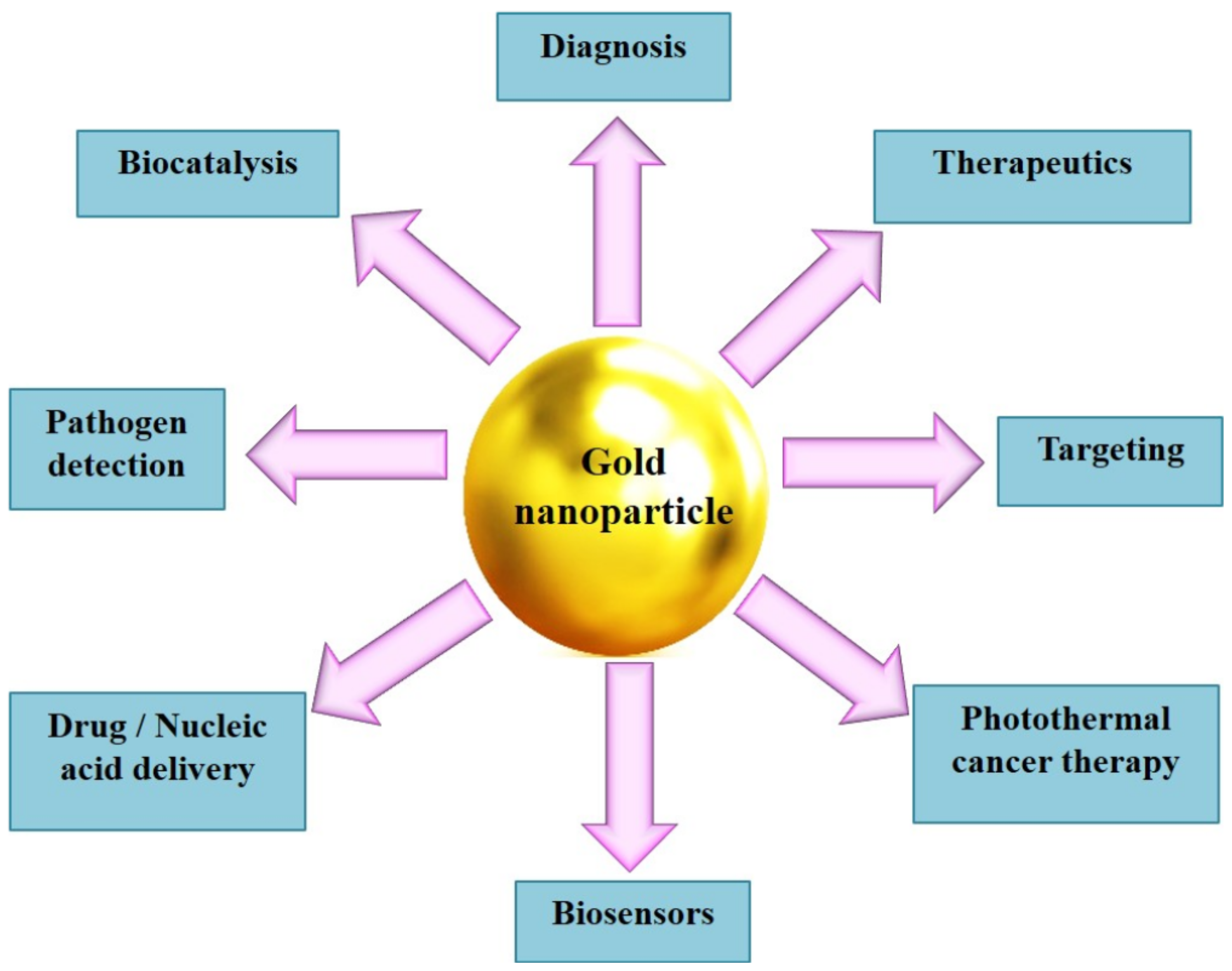
7. Biosynthesized AuNPs in Cancer Theranostics: Imaging, Drug Delivery, and Treatment

Cancer is a disease characterized by abnormal and unrestricted cell growth with potential to spread to other parts of the body. The World Health Organization (WHO) reported nearly 10 million deaths in 2020 and this is expected to increase to an estimated 12 million deaths by 2030 [129]. Among the different types of nanomaterials used to treat cancer, biosynthesized AuNPs functionalized with targeting ligands and anticancer drugs are considered as promising candidates in diagnosis and cancer therapy [24,130]. Kim et al. [131] explored this concept to prove the efficacy of AuNPs in treating glioblastoma multiforme (GBM), the most common primary grade 4 brain tumor. They demonstrated that the oral delivery of AuNPs conjugated with milk protein lactoferrin and polyethylene glycol (PEG), a biocompatible polymer, was able to cross the blood–brain barrier and bind with lactoferrin receptors that are highly expressed in the brain tumor cells in mice models. Further, when irradiated with laser light, the administered AuNPs increased the temperature in GBM due to photothermiaproperties and induced a significant reduction in the tumor volume. A schematic illustration for oral absorption of lactoferrin (Lf)–PEG-conjugated AuNPs and the mechanism of action of Lf–PEG–AuNP targeting Glioblastoma multiforme (GBM) through lactoferrin receptor pathway of the small intestine, the blood–brain barrier, and GBM cells is given in Ref. [131].
AuNPs, either individually or in combination with other treatment modalities such as radio/chemotherapy, have the ability to induce hyperthermia or deliver the drug in the targeted region or cell to produce a synergetic effect and thus help to facilitate cancer treatment. For instance, Rezaeian et al. [132] used a green chemistry approach to synthesize curcumin-coated AuNPs and performed in vitro study to the compare nanoparticle-mediated photothermal therapy and radiofrequency electric field hyperthermia on mouse colorectal cancer (CT26) cell lines. The results have shown that the NPs induced apoptosis cell death considerably using both photothermal therapy and radiofrequency electric field hyperthermia treatments. Another study used the green chemistry method to synthesize stable AuNPs coupled with 5-Fluorouracil, a chemotherapeutic drug that is widely used for the treatment of liver cancer [133]. This study focused on in vivo toxicity induced by AuNPs in the zebrafish embryo model, in vitro drug release behavior, and efficacy of the NPs in human liver cancer (HepG2) cell lines. In vivo biodistribution analysis indicated that a higher amount of AuNPs accumulated in the liver induced significant cytotoxicity in HepG2 cell lines, which signifies that the AuNPs could be used as a tool for both imaging and targeted drug delivery with minimal side effects of liver cancer. Various reports specifying anticancer activity of AuNPs biosynthesized from different plant sources are mentioned in Table 2.

8. Bioapplications of Medicinal-Plants-Based AuNPs

Medicinal plants, which are widely used in traditional health practices, play a vital role in the treatment of various kinds of human diseases. Their extracts from different plant parts such as leaves, flowers, and roots are used in the development of novel therapeutic drugs [154,155]. It is estimated that above 60% of anticancer drugs that are currently used for cancer treatment are isolated from medicinal plants and almost 3000 medicinal plants worldwide have been reported to have anticancer properties [156,157]. Medicinal plants are rich in various phytoconstituents including polyphenols, flavonoids, glycosides, terpenoids, fatty acids, alkaloids, saponins, and tannins [136,158]. When these materials are used as a source for NPs synthesis, the surface of the NPs is either coated or conjugated with these beneficial compounds. Hence, NPs synthesized using medicinal plant extracts show interesting antibacterial, anti-inflammatory, antidiabetic, and cytotoxic properties [159].
Further, AuNPs synthesized via different green techniques using environmentally friendly reagents exhibit excellent immune response regulation and efficacy in therapy against immune system-associated diseases such as cancer, inflammatory, and autoimmune diseases [160]. However, scientific research on the anticancer properties of medicinal plants is limited. The exact mechanism and the key components of the plant extracts that induce biomedical effects are not well explored. Wang et al. [137] reported the green synthesis of AuNPs using a medicinal plant Scutellaria barbata, which is widely used in the Chinese system of medicine to treat various human ailments. Biosynthesized AuNPs showed effective anticancer activity against human pancreatic cancer cell lines (PANC-1).
Geetha et al. [138] revealed a simple, cost-effective, and one-step process to biosynthesize AuNPs using flower extract of a pharmacologically important tree Couroupita guianensis, commonly known as cannon ball tree, which has innumerable medicinal applications including antibiotic, antiseptic, and anti-inflammatory activity. The anticancer potential of biosynthesized AuNPs was evaluated using a variety of techniques such as MTT assay, DNA fragmentation, apoptosis by 4,6-diamidino-2-phenylindole (DAPI), a fluorescent staining, and comet assay for DNA damage, against human leukemia cell (HL-60) line. Leukemia is a type of blood cancer and a leading cause of cancer-related mortality worldwide [161]. Becerril et al. [162] reported the green synthesis of AuNPs using the aqueous extract from Turnera diffusa, a native desert plant used for traditional medicine in Mexico. This plant has great pharmacological significance including anti-obesity, antioxidant, antibacterial, anti-inflammatory, antidiabetic, antimycotic, and cytotoxic activities. The cytotoxicity and immunomodulatory effects of AuNPs synthesized using this plant extract were investigated on the leukocytes of longfin yellowtail Seriola rivoliana, a marine fish, and antibacterial activity against Vibrio parahaemolyticus and Aeromonas hydrophila, Gram-negative bacteria. The results indicated that the AuNPs increased the phagocytosis activity, attenuated the reactive oxygen species in leukocytes production, and increased the cellular antibacterial mechanism mediated by nitric oxide production.
Yasmin et al. [163] employed green techniques for rapid synthesis of spherical AuNPs in the size range of 16–30 nm using Hibiscus rosasinensis, a medicinal plant that has a lot of beneficial applications such as anti-infectious, anthelmintic, anti-inflammatory, diuretic, and antipyretic properties. The synthesized particles were found to be stable for up to a few months and have the potential to be used for medical and biosensor applications. A recent study reported the synthesis of AuNPs using the aqueous Mentha longifolia leaf extract. The major constituents of this plant include polyphenols, alkaloids, organic acids, terpenoids, etc., and this plant has been used as an antihypertensive and antitussive drug in traditional medicine. The biosynthesized nanoparticles were found to be effective against various breast cancer cell lines, such as breast adenocarcinoma (MCF7), breast carcinoma (Hs 578Bst), breast infiltrating ductal cell carcinoma (Hs 319.T), and breast infiltrating lobular carcinoma (UACC-3133) cell lines, without causing cytotoxicity against a normal cell line (HUVEC). Therefore, AuNPs synthesized using Mentha longifolia leaf aqueous extract can be tested as an anti-breast cancer drug in humans in the near future [153]. Another report described the biogenic synthesis of AuNPs using the Jasminum auriculatum leaf extract. The leaves of this plant have numerous medicinal applications such as antilithiatic, wound healing activity, and diuretic activity. They are used in the treatment of leprosy, skin diseases, ulcers, and wounds. The MTT assay performed using these NPs against human cervical cancer (HeLa) cells revealed significant anticancer activity without the requirement of doping additional molecules [134].
Hasan et al. [135] biosynthesized AuNPs from Turbinaria decurrens, which is an Egyptian marine brown macroalga, which has a diverse group of phytochemicals with unique bioactivities and is widely used as food and medicine. The authors compared the chemical composition and antioxidant and anticancer activities of both the hydromethanolic extract (HME) of this plant and the HME–AuNPs on three different cancer cell lines (MCF-7, HEPG-2, and HCT-116) using MTT assay. The results showed the strong anticancer activity of AuNPs against all the three studied cell lines. Their findings indicated that the biosynthesized AuNPs could be used as a source for the discovery of novel therapeutic agents in the biomedical field to treat oxidative stress-related diseases, particularly cancer. Hosny et al. [136] explained a phytofabrication technique to synthesize AuNPs that remained stable for up to three months using the aqueous leaf extract of Tecoma capensis, a flowering plant commonly found in tropical and subtropical areas of Africa. The anticancer efficacy of T. capensis–AuNPs was tested against human breast cancer cell line (MCF7) using MTT assay and the results revealed the excellent potency of AuNPs in preventing the development and proliferation of MCF7 cells. All these studies explain that the synthesis of AuNPs based on medicinal plants is beneficial to express their health and medical benefits along with their multifunctional potential in treating different diseases, including cancer.

9. Challenges and Future Prospects

In the recent decade, the green synthesis approach has been successfully used to synthesize a variety of NPs, including AuNPs, with varying morphology and properties. Numerous research articles have been published worldwide on the interdisciplinary routes in the synthesis of NPs from different plants and microorganisms. However, there are several limitations and drawbacks in the green synthesis method which limit their large-scale production for commercial purposes and diminish their subsequent applications in biotechnology and nanomedicine. The current challenges are summarized below:
  • Lack of sufficient data to control the size and shape of NPs: Many reports have demonstrated the formation of AuNPs with different morphologies during the synthesis process. The formation of NPs with uniform morphology and narrow size distribution is essential for pharmacological and biological applications. Proper knowledge to optimize the reaction mixture, experimental time, pH, temperature, rotational speed, concentrations of chloroauric acid concentrations, etc., have to be taken into care to solve this problem.
  • Lack of standardized protocols to reproduce NPs with the same characteristics: NPs from different parts even of the same plant have variations in their structure and properties, as, e.g., AuNPs synthesized from different plant parts exhibit different levels of cytotoxicity due to the difference in the antioxidant/metabolite contents.
  • Lack of clear knowledge on the mechanism of NPs synthesis is a major drawback, and identification of key components present in different metabolites from plants and microbial sources that play an active role in the synthesis of NPs are challenging.
  • Separation and purification of NPs from the complex reaction mixture is another important aspect that still remains a hurdle.
  • Scalability of synthesized NPs from a laboratory approach to meet the huge demands in the industrial and pharmaceutical scale is a major concern.
  • A detailed toxicological study of the biosynthesized NPs is crucial to enhance their scope in diverse fields.
  • Technical barriers and regulatory policies for the commercial synthesis of NPs limit the scale-up process, which needs to be overcome.
  • Stability and functionalization: More research on green synthesis is required to surface-modify the synthesized NPs to improve their stability in biological media and functionalize them with specific antibodies or peptides to improve their applications in drug delivery and cancer therapy.
Even though the green synthesis approach is very popular, overcoming the above challenges could improve the global acceptability and adaptability of the commercial synthesis of AuNPs. Future research and development in this sector should be directed towards overcoming the present hurdles and coming up with novel standardized protocols, designing smart and safe AuNPs functionalized with different biomolecules, and targeting moieties for multifunctional applications.

10. Conclusions

Due to their unique characteristics, AuNPs have enormous applications in various fields such as electronics, catalysis, optics, sensors, and biology. Though different physicochemical methods are used in the synthesis of NPs, the green synthesis method is a promising approach to produce different types of NPs, including gold nanomaterials, in a simple, eco-friendly, and cost-effective manner. This method has several advantages over the conventional physical and chemical methods used for NPs synthesis, such as safety, and does not involve the use of hazardous chemicals or the addition of external harmful substances during the synthesis/durability of NPs. Different parts of plants and a variety of microbes, including bacteria, fungi, algae, and yeast, are used as a natural source in the biosynthesis process. Further, NPs synthesized using biological extracts have several beneficial properties, such as high anticancer, antimicrobial, anti-inflammatory, antioxidant, and catalytic activity, etc., which find exciting applications in nanomedicine.
This review article focuses on the “state-of-the-art” research on the “green synthesis” of AuNPs, different sources of green materials, with special emphasis on biosynthesis of NPs from different parts of the plant, intracellular and extracellular synthesis from microbes, and their overall applications. Further, the advantage of the green synthesis method over the conventional chemical synthesis methods, current challenges in this field, and the prospects are discussed in detail. Green synthesis is an emerging field and current research on the biogenesis of AuNPs, characterization, and functionalization of the synthesized particles are still in the developing phase. A thorough understanding of the basic principles of green chemistry and more research work is required to gain sufficient knowledge in this field. Addressing the current challenges in this field and overcoming them with standardized protocols and innovative techniques can revolutionize the synthesis of AuNPs on both laboratory and commercial scales.

Author Contributions

P.B.S.—writing, conceived the study design, data collection, and prepared an original draft; J.G.—review and editing manuscript; H.C.—general guidance, project directing, and manuscript revisions. All authors have read and agreed to the published version of the manuscript.

Funding

We gratefully acknowledge the financial support from the project KP-06-DB-8/01.12.2020 with the Bulgarian National Science Fund under the National Scientific Program P. Beron 2020 and Grant No. D01-229/27.10.2021 from the Ministry of Education and Science of Bulgaria.

Institutional Review Board Statement

Not applicable.

Informed Consent Statement

Not applicable.

Data Availability Statement

The data generated within this research is included in the paper.

Conflicts of Interest

The authors declare no conflict of interest.

References

  1. Sarfraz, N.; Khan, I. Plasmonic Gold Nanoparticles (AuNPs): Properties, Synthesis and their Advanced Energy, Environmental and Biomedical Applications. Chem. Asian J. 2021, 16, 720–742. [Google Scholar] [CrossRef] [PubMed]
  2. Hammami, I.; Alabdallah, N.M.; Jomaa, A.A.; Kamoun, M. Gold nanoparticles: Synthesis properties and applications. J. King Saud-Univ. Sci. 2021, 33, 101560. [Google Scholar] [CrossRef]
  3. Amina, S.J.; Guo, B. Review on the Synthesis and Functionalization of Gold Nanoparticles as a Drug Delivery Vehicle. Int. J. Nanomed. 2020, 15, 9823–9857. [Google Scholar] [CrossRef] [PubMed]
  4. Hu, X.; Zhang, Y.; Ding, T.; Liu, J.; Zhao, H. Multifunctional Gold Nanoparticles: A Novel Nanomaterial for Various Medical Applications and Biological Activities. Front. Bioeng. Biotechnol. 2020, 8, 990. [Google Scholar] [CrossRef] [PubMed]
  5. Menon, S.; Rajeshkumar, S.; Kumar, V. A review on biogenic synthesis of gold nanoparticles, characterization, and its applications. Resour.-Effic. Technol. 2017, 3, 516–527. [Google Scholar] [CrossRef]
  6. Tian, F.; Bonnier, F.; Casey, A.; Shanahan, A.E.; Byrne, H.J. Surface enhanced Raman scattering with gold nanoparticles: Effect of particle shape. Anal. Methods 2014, 6, 9116–9123. [Google Scholar] [CrossRef] [Green Version]
  7. Bansal, S.A.; Kumar, V.; Karimi, J.; Singh, A.P.; Kumar, S. Role of gold nanoparticles in advanced biomedical applications. Nanoscale Adv. 2020, 2, 3764–3787. [Google Scholar] [CrossRef]
  8. Aldewachi, H.; Chalati, T.; Woodroofe, M.N.; Bricklebank, N.; Sharrack, B.; Gardiner, P. Gold nanoparticle-based colorimetric biosensors. Nanoscale 2018, 10, 18–33. [Google Scholar] [CrossRef] [PubMed] [Green Version]
  9. Kulabhusan, P.K.; Tripathi, A.; Kant, K. Gold Nanoparticles and Plant Pathogens: An Overview and Prospective for Biosensing in Forestry. Sensors 2022, 22, 1259. [Google Scholar] [CrossRef]
  10. António, M.; Vitorino, R.; Daniel-da Silva, A.L. Gold nanoparticles-based assays for biodetection in urine. Talanta 2021, 230, 122345. [Google Scholar] [CrossRef] [PubMed]
  11. Sibuyi, N.R.S.; Moabelo, K.L.; Fadaka, A.O.; Meyer, S.; Onani, M.O.; Madiehe, A.M.; Meyer, M. Multifunctional Gold Nanoparticles for Improved Diagnostic and Therapeutic Applications: A Review. Nanoscale Res. Lett. 2021, 16, 174. [Google Scholar] [CrossRef] [PubMed]
  12. Kato, Y.; Kikuchi, F.; Imura, Y.; Yoshimura, E.; Suzuki, M. Various Shapes of Gold Nanoparticles Synthesized by Glycolipids Extracted from Lactobacillus casei. In Biomineralization; Endo, K., Kogure, T., Nagasawa, H., Eds.; Springer: Singapore, 2018; pp. 259–265. [Google Scholar] [CrossRef]
  13. Luo, D.; Wang, X.; Burda, C.; Basilion, J.P. Recent Development of Gold Nanoparticles as Contrast Agents for Cancer Diagnosis. Cancers 2021, 13, 1825. [Google Scholar] [CrossRef]
  14. Ko, W.C.; Wang, S.J.; Hsiao, C.Y.; Hung, C.T.; Hsu, Y.J.; Chang, D.C.; Hung, C.F. Pharmacological Role of Functionalized Gold Nanoparticles in Disease Applications. Molecules 2022, 27, 1551. [Google Scholar] [CrossRef] [PubMed]
  15. Santhosh, P.B.; Thomas, N.; Sudhakar, S.; Chadha, A.; Mani, E. Phospholipid stabilized gold nanorods: Towards improved colloidal stability and biocompatibility. Phys. Chem. Chem. Phys. 2017, 19, 18494–18504. [Google Scholar] [CrossRef] [PubMed]
  16. Zhang, L.; Han, F. Protein coated gold nanoparticles as template for the directed synthesis of highly fluorescent gold nanoclusters. Nanotechnology 2018, 29, 165702. [Google Scholar] [CrossRef] [PubMed]
  17. Vines, J.B.; Yoon, J.H.; Ryu, N.E.; Lim, D.J.; Park, H. Gold Nanoparticles for Photothermal Cancer Therapy. Front. Chem. 2019, 7, 167. [Google Scholar] [CrossRef] [PubMed] [Green Version]
  18. Mandhata, C.P.; Sahoo, C.R.; Padhy, R.N. Biomedical Applications of Biosynthesized Gold Nanoparticles from Cyanobacteria: An Overview. Biol. Trace Elem. Res. 2022. [Google Scholar] [CrossRef]
  19. Lee, K.X.; Shameli, K.; Yew, Y.P.; Teow, S.Y.; Jahangirian, H.; Rafiee-Moghaddam, R.; Webster, T.J. Recent Developments in the Facile Bio-Synthesis of Gold Nanoparticles (AuNPs) and Their Biomedical Applications. Int. J. Nanomed. 2020, 15, 275–300. [Google Scholar] [CrossRef] [PubMed]
  20. Dong, J.; Carpinone, P.L.; Pyrgiotakis, G.; Demokritou, P.; Moudgil, B.M. Synthesis of Precision Gold Nanoparticles Using Turkevich Method. KONA Powder Part. J. 2020, 37, 224–232. [Google Scholar] [CrossRef] [PubMed] [Green Version]
  21. Jia, Y.P.; Shi, K.; Liao, J.F.; Peng, J.R.; Hao, Y.; Qu, Y.; Chen, L.J.; Liu, L.; Yuan, X.; Qian, Z.Y.; et al. Effects of Cetyltrimethylammonium Bromide on the Toxicity of Gold Nanorods Both In Vitro and In Vivo: Molecular Origin of Cytotoxicity and Inflammation. Small Methods 2020, 4, 1900799. [Google Scholar] [CrossRef]
  22. Locatelli, E.; Monaco, I.; Franchini, M.C. Surface modifications of gold nanorods for applications in nanomedicine. RSC Adv. 2015, 5, 21681–21699. [Google Scholar] [CrossRef]
  23. Javed, R.; Zia, M.; Naz, S.; Aisida, S.O.; Ain, N.u.; Ao, Q. Role of capping agents in the application of nanoparticles in biomedicine and environmental remediation: Recent trends and future prospects. J. Nanobiotechnol. 2020, 18, 172. [Google Scholar] [CrossRef] [PubMed]
  24. Mikhailova, E.O. Gold Nanoparticles: Biosynthesis and Potential of Biomedical Application. J. Funct. Biomater. 2021, 12, 70. [Google Scholar] [CrossRef]
  25. Gu, X.; Xu, Z.; Gu, L.; Xu, H.; Han, F.; Chen, B.; Pan, X. Preparation and antibacterial properties of gold nanoparticles: A review. Environ. Chem. Lett. 2021, 19, 167–187. [Google Scholar] [CrossRef]
  26. Chowdhury, N.K.; Choudhury, R.; Gogoi, B.; Chang, C.M.; Pandey, R.P. Microbial synthesis of gold nanoparticles and their application. Curr. Drug Targets 2022, 23. [Google Scholar] [CrossRef]
  27. Ghosh, S.; Ahmad, R.; Zeyaullah, M.; Khare, S.K. Microbial Nano-Factories: Synthesis and Biomedical Applications. Front. Chem. 2021, 9, 626834. [Google Scholar] [CrossRef]
  28. Soltys, L.; Olkhovyy, O.; Tatarchuk, T.; Naushad, M. Green Synthesis of Metal and Metal Oxide Nanoparticles: Principles of Green Chemistry and Raw Materials. Magnetochemistry 2021, 7, 145. [Google Scholar] [CrossRef]
  29. Tatarchuk, T.; Danyliuk, N.; Shyichuk, A.; Kotsyubynsky, V.; Lapchuk, I.; Mandzyuk, V. Green synthesis of cobalt ferrite using grape extract: The impact of cation distribution and inversion degree on the catalytic activity in the decomposition of hydrogen peroxide. Emergent Mater. 2021, 5, 89–103. [Google Scholar] [CrossRef]
  30. Tatarchuk, T.; Shyichuk, A.; Sojka, Z.; Gryboś, J.; Naushad, M.; Kotsyubynsky, V.; Kowalska, M.; Kwiatkowska-Marks, S.; Danyliuk, N. Green synthesis, structure, cations distribution and bonding characteristics of superparamagnetic cobalt-zinc ferrites nanoparticles for Pb(II) adsorption and magnetic hyperthermia applications. J. Mol. Liq. 2021, 328, 115375. [Google Scholar] [CrossRef]
  31. Rattan, R.; Shukla, S.; Sharma, B.; Bhat, M. A Mini-Review on Lichen-Based Nanoparticles and Their Applications as Antimicrobial Agents. Front. Microbiol. 2021, 12, 336. [Google Scholar] [CrossRef]
  32. Can, M. Green gold nanoparticles from plant-derived materials: An overview of the reaction synthesis types, conditions, and applications. Rev. Chem. Eng. 2020, 36, 859–877. [Google Scholar] [CrossRef]
  33. Teimuri-mofrad, R.; Hadi, R.; Tahmasebi, B.; Farhoudian, S.; Mehravar, M.; Nasiri, R. Green synthesis of gold nanoparticles using plant extract: Mini-review. Nanochem. Res. 2017, 2, 8–19. [Google Scholar] [CrossRef]
  34. Rajan, A.; Vilas, V.; Philip, D. Studies on catalytic, antioxidant, antibacterial and anticancer activities of biogenic gold nanoparticles. J. Mol. Liq. 2015, 212, 331–339. [Google Scholar] [CrossRef]
  35. Yang, N.; WeiHong, L.; Hao, L. Biosynthesis of Au nanoparticles using agricultural waste mango peel extract and its in vitro cytotoxic effect on two normal cells. Mater. Lett. 2014, 134, 67–70. [Google Scholar] [CrossRef]
  36. Khalil, M.M.H.; Ismail, E.H.; El-Magdoub, F. Biosynthesis of Au nanoparticles using olive leaf extract: 1st Nano Updates. Arab. J. Chem. 2012, 5, 431–437. [Google Scholar] [CrossRef] [Green Version]
  37. Sujitha, M.V.; Kannan, S. Green synthesis of gold nanoparticles using Citrus fruits (Citrus limon, Citrus reticulata and Citrus sinensis) aqueous extract and its characterization. Spectrochim. Acta A Part Mol. Biomol. Spectrosc. 2013, 102, 15–23. [Google Scholar] [CrossRef]
  38. Abhijith, K.S.; Thakur, M.S. Application of green synthesis of gold nanoparticles for sensitive detection of aflatoxin B1 based on metal enhanced fluorescence. Anal. Methods 2012, 4, 4250–4256. [Google Scholar] [CrossRef]
  39. Vijayakumar, S.; Vaseeharan, B.; Malaikozhundan, B.; Gopi, N.; Ekambaram, P.; Pachaiappan, R.; Velusamy, P.; Murugan, K.; Benelli, G.; Suresh Kumar, R.; et al. Therapeutic effects of gold nanoparticles synthesized using Musa paradisiaca peel extract against multiple antibiotic resistant Enterococcus faecalis biofilms and human lung cancer cells (A549). Microb. Pathog. 2017, 102, 173–183. [Google Scholar] [CrossRef]
  40. Velmurugan, P.; Anbalagan, K.; Manosathyadevan, M.; Lee, K.J.; Cho, M.; Lee, S.M.; Park, J.H.; Oh, S.G.; Bang, K.S.; Oh, B.T. Green synthesis of silver and gold nanoparticles using Zingiber officinale root extract and antibacterial activity of silver nanoparticles against food pathogens. Bioprocess Biosyst. Eng. 2014, 37, 1935–1943. [Google Scholar] [CrossRef]
  41. Fazal, S.; Jayasree, A.; Sasidharan, S.; Koyakutty, M.; Nair, S.V.; Menon, D. Green Synthesis of Anisotropic Gold Nanoparticles for Photothermal Therapy of Cancer. ACS Appl. Mater. Interfaces 2014, 6, 8080–8089. [Google Scholar] [CrossRef]
  42. Yuan, C.G.; Huo, C.; Yu, S.; Gui, B. Biosynthesis of gold nanoparticles using Capsicum annuum var. grossum pulp extract and its catalytic activity. Phys. E Low-Dimens. Syst. Nanostruct. 2017, 85, 19–26. [Google Scholar] [CrossRef]
  43. Yu, J.; Xu, D.; Guan, H.N.; Wang, C.; Huang, L.K.; Chi, D.F. Facile one-step green synthesis of gold nanoparticles using Citrus maxima aqueous extracts and its catalytic activity. Mater. Lett. 2016, 166, 110–112. [Google Scholar] [CrossRef]
  44. Geethalakshmi, R.; Sarada, D. Characterization and antimicrobial activity of gold and silver nanoparticles synthesized using saponin isolated from Trianthema decandra L. Ind. Crop. Prod. 2013, 51, 107–115. [Google Scholar] [CrossRef]
  45. Poojary, M.M.; Passamonti, P.; Adhikari, A.V. Green Synthesis of Silver and Gold Nanoparticles Using Root Bark Extract of Mammea suriga: Characterization, Process Optimization, and Their Antibacterial Activity. BioNanoScience 2016, 6, 110–120. [Google Scholar] [CrossRef]
  46. Jayaseelan, C.; Ramkumar, R.; Rahuman, A.A.; Perumal, P. Green synthesis of gold nanoparticles using seed aqueous extract of Abelmoschus esculentus and its antifungal activity. Ind. Crop. Prod. 2013, 45, 423–429. [Google Scholar] [CrossRef]
  47. Suresh, A.K.; Pelletier, D.A.; Wang, W.; Broich, M.L.; Moon, J.W.; Gu, B.; Allison, D.P.; Joy, D.C.; Phelps, T.J.; Doktycz, M.J. Biofabrication of discrete spherical gold nanoparticles using the metal-reducing bacterium Shewanella oneidensis. Acta Biomater. 2011, 7, 2148–2152. [Google Scholar] [CrossRef]
  48. Vinay Gopal, J.; Thenmozhi, M.; Kannabiran, K.; Rajakumar, G.; Velayutham, K.; Rahuman, A.A. Actinobacteria mediated synthesis of gold nanoparticles using Streptomyces sp. VITDDK3 and its antifungal activity. Mater. Lett. 2013, 93, 360–362. [Google Scholar] [CrossRef]
  49. Bennur, T.; Khan, Z.; Kshirsagar, R.; Javdekar, V.; Zinjarde, S. Biogenic gold nanoparticles from the Actinomycete Gordonia amarae: Application in rapid sensing of copper ions. Sens. Actuators B Chem. 2016, 233, 684–690. [Google Scholar] [CrossRef]
  50. Luo, P.; Liu, Y.; Xia, Y.; Xu, H.; Xie, G. Aptamer biosensor for sensitive detection of toxin A of Clostridium difficile using gold nanoparticles synthesized by Bacillus stearothermophilus. Biosens. Bioelectron. 2014, 54, 217–221. [Google Scholar] [CrossRef]
  51. Syed, B.; Prasad, N.M.N.; Satisha, S. Endogenic mediated synthesis of gold nanoparticles bearing bactericidal activity. J. Microsc. Ultrastruct. 2016, 4, 162. [Google Scholar] [CrossRef] [Green Version]
  52. Nangia, Y.; Wangoo, N.; Sharma, S.; Wu, J.S.; Dravid, V.; Shekhawat, G.S.; Raman Suri, C. Facile biosynthesis of phosphate capped gold nanoparticles by a bacterial isolate Stenotrophomonas maltophilia. Appl. Phys. Lett. 2009, 94, 233901. [Google Scholar] [CrossRef]
  53. Singh, P.; Singh, H.; Kim, Y.J.; Mathiyalagan, R.; Wang, C.; Yang, D.C. Extracellular synthesis of silver and gold nanoparticles by Sporosarcina koreensis DC4 and their biological applications. Enzym. Microb. Technol. 2016, 86, 75–83. [Google Scholar] [CrossRef] [PubMed]
  54. Das, S.K.; Das, A.R.; Guha, A.K. Gold Nanoparticles: Microbial Synthesis and Application in Water Hygiene Management. Langmuir 2009, 25, 8192–8199. [Google Scholar] [CrossRef] [PubMed]
  55. Dasaratrao Sawle, B.; Salimath, B.; Deshpande, R.; Dhondojirao Bedre, M.; Krishnamurthy Prabhakar, B.; Venkataraman, A. Biosynthesis and stabilization of Au and Au–Ag alloy nanoparticles by fungus, Fusarium semitectum. Sci. Technol. Adv. Mater. 2008, 9, 035012. [Google Scholar] [CrossRef] [PubMed]
  56. Chauhan, A.; Zubair, S.; Tufail, S.; Sherwani, A.; Sajid, M.; Raman, S.C.; Azam, A.; Owais, M. Fungus-mediated biological synthesis of gold nanoparticles: Potential in detection of liver cancer. Int. J. Nanomed. 2011, 6, 2305–2319. [Google Scholar] [CrossRef] [Green Version]
  57. Philip, D. Biosynthesis of Au, Ag and Au–Ag nanoparticles using edible mushroom extract. Spectrochim. Acta Part A Mol. Biomol. Spectrosc. 2009, 73, 374–381. [Google Scholar] [CrossRef]
  58. Kumar, S.A.; Peter, Y.A.; Nadeau, J.L. Facile biosynthesis, separation and conjugation of gold nanoparticles to doxorubicin. Nanotechnology 2008, 19, 495101. [Google Scholar] [CrossRef] [Green Version]
  59. Mishra, A.; Tripathy, S.K.; Wahab, R.; Jeong, S.H.; Hwang, I.; Yang, Y.B.; Kim, Y.S.; Shin, H.S.; Yun, S.I. Microbial synthesis of gold nanoparticles using the fungus Penicillium brevicompactum and their cytotoxic effects against mouse mayo blast cancer C2C12 cells. Appl. Microbiol. Biotechnol. 2011, 92, 617–630. [Google Scholar] [CrossRef] [PubMed]
  60. Mukherjee, P.; Ahmad, A.; Mandal, D.; Senapati, S.; Sainkar, S.R.; Khan, M.I.; Ramani, R.; Parischa, R.; Ajayakumar, P.V.; Alam, M.; et al. Bioreduction of AuCl4-Ions by the Fungus, Verticillium sp. and Surface Trapping of the Gold Nanoparticles Formed. Angew. Chem. Int. Ed. 2001, 40, 3585–3588. [Google Scholar] [CrossRef]
  61. Sarkar, J.; Ray, S.; Chattopadhyay, D.; Laskar, A.; Acharya, K. Mycogenesis of gold nanoparticles using a phytopathogen Alternaria alternata. Bioprocess Biosyst. Eng. 2012, 35, 637–643. [Google Scholar] [CrossRef] [PubMed]
  62. Gericke, M.; Pinches, A. Microbial production of gold nanoparticles. Gold Bull. 2006, 39, 22–28. [Google Scholar] [CrossRef] [Green Version]
  63. Shankar, S.S.; Ahmad, A.; Pasricha, R.; Khan, M.I.; Kumar, R.; Sastry, M. Immobilization of biogenic gold nanoparticles in thermally evaporated fatty acid and amine thin films. J. Colloid Interface Sci. 2004, 274, 69–75. [Google Scholar] [CrossRef]
  64. Zhang, X.; He, X.; Wang, K.; Yang, X. Different Active Biomolecules Involved in Biosynthesis of Gold Nanoparticles by Three Fungus Species. J. Biomed. Nanotechnol. 2011, 7, 245–254. [Google Scholar] [CrossRef]
  65. Gericke, M.; Pinches, A. Biological synthesis of metal nanoparticles. Hydrometallurgy 2006, 83, 132. [Google Scholar] [CrossRef]
  66. Pimprikar, P.S.; Joshi, S.S.; Kumar, A.R.; Zinjarde, S.S.; Kulkarni, S.K. Influence of biomass and gold salt concentration on nanoparticle synthesis by the tropical marine yeast Yarrowia lipolytica NCIM 3589. Colloids Surfaces B Biointerfaces 2009, 74, 309–316. [Google Scholar] [CrossRef] [PubMed]
  67. Naveena, B.; Prakash, S. Biological Synthesis of Gold Nanoparticles Using Marine Algae Gracilaria Corticata and its Application as a Potent Antimicrobial and Antioxidant Agent. Asian J. Pharm. Clin. Res. 2013, 6, 179. [Google Scholar]
  68. Ogi, T.; Saitoh, N.; Nomura, T.; Konishi, Y. Room-temperature synthesis of gold nanoparticles and nanoplates using Shewanella algae cell extract. J. Nanopart. Res. 2010, 12, 2531–2539. [Google Scholar] [CrossRef]
  69. Usman, A.I.; Aziz, A.A.; Noqta, O.A. Application of Green Synthesis of Gold Nanoparticles: A Review. J. Teknol. 2019, 81, 171–182. [Google Scholar] [CrossRef] [Green Version]
  70. Nadeem, M.; Abbasi, B.H.; Younas, M.; Ahmad, W.; Khan, T. A review of the green syntheses and anti-microbial applications of gold nanoparticles. Green Chem. Lett. Rev. 2017, 10, 216–227. [Google Scholar] [CrossRef] [Green Version]
  71. Kuppusamy, P.; Yusoff, M.M.; Maniam, G.P.; Govindan, N. Biosynthesis of metallic nanoparticles using plant derivatives and their new avenues in pharmacological applications—An updated report. Saudi Pharm. J. 2016, 24, 473–484. [Google Scholar] [CrossRef]
  72. Ahmed, S.; Annu; Ikram, S.; Yudha, S.S. Biosynthesis of gold nanoparticles: A green approach. J. Photochem. Photobiol. B Biol. 2016, 161, 141–153. [Google Scholar] [CrossRef] [PubMed]
  73. Singh, J.; Dutta, T.; Kim, K.H.; Rawat, M.; Samddar, P.; Kumar, P. ‘Green’ synthesis of metals and their oxide nanoparticles: Applications for environmental remediation. J. Nanobiotechnol. 2018, 16, 84. [Google Scholar] [CrossRef] [PubMed]
  74. Drummer, S.; Madzimbamuto, T.; Chowdhury, M. Green Synthesis of Transition-Metal Nanoparticles and Their Oxides: A Review. Materials 2021, 14, 2700. [Google Scholar] [CrossRef] [PubMed]
  75. Islam, N.U.; Jalil, K.; Shahid, M.; Rauf, A.; Muhammad, N.; Khan, A.; Shah, M.R.; Khan, M.A. Green synthesis and biological activities of gold nanoparticles functionalized with Salix alba. Arab. J. Chem. 2019, 12, 2914–2925. [Google Scholar] [CrossRef] [Green Version]
  76. Narayanan, K.B.; Sakthivel, N. Coriander leaf mediated biosynthesis of gold nanoparticles. Mater. Lett. 2008, 62, 4588–4590. [Google Scholar] [CrossRef]
  77. Boruah, S.K.; Boruah, P.K.; Sarma, P.; Medhi, C.; Medhi, O.K. Green Synthesis Of Gold Nanoparticles Using Camellia Sinensis And Kinetics Of The Reaction. Adv. Mater. Lett. 2012, 3, 481–486. [Google Scholar] [CrossRef]
  78. Chen, J.; Li, Y.; Fang, G.; Cao, Z.; Shang, Y.; Alfarraj, S.; Ali Alharbi, S.; Li, J.; Yang, S.; Duan, X. Green synthesis, characterization, cytotoxicity, antioxidant, and anti-human ovarian cancer activities of Curcumae kwangsiensis leaf aqueous extract green-synthesized gold nanoparticles. Arab. J. Chem. 2021, 14, 103000. [Google Scholar] [CrossRef]
  79. Chahardoli, A.; Karimi, N.; Sadeghi, F.; Fattahi, A. Green approach for synthesis of gold nanoparticles from Nigella arvensis leaf extract and evaluation of their antibacterial, antioxidant, cytotoxicity and catalytic activities. Artif. Cells Nanomed. Biotechnol. 2018, 46, 579–588. [Google Scholar] [CrossRef] [PubMed] [Green Version]
  80. Bahram, M.; Mohammadzadeh, E. Green synthesis of gold nanoparticles with willow tree bark extract: A sensitive colourimetric sensor for cysteine detection. Anal. Methods 2014, 6, 6916–6924. [Google Scholar] [CrossRef]
  81. Rodríguez-León, E.; Rodríguez-Vázquez, B.E.; Martínez-Higuera, A.; Rodríguez-Beas, C.; Larios-Rodríguez, E.; Navarro, R.E.; López-Esparza, R.; Iñiguez-Palomares, R.A. Synthesis of Gold Nanoparticles Using Mimosa tenuiflora Extract, Assessments of Cytotoxicity, Cellular Uptake, and Catalysis. Nanoscale Res. Lett. 2019, 14, 334. [Google Scholar] [CrossRef] [PubMed] [Green Version]
  82. ElMitwalli, O.S.; Barakat, O.A.; Daoud, R.M.; Akhtar, S.; Henari, F.Z. Green synthesis of gold nanoparticles using cinnamon bark extract, characterization, and fluorescence activity in Au/eosin Y assemblies. J. Nanopart. Res. 2020, 22, 309. [Google Scholar] [CrossRef]
  83. Wang, B.; Yang, G.; Chen, J.; Fang, G. Green Synthesis and Characterization of Gold Nanoparticles Using Lignin Nanoparticles. Nanomaterials 2020, 10, 1869. [Google Scholar] [CrossRef]
  84. Parida, U.K.; Bindhani, B.K.; Nayak, P. Green Synthesis and Characterization of Gold Nanoparticles Using Onion (Allium cepa) Extract. World J. Nano Sci. Eng. 2011, 1, 93–98. [Google Scholar] [CrossRef] [Green Version]
  85. Xin Lee, K.; Shameli, K.; Miyake, M.; Kuwano, N.; Bt Ahmad Khairudin, N.B.; Bt Mohamad, S.E.; Yew, Y.P. Green Synthesis of Gold Nanoparticles Using Aqueous Extract of Garcinia mangostana Fruit Peels. J. Nanomater. 2016, 2016, e8489094. [Google Scholar] [CrossRef] [Green Version]
  86. Bogireddy, N.K.R.; Pal, U.; Gomez, L.M.; Agarwal, V. Size controlled green synthesis of gold nanoparticles using Coffea arabica seed extract and their catalytic performance in 4-nitrophenol reduction. RSC Adv. 2018, 8, 24819–24826. [Google Scholar] [CrossRef] [Green Version]
  87. Oueslati, M.H.; Ben Tahar, L.; Harrath, A.H. Synthesis of ultra-small gold nanoparticles by polyphenol extracted from Salvia officinalis and efficiency for catalytic reduction of p-nitrophenol and methylene blue. Green Chem. Lett. Rev. 2020, 13, 18–26. [Google Scholar] [CrossRef] [Green Version]
  88. Anbu, P.; Gopinath, S.C.; Jayanthi, S. Synthesis of gold nanoparticles using Platycodon grandiflorum extract and its antipathogenic activity under optimal conditions. Nanomater. Nanotechnol. 2020, 10, 1–9. [Google Scholar] [CrossRef]
  89. Sett, A.; Gadewar, M.; Sharma, P.; Deka, M.; Bora, U. Green synthesis of gold nanoparticles using aqueous extract of Dillenia indica. Adv. Nat. Sci. Nanosci. Nanotechnol. 2016, 7, 025005. [Google Scholar] [CrossRef]
  90. Elia, P.; Zach, R.; Hazan, S.; Kolusheva, S.; Porat, Z.; Zeiri, Y. Green synthesis of gold nanoparticles using plant extracts as reducing agents. Int. J. Nanomed. 2014, 9, 4007–4021. [Google Scholar] [CrossRef] [Green Version]
  91. Chen, R.; Wu, J.; Li, H.; Cheng, G.; Lu, Z.; Che, C.M. Fabrication of gold nanoparticles with different morphologies in HEPES buffer. Rare Met. 2010, 29, 180–186. [Google Scholar] [CrossRef] [Green Version]
  92. Tsuji, M.; Miyamae, N.; Hashimoto, M.; Nishio, M.; Hikino, S.; Ishigami, N.; Tanaka, I. Shape and size controlled synthesis of gold nanocrystals using oxidative etching by AuCl4 and Cl anions in microwave-polyol process. Colloids Surfaces A Physicochem. Eng. Asp. 2007, 302, 587–598. [Google Scholar] [CrossRef]
  93. Rao, Y.; Inwati, G.K.; Singh, M. Green synthesis of capped gold nanoparticles and their effect on Gram-positive and Gram-negative bacteria. Future Sci. OA 2017, 3, FSO239. [Google Scholar] [CrossRef] [Green Version]
  94. Sharma, N.; Pinnaka, A.K.; Raje, M.; FNU, A.; Bhattacharyya, M.S.; Choudhury, A.R. Exploitation of marine bacteria for production of gold nanoparticles. Microb. Cell Factories 2012, 11, 86. [Google Scholar] [CrossRef] [Green Version]
  95. Akintelu, S.A.; Yao, B.; Folorunso, A.S. Bioremediation and pharmacological applications of gold nanoparticles synthesized from plant materials. Heliyon 2021, 7, e06591. [Google Scholar] [CrossRef]
  96. Siddiqi, K.S.; Husen, A. Fabrication of Metal Nanoparticles from Fungi and Metal Salts: Scope and Application. Nanoscale Res. Lett. 2016, 11, 98. [Google Scholar] [CrossRef] [Green Version]
  97. Francis, S.; Koshy, E.P.; Mathew, B. Microwave aided and plant reduced gold nanoparticles as talented dye degradation catalysts. Sci. Iran. 2019, 26, 1944–1950. [Google Scholar] [CrossRef] [Green Version]
  98. Rautray, S.; Rajananthini, A.U. Therapeutic Potential of Green, Synthesized Gold Nanoparticles. BioPharm. Int. 2020, 33, 30–38. [Google Scholar]
  99. Vaishaly, A.; Mathew, B.; Krishnamurthy, N.; Krishnamurthy, T. Bioaccumulation of heavy metals by fungi. Int. J. Environ. Chem. Chromatogr. 2015, 1, 15–21. [Google Scholar]
  100. Dikshit, P.K.; Kumar, J.; Das, A.K.; Sadhu, S.; Sharma, S.; Singh, S.; Gupta, P.K.; Kim, B.S. Green Synthesis of Metallic Nanoparticles: Applications and Limitations. Catalysts 2021, 11, 902. [Google Scholar] [CrossRef]
  101. Soni, N.; Prakash, S. Synthesis of gold nanoparticles by the fungus Aspergillus niger and its efficacy against mosquito larvae. RIP 2012, 2, 1–7. [Google Scholar] [CrossRef] [Green Version]
  102. Du, L.; Xian, L.; Feng, J.X. Rapid extra-/intracellular biosynthesis of gold nanoparticles by the fungus Penicillium sp. J. Nanopart. Res. 2011, 13, 921–930. [Google Scholar] [CrossRef]
  103. Verma, V.C.; Kharwar, R.N.; Singh, S.K.; Solanki, R.; Prakash, S. Erratum to: Biofabrication of anisotropic gold nanotriangles using extract of endophytic Aspergillus clavatus as a dual functional reductant and stabilizer. Nanoscale Res. Lett. 2011, 6, 261. [Google Scholar] [CrossRef] [PubMed] [Green Version]
  104. Priyadarshini, E.; Pradhan, N.; Sukla, L.B.; Panda, P.K. Controlled Synthesis of Gold Nanoparticles Using Aspergillus terreus IF0 and Its Antibacterial Potential against Gram Negative Pathogenic Bacteria. J. Nanotechnol. 2014, 2014, e653198. [Google Scholar] [CrossRef] [Green Version]
  105. Nguyen, T.H.A.; Nguyen, V.C.; Phan, T.N.H.; Le, V.T.; Vasseghian, Y.; Trubitsyn, M.A.; Nguyen, A.T.; Chau, T.P.; Doan, V.D. Novel biogenic silver and gold nanoparticles for multifunctional applications: Green synthesis, catalytic and antibacterial activity, and colorimetric detection of Fe(III) ions. Chemosphere 2022, 287, 132271. [Google Scholar] [CrossRef]
  106. Sastry, M.; Ahmad, A.; Khan, M.; Kumar, R. Biosynthesis of metal nanoparticles using fungi and actinomycete. Curr. Sci. 2003, 85, 162–170. [Google Scholar]
  107. Dhanasekar, N.N.; Rahul, G.R.; Narayanan, K.B.; Sakthivel, G.R.a.N. Green Chemistry Approach for the Synthesis of Gold Nanoparticles Using the Fungus Alternaria sp. J. Microbiol. Biotechnol. 2015, 25, 1129–1135. [Google Scholar] [CrossRef]
  108. Molnár, Z.; Bódai, V.; Szakacs, G.; Erdélyi, B.; Fogarassy, Z.; Sáfrán, G.; Varga, T.; Kónya, Z.; Tóth-Szeles, E.; Szűcs, R.; et al. Green synthesis of gold nanoparticles by thermophilic filamentous fungi. Sci. Rep. 2018, 8, 3943. [Google Scholar] [CrossRef]
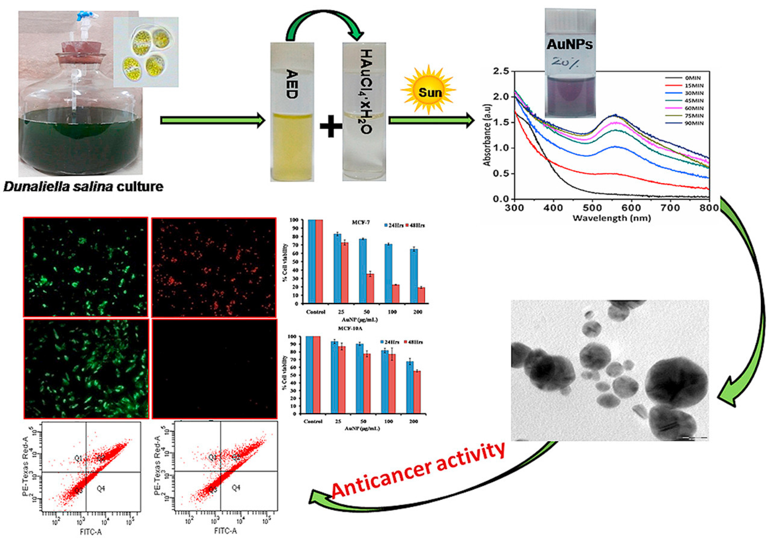
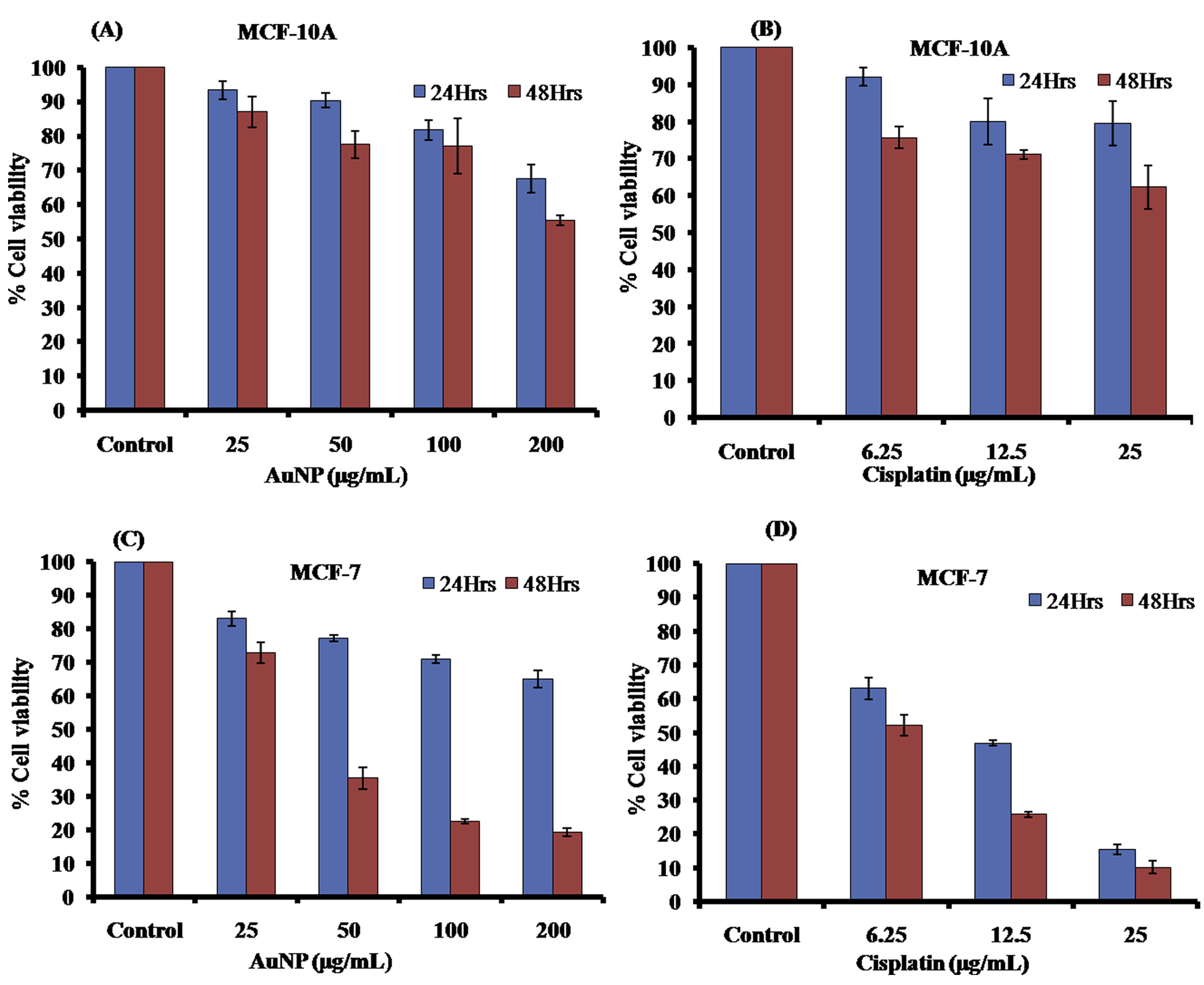
  109. Singh, A.K.; Tiwari, R.; Singh, V.K.; Singh, P.; Khadim, S.R.; Singh, U.; Laxmi; Srivastava, V.; Hasan, S.H.; Asthana, R.K. Green synthesis of gold nanoparticles from Dunaliella salina, its characterization and in vitro anticancer activity on breast cancer cell line. J. Drug Deliv. Sci. Technol. 2019, 51, 164–176. [Google Scholar] [CrossRef]
  110. Chellapandian, C.; Ramkumar, B.; Puja, P.; Shanmuganathan, R.; Pugazhendhi, A.; Kumar, P. Gold nanoparticles using red seaweed Gracilaria verrucosa: Green synthesis, characterization and biocompatibility studies. Process Biochem. 2019, 80, 58–63. [Google Scholar] [CrossRef]
  111. Jorquera, M.; Martínez, O.; Maruyama, F.; Marschner, P.; Mora, M.d.l.L. Current and Future Biotechnological Applications of Bacterial Phytases and Phytase-Producing Bacteria. Microbes Environ. 2008, 23, 182–191. [Google Scholar] [CrossRef] [Green Version]
  112. Grasso, G.; Zane, D.; Dragone, R. Microbial Nanotechnology: Challenges and Prospects for Green Biocatalytic Synthesis of Nanoscale Materials for Sensoristic and Biomedical Applications. Nanomaterials 2020, 10, 11. [Google Scholar] [CrossRef] [Green Version]
  113. Brown, S.; Sarikaya, M.; Johnson, E. A genetic analysis of crystal growth11Edited by M. Gottesman. J. Mol. Biol. 2000, 299, 725–735. [Google Scholar] [CrossRef] [PubMed]
  114. Nair, B.; Pradeep, T. Coalescence of Nanoclusters and Formation of Submicron Crystallites Assisted by Lactobacillus Strains. Cryst. Growth Des. 2002, 2, 293–298. [Google Scholar] [CrossRef]
  115. Husseiny, M.I.; El-Aziz, M.A.; Badr, Y.; Mahmoud, M.A. Biosynthesis of gold nanoparticles using Pseudomonas aeruginosa. Spectrochim. Acta Part A Mol. Biomol. Spectrosc. 2007, 67, 1003–1006. [Google Scholar] [CrossRef]
  116. Kumari, M.; Mishra, A.; Pandey, S.; Singh, S.P.; Chaudhry, V.; Mudiam, M.K.R.; Shukla, S.; Kakkar, P.; Nautiyal, C.S. Physico-Chemical Condition Optimization during Biosynthesis lead to development of Improved and Catalytically Efficient Gold Nano Particles. Sci. Rep. 2016, 6, 27575. [Google Scholar] [CrossRef]
  117. He, S.; Guo, Z.; Zhang, Y.; Zhang, S.; Wang, J.; Gu, N. Biosynthesis of gold nanoparticles using the bacteria Rhodopseudomonas capsulata. Mater. Lett. 2007, 61, 3984–3987. [Google Scholar] [CrossRef]
  118. Sathiyanarayanan, G.; Kiran, G.S.; Selvin, J.; Saibaba, G. Optimization of polyhydroxybutyrate production by marine Bacillus megaterium MSBN04 under solid state culture. Int. J. Biol. Macromol. 2013, 60, 253–261. [Google Scholar] [CrossRef]
  119. Nadaf, N.Y.; Kanase, S.S. Biosynthesis of gold nanoparticles by Bacillus marisflavi and its potential in catalytic dye degradation. Arab. J. Chem. 2019, 12, 4806–4814. [Google Scholar] [CrossRef] [Green Version]
  120. Ahmad, A.; Senapati, S.; Khan, M.I.; Kumar, R.; Ramani, R.; Srinivas, V.; Sastry, M. Intracellular synthesis of gold nanoparticles by a novel alkalotolerant actinomycete, Rhodococcusspecies. Nanotechnology 2003, 14, 824–828. [Google Scholar] [CrossRef]
  121. Correa-Llantén, D.N.; Muñoz-Ibacache, S.A.; Castro, M.E.; Muñoz, P.A.; Blamey, J.M. Gold nanoparticles synthesized by Geobacillus sp. strain ID17 a thermophilic bacterium isolated from Deception Island, Antarctica. Microb. Cell Factories 2013, 12, 75. [Google Scholar] [CrossRef] [Green Version]
  122. Kunoh, T.; Takeda, M.; Matsumoto, S.; Suzuki, I.; Takano, M.; Kunoh, H.; Takada, J. Green Synthesis of Gold Nanoparticles Coupled with Nucleic Acid Oxidation. ACS Sustain. Chem. Eng. 2018, 6, 364–373. [Google Scholar] [CrossRef] [Green Version]
  123. Li, J.; Li, Q.; Ma, X.; Tian, B.; Li, T.; Yu, J.; Dai, S.; Weng, Y.; Hua, Y. Biosynthesis of gold nanoparticles by the extreme bacterium Deinococcus radiodurans and an evaluation of their antibacterial properties. Int. J. Nanomed. 2016, 11, 5931–5944. [Google Scholar] [CrossRef] [PubMed] [Green Version]
  124. Gholami-Shabani, M.; Shams-Ghahfarokhi, M.; Gholami-Shabani, Z.; Akbarzadeh, A.; Riazi, G.; Ajdari, S.; Amani, A.; Razzaghi-Abyaneh, M. Enzymatic synthesis of gold nanoparticles using sulfite reductase purified from Escherichia coli: A green eco-friendly approach. Process Biochem. 2015, 50, 1076–1085. [Google Scholar] [CrossRef]
  125. Vinodhini, S.; Vithiya, B.S.M.; Prasad, T.A.A. Green synthesis of silver nanoparticles by employing the Allium fistulosum, Tabernaemontana divaricate and Basella alba leaf extracts for antimicrobial applications. J. King Saud Univ. Sci. 2022, 34, 101939. [Google Scholar] [CrossRef]
  126. Tyagi, P.K.; Gupta, S.; Tyagi, S.; Kumar, M.; Pandiselvam, R.; Daştan, S.D.; Sharifi-Rad, J.; Gola, D.; Arya, A. Green Synthesis of Iron Nanoparticles from Spinach Leaf and Banana Peel Aqueous Extracts and Evaluation of Antibacterial Potential. J. Nanomater. 2021, 2021, 4871453. [Google Scholar] [CrossRef]
  127. Iqbal, J.; Abbasi, B.A.; Yaseen, T.; Zahra, S.A.; Shahbaz, A.; Shah, S.A.; Uddin, S.; Ma, X.; Raouf, B.; Kanwal, S.; et al. Green synthesis of zinc oxide nanoparticles using Elaeagnus angustifolia L. leaf extracts and their multiple in vitro biological applications. Sci. Rep. 2021, 11, 20988. [Google Scholar] [CrossRef]
  128. Gulbagca, F.; Aygün, A.; Gülcan, M.; Ozdemir, S.; Gonca, S.; Şen, F. Green synthesis of palladium nanoparticles: Preparation, characterization, and investigation of antioxidant, antimicrobial, anticancer, and DNA cleavage activities. Appl. Organomet. Chem. 2021, 35, e6272. [Google Scholar] [CrossRef]
  129. Ferlay, J.; Colombet, M.; Soerjomataram, I.; Parkin, D.M.; Piñeros, M.; Znaor, A.; Bray, F. Cancer statistics for the year 2020: An overview. Int. J. Cancer 2021, 149, 778–789. [Google Scholar] [CrossRef]
  130. Bharadwaj, K.K.; Rabha, B.; Pati, S.; Sarkar, T.; Choudhury, B.K.; Barman, A.; Bhattacharjya, D.; Srivastava, A.; Baishya, D.; Edinur, H.A.; et al. Green Synthesis of Gold Nanoparticles Using Plant Extracts as Beneficial Prospect for Cancer Theranostics. Molecules 2021, 26, 6389. [Google Scholar] [CrossRef]
  131. Kim, H.S.; Lee, S.J.; Lee, D.Y. Milk protein-shelled gold nanoparticles with gastrointestinally active absorption for aurotherapy to brain tumor. Bioact. Mater. 2022, 8, 35–48. [Google Scholar] [CrossRef]
  132. Rezaeian, A.; Amini, S.M.; Najafabadi, M.R.H.; Farsangi, Z.J.; Samadian, H. Plasmonic hyperthermia or radiofrequency electric field hyperthermia of cancerous cells through green-synthesized curcumin-coated gold nanoparticles. Lasers Med. Sci. 2021, 37, 1333–1341. [Google Scholar] [CrossRef] [PubMed]
  133. Ganeshkumar, M.; Ponrasu, T.; Raja, M.D.; Subamekala, M.K.; Suguna, L. Green synthesis of pullulan stabilized gold nanoparticles for cancer targeted drug delivery. Spectrochim. Acta Part A Mol. Biomol. Spectrosc. 2014, 130, 64–71. [Google Scholar] [CrossRef] [PubMed]
  134. Balasubramanian, S.; Kala, S.M.J.; Pushparaj, T.L. Biogenic synthesis of gold nanoparticles using Jasminum auriculatum leaf extract and their catalytic, antimicrobial and anticancer activities. J. Drug Deliv. Sci. Technol. 2020, 57, 101620. [Google Scholar] [CrossRef]
  135. Hasan, E.A.; El-Hashash, M.A.; Zahran, M.K.; El-Rafie, H.M. Comparative study of chemical composition, antioxidant and anticancer activities of both Turbinaria decurrens Bory methanol extract and its biosynthesized gold nanoparticles. J. Drug Deliv. Sci. Technol. 2022, 67, 103005. [Google Scholar] [CrossRef]
  136. Hosny, M.; Fawzy, M.; El-Badry, Y.A.; Hussein, E.E.; Eltaweil, A.S. Plant-assisted synthesis of gold nanoparticles for photocatalytic, anticancer, and antioxidant applications. J. Saudi Chem. Soc. 2022, 26, 101419. [Google Scholar] [CrossRef]
  137. Wang, L.; Xu, J.; Yan, Y.; Liu, H.; Karunakaran, T.; Li, F. Green synthesis of gold nanoparticles from Scutellaria barbata and its anticancer activity in pancreatic cancer cell (PANC-1). Artif. Cells Nanomed. Biotechnol. 2019, 47, 1617–1627. [Google Scholar] [CrossRef] [PubMed] [Green Version]
  138. Geetha, R.; Ashokkumar, T.; Tamilselvan, S.; Govindaraju, K.; Sadiq, M.; Singaravelu, G. Green synthesis of gold nanoparticles and their anticancer activity. Cancer Nano 2013, 4, 91–98. [Google Scholar] [CrossRef] [Green Version]
  139. Muthukumar, T.; Sudhakumari; Sambandam, B.; Aravinthan, A.; Sastry, T.P.; Kim, J.H. Green synthesis of gold nanoparticles and their enhanced synergistic antitumor activity using HepG2 and MCF7 cells and its antibacterial effects. Process Biochem. 2016, 51, 384–391. [Google Scholar] [CrossRef]
  140. Reddy, M. Synthesis of gold nanoparticles from the flower extracts of tabebuia argentiea and their anticancer activity. Int. J. Pharm. Biol. Sci. 2017, 8, 379–383. [Google Scholar]
  141. Al Saqr, A.; Khafagy, E.S.; Alalaiwe, A.; Aldawsari, M.F.; Alshahrani, S.M.; Anwer, M.K.; Khan, S.; Lila, A.S.A.; Arab, H.H.; Hegazy, W.A.H. Synthesis of Gold Nanoparticles by Using Green Machinery: Characterization and In Vitro Toxicity. Nanomaterials 2021, 11, 808. [Google Scholar] [CrossRef]
  142. Dhayalan, M.; Denison, M.I.J.; Ayyar, M.; Gandhi, N.N.; Krishnan, K.; Abdulhadi, B. Biogenic synthesis, characterization of gold and silver nanoparticles from Coleus forskohlii and their clinical importance. J. Photochem. Photobiol. B Biol. 2018, 183, 251–257. [Google Scholar] [CrossRef] [PubMed]
  143. Sun, B.; Hu, N.; Han, L.; Pi, Y.; Gao, Y.; Chen, K. Anticancer activity of green synthesised gold nanoparticles from Marsdenia tenacissima inhibits A549 cell proliferation through the apoptotic pathway. Artif. Cells Nanomed. Biotechnol. 2019, 47, 4012–4019. [Google Scholar] [CrossRef] [Green Version]
  144. Santos, N.M.; Gomes, A.S.; Cavalcante, D.G.; Santos, L.F.; Teixeira, S.R.; Cabrera, F.C.; Job, A.E. Green synthesis of colloidal gold nanoparticles using latex from Hevea brasiliensis and evaluation of their in vitro cytotoxicity and genotoxicity. IET Nanobiotechnol. 2019, 13, 307–315. [Google Scholar] [CrossRef] [PubMed]
  145. Balasubramani, G.; Ramkumar, R.; Krishnaveni, N.; Pazhanimuthu, A.; Natarajan, T.; Sowmiya, R.; Perumal, P. Structural characterization, antioxidant and anticancer properties of gold nanoparticles synthesized from leaf extract (decoction) of Antigonon leptopus Hook. & Arn. J. Trace Elem. Med. Biol. 2015, 30, 83–89. [Google Scholar] [CrossRef] [PubMed]
  146. Vijayan, R.; Joseph, S.; Mathew, B. Anticancer, antimicrobial, antioxidant, and catalytic activities of green-synthesized silver and gold nanoparticles using Bauhinia purpurea leaf extract. Bioprocess Biosyst. Eng. 2019, 42, 305–319. [Google Scholar] [CrossRef] [PubMed]
  147. El-Borady, O.M.; Ayat, M.S.; Shabrawy, M.A.; Millet, P. Green synthesis of gold nanoparticles using Parsley leaves extract and their applications as an alternative catalytic, antioxidant, anticancer, and antibacterial agents. Adv. Powder Technol. 2020, 31, 4390–4400. [Google Scholar] [CrossRef]
  148. Vijayan, R.; Joseph, S.; Mathew, B. Indigofera tinctoria leaf extract mediated green synthesis of silver and gold nanoparticles and assessment of their anticancer, antimicrobial, antioxidant and catalytic properties. Artif. Cells Nanomed. Biotechnol. 2018, 46, 861–871. [Google Scholar] [CrossRef] [PubMed] [Green Version]
  149. Patil, M.P.; Bayaraa, E.; Subedi, P.; Piad, L.L.A.; Tarte, N.H.; Kim, G.D. Biogenic synthesis, characterization of gold nanoparticles using Lonicera japonica and their anticancer activity on HeLa cells. J. Drug Deliv. Sci. Technol. 2019, 51, 83–90. [Google Scholar] [CrossRef]
  150. Jeyaraj, M.; Arun, R.; Sathishkumar, G.; MubarakAli, D.; Rajesh, M.; Sivanandhan, G.; Kapildev, G.; Manickavasagam, M.; Thajuddin, N.; Ganapathi, A. An evidence on G2/M arrest, DNA damage and caspase mediated apoptotic effect of biosynthesized gold nanoparticles on human cervical carcinoma cells (HeLa). Mater. Res. Bull. 2014, 52, 15–24. [Google Scholar] [CrossRef]
  151. Arunachalam, P.K.; Arun, L.; Annamalai, D.S.K.; Arunachalam, A. Biofunctionalized Gold Nanoparticles Synthesis from Gymnema sylvestre And Its Preliminary Anticancer Activity. Int. J. Pharm. Pharm. Sci. 2014, 6, 423–430. [Google Scholar]
  152. Botteon, C.E.A.; Silva, L.B.; Ccana-Ccapatinta, G.V.; Silva, T.S.; Ambrosio, S.R.; Veneziani, R.C.S.; Bastos, J.K.; Marcato, P.D. Biosynthesis and characterization of gold nanoparticles using Brazilian red propolis and evaluation of its antimicrobial and anticancer activities. Sci. Rep. 2021, 11, 1974. [Google Scholar] [CrossRef] [PubMed]
  153. Li, S.; Al-Misned, F.A.; El-Serehy, H.A.; Yang, L. Green synthesis of gold nanoparticles using aqueous extract of Mentha Longifolia leaf and investigation of its anti-human breast carcinoma properties in the in vitro condition. Arab. J. Chem. 2021, 14, 102931. [Google Scholar] [CrossRef]
  154. Chen, X.; Martin, C.; Chen, W.; Chen, X.; Martin, C.; Chen, W. Medicinal Plant Biology: A new era for medicinal plant research. Med. Plant Biol. 2022, 1, 1. [Google Scholar] [CrossRef]
  155. Achour, S.; Chebaibi, M.; Essabouni, H.; Bourhia, M.; Ouahmane, L.; Mohammad Salamatullah, A.; A M Aboul-Soud, M.; Giesy, J.P. Ethnobotanical Study of Medicinal Plants Used as Therapeutic Agents to Manage Diseases of Humans. Evid.-Based Complement. Altern. Med. 2022, 2022, e4104772. [Google Scholar] [CrossRef]
  156. Muddapur, U.M.; Alshehri, S.; Ghoneim, M.M.; Mahnashi, M.H.; Alshahrani, M.A.; Khan, A.A.; Iqubal, S.M.S.; Bahafi, A.; More, S.S.; Shaikh, I.A.; et al. Plant-Based Synthesis of Gold Nanoparticles and Theranostic Applications: A Review. Molecules 2022, 27, 1391. [Google Scholar] [CrossRef]
  157. Tilaoui, M.; Ait Mouse, H.; Zyad, A. Update and New Insights on Future Cancer Drug Candidates From Plant-Based Alkaloids. Front. Pharmacol. 2021, 12, 719694. [Google Scholar] [CrossRef] [PubMed]
  158. Hiba, H.; Thoppil, J.E. Medicinal herbs as a panacea for biogenic silver nanoparticles. Bull. Natl. Res. Cent. 2022, 46, 9. [Google Scholar] [CrossRef]
  159. Oyenihi, O.R.; Oyenihi, A.B.; Erhabor, J.O.; Matsabisa, M.G.; Oguntibeju, O.O. Unravelling the Anticancer Mechanisms of Traditional Herbal Medicines with Metabolomics. Molecules 2021, 26, 6541. [Google Scholar] [CrossRef] [PubMed]
  160. Cai, F.; Li, S.; Huang, H.; Iqbal, J.; Wang, C.; Jiang, X. Green synthesis of gold nanoparticles for immune response regulation: Mechanisms, applications, and perspectives. J. Biomed. Mater. Res. Part A 2022, 110, 424–442. [Google Scholar] [CrossRef]
  161. Lin, X.; Wang, J.; Huang, X.; Wang, H.; Li, F.; Ye, W.; Huang, S.; Pan, J.; Ling, Q.; Wei, W.; et al. Global, regional, and national burdens of leukemia from 1990 to 2017: A systematic analysis of the global burden of disease 2017 study. Aging 2021, 13, 10468–10489. [Google Scholar] [CrossRef]
  162. Reyes-Becerril, M.; Ruvalcaba, F.; Sanchez, V.; López, M.G.; Silva-Jara, J.; Hernandez-Adame, L.; Angulo, C. Green synthesis of gold nanoparticles using Turnera diffusa Willd enhanced antimicrobial properties and immune response in Longfin yellowtail leukocytes. Aquac. Res. 2021, 52, 3391–3402. [Google Scholar] [CrossRef]
  163. Yasmin, A.; Ramesh, K.; Rajeshkumar, S. Optimization and stabilization of gold nanoparticles by using herbal plant extract with microwave heating. Nano Converg. 2014, 1, 12. [Google Scholar] [CrossRef] [PubMed] [Green Version]
Figure 1. Multifunctional applications of gold nanoparticles.
Chemistry 04 00026 g001
Figure 2. Top-down and bottom-up approaches for nanoparticles synthesis.
Chemistry 04 00026 g002
Figure 3. Different methods for synthesis of gold nanoparticles.
Chemistry 04 00026 g003
Figure 5. Schematic mechanism of (a) extracellular and (b) intracellular AuNPs biosynthesis.
Chemistry 04 00026 g005
Figure 6. (A,B) TEM images of AuNPs synthesized by leaf extract of coriander. Reprinted with permission from Ref. [76]. Copyright 2008 Elsevier.
Chemistry 04 00026 g006
Figure 7. Green synthesis of gold nanoparticles from Dunaliella salina, UV–Vis characterization spectra, and in vitro anticancer activity on breast cancer cell line. Reprinted with permission from Ref. [109]. Copyright 2019 Elsevier.
Chemistry 04 00026 g007
Figure 8. Cell viability using MTT assay after treatment with AuNPs and cisplatin on MCF 10A (A,B) and MCF 7 cell lines (C,D). Reprinted with permission from Ref. [109]. Copyright 2019 Elsevier.
Chemistry 04 00026 g008
Figure 9. Biocompatibility of HEK-239 cells with AuNPs under fluorescence microscope evidenced by Trypan blue exclusion and AO/EB staining. (a) Control cells appear green with no evidence of cell death. (b) Cells treated with AuNPs (100 μ g/mL) appear similar to control cells. Reprinted with permission from Ref. [110]. Copyright 2019 Elsevier.
Chemistry 04 00026 g009
Table 1. Green synthesis of AuNPs using different bioagents and their applications.
BioagentSize (nm)ShapeApplicationRef.
Areca catechu13.7SphericalAnticancer, Antibacterial, Antioxidant, Catalyst[34]
Mangifera indica Linn6–18SphericalDrug delivery[35]
Olive leaves50–100Spherical, Triangular, HexagonalAntioxidant[36]
Citrus limon15–80Spherical, TriangularAnticancer, Antimicrobial, Anti-inflammatory[37]
Coreopsis lanceolata20–30SphericalDetections of aflatoxins[38]
Musa paradisiaca<50Spherical, TriangularAnticancer[39]
Zingiber officinale5–20Spherical, Triangular, HexagonalAntibacterial[40]
Cocoa extract150–200Spherical, prismatic, RodPhotothermal Therapy, contrast agents[41]
Capsicum annuum var. grossum6–37Quasi-spherical, Triangular, HexagonalCatalyst[42]
Citrus maxima25SphericalCatalyst[43]
Trianthema decandra17.9–79.9Spherical, Hexagonal, CubicalAntimicrobial[44]
Mammea suriga22–50Spherical, SquareAntibacterial[45]
Abelmoschus esculentus45–75SphericalAntifungal[46]
Shewanella oneidensis2–50SphericalAntibacterial[47]
Streptomyces sp.90CubicalAntifungal[48]
Gordonia amara15–40Spherical, PolycrystallineBiosensor[49]
Bacillus stearothermophilus5–30Spherical, Triangular and otherBiosensor[50]
Pseudomonas fluorescens5–50SphericalBactericidal[51]
Stenotrophomonas malophilia40SphericalBioremediation[52]
Sporosarcina koreensis DC430–50SphericalCatalyst[53]
Rhizopus oryzae10NanocrystallinePesticides[54]
Fusarium semitectum18–50SphericalOptoelectronics[55]
Candida albicans20–80Spherical, Non-sphericalDetection of liver cancer[56]
Volvariella volvacea20–150Spherical, Triangular, HexagonalTherapeutic[57]
Helminthosporum solani2–70PolydispersedAnticancer drug[58]
Penicillium brevicompactum10–50SphericalAnticancer[59]
Verticillium sp.20SphericalBiomedical[60]
Alternaria alternata12 ± 5Spherical, TriangularBiomedical[61]
Verticillium luteoalbum<10Spherical, Triangular, HexagonalOptics and Sensor[62]
Fusarium oxysporum46–70Spherical, TriangularBiomedical[63]
Aureobasidium pullulans29 ± 6SphericalBiomedical[64]
Pichia jadinii10–100Spherical, Triangular, HexagonalOptics and Sensor[65]
Yarrowia lipolytica7.5–27Spherical, Triangular, HexagonalBiomedical[66]
Gracilaria corticata5SphericalAntibacterial, antioxidant[67]
Shewanella algae9.6–200Spherical, NanoplatesBiomedical[68]
Table 2. Green synthesis of AuNPs using different bioagents and their applications.
PlantExtract SourceAnticancer TypeSize (nm)Ref.
Jasminum auriculatumleaf extractHuman cervical cancer cell line (HeLa)8–37[134]
Turbinaria decurrenshydromethanolic extractMCF-7, HEPG-2, and HCT-116 cell lines10–19[135]
Tecoma capensisleaf extractHuman breast cancer cell line (MCF7)10–35[136]
Scutellaria barbataaqueous extract of plantHuman pancreatic cancer cell lines (PANC-1)400–1000[137]
Couroupita guianensisflower extractHuman leukemia cell line (HL-60)7–48[138]
Catharanthus roseusleaf extractMCF7 and HepG2 cell line15–28[139]
Tabebuia argentieaflower extractLiver cancer cell line (HEPG2)56[140]
Benincasa hispidaaqueous extractHuman cervical cancer cell line (HeLa)22.18 ± 2[141]
Coleous forskohliiroot extractLiver cancer cell line (HEPG2)10–30[142]
Orchidwhole plant extractBreast cancer cell line (AMJ 13)14–50[143]
Hevea brasiliensislatex extractOvarian cell line (CHO-K1)9[144]
Antigonon leptopusleaf extractHuman breast cancer (MCF-7) cells13–28[145]
Bauhinia purpurealeaf extractLung carcinoma cell line (A549)20–100[146]
Petroselinum crispumleaf extractHuman colorectal cell line (COLO- 201)20–80[147]
Indigofera tinctorialeaf extractLung carcinoma cell line (A549)6–29[148]
Lonicera 4 japonicaflower extractHuman cervical cancer cell line (HeLa)10–40[149]
Podophyllum hexandrumleaf extractHuman cervical cancer cell line (HeLa)5–35[150]
Gymnema sylvestreleaf extractHuma colorectal cancer cell line (HT29)72.8[151]
Brazilian Red Propolishydroethanolic extractBladder (T24), prostate (PC-3) cancer cell line8–15[152]
Mentha Longifolialeaf extractBreast cancer cell lines (MCF7, Hs 578Bst, Hs 319.T, UACC-3133)36.4[153]
Publisher’s Note: MDPI stays neutral with regard to jurisdictional claims in published maps and institutional affiliations.

