Review

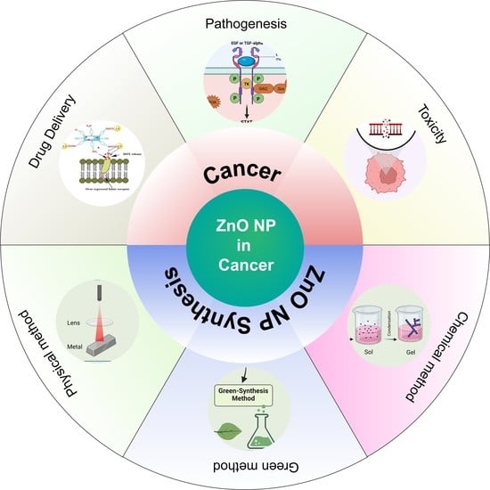
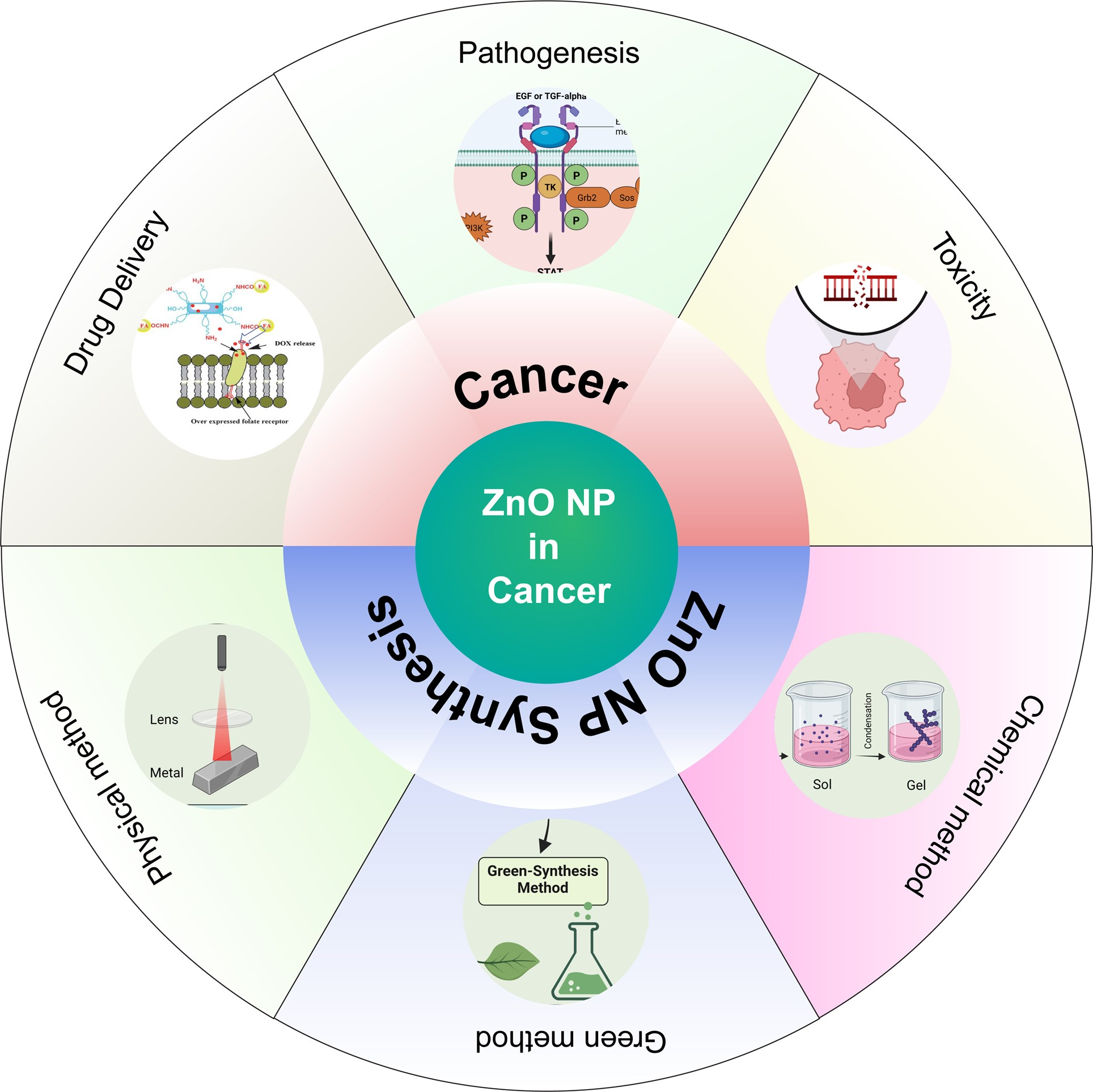
Emerging Trends in the Application of Green Synthesized Biocompatible ZnO Nanoparticles for Translational Paradigm in Cancer Therapy

1
School of Biotechnology, KIIT University, Bhubaneswar 751024, Odisha, India
2
Condensed Matter Theory Group, Materials Theory Division, Department of Physics and Astronomy, Uppsala University, Box 516, SE-75120 Uppsala, Sweden
*
Authors to whom correspondence should be addressed.
J. Nanotheranostics 2023, 4(3), 248-279; https://doi.org/10.3390/jnt4030012
Submission received: 21 May 2023 / Revised: 16 June 2023 / Accepted: 28 June 2023 / Published: 1 July 2023
(This article belongs to the Special Issue Role of Nano-Bio Interaction for Nanotheranostic Applications)

Abstract

:
Zinc oxide nanomaterials have been the cynosure of this decade because of their immense potential in different biomedical applications. It includes their usage in the prognosis and treatment of different infectious and cellular diseases, owing to their peculiar physiochemical properties such as variable shape, size, and surface charge etc. Increasing demand and usage of the ZnO nanomaterials raise concerns about their cellular and molecular toxicity and their biocompatibility with human cells. This review comprehensively details their physiochemical properties for usage in biomedical applications. Furthermore, the toxicological concerns of ZnO nanomaterials with different types of cellular systems have been reviewed. Moreover, the biomedical and biocompatible efficacy of ZnO nanomaterials for cancer specific pathways has been discussed. This review offers insights into the current scenario of ZnO nanomaterials usage and signifies their potential future extension usage on different types of biomedical and environmental applications.

Graphical Abstract

1. Introduction

Cancer is a devastating disease affecting millions of people worldwide which has necessitated the exploration of novel and effective therapeutic approaches [1]. Traditional treatments, such as chemotherapy and radiation therapy, often encounter limitations such as drug resistance and toxicity to healthy tissues [2]. Therefore, there is a pressing need to develop innovative cancer therapies that can overcome these challenges. In recent years, nanoparticles, specifically zinc oxide nanoparticles (ZnO NPs), have emerged as a promising area of research in cancer treatment [3]. ZnO NPs possess unique characteristics that make them highly suitable for cancer therapy [4]. Their high surface area-to-volume ratio enhances reactivity and improves drug delivery efficiency. Moreover, their small size enables functionalization for targeted delivery to cancer cells while sparing healthy cells from harm. Additionally, ZnO NPs exhibit photocatalytic properties, which can be harnessed for photodynamic therapy [4]. Studies have shown that ZnO NPs can inhibit the progression of cancer cells by inducing apoptosis, cell cycle arrest, and DNA damage [5]. Furthermore, they have the potential to enhance the efficacy of traditional cancer treatments, such as chemotherapy and radiation therapy, by increasing drug uptake and improving the delivery of therapeutic agents to cancer cells [5]. Although ZnO NPs offer significant promise in cancer therapy, addressing potential risks and concerns is crucial for their clinical application. One major concern is the potential toxicity of ZnO NPs, which can vary depending on size, shape, and surface charge [6]. A comprehensive understanding of the interactions between ZnO NPs and cells or tissues is still lacking, necessitating further investigation to elucidate their biological effects. Another concern is the possibility of ZnO NPs accumulating in organs and tissues, which may lead to long-term health effects [7]. In light of these considerations, it is clear that ZnO NPs have shown great potential as cancer therapies, owing to their unique properties and ability to enhance the effectiveness of conventional treatments. However, addressing the identified risks and concerns and conducting further research to fully comprehend the biological effects of ZnO NPs are imperative for the development of safe and efficient methods for cancer therapy [8].
Moreover, ZnO NPs have demonstrated great potential in drug delivery applications, capitalizing on their distinctive physicochemical characteristics, such as a large surface area to volume ratio, biocompatibility, and photostability [9]. ZnO NPs can effectively carry various drugs, including anti-cancer agents, antibiotics, and anti-inflammatory drugs. Different strategies for drug delivery use ZnO NPs, such as surface loading of drugs onto the nanoparticles and encapsulating drugs within the NPs. These approaches have shown successful delivery of drugs such as doxorubicin, paclitaxel, curcumin, ibuprofen, and methotrexate [8,10].
Furthermore, functionalizing ZnO NPs with targeting ligands, such as antibodies, peptides, or aptamers, have been explored to enhance their specificity for particular cell or tissue types, thereby improving drug efficacy and minimizing side effects [11]. The ability of ZnO NPs to deliver drugs with enhanced targeting and controlled release has the potential to revolutionize drug delivery systems, improving the delivery of drugs and reducing their adverse effects [12]. However, further research is necessary to fully understand the safety and efficacy of these approaches [13]. In recent studies, zinc oxide quantum dots (ZnO QDs) have also shown promise as pH-responsive pore-blockers and drug delivery agents [14]. These studies have demonstrated the successful doxorubicin (DOX) delivery to cancer cells using ZnO QDs, resulting in efficient drug release in intracellular compartments and increased cytotoxicity [15]. Additionally, ZnO-functionalized upconverting nano theranostic agents have shown the potential for targeted drug delivery and real-time monitoring of treatment efficacy through their multi-modality imaging capabilities.
Utilizing ZnO NPs and their derivatives in cancer therapy and drug delivery holds significant promise. These nanoparticles offer unique properties that can enhance the effectiveness of traditional treatments and enable targeted drug delivery. However, it is crucial to address potential risks and concerns associated with their use and conduct further research to fully understand their biological effects and develop safe and efficient cancer therapy and drug delivery methods. In overcoming these challenges, ZnO NPs have the potential to revolutionize cancer treatment and improve patient outcomes [16,17].

2. Zinc Oxide Nanoparticles and Their Physiochemical Properties

Owing to the unique optical properties of ZnO NPs, they have been considered an important and distinctive tool for different biomedical and environmental applications. Some of these include the following.

2.1. Size and Shape

Zinc oxide nanoparticles (ZnO NPs) have garnered significant attention due to their distinctive properties associated with their size and shape. The physical and chemical characteristics of ZnO NPs are greatly influenced by their size, shape, and surface area. Specifically, the morphological features of ZnO NPs play a crucial role in determining their optical, electronic, and magnetic properties. For instance, spherical ZnO NPs exhibit a higher surface area-to-volume ratio compared to rod-shaped ones, resulting in enhanced reactivity and light absorption capabilities. Moreover, ZnO NPs possess an elevated surface area-to-volume ratio, rendering them highly reactive and effective in diverse applications such as catalysis, sensors, and biomedical applications. However, it is important to note that the size of ZnO NPs can also impact their toxicity and biological activity, underscoring the need to optimize the nanoparticles’ size and structure for specific applications [18,19].
The characterization of ZnO NPs in different dimensions (1D, 2D, and 3D) has been categorized using various standard techniques. The details can be specified as the following.

2.1.1. One-Dimensional (1D)

ZnO NPs have unique characteristics that make them promising candidates for various applications, such as biosensors, photocatalysts, and optoelectronics [20]. The ID nanostructure of ZnO NPs includes nanowires, nanorods, nanotubes, nanoribbons, nanobelts, or nanocables [21]. To fully utilize their potential, it is essential to understand their physical and chemical properties at the nanoscale level. Transmission electron microscopy (TEM) is a powerful technique for visualizing an individual ZnO NP and determining its size and shape [22]. Scanning electron microscopy (SEM) also provides a 3D visualization of the ZnO NP and can reveal its morphology and distribution. X-ray diffraction (XRD) is a valuable tool to analyze the crystal structure of ZnO NP and determine its orientation and phase. Fourier-transform infrared (FTIR) spectroscopy helps study the chemical composition of ZnO NP, such as the presence of functional groups or defects. Raman spectroscopy is also employed to investigate the structural and vibrational properties of ZnO NP [23]. Overall, ZnO 1D characterization with various techniques is crucial in understanding the properties of ZnO NPs and optimizing their performance in various applications [24].

2.1.2. Two-Dimensional (2D)

Recently, there has been a lot of interest in ZnO NPs because of their unique properties and potential uses in several areas, including energy, biomedicine, optics, and technology. ZnO NPs have a wide range of sizes, shapes, and surface chemistries, which can significantly affect their physical and chemical properties. Characterization of ZnO NPs in 2D (structures including nanosheets, nanowalls, nanoflakes, nanoplates, nanodisks) involves understanding their size, shape, surface area, and crystal structure. TEM and SEM are two standard techniques for analyzing ZnO NPs in 2D [23]. TEM provides high-resolution images of individual NPs, allowing for accurate size and shape measurements. SEM includes information on the overall size distribution and morphology of the NPs. XRD and FTIR spectroscopy are used to determine the crystal structure and chemical composition of ZnO NPs. XRD provides information on the crystal structure and phase of the NPs, while FTIR is used to identify chemical functional groups present on the surface of the NPs. Using Brunauer–Emmett–Teller (BET) analysis, surface area analysis provides information on the specific surface area of the NPs, which is essential for applications such as catalysis and gas sensing [23]. Additionally, techniques such as DLS and zeta potential analysis can be used to determine the size distribution and surface charge of ZnO NPs in solution.

2.1.3. Three-Dimensional (3D)

The 3D nanostructures of zinc oxide nanoparticles include nanoflowers, agglomeration of nanorods, nanospheres, hollow spheres, and tetrapod shaped etc. ZnO NPs are widely used in many fields due to their unique properties, high surface area, high reactivity, and excellent photocatalytic activity. Characterizing ZnO NPs in three dimensions (3D) is crucial to understanding their properties and behavior in different environments. One of the techniques used to characterize ZnO NPs in 3D is transmission electron microscopy (TEM). TEM provides high-resolution images of the particle’s structure and morphology in 2D, and tomography can generate 3D reconstructions of the particle’s shape and size distribution. Another technique that can be used for 3D characterization is X-ray computed tomography (CT) [23]. X-ray CT is a non-destructive process that can obtain 3D descriptions of the particle’s size, shape, and distribution. This technique can also provide information about the particle’s density, porosity, and chemical composition. Additionally, confocal microscopy can be used to obtain 3D images of ZnO NPs in solution. This technique provides information about the particle’s distribution and aggregation behavior and the effect of environmental factors such as pH and temperature. The 3D characterization of ZnO NPs is essential for understanding their behavior and properties in different environments [25].
Overall, the characterization of ZnO NPs in different dimensions involves a combination of microscopy, spectroscopy, and diffraction techniques to determine their physical and chemical properties [23].

2.2. Surface Area

The surface area of ZnO NPs is crucial in determining their properties and applications. They have a large surface-to-volume ratio, and so ZnO NPs have more surface atoms available for reactions, making them more reactive than their bulk counterparts [18]. This increased surface area can enhance their catalytic, photocatalytic, and sensing properties, making them attractive for various applications. The surface area of ZnO NPs also affects their strength and biocompatibility [26]. Large surface areas can increase the surface energy and reactivity of the nanoparticles, leading to accumulation and instability. On the other hand, smaller ZnO NPs with a large surface area can interact with biological molecules and cells more efficiently, making them useful in biomedical applications [27]. For example, a study investigated the impact of surface area on the efficacy of zinc oxide nanoparticles for treating lung cancer in mice. They prepared two types of nanoparticles: large-sized particles with a low surface area, and small-sized particles with a high surface area. The mice were divided into two groups, with each group receiving one type of nanoparticle treatment. Results showed that the group treated with small-sized nanoparticles, having a higher surface area, exhibited a greater reduction in tumor size compared to the group treated with large-sized nanoparticles. This suggests that surface area influences the therapeutic effectiveness of zinc oxide nanoparticles in lung cancer treatment [28]. Moreover, the surface area of ZnO NPs can be modified through various surface modification techniques, including surface functionalization and doping, which can alter their physicochemical properties and improve their performance in specific applications. Therefore, controlling and optimizing the surface area of ZnO NPs is crucial for designing and developing their applications in various fields.

2.3. Electrical and Optical Properties

Zinc oxide nanoparticles (ZnO NPs) possess unique optical and electrical properties that make them highly desirable for various applications. Regarding optical properties, ZnO NPs have a wide bandgap, allowing them to absorb ultraviolet (UV) light effectively. These characteristics benefit applications such as photocatalysis and photovoltaics, enabling efficient solar energy conversion into electricity [29]. The optical properties of ZnO NPs can be altered by adjusting their size and form, leading to a blue shift in the absorption edge known as quantum confinement [30]. Additionally, certain sizes and shapes of ZnO NPs exhibit the surface plasmon resonance (SPR) effect, which results in strong absorption and scattering of light in the visible and near-infrared regions. This makes ZnO NPs useful in biosensing and imaging applications. The electrical properties of ZnO NPs are also important. They are semiconducting materials with a wide bandgap, capable of efficiently absorbing and emitting UV light [31]. Doping ZnO NPs with impurities can further modify their electrical properties, enabling n-type or p-type conductivity and facilitating their use in electronic devices such as transistors, LEDs, and solar cells. The size, shape, and surface area of ZnO NPs also influence their electrical characteristics, with nanorods and nanowires displaying anisotropic properties and small NPs exhibiting size-dependent electrical behavior. Through manipulating the dimension, form, and surface properties of ZnO NPs, their optical and electrical characteristics can be fine-tuned to suit various applications in fields such as photocatalysis, photovoltaics, and optoelectronics [32].

2.4. Thermal Stability

The thermal properties of ZnO NPs are essential for their applications in various fields, including electronics, catalysis, and energy storage. ZnO NPs have a high melting point of approximately 1975 °C and a low thermal conductivity, making them good thermal insulators. The thermal conductivity of ZnO NPs will be enhanced by doping with elements such as aluminum, gallium, or indium, which can also modify their electronic and optical properties [33]. These doped ZnO NPs can be used in thermoelectric applications to convert heat energy into electrical energy. Furthermore, the thermal stability of ZnO NPs can be improved by surface modification techniques such as coating with a protective layer or functionalization with organic or inorganic molecules. These modifications can also enhance their dispersion and prevent accumulation, improving thermal conductivity and stability. In addition, ZnO NPs can be used as thermal energy storage materials due to their high specific heat capacity, which can store thermal energy efficiently. This property makes them useful in solar energy storage and waste heat recovery applications. In conclusion, the thermal properties of ZnO NPs are crucial for their applications in various fields, and their properties can be tailored by doping surface modification and size control.

2.5. Magnetic Properties

ZnO NPs are not magnetic in their pure form because they have no unpaired electrons in their crystal structure. However, the magnetic characteristics of ZnO NPs can be induced by doping with magnetic ions or by forming magnetic nanocomposites with other materials. Doping ZnO NPs with magnetic ions such as iron, cobalt, or nickel can introduce magnetic moments to the crystal structure, leading to magnetic properties. These magnetic ZnO NPs can be used in magnetic data storage, sensors, and biomedical applications. In addition, magnetic nanocomposites can be formed by incorporating ZnO NPs into magnetic materials such as iron oxide or cobalt oxide. These composites exhibit unique magnetic properties that can be tailored by controlling the nanoparticles’ size, shape, and composition. Magnetic ZnO nanocomposites can be used in magnetic hyperthermia, drug delivery, and MRI [34]. Moreover, their shape and size can also influence the magnetic properties of ZnO NPs. For example, ZnO nanorods can exhibit anisotropic magnetic properties due to their elongated shape. Additionally, smaller ZnO NPs have a larger surface area-to-volume ratio, which can increase their magnetic moments and enhance their magnetic properties. The magnetic characteristics of ZnO NPs can be induced by doping, forming magnetic nanocomposites, and controlling their size and shape. These magnetic properties make ZnO NPs attractive for data storage, sensors, and biomedicine applications [33].

2.6. ROS Inducing Property

ZnO NPs have attracted significant attention as antibacterial agents due to their unique characteristics. ZnO NPs can prevent the growth of many bacteria, including Gram-positive and Gram-negative bacteria, making them an attractive alternative to conventional antibiotics. ZnO NPs’ antibacterial characteristics are primarily attributed to their ability to generate ROS upon UV or visible light exposure. ROS can cause oxidative stress and damage bacterial cells, inhibiting bacterial growth and cell death [35,36]. Moreover, ZnO NPs can also interact with bacterial cell membranes, disrupting their structure and function. ZnO NPs can penetrate the cell membrane and interact with intracellular components, and inhibit bacterial growth [37].
Furthermore, the antibacterial properties of ZnO NPs can be enhanced by size and shape control, as smaller particles and particles with a higher surface area-to-volume ratio can exhibit more significant antibacterial activity. ZnO NPs exhibit solid antibacterial properties due to their ability to generate ROS and interact with bacterial cell membranes. Both Gram-positive and Gram-negative bacteria strains are susceptible to the ZnO NPs. ZnO nanoparticles have been demonstrated to be less effective against Gram-positive bacteria, according to the available research. Gram-negative bacteria have a modest advantage in resistance because of the unique structure of their cell walls [38]. Gram-negative bacteria, in contrast to Gram-positive bacteria, have an extra outer membrane that includes lipopolysaccharides (LPS). LPS has been shown to boost bacterial resistance, especially to antibiotics, by enhancing the barrier characteristics of the outer membrane [39]. The size and shape of ZnO NPs can also influence their antibacterial activity. Gram-positive bacteria such as Bacillus subtilis and Staphylococcus aureus as well as Gram-negative bacteria such as Pseudomonas aeruginosa, Campylobacter jejuni, and Escherichia coli have been used to investigate ZnO NPs’ antibacterial activity [40]. Upon reducing the size of the ZnONP to the nano range the bacteriostatic effect was enhanced against S. aureus, E. coli, P. aeruginosa, and Pseudomonas fluorescens [41]. These properties make ZnO NPs attractive for various antibacterial applications, including wound healing, food packaging, and water purification [42]. Overall, the physical properties of ZnO NPs make them a promising material for several uses, including electronics, photonics, catalysis, and biomedical applications [43].

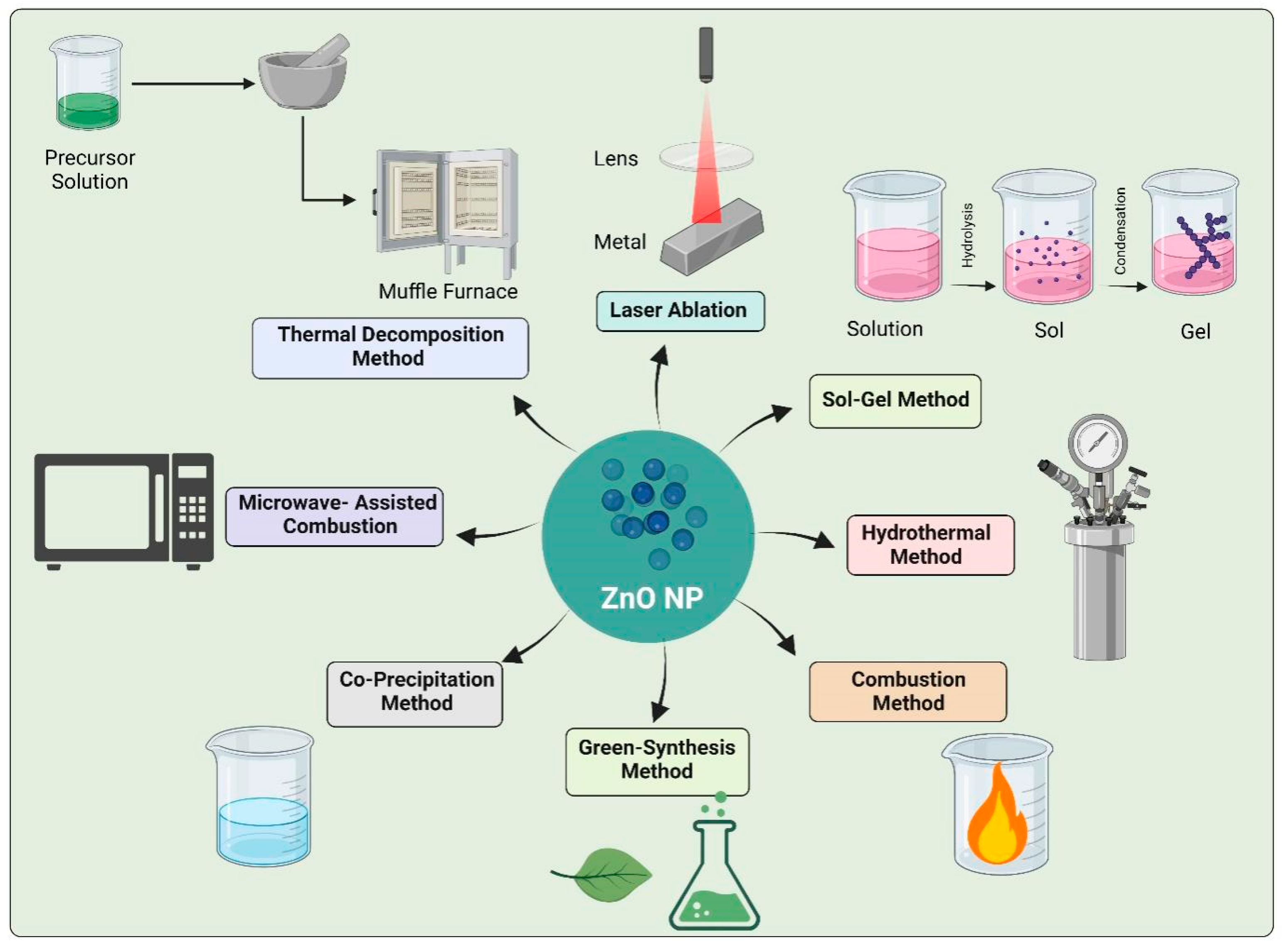
3. Synthesis of Zinc Oxide Nanoparticles

3.1. Physical Methods

There are several physical methods for synthesizing zinc oxide ZnO NPs, each with advantages and disadvantages (Figure 1). Some common types are Laser ablation: Laser ablation is a promising technique for synthesizing ZnO nanoparticles (NPs). In this method, a high-power laser is used to ablate a ZnO target in a liquid or gas environment, forming NPs. The properties of the NPs, such as dimension, form, and crystallinity, can be tuned by adjusting the laser energy, ablation time, and other parameters. One of the advantages of laser ablation is that it can produce high-quality ZnO NPs with narrow size distributions and well-controlled shapes. The resulting NPs have potential applications in various fields, including optoelectronics, catalysis, and biomedicine [44]. Moreover, laser ablation is a relatively simple and scalable method for NP synthesis. It does not require toxic chemicals or high temperatures, often used in other forms of NP synthesis, making it a more environmentally friendly approach. Laser ablation is a promising technique for synthesizing ZnO NPs with tunable properties, which could have critical applications in various fields [45]. Thermal evaporation: Thermal evaporation is a commonly used technique for synthesizing ZnO NPs. In this method, a ZnO source material is heated to a high temperature, causing it to vaporize and condense onto a substrate to form NPs. The properties of the resulting NPs can be controlled through adjusting the parameters of the thermal evaporation process, such as the temperature, pressure, and duration of the evaporation. Optimizing these parameters makes it possible to produce ZnO NPs with specific sizes, shapes, and crystalline structures. Thermal evaporation is a relatively simple and low-cost method for NP synthesis, which has been widely used in various applications, including electronics, catalysis, and biomedical engineering. However, one of the disadvantages of this method is that it may require high temperatures, which can limit the types of substrates that can be used. Overall, thermal evaporation is a promising technique for synthesizing ZnO NPs with controlled properties. However, it is essential to carefully optimize the process parameters to achieve the desired properties of the NPs [46]. Sputtering: It is a physical vapor deposition technique that can synthesize ZnO nanoparticles (NPs). In this method, a ZnO target material is bombarded with high-energy ions, causing atoms or clusters of particles to be ejected from the surface of the target and deposited onto a substrate to form NPs. The properties of the resulting NPs can be controlled through adjusting the parameters of the sputtering process, such as the gas pressure, target material, and deposition time. Optimizing these parameters makes it possible to produce ZnO NPs with specific sizes, shapes, and crystalline structures. Sputtering is a versatile and flexible method for NP synthesis, which has been used in a wide range of applications, including optoelectronics, energy, and sense. However, one of the disadvantages of this method is that it may require expensive equipment and high vacuum conditions, which can increase the cost and complexity of the process. Sputtering is a promising technique for synthesizing ZnO NPs with controlled properties [47]. However, it is essential to carefully optimize the process parameters to achieve the desired properties of the NPs (Rashid et al., 2015). Microwave-assisted method: This method is widely used for preparing nanomaterials as it has several advantages over conventional methods. One of the commonly synthesized nanoparticles utilizing this method is ZnO NP. The synthesis of ZnO NP using the microwave-assisted technique includes using microwave radiation to initiate the reaction between zinc acetate or zinc nitrate and a base such as sodium hydroxide or ammonium hydroxide. The response occurs in a suitable solvent such as water or ethanol under controlled microwave conditions such as temperature and power. One of the main advantages of this technique is the fast and efficient production of ZnO nanoparticles in a short amount of time. Depending on the reaction conditions used, the method can be completed within minutes to hours. Another advantage is the ability to obtain nanoparticles with controlled size and morphology through adjusting the reaction parameters [48].
The mechanism of the reaction involves the heating of the reactants by microwave radiation, which leads to the development of nuclei. These nuclei grow by adding precursor molecules until the nanoparticles reach their desired size. The dimension of the nanoparticles can be controlled through adjusting the power, reaction time, and concentration of the precursor molecules. Moreover, the microwave-assisted method for synthesizing ZnO nanoparticles can be scaled up quickly, making it suitable for large-scale production. Additionally, the process is a green synthesis approach as it does not require toxic solvents or harsh reaction conditions. It is a sustainable and eco-friendly method for the synthesis of nanoparticles. The microwave-assisted method is promising for synthesizing ZnO nanoparticles as it has several advantages over conventional methods. The process offers a fast, efficient, and controlled synthesis of nanoparticles with a high degree of reproducibility. Its scalability and eco-friendliness make it suitable for large-scale production of ZnO nanoparticles for various applications in nanomedicine, electronics, and energy conversion [48]. The limitations of these physical methods for synthesizing ZnO NPs include the need for expensive equipment, specialized expertise, and the potential formation of defects and impurities in the NPs. Moreover, some of these methods may generate hazardous waste and consume large amounts of energy, which can be environmentally unfriendly. Therefore, developing sustainable and green synthesis methods for ZnO NPs is essential to overcome these drawbacks and ensure the safe and efficient production of ZnO NPs for various applications.

3.2. Chemical Methods

Chemical methods for synthesizing zinc oxide (ZnO) nanoparticles (NPs) involve using chemical reagents to reduce and stabilize ZnO particles (Figure 1). The most commonly used one are Sol-gel method: This method involves mixing a ZnO precursor with a solvent to form a gel, which is then dried and calcined to produce ZnO NPs. The advantage of this method is that it can make well-controlled and highly pure NPs. However, it can be time-consuming, and solvents can be hazardous and environmentally unfriendly. Ba-Abbad et al. used Sm3+ coated ZnONP of size 12.7–18.9 nm, and zinc acetate was added to oxalic acid and samarium nitrate at 65 °C to form a mixture. The mixture was calcined at 400 °C for two hours after being oven dried at 80 °C overnight [49]. The co-precipitation method: It is a widely used method for synthesizing ZnO NPs. This method involves the precipitation of zinc and hydroxide ions in a solution under controlled conditions, forming ZnO NPs. In the co-precipitation method, a zinc precursor compound such as zinc nitrate or zinc acetate is dissolved in a solvent and mixed with a base such as sodium hydroxide or ammonium hydroxide [50]. The pH of the solution is adjusted to a specific range, typically between 7 and 11, to initiate the precipitation of the zinc ions. The resulting mixture is stirred or sonicated for a certain period to promote the formation of ZnO NPs. One of the advantages of the co-precipitation method is its simplicity and low cost. The required materials are readily available, and the synthesis process is relatively easy [50]. This method can also produce many ZnO NPs with high yields. Hydrothermal method: This method involves using high-temperature and high-pressure conditions to facilitate the nucleation and growth of ZnO nanoparticles. The hydrothermal method for synthesizing ZnO nanoparticles consists of the reaction of a zinc salt, such as zinc nitrate or zinc acetate, with a base, such as sodium hydroxide or potassium hydroxide, in the presence of water or a suitable solvent, such as ethanol or methanol. The reaction occurs in a sealed vessel, which is then heated to a high temperature (usually between 100–200 °C) for a certain amount of time (typically a few hours to a day). During the reaction, the hydrothermal conditions create a highly pressurized and heated environment that promotes the nucleation and growth of ZnO nanoparticles [51]. The size and morphology of the nanoparticles can be organized through adjusting the reaction conditions, such as the temperature, pressure, and reaction time. One of the advantages of the hydrothermal method is its ability to produce highly crystalline and uniform ZnO nanoparticles with a narrow size distribution. The high-temperature and high-pressure conditions of the reaction facilitate the formation of well-defined crystalline structures with high purity and homogeneity. Another advantage of the hydrothermal method is its versatility in producing ZnO nanoparticles with different morphologies, such as nanorods, nanowires, and nanospheres. This morphology control is achieved through adjusting the response circumstances, such as the type and concentration of the precursors, the solvent used, and the reaction temperature and time [51]. Furthermore, the hydrothermal method is a green synthesis approach since it does not require toxic solvents or harsh reaction conditions, and the by-products can be easily disposed of. This method is also easily scalable, making it suitable for the large-scale production of ZnO nanoparticles [52]. The hydrothermal technique is widely used for synthesizing ZnO nanoparticles due to its several advantages, such as producing highly crystalline and uniform nanoparticles with controlled size and morphology. The method is versatile, eco-friendly, and scalable, making it a promising approach for the large-scale production of ZnO nanoparticles for several applications in catalysis, optics, and biomedicine fields.
The limitations of these chemical methods for synthesizing ZnO NPs include the potential formation of impurities and the use of hazardous chemicals, which can be environmentally unfriendly. Moreover, some of these methods may require specialized equipment and can be time-consuming, limiting their scalability and applicability. Therefore, developing sustainable and green chemical methods for synthesizing ZnO NPs is essential to overcome these drawbacks and ensure the safe and efficient production of ZnO NPs for various applications.

3.3. Bio-Synthesis

Green synthesis of ZnO NPs is gaining attention due to the potential to produce biocompatible and environmentally friendly materials (Figure 1). Various methods have been developed to synthesize ZnO NPs using plant extracts, microorganisms, green chemistry principles, and microwave-assisted synthesis.

3.3.1. Plant-Mediated Synthesis

Plants are commonly utilized as a biological substrate to synthesize nanoparticles containing metallic ions due to their cost-effectiveness, ease of processing, and reduced toxicity compared to microorganisms [53,54,55]. The use of plant-based substrates eliminates exposure to hazardous microorganisms and the accompanying health risks and safety concerns. Obtaining plant extracts for this purpose is a straightforward process that involves exposing the plant to a solvent, such as distilled water or ethanol [56,57]. Various parts of the plant, such as leaves, roots, seeds, and fruits, have been utilized for the synthesis of zinc oxide nanoparticles [58,59]. It is well-established that plants contain high concentrations of active compounds, including methylxanthines, phenolic acids, flavonoids, and saponins, which possess antioxidant properties that can neutralize reactive oxygen species (ROS) and free radicals, as well as chelate metals [60,61]. As a result, these antioxidants are believed to play a crucial role in the green synthesis of metal and metal oxide nanoparticles, as they have the ability to reduce or chelate metal ions and serve as stabilizers for the nanoparticles produced [62,63,64]. Despite this knowledge of the phytochemical properties of antioxidants, plant extracts contain a vast array of these active compounds in varying concentrations [65]. Consequently, it can be challenging to determine the exact amount of all molecules extracted from the plant, making it difficult to define a precise mechanism for the biosynthesis of metal and metal oxide nanoparticles using plant-based substrates [66,67,68]. The proposed mechanism route for the green synthesis of ZnO NPs using plant-based substrates highlights the important role that antioxidants play in the process. Specifically, it is suggested that the active compounds present in the plant extract can react with a zinc salt to reduce or form complexes with the metal. Nava et al. proposed a mechanism route based on the chemical properties of the flavonoids, limonoids, and carotenoids that are found in the fruit peels used for obtaining the ZnO NPs. These antioxidants are believed to chelate the zinc (II) ions and form metal-coordinated complexes, which are further thermally treated to degrade the complex and form zinc oxide nanoparticles with an average size of 9.7 nm. It should be noted that while this proposed mechanism is based on experimental data, the exact mechanism for the green synthesis of ZnO NPs using plant-based substrates is still an active area of research and remains to be fully understood [69]. In 2017, Matinise and colleagues proposed a comparable route where the antioxidants present in Moringa oleifera leaves also function as chelators for zinc (II) ions, resulting in the formation of zinc oxide after calcination [70]. Fourier transform infrared spectroscopy (FTIR) was employed to analyze both the plant extract and ZnO NPs obtained through different temperature treatments (100 °C and 500 °C). The vegetal extract exhibited absorption bands characteristic of bioactive compounds, while the ZnO synthesized at 100 °C displayed hydroxyl (–OH) stretching bands that might indicate the formation of zinc complexes with antioxidants during the synthesis. These results are consistent with those of other studies in which FTIR absorption bands typical of bioactive compounds were detected in green-synthesized ZnO NPs [71]. On the other hand, studies suggest that Eclipta alba leaves can be used for producing ZnO NPs by reducing zinc (II) ions to metallic zinc instead of forming coordinated complexes with the active plant compounds. After the bio-reduction of zinc, the metallic zinc then reacts with the dissolved oxygen present in the solution to form the ZnO nuclei. The plant compounds are believed to act as stabilizers to prevent agglomeration of particles and crystal growth [72] proposed that ascorbic acid bioreduces the zinc (II) ions when using Lycopersicon esculentum extract to obtain ZnO NPs. Gupta et al. also biosynthesized ZnO NPs using plant extract and suggested that the metabolites present in the substrate are responsible for both the reduction of metallic ions and particle stabilization [73].
Plant extracts are used in the plant-mediated synthesis technique as reducing and capping agents to create ZnO NPs (refer to Table 1). The plant extracts also work as limiting agents to prevent accumulation and control the dimension and form of the nanoparticles. Various plant extracts, such as Aloe vera, Azadirachta indica, and Terminalia chebula, have synthesized ZnO NPs. This method’s advantages include using natural and renewable resources and producing NPs with tunable properties. Aminuzzaman et al. employed a Garcinia mangostana (G. mangostana) fruit pericarp to synthesize spherical ZnO nanoparticles (NPs) with an average dimension of zinc nitrate hexahydrate of 21 nm. To produce the ZnO NPs, the reaction mixture was heated in a furnace for 2 hours at 400 °C after being magnetically agitated for a short period at 70 to 80 °C [74]. Later, Sharmila et al. utilized Tecoma castanifolia leaf extract to synthesize spherical ZnO nanoparticles (NPs) with a size range of 70–75 nm from zinc sulfate after 4 days of room temperature incubation; the reaction mélange was centrifuged cleaned and desiccated in an oven to produce the desired nanoparticles [75]. Pillai et al. have discovered a new method for producing ZnO NPs with a dimension of 47 nm using a combination of zinc nitrate and plant preparations from Cinnamomum tamala, Beta vulgaris, Cinnamomum verum, and Brassica oleracea var. italica. The process involves heating the resulting product to 600 °C to create an off-white paste, which is then heated at 400 °C for 2 hours in a muffle furnace to produce the desired ZnO nanopowder. This innovative method could have significant implications for the production of ZnO NPs, as it is an eco-friendlier way of producing the material.
Green synthesized zinc oxide nanoparticles (ZnO NPs) offer several advantages, such as reduced toxicity and increased biocompatibility compared to conventional synthesized ZnO NPs. Some applications of green-synthesized ZnO NPs in cancer research are as follows.
Photodynamic Therapy (PDT): ZnO NPs synthesized via green methods can be used in PDT, a non-invasive treatment approach for cancer. ZnO NPs possess excellent photocatalytic properties and can generate reactive oxygen species (ROS) when exposed to light. Upon light irradiation, the ZnO NPs produce ROS, which can induce oxidative stress and selectively kill cancer cells, sparing healthy tissues [76]. Imaging and Theranostic: ZnO NPs synthesized using green methods can be used as contrast agents for various imaging techniques, including magnetic resonance imaging (MRI), computed tomography (CT), and fluorescence imaging. Their inherent optical and magnetic properties make them suitable for cancer imaging and theranostic applications, enabling simultaneous diagnosis and therapy [77].
Table 1. List of green synthesized ZnO nanoparticles from different plant parts.
Plants Used for Green SynthesisPlant Part Used for SynthesisMorphology of ZnO Nanoparticles Size Range (nm)References
Myristica fragransFruit ExtractsSpherical- to hexagonal-shaped particles43.3 to 83.1 nm (SEM)[78]
Cassia fistulaLeaf extracts Spherical/agglomerated0.1 nm to 10,000 nm (SEM)[79]
Melia azadarachLeaf extracts Spherical/agglomerated0.1 nm to 10,000 nm (SEM)[79]
Elaeagnus angustifoliaLeaf extracts Spherical/agglomerated~26 nm (TEM)[80]
Geranium wallichianumLeaf Extracts Hexagonal~18 nm (TEM)[81]
Euphorbia heterophylla (L.)Leaf Extracts Hexagonal~40 nm (SEM)[82]
Moringa oleiferaGum Agglomerated~60 nm (SEM)[83]
Azadirachta indicaLeaf Extracts Hexagonal10–30 nm (TEM)[84]
Azadirachta indicaLeaf Extracts Spherical18 nm (XRD)[85]
Agathosma betulinaLeaf ExtractsQuasi-spherical agglomerates15.8 nm (TEM), 12–26 nm (HRTEM)[86]
Rosa caninaFruit extractSpherical<50 nm (SEM)[87]
Cocus nuciferaCoconut waterSpherical and predominantly
hexagonal without any
agglomeration
20–80 nm (TEM), 21.2 nm (XRD)[87]
Vitex negundoFlowers Hexagonal38.17 nm (XRD) [88]
Vitex negundoLeaf ExtractsSpherical75–80 nm (SEM & EDX), 38.17nm (XRD)[88]
Solanum nigrumLeaf extract Wurtzite hexagonal, quasi-spherical20–30 nm (XRD and
FE-SEM), 29.79 nm (TEM)
[89]
GossypiumCellulosic fiber Wurtzite, spherical, nano rod13 nm (XRD)[89]
Pongamia pinnataFresh leavesSpherical, hexagonal, nano rod26 nm (XRD), Agglomeration of
100 nm (DLS, SEM, TEM)
[88]
Plectranthus amboinicusLeaf extract Rod shape nanoparticle with
agglomerates
50–180 nm (SEM)[90]
Phyllanthus niruriLeaf ExtractsHexagonal wurtzite, quasi-spherical25.61 nm (FE-SEM & XRD)[91]
Nephelium lappaceum L.Fruit peels Needle-shaped forming
agglomerate
50.95 nm (XRD)[92]
Anisochilus carnosusLeaf extract Hexagonal wurtzite, quasi-spherical20–40 nm (FE-SEM), 30–40 nm (TEM)[93]
Trifolium pratenseFlower ExtractSpherical60–70 nm (XRD)[94]
Aloe veraFreeze-dried
leaf peel
Spherical, hexagonal25–65 nm (SEM & TEM)[95]

3.3.2. Microbial-Mediated Synthesis

Microbial-mediated synthesis involves using microorganisms such as bacteria, fungi, and algae to synthesize ZnO NPs. The microbial cells secrete enzymes that act as reducing agents to transform metal ions into metal nanoparticles [96,97]. This method has the advantage of using low-cost and readily available microbial cells as reducing agents. Varying cultural circumstances and growth media can regulate the scope and shape of the ZnO NPs. Moghaddam et al. synthesized irregularly shaped zinc oxide nanoparticles (ZnO NPs) ranging in size from 10 to 61 nanometers. They used zinc acetate as the starting material and employed a yeast strain called Pichia kudriavzevii, separate from sour plums. The reaction combination was incubated at 35 °C for 12 to 36 h to complete the synthesis. After incubation, the resulting ZnO NPs were centrifuged from the reaction mixture and desiccated for 6 h at 150 °C [98].
These green chemistry-based synthesis procedures involve 3R principles for the synthesis of NPs. For example, water-based synthesis methods that use water as the solvent and a reducing agent have been developed. These methods eliminate the use of hazardous chemicals and reduce the surrounding effects of the synthesis process [99]. Other green chemistry-based methods include the use of biodegradable surfactants and non-toxic stabilizers. Amith et al. conducted a study to synthesize zinc oxide nanoparticles (ZnO NPs) with hexagonal or pyramidal shapes using a rice-based biotemplate and a co-precipitation method. The researchers combined aqueous zinc acetate and sodium hydroxide solution with uncooked rice flour to form a reaction mixture. The resulting residue was placed in an autoclave and heated at the prevailing weather conditions for 1 h. After the reaction was complete, the ZnO NPs were purified and dried in an oven at 100 °C for 1 h. The unique approach of utilizing a rice-based biotemplate to synthesize ZnO NPs with different shapes could have potential applications in various fields such as medicine and electronics. Additionally, the co-precipitation method used in this study is a widely used technique for synthesizing nanoparticles and has been shown to produce high-quality nanoparticles with improved properties [100].
Microbial-mediated synthesis refers to the use of microorganisms to produce nanoparticles (NPs) through biological processes. ZnO NPs have been of great interest in various fields, including cancer research, due to their unique properties and potential applications. ZnO Np can be engineered to have a large surface area, allowing for enhanced interactions with immune cells. The increased surface area enables better delivery of therapeutic agents to immune cells, facilitating their activation and subsequent targeting of cancer cells [101].

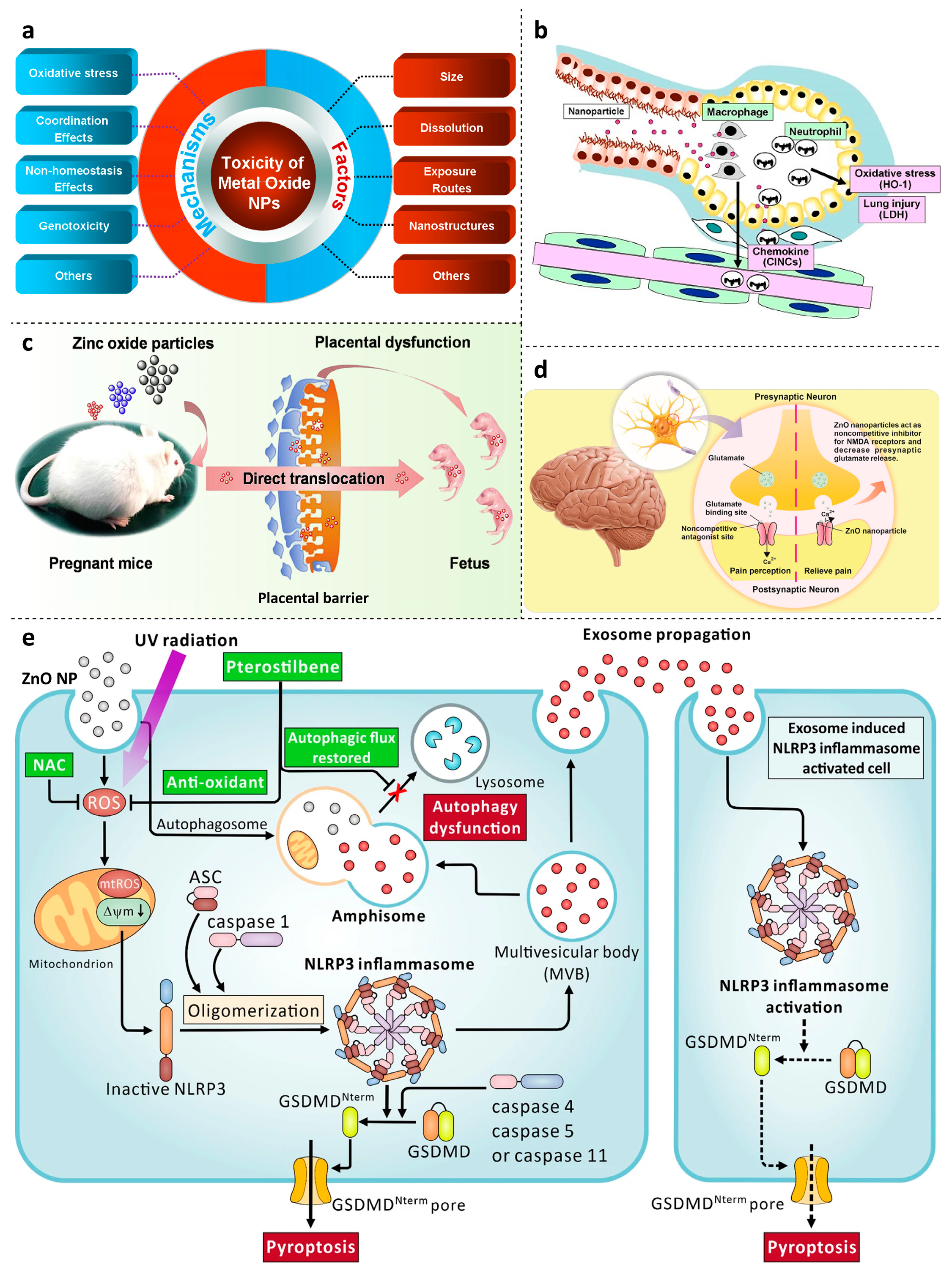
4. Toxicology Concerns of ZnO Nanoparticles

Although ZnO NPs have many potential applications in various fields, their potential toxicity is a significant concern. Some critical toxicological concerns of ZnO NPs are as follows.

4.1. Pulmonary Toxicity

Pulmonary toxicity is a potential toxicology concern of zinc oxide (ZnO) nanoparticles (Figure 2). ZnO nanoparticles can be released into the air during manufacturing, handling, or use and inhaled into the lungs, where they can cause respiratory and systemic effects [102,103]. ZnO nanoparticles can penetrate the respiratory system and deposit in the alveolar regions of the lungs. Once inside the lung, they can induce toxicity by generating ROS and causing oxidative stress, leading to inflammation, DNA damage, and cell death [5]. Moreover, ZnO nanoparticles may prevent the lung cells’ mitochondria from operating normally, leading to further damage. Another concern is the potential for ZnO nanoparticles to cause pulmonary fibrosis, a condition where scar tissue forms in the lungs, leading to difficulty in breathing [104]. Studies have shown that exposure to ZnO nanoparticles can induce the production of pro-fibrotic factors, leading to scar tissue formation in the lungs [105]. Additionally, the small size and high surface area of ZnO nanoparticles can make them more reactive and able to interact with biological molecules in the lungs, leading to an inflammatory response [106]. Furthermore, the ability of ZnO nanoparticles to enter the bloodstream from the lungs can lead to systemic toxicity. However, it is essential to note that the toxicity of ZnO nanoparticles can be mitigated by modifying their physicochemical properties. For example, using larger-sized nanoparticles can reduce their ability to penetrate the respiratory system and interact with the underlying cells [107]. Coating ZnO nanoparticles with biocompatible materials can reduce their toxicity and improve their biocompatibility [33]. The potential inhalation toxicity of ZnO nanoparticles is a concern that needs to be considered as their use continues to increase. ZnO nanoparticles can penetrate the respiratory system and cause respiratory and systemic effects through various mechanisms. Further research is needed to fully understand the potential risks of ZnO nanoparticles to human health and the environment.
Skin toxicity: ZnO nanoparticles have gained significant attention in nanotechnology due to their unique properties and potential applications in various industries. However, their increasing use has raised concerns about their potential toxicity, primarily related to skin exposure. Studies have shown that ZnO nanoparticles can penetrate the skin and reach the underlying cells, where they can induce toxicity [108]. The toxicity of ZnO nanoparticles to the skin depends on various factors, such as their size, concentration, surface area, and coating material [109].
One of the main concerns regarding the skin toxicity of ZnO nanoparticles is their ability to generate reactive oxygen species (ROS) upon exposure to light. ROS can cause oxidative stress, damaging the cellular structures and leading to inflammation and cell death [110]. Additionally, ZnO nanoparticles can induce DNA damage and interfere with the normal cell cycle [111]. Another concern is the potential for ZnO nanoparticles to cause skin irritation or sensitization. The small size and high surface area of nanoparticles make them more reactive and able to interact with skin cells, leading to an inflammatory response. Furthermore, the ability of ZnO nanoparticles to penetrate the skin can lead to systemic toxicity if absorbed into the bloodstream [112]. However, it is essential to note that the toxicity of ZnO nanoparticles can be mitigated by modifying their physicochemical properties. For example, coating ZnO nanoparticles with biocompatible materials can reduce their toxicity and improve their biocompatibility. Additionally, using larger-sized nanoparticles can reduce their ability to penetrate the skin and interact with the underlying cells [113].
The skin toxicity of ZnO nanoparticles is a concern that needs to be addressed as their use continues to increase (Figure 2). The toxicity depends on various factors, such as their size, concentration, and surface area, and can be mitigated by modifying their physicochemical properties. Further research is needed to fully understand the potential risks of ZnO nanoparticles to human health and the environment.

4.2. Genotoxicity

Zinc oxide (ZnO) nanoparticles pose potential toxicology concerns, particularly regarding genotoxicity. Research has shown that ZnO nanoparticles can cause DNA damage, which can result in genetic mutations and potentially contribute to cancer development (Figure 2) [5]. The genotoxic effects of ZnO nanoparticles are primarily attributed to the generation of reactive oxygen species (ROS), which can lead to oxidative stress and DNA damage [110]. Additionally, these nanoparticles have been found to induce micronuclei formation, which are small nuclei that appear when chromosomes are damaged or disrupted. The presence of micronuclei indicates genotoxicity and can increase the risk of genetic mutations [114]. Moreover, ZnO nanoparticles can interfere with the normal cell cycle, leading to abnormal cell growth, division, and genetic mutations. Studies have demonstrated that exposure to ZnO nanoparticles can result in cell cycle arrest and apoptosis. The extent of genotoxicity associated with ZnO nanoparticles depends on factors such as their size, concentration, and surface area [6]. Smaller nanoparticles tend to exhibit greater genotoxicity compared to larger ones [115]. Additionally, impurities or contaminants in the nanoparticles can enhance their genotoxic effects. However, it is worth noting that modifying the properties of ZnO nanoparticles, such as coating them with biocompatible materials, can reduce their toxicity and enhance their biocompatibility, potentially mitigating their genotoxicity. The potential genotoxicity of ZnO nanoparticles is a significant concern that should be considered as their usage continues to rise. Further research is necessary to comprehensively understand the potential risks of ZnO nanoparticles to human health and the environment. For example, a study on mice explored the effects of ZnO nanoparticles on neurotoxicity and cancer treatment. The study discovered that while ZnO nanoparticles exhibited anticancer activity through inhibiting tumor growth and inducing apoptosis in cancer cells, they also displayed neurotoxic effects. These nanoparticles could cross the blood-brain barrier and accumulate in the brain, causing oxidative stress, DNA damage, and inflammation in neural cells. These neurotoxic effects raised concerns regarding their potential impact on the overall system’s overall health [116].

4.3. Neurotoxicity

Neurotoxicity is another potential toxicology concern of zinc oxide (ZnO) nanoparticles. ZnO nanoparticles have been shown to cross the blood-brain barrier and accumulate in the brain, where they can induce neuronal damage and affect brain function (Figure 2). One of the mechanisms of ZnO nanoparticle-induced neurotoxicity is generating reactive oxygen species (ROS) in the brain. ROS can cause oxidative stress, damaging neuronal cells and leading to inflammation and cell death [108]. Additionally, ZnO nanoparticles can interfere with the normal functioning of the mitochondria, which are the energy-producing organelles in cells, leading to further damage to the neuronal cells. Moreover, ZnO nanoparticles can affect the neurotransmitter system, which is responsible for the communication between the neurons in the brain. Studies have shown that ZnO nanoparticles can disrupt the expected levels of neurotransmitters such as dopamine, acetylcholine, and glutamate, leading to alterations in brain function [117]. Another potential mechanism of ZnO nanoparticle-induced neurotoxicity is the induction of apoptosis, which is programmed cell death. Apoptosis can lead to the loss of neuronal cells and affect brain function. However, it is essential to note that the toxicity of ZnO nanoparticles can depend on their size, concentration, and surface area, among other factors. Smaller nanoparticles are more toxic to the brain than larger ones [118]. Coating ZnO nanoparticles with biocompatible materials can reduce their toxicity and improve their biocompatibility. For example, the potential neurotoxicity of ZnO nanoparticles was conducted in a study on laboratory mice. In the study, mice were exposed to ZnO nanoparticles through inhalation, and their brain function was subsequently assessed. The researchers found that the inhalation of ZnO nanoparticles led to the accumulation of these particles in the brain tissue of the mice. This accumulation resulted in increased levels of reactive oxygen species (ROS) within the brain, leading to oxidative stress and neuronal damage. The mice exhibited impaired cognitive function and behavioral abnormalities, indicating the adverse effects of ZnO nanoparticles on brain function [119]. Furthermore, the study demonstrated that ZnO nanoparticles interfered with the neurotransmitter system in the brain. Specifically, they disrupted the normal levels of dopamine, a neurotransmitter involved in mood regulation and movement control. This disruption in dopamine levels could contribute to neurological disorders such as Parkinson’s disease. The potential neurotoxic effects of ZnO nanoparticles include their ability to cross the blood-brain barrier, induce oxidative stress, and disrupt neurotransmitter function, ultimately affecting brain health and function [119].
The potential neurotoxicity of ZnO nanoparticles is a concern that needs to be considered as their use continues to increase. ZnO nanoparticles can cross the blood-brain barrier and accumulate in the brain, where they can induce neuronal damage and affect brain function through various mechanisms. Further research is needed to fully understand the potential risks of ZnO nanoparticles to human health and the environment.

4.4. Reproductive Toxicity

Reproductive toxicity is another potential toxicology concern of zinc oxide (ZnO) nanoparticles (Figure 2). ZnO nanoparticles have been shown to adversely affect male and female reproductive systems, leading to possible infertility and developmental defects [120]. ZnO nanoparticles can accumulate in reproductive organs such as the testes and ovaries, leading to structural damage and reduced fertility [121]. In male rats, exposure to ZnO nanoparticles has been shown to cause a decrease in sperm count and motility, as well as changes in the morphology of the testes [122]. Similarly, in female rats, exposure to ZnO nanoparticles has been shown to lead to a decrease in fertility and alterations in the morphology of the ovaries. Moreover, ZnO nanoparticles can cross the placenta and accumulate in fetal tissues, potentially leading to developmental defects [123]. Studies have shown that exposure to ZnO nanoparticles during pregnancy can lead to fetal growth retardation and alterations in fetal development, including skeletal abnormalities and altered brain development [124]. However, it is essential to note that the reproductive toxicity of ZnO nanoparticles can be reduced by modifying their properties. Further research is needed to fully understand the potential risks of ZnO nanoparticles to human health and the environment.
Conducting thorough toxicological studies to assess the potential risks of ZnO NPs and develop strategies to minimize their toxicity is essential [125]. Such studies should include in vitro and in vivo models and environmental and occupational exposure assessments to ensure the safe and sustainable use of ZnO NPs in various applications.
Figure 2. Schematic diagram showing the toxicological concern of Zinc oxide NPs. (a) Toxicology of Zinc oxide nanoparticles is influenced by a number of critical factors, including particle size, solubility, exposure routes, structures, and others. Some of the damaging processes that depict the toxic impact include oxidative stress, non-homeostasis, genotoxicity, and coordination problems [126]. (b) zinc oxide nanoparticles that cause lung damage [102] (c) Influence of ZnO nanoparticles size on toxicities to the developing fetus and placenta (reprinted form reference [127], copyright Elsevier 2019) (d) ZnO nanoparticles that have pain-relieving properties. Glutamate release is decreased by ZnO nanoparticles. They are not competing NMDA glutamate receptor inhibitors, so they reduce the capacity of glutamate to have an impact on these receptors [128] (e) Skin toxicity is brought on by ZnONPs and UVB, which also inhibits PT [129].
Jnt 04 00012 g002

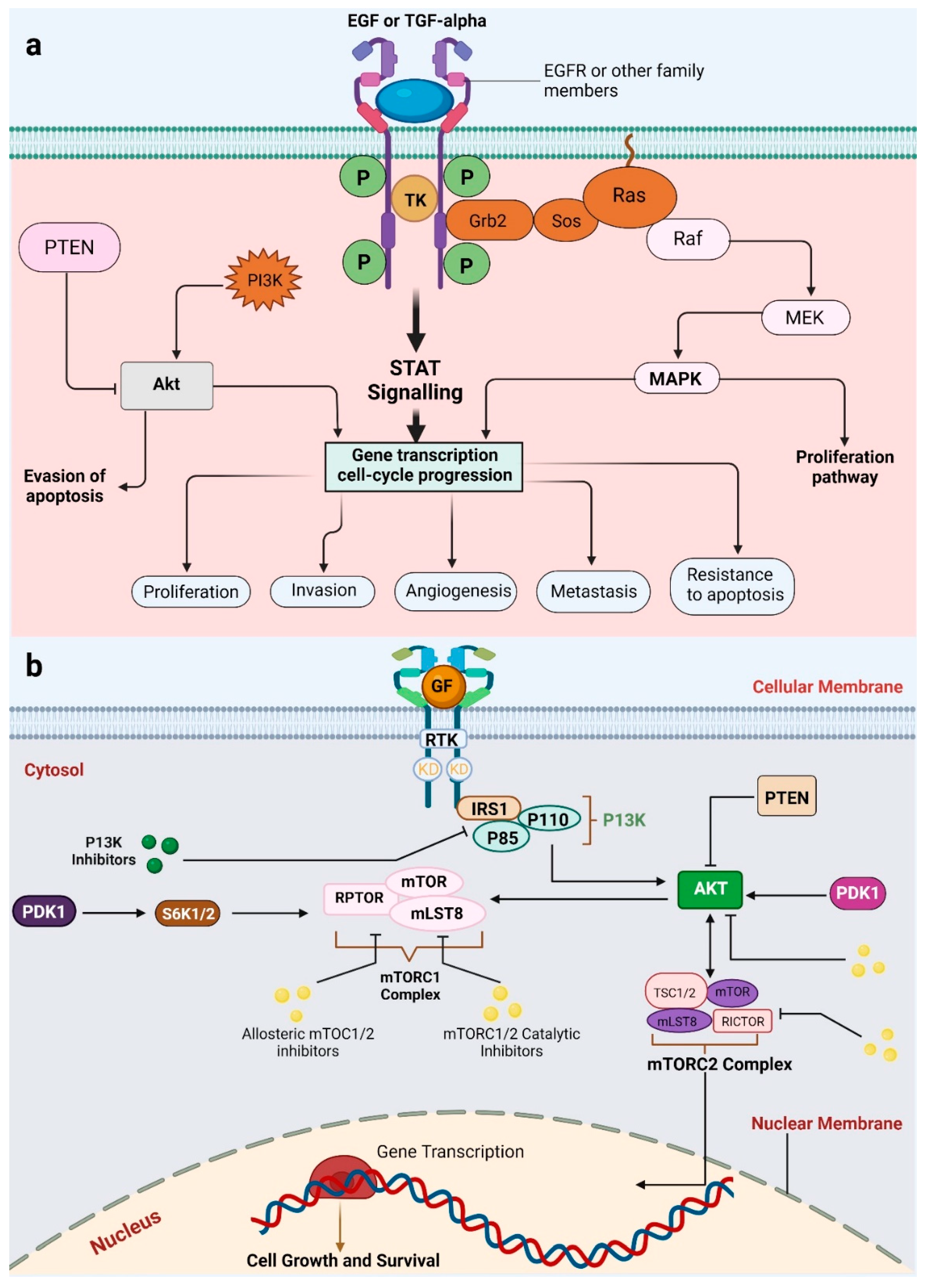
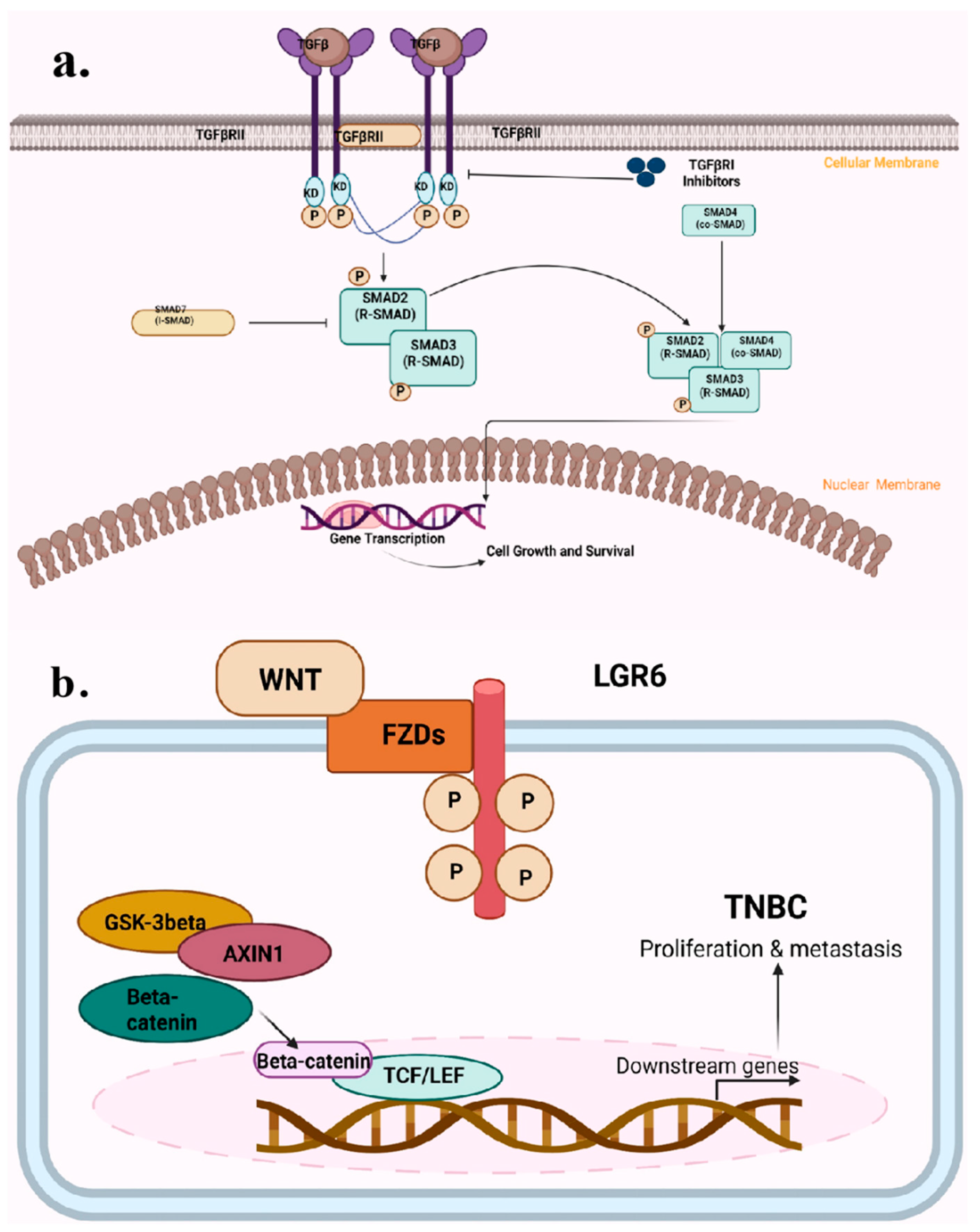
5. Pathogenesis of Cancer
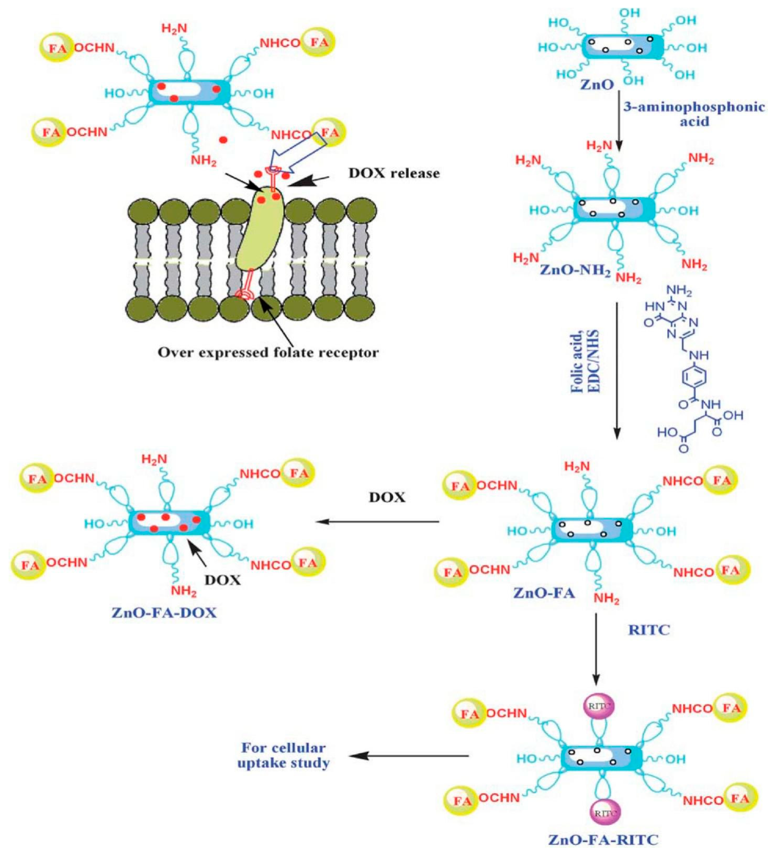
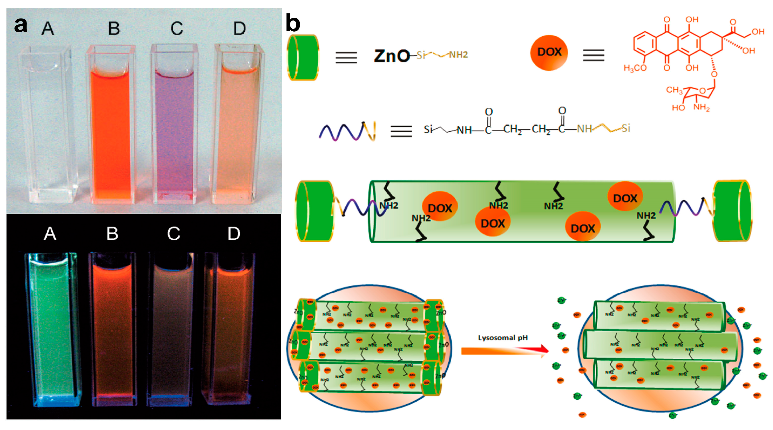
Cancer is a very complex disease that occurs from a combination of genetic and environmental factors. The pathogenesis of cancer involves the accumulation of specific genetic mutations or alterations that result in uncontrolled cell growth and division, leading to the development of tumors. These tumors can be either benign or malignant, and their behavior depends on various factors such as the type of cell, location, and cancer stage. The process of cancer development begins with a genetic mutation in a single cell that leads to uncontrolled cell growth. This mutation can be caused by various factors, such as exposure to radiation, chemicals, viruses, or inherited genetic defects [130]. Once a cell acquires a conversion, it can divide uncontrollably, forming a small cluster of abnormal cells known as a tumor. Over time, these abnormal cells can acquire additional mutations that further increase their growth and survival advantages, forming a giant tumor. Some of these mutations may confer the potentiality of the tumor cells to permeate nearby tissues, enter the bloodstream, and spread to other parts of the body, a process called metastasis. Cancer consists of certain hallmarks, including apoptosis evasion, sustained proliferation, and resistance over growth suppressors, and those result from genetic and epigenetic alterations that drive cancer pathogenesis. These alterations can occur in genes that control cell proliferation, cell death, DNA repair, and other critical cellular processes. The underlying molecular mechanisms of this disease involve dysregulation of several signaling pathways, including EGFR, RAS-MAPK, Wnt/β-catenin, PI3K/AKT/mTOR, and TGF-β and TP53 pathways. Mutations or dysregulation of these pathways leads to abnormal growth of cells, contributing to cancer development. Additionally, the tumor microenvironment, which includes surrounding cells, extracellular matrix, and signaling molecules, plays a pivotal role in cancer pathogenesis. The tumor microenvironment can provide nutrients, oxygen, and growth factors to cancer cells and help them evade the immune system and resist therapy.
EGFR signaling is one of the commonly altered pathways in most cancer types (Figure 3a). EGFR mutations can lead to the overexpression of the EGFR protein, which activates downstream signaling cascades, including the PI3K/AKT/mTOR and RAS/RAF/MAPK pathways, resulting in uncontrolled cell proliferation (Figure 3b). The EGFR or epidermal growth factor receptor is a well-known RTK or receptor tyrosine kinase family member, including ERBB2, ERBB3, and ERBB4 [131]. The activation of tyrosine kinases (TKs) and receptor transphosphorylation are downstream ligand binding events. This activates pathways such as Ras, phosphatidylinositol-3-kinase, and MAP kinase by providing docking sites for various cytoplasmic signaling molecules (PI3K) [131]. Mutations in EGFR and TK include insertions, deletions, and activating point mutations, all targeting essential areas of the TK domain linked with downstream signaling. Deletions in exon 19 and the point mutation (L858R) in exon 21 account for about 85% of conversions [132].
The RAS-MAPK pathway is a crucial signaling cascade in cell proliferation, survival, and differentiation. Dysregulation of this pathway has been involved in the pathogenesis of numerous cancers, making it a good target for cancer therapy. The binding of growth factors, such as epidermal growth factor (EGF), to their cell surface receptors activates the RAS-MAPK pathway. This binding initiates a cascade of events that triggers the RAS protein, a small GTPase that serves as a molecular switch. Activated RAS recruits and starts a series of downstream effectors, including RAF, MEK, and ERK, ultimately leading to the phosphorylation of various substrates involved in cell proliferation and survival. Dysregulation of the RAS-MAPK pathway occurs through multiple mechanisms, including mutations in RAS, RAF, MEK, and ERK, as well as alterations in upstream regulators and feedback loops. These alterations result in the constitutive activation of this pathway, promoting uncontrolled cell growth, survival, and invasion, all hallmarks of cancer [133].
Mutations in RAS are among the most common oncogenic alterations in human cancer. Mutant RAS proteins have a decreased ability to hydrolyze GTP and remain in their active, GTP-bound form, leading to persistent activation of downstream effectors. Mutations in RAS can be found in a wide range of cancers, including lung, colorectal, and pancreatic cancer [134]. In addition to mutations in RAS, alterations in upstream regulators of the RAS-MAPK pathway can also contribute to cancer pathogenesis. For example, overexpression of growth factor receptors, such as the EGF receptor (EGFR), can activate the RAS-MAPK pathway and promote cell proliferation and survival. EGFR overexpression is commonly observed in many solid tumors, including breast and lung cancer. The RAF family of proteins, including ARAF, BRAF, and CRAF, are downstream effectors of RAS that can also be mutated in cancer. BRAF mutations are prevalent in melanoma and can activate the RAS-MAPK pathway without upstream RAS activation. MEK1 and MEK2, downstream effectors of RAF, are frequently mutated in some cancers, including melanoma and lung cancer. ERK, the final effector of the RAS-MAPK pathway, can also be activated by other signaling pathways, such as the PI3K-Akt pathway, which can contribute to its dysregulation in cancer. ERK can phosphorylate many substrates involved in cell proliferation, survival, and invasion, including transcription factors, kinases, and cytoskeletal proteins [135,136].
Certain forms of cancer are driven by point mutations in the RAS proto-oncogenes (HRAS, KRAS, and NRAS), which encodes proteins localized on the cell’s plasma membrane and promotes uncontrolled cell proliferation. KRAS is the most often mutated RAS gene in lung cancer, typically at codon 12 and sometimes at codons 13 and 61 [137]. Activating mutations in KRAS can activate the downstream signaling pathways, such as the PI3K/AKT/mTOR and RAS/RAF/MAPK pathways, leading to cell growth and division [138]. Most KRAS mutations are G-T transversions, typical of the heavy DNA adducts from the nitrosamines and polycyclic hydrocarbons in tobacco smoke. Finally, nuclear proto-oncogenes such as MYC are activated by RAS signaling, and their transcriptional activation of downstream genes drives cell growth. MYC, MYCN, and MYCL are the main factors in the MYC family [139].
The TGF-β, or transforming growth factor beta pathway, is a crucial regulator of cancer pathogenesis (Figure 4). TGF-β is a cytokine that plays a vital role in numerous physiological processes, including cell proliferation, migration, and differentiation. However, dysregulation of the TGF-β pathway is frequently observed in many types of cancer, and it can contribute to tumor growth, invasion, and metastasis. The TGF-β pathway regulates cancer pathogenesis through various mechanisms [140]. First, it controls cell proliferation by regulating the cell cycle and inducing cell cycle arrest in response to DNA damage. TGF-β signaling also promotes apoptosis by activating the Bcl-2 family of pro-apoptotic proteins. Dysregulation of TGF-β signaling can lead to uncontrolled cell proliferation and reduced apoptosis, which are hallmark features of cancer cells. The TGF-β pathway regulates cancer pathogenesis by controlling cell migration and invasion. TGF-β promotes EMT or epithelial-to-mesenchymal transition, in which epithelial cells lose cell-cell adhesion and adopt a mesenchymal phenotype, allowing them to migrate and infiltrate the adjacent tissues. Dysregulation of TGF-β signaling can result in sustained EMT, leading to increased invasion and metastasis [140]. Second, the TGF-β pathway regulates the immune system by controlling the differentiation and activation of immune cells. TGF-β can suppress the immune response by inhibiting T cell activation and promoting the differentiation of regulatory T cells (Tregs). Tregs are a resistant cell type that inhibits the immunological response and promotes immune tolerance, allowing cancer cells to evade the immune system. Dysregulation of TGF-β signaling can contribute to immune suppression and immune evasion by cancer cells [141]. The TGF-β pathway regulates cancer pathogenesis by interacting with other molecular pathways. For example, TGF-β may activate the SMAD transcription factor family, which can promote EMT and contribute to the migration and invasion of cancer cells. TGF-β may also trigger the PI3K-Akt-mTOR pathway, which is frequently dysregulated in cancer and contributes to the survival and proliferation of cancer cells [142]. Furthermore, the TGF-β pathway plays an essential role in the tumor microenvironment. TGF-β can control the production of extracellular matrix (ECM) proteins, which can promote tumor growth and invasion. TGF-β can also promote the recruitment of immune cells to the tumor microenvironment, contributing to immune suppression and tumor growth [142].
The PI3K/AKT/mTOR pathway also regulates cell growth, survival, and metabolism in many types of cancer. Dysregulation of this signaling can lead to uncontrolled cell proliferation, a hallmark of cancer [142]. The catalytic and regulatory subunits of PI3Ks make up the heterodimeric structure of these lipid kinases. Somatic mutations in the PIK3CA or phosphoinositide-3-kinase, catalytic, alpha polypeptide gene have been detected in only the regulatory component p85a of human malignancies. These mutations occur almost exclusively in the helical or kinase domains of PIK3CA.
The Wnt-β-catenin signaling is one of the critical signaling cascades involved in many developmental processes, including cell proliferation, differentiation, and tissue homeostasis (Figure 4). Abnormal activation of the Wnt-β-catenin pathway has been reported in various types of cancer, including colorectal, breast, lung, and liver cancer. Wnt ligands activate the Wnt-β-catenin course by binding to Frizzled receptors on the cell surface, which leads to the cytoplasmic protein Disheveled (Dvl) recruitment, and the inhibition of the destruction complex, which is composed of Axin, APC, and GSK-3β. This inhibition stabilizes and accumulates β-catenin in the cytoplasm, where it can interact with TCF/LEF transcription factors to translocate to the nucleus and activate target genes. Mutations in the Wnt pathway components can lead to aberrant activation of the pathway in cancer. For example, β-catenin accumulates in the cytoplasm and nucleus when mutations in APC, a negative pathway regulator, are prevalent in colorectal cancer.
Similarly, mutations in β-catenin itself can result in its stabilization and activation of the pathway, which has been found in several types of cancer, including hepatocellular carcinoma. Activation of the Wnt-β-catenin pathway can also contribute to cancer pathogenesis by promoting cell proliferation, survival, and migration. Activation of the path in cancer cells results in the overexpression of target genes, including c-Myc and Cyclin D1, which drive cell cycle progression and proliferation. In addition, activation of the system can suppress apoptosis by upregulating anti-apoptotic genes such as Bcl-2. Furthermore, the Wnt-β-catenin pathway can increase cancer cell motility and invasion by upregulating Snail and Slug genes involved in epithelial-mesenchymal transition (EMT). EMT is a process in which epithelial cells lose their cell-cell adhesion and acquire mesenchymal properties, allowing them to move and invade adjacent tissues. In numerous kinds of cancer, activation of the Wnt-β-catenin pathway has been demonstrated to induce EMT and metastasis [143,144]. Genomic integrity is protected by tumor suppressor genes such as p53, even when exposed to carcinogens and UV radiation. Rapid overexpression of p53 due to DNA damage or hypoxia enables it to act as a sequence-specific transcription factor, modulating the G1/S cell cycle transition, the G2/M DNA damage checkpoint, and apoptosis by regulating the expression of target genes such as MDM2, p21, BAX and GADD45 [145]. The accumulation of several mutations and the subsequent evolution of a cancer cell is primarily facilitated by p53 malfunction, which allows genetically damaged cells to survive abnormally. p21 is a p53-responsive gene that inhibits cyclin/cyclin-dependent kinase (CDK) complexes during the G1 phase of the cell cycle (P21 RNA and Protein Expression in Non-Small Cell Lung Carcinomas: Evidence of P53-Independent Expression and Association with Tumoral Differentiation—PubMed, n.d.).
Furthermore, epigenetic modifications are critical in controlling gene expression and cellular function. These modifications can be heritable and reversible and can occur in response to environmental cues, including carcinogen exposure. Dysregulated epigenetic mechanisms can contribute to cancer aetiology by affecting the expression of genes involved in critical cellular processes such as cell proliferation, DNA repair, and apoptosis [146]. DNA methylation, which consists of adding a methyl group to cytosine residues in CpG dinucleotides, is one of the most well-studied epigenetic alterations in cancer. Gene silence can occur from hypermethylation of promoter regions, whereas gene activation might result from hypomethylation. Global hypomethylation and site-specific hypermethylation are frequent in cancer, resulting in abnormal gene expression and facilitating oncogenesis. Hypermethylation of tumor suppressor genes’ promoter regions, such as p16INK4a and BRCA1, can result in their silence and contribute to cancer aetiology.
On the other hand, hypomethylation of oncogenes such as MYC and RAS can lead to their activation and promote cancer cell proliferation [147]. Histone modifications are another critical epigenetic mechanism that regulates gene expression. These alterations include acetylation, methylation, phosphorylation, and ubiquitination of specific amino acid residues on histone proteins. Histone acetylation, which includes adding an acetyl group to lysine residues, is related to gene activation.
In contrast, histone methylation, depending on the individual lysine residue and the degree of methylation, can result in either gene activation or repression. Dysregulation of histone modifications can contribute to cancer pathogenesis by altering gene expression in cell cycle regulation, DNA damage response, and apoptosis. For example, in many cancers, the H3K9me3 repressive mark is lost, leading to aberrant expression of oncogenes such as MYC and EZH2 [148]. Noncoding RNAs (ncRNAs), which include long noncoding RNAs (lncRNAs) and microRNAs (miRNAs), are also crucial in regulating gene expression and cellular function. miRNAs are short, noncoding RNAs that bind to specific messenger RNAs (mRNAs) and either promote or prevent translation, resulting in post-transcriptional gene control. miRNA dysregulation has been linked to various cancers and has been shown to contribute to cancer pathogenesis by affecting the expression of oncogenes or tumor suppressor genes. lncRNAs are larger noncoding RNAs that have diverse functions in gene regulation. Dysregulation of lncRNAs has been associated with cancer pathogenesis, promoting tumor growth, metastasis, and drug resistance. The lncRNA HOTAIR, for example, has been found to enhance breast cancer metastasis by modulating the expression of EMT transition genes.
The body’s normal response to cellular and DNA damage, also known as programmed cell death or apoptosis, is commonly evaded by tumor cells [99]. Compared to adenocarcinoma (~10%), squamous cell carcinoma, accounting for 25–35% of lung cancer, is more likely to express the BCL-2 anti-apoptotic proto-oncogene. (Bcl-2 Protein Expression in Lung Cancer and Close Correlation with Neuroendocrine Differentiation—PubMed, n.d.), 10.1056/NEJM199309023291003). Furthermore, angiogenesis is mediated by a diverse group of inducers and inhibitors that collectively regulate endothelial cell proliferation and migration, both of which are required for tumor angiogenesis. VEGF, or vascular endothelial growth factor, and essential fibroblast growth factor (bFGF) are two critical growth factors in the generation of angiogenesis [149]. ZnO nanoparticles, because of their potential for cancer suppression, have sparked considerable attention. Within cancer cells, ZnO nanoparticles have been shown to enhance the formation of reactive oxygen species, which can result in cell homeostasis [150]. The production of ROS because of the ZnO nanoparticle is because of its semiconductor nature. The production of ROS in cells causes DNA fragmentation, which activates apoptotic pathways, potentially inhibiting metastasis. The cytotoxicity of ZnO nanoparticles is explained by the intracellular release of zinc ions, which further promotes ROS generation, creating disequilibrium in zinc-mediated protein activity and cancer cell death [151]. In an experiment conducted by Ng et al. [111], they studied the interaction between the ZnO oxide nanoparticle and a tumor suppressive pathway which centred around p53. Here, the researchers found that the administration of ZnO nanoparticles to induce apoptosis in the cells requires p53 to act as a molecular mechanism to prevent carcinogenesis when DNA damage occurs. ZnO nanoparticles are believed to stop S phase cell cycle arrest in T24 (bladder cancer) cells even at low dosages and cause late apoptosis, which inhibits cellular migration and aggressive potential due to histone methylation changes. Zhang et al. identified the mechanism behind the anticancer impact of ZnO nanoparticles, which involves a reduction in the levels of histone methyltransferase EZH2 and H3K27m 3 [152]. ZnO nanoparticles have the potential to interfere with the signaling cascade mechanism of the PI3K/Akt pathway and the NF- κB pathway, hindering cell proliferation, and disrupting the cancer cell signaling promoting cell apoptosis [153]. It can inhibit the development of new blood cells for cancer metastasis by interfering with the angiogenesis signaling system which proves to be an essential property for the suppression of cancer [8]. ZnO nanoparticles can inhibit endothelial cell recruitment and growth, limiting blood flow to the cancer cells. This interruption of tumor vascularization helps to limit cancer development and metastasis overall [154]. The ZnO nanoparticle can restrict the tumor’s nutrition and oxygen supply inducing mitochondrial dysfunction and causing energy depletion and cell death [155]. ZnO nanoparticles can be employed as a promising treatment option targeting the mitochondria for suppressing cancer development.

6. Drug Delivery

ZnO NPs have shown potential in drug delivery applications owing to their distinct physicochemical characteristics, including a large surface area to volume ratio, biocompatibility, and photostability. ZnO NPs can carry various drugs, including anticancer agents, antibiotics, and anti-inflammatory drugs. One approach for drug delivery using ZnO NPs involves loading the medicine onto the surface of the nanoparticles. The drug can be adsorbed onto the surface of ZnO NPs through physical adsorption, electrostatic interaction, or covalent bonding. This approach has been used to deliver doxorubicin, paclitaxel, and curcumin. Another strategy involves encapsulating the drug within ZnO NPs. This can be achieved through various methods, such as co-precipitation, sol-gel, and hydrothermal synthesis [51]. This approach has been used to deliver drugs such as ibuprofen and methotrexate. ZnO NPs can also be functionalized with targeting ligands such as antibodies, peptides, or aptamers to enhance their specificity for a particular cell or tissue type. This can improve the efficacy of the drug and reduce side effects. Overall, drug delivery using ZnO NPs shows promise as a potential strategy for improving the delivery of drugs and reducing their side effects. However, further research is needed to understand the safety and efficacy of this approach entirely.
The potential application of zinc oxide quantum dots (ZnO QDs) as a pH-responsive pore-blocker to stop the flow of enriched doxorubicin (DOX) from mesoporous nanoparticles of silica was investigated by Muhammad et al. DOX was effectively filled into MSN’s pores that resembled channels. The holes in the MSN were then sealed using amine-functionalized ZnO QDs (NH2-ZnOQDs), which formed covalent amide bonds with the carboxylic acid (COOH) groups on the MSN’s top surface (Figure 5). The ZnO QD lids were quickly broken down in the marginally acidic intracellular compartment after the nano assemblies were absorbed into HeLa cells, unleashing DOX into the cytosol. The ZnO nanoparticles also showed intrinsic anticancer activity, which had a synergistic antitumor impact in HeLa cells and had a synergistic antitumor effect [156]. Mitra et al. created ZnO nanorods with folic acid functionality for doxorubicin distribution that is targeted and pH-responsive. (DOX). In MDA-MB-231 cells that overexpress the folate receptor, the DOX-loaded nano-carrier showed more significant cytotoxicity than free DOX, demonstrating efficient absorption by the cancer cells. Furthermore, in vivo tests revealed no evidence of systemic harm linked to the nano-carrier [157]. Zhang et al. created a pH-responsive drug delivery system for doxorubicin (DOX) utilizing a core shell that is a safe and harmless nano-carrier consisting of ZnO quantum dots (QDs) protected by a non-toxic polyacrylamide shell (Figure 6). The ZnO@polymer QDs were stable, did not decompose at physiological pH, and could cross the human glioblastoma (U251) cell membrane through endocytosis. Once inside the cells, the ZnO@polymer QDs (ZnO@polymer-DOX) filled with DOX broke down in the lysosomes or endosomes, releasing DOX that then moved to the nucleus. Compared to free DOX, which penetrates cells via reversible passive diffusion, the ZnO@polymer-DOX showed more significant cytotoxicity [158].
Tan et al. developed a novel drug delivery system for doxorubicin (DOX) that utilized ZnO@poly(N-isopropyl acrylamide) nanohybrids to achieve a release that responds to both pH and temperature. N-isopropyl acrylamide is a thermally responsive material that exhibits unique properties when it comes to drug delivery. When heated, it undergoes shrinkage from its expanded hydrophilic state to a hydrophobic state, which leads to the release of DOX from its surface. This phenomenon is highly advantageous for controlled drug release. Additionally, low pH facilitates the dissolution of the ZnO core, which further aids in the release of the trapped DOX. [159]. Zhang and the team have developed a revolutionary drug delivery system that utilizes mesoporous silica nanoparticles (MSN) for targeted and charge-reversal drug delivery. This multifunctional system improves cellular uptake and pH-responsive discharge of doxorubicin (DOX) using ZnO quantum dots (QDs). The key to the system’s success is its ability to facilitate escape from endosomes. The MSNs have been modified with a cell-penetrating decalysene peptide coated in negatively charged acid-labile-carboxylic amides, while the positively charged ZnOQDs are used to cap the DOX-loaded MSN pores via electrostatic interactions. Upon entering the acidic endosome of cancer cells, the ZnOQDs dissolve and the acid-labile β-carboxylic amides undergo hydrolysis to produce the active peptide form, leading to the uncapping of MSN pores and the release of DOX into the cytosol. The ZnO@MSN DDS system has shown a synergistic anticancer effect in HepG2 cells, indicating promising potential for targeted cancer therapy. This is a significant breakthrough in drug delivery technology and could revolutionize cancer treatment in the future [160].
Drug delivery systems that can both deliver drugs to target cells and provide real-time feedback on drug efficacy are highly desirable (Table 2). In a recent study by Wang et al., ZnOQDs were used as a pore-blocking agent in DOX-loaded MSNs by forming an amide bond. The researchers have also developed ZnO-functionalized upconverting nano theranostic agents using lanthanide-doped upconverting nanoparticles (UCNPs) as the core and MSN layer as the outer shell. These agents have demonstrated several desirable properties, including capable internalization in HeLa cells, on-demand pH-triggered drug release, and multi-modality imaging capabilities. The imaging properties include computed tomography, upconversion luminescence, and magnetic resonance imaging, making these agents valuable tools for in vivo diagnosis and monitoring of cancer. Overall, the combination of drug delivery and imaging capabilities in these ZnO-functionalized upconverting nano theranostic agents holds great potential for the development of effective and personalized cancer therapies. This research demonstrates the potential of ZnO-functionalized upconverting nano theranostic agents as a promising strategy for targeted drug delivery and real-time monitoring of treatment efficacy [161]. Cai et al. conducted a study on the development of a pH-responsive drug delivery system using hyaluronic acid (HA) functionalized zinc oxide quantum dots (ZnOQDs) for delivering the anticancer drug doxorubicin (DOX) to A549 cells. DOX was loaded onto the surface of ZnOQDs by forming a six-membered chelate between Zn2+ and the oxygenated functional groups of the anthraquinone moiety in DOX [162].
The results showed that the HA-functionalized ZnOQD-DOX complex exhibited more significant cytotoxicity than non-targeted ZnOQD-DOX due to the better cellular uptake of the drug. The study suggests that the HA-functionalized ZnOQD-DOX complex could serve as a promising pH-responsive drug delivery system for targeted cancer therapy.
While inflammation restores cellular homeostasis and tissue microenvironment, chronic inflammation can cause serious health complications such as cancer, arthritis, asthma, pancreatitis, and inflammatory bowel diseases [163,164]. Inflammation remains critical and the infiltration of the leukocytes and the inflammatory cytokines generally promotes tumor development [165]. ZnO nanoparticles owing to their excellent biomedical applications, play a crucial role in homeostasis, maintaining cell redox balance, enzyme regulation, and DNA and protein synthesis machinery regulation [166]. Several studies have shown that the ZnO nanoparticle can suppress the release of pro-inflammatory cytokines such as IL-β and TNF-α and myeloperoxidase, which results in the anti-inflammatory effect of the nanoparticle [167]. The peculiar ability that ZnO nanoparticles can generate pro-inflammatory cytokines at low concentrations causes an appropriate amount of cell death, implying suppression of tumor necrosis factor at a certain dose [27]. In a study by Mohammad et al., green synthesized ZnO NPs were assessed for anti-inflammatory and anti-angiogenesis properties. Using a real-time quantitative polymerase chain reaction, a shift in the expression of the angiogenesis gene, VEGF, and VEGFR, as well as a reduction in inflammation at regular intervals was discovered [168]. The study revealed that ZnO nanoparticles reduced IL-10 levels while increasing IL-1B gene expression. ROS production causes inflammation by causing lipid peroxidation to damage the phospholipid membrane. ZnO NPs were seen to reduce the expression of IL-1β, IL-6, and TNF-α in a dose-dependent manner in RAW 264.7 cells. ROS causes inflammation by initiating lipid peroxidation in the phospholipid membrane [169,170]. Controlling ROS formation, therefore, helps ZnO NP anti-inflammatory activity ZnO NPs reduce the expression of pro-inflammatory genes and proteins, allowing for the treatment of inflammatory illnesses.

7. Future Perspective

The use of Zinc Oxide nanoparticles synthesized through green approaches for cancer therapy holds great potential for the future. Researchers are continually exploring different green synthesis methods to produce Zinc Oxide nanoparticles with improved properties, such as increased selectivity, reduced toxicity, and enhanced stability.
Moreover, combining Zinc Oxide nanoparticles with other therapies, such as chemotherapy, radiation therapy, or immunotherapy, may improve cancer treatment outcomes. Another potential application of Zinc Oxide nanoparticles is in the early detection of cancer. CT scans could potentially be used to visualize the distribution of zinc oxide nanoparticles in tumor tissues, helping to assess their accumulation and potential therapeutic effects. These nanoparticles can be designed to bind specifically to cancer cells and used in imaging techniques, enabling early detection of cancerous cells before they spread. However, different studies are required to completely appreciate Zinc Oxide nanoparticles’ safety and toxicity in humans and optimize their use in cancer therapy. It is essential to continue exploring the potential of green synthesis approaches for producing Zinc oxide nanoparticles, as these methods are more sustainable and eco-friendly than traditional ones. In summary, the future of targeting cancer with Zinc oxide nanoparticles using green synthesis approaches is promising, and continued research in this area has the potential to lead to significant improvements in cancer treatment and detection.

8. Conclusions

Zinc oxide nanoparticles have emerged as promising candidates for drug delivery in cancer treatment due to their unique physicochemical properties and potential for targeted therapy. These nanoparticles possess a high surface area-to-volume ratio, allowing for efficient drug loading and delivery. They can be easily functionalized to specifically target cancer cells, minimizing off-target effects and reducing the dosage required. One advantage of using zinc oxide nanoparticles is their excellent biocompatibility. They can be synthesized using green methods, which involve the use of natural extracts or biomolecules as reducing and stabilizing agents. This preserves the biocompatibility and bioactivity of the nanoparticles, minimizing potential toxicity, and adverse reactions. Green synthesis methods also align with environmentally friendly practices, making them suitable for biomedical applications. The ability to engineer zinc oxide nanoparticles for selective targeting of cancer cells is a key aspect of their potential in drug delivery. These nanoparticles can be designed to accumulate in tumor sites, delivering anticancer drugs directly to the cancer cells. Upon exposure to light, the nanoparticles can generate reactive oxygen species, leading to localized cell damage and tumor regression. Furthermore, zinc oxide nanoparticles allow for controlled drug release through surface modifications. This controlled release mechanism enables sustained and targeted drug delivery, improving drug bioavailability, reducing systemic toxicity, and enhancing the therapeutic outcome in cancer treatment. Despite the numerous advantages, there are still challenges to address. Further research is necessary to optimize synthesis protocols, enhance stability, control drug release kinetics, and evaluate long-term biocompatibility and safety aspects. Standardized protocols for the production and characterization of green-synthesized zinc oxide nanoparticles are also needed to ensure reproducibility and comparability of results across different studies. The use of green-synthesized zinc oxide nanoparticles for drug delivery in cancer treatment holds great potential for improving outcomes. Their unique properties, environmentally friendly synthesis approach, and targeted drug delivery capabilities offer new possibilities for effective and safer cancer therapies. Continued research and development in this field will contribute to the advancement of nanomedicine and personalized cancer treatment strategies.

Author Contributions

The manuscript was written through contributions of all authors. Conceptualization: S.S.N., B.G. and S.K.V.; Illustrations: F.Z.S., D.S., P.K.P., A.N., E.J. and A.S.; Literature analyses: E.J., S.K.V. and A.C., writing (original draft preparation): S.S.N., B.G., A.N., F.Z.S. and D.S.; writing (review and editing): P.K.P., S.K.V., S.K.V., A.S. and M.S.; supervision: M.S. and S.K.V.; funding acquisition: S.K.V. and M.S. All authors have read and agreed to the published version of the manuscript.

Funding

We acknowledge infrastructure support available through DBT-BUILDER program (BT/INF/22/SP42155/2021) at KIIT UNIVERSITY.

Data Availability Statement

Not Applicable.

Conflicts of Interest

The authors declare no conflict of interest.

References

  1. Gomez, G.G.; Wykosky, J.; Zanca, C.; Furnari, F.B.; Cavenee, W.K. Therapeutic resistance in cancer: microRNA regulation of EGFR signaling networks. Cancer Biol. Med. 2013, 10, 192–205. [Google Scholar] [CrossRef] [PubMed]
  2. Cheng, Z.; Li, M.; Dey, R.; Chen, Y. Nanomaterials for cancer therapy: Current progress and perspectives. J. Hematol. Oncol. 2021, 14, 85. [Google Scholar] [CrossRef] [PubMed]
  3. Mishra, Y.K.; Adelung, R. ZnO tetrapod materials for functional applications. Mater. Today 2018, 21, 631–651. [Google Scholar] [CrossRef]
  4. Bisht, G.; Rayamajhi, S. ZnO Nanoparticles: A Promising Anticancer Agent. Nanobiomedicine 2016, 3, 9. [Google Scholar] [CrossRef]
  5. Lu, X.; Zhu, T.; Chen, C.; Liu, Y. Right or Left: The Role of Nanoparticles in Pulmonary Diseases. Int. J. Mol. Sci. 2014, 15, 17577–17600. [Google Scholar] [CrossRef] [Green Version]
  6. Jiang, J.; Pi, J.; Cai, J. The Advancing of Zinc Oxide Nanoparticles for Biomedical Applications. Bioinorg. Chem. Appl. 2018, 2018, 1062562. [Google Scholar] [CrossRef]
  7. Choi, S.J.; Choy, J.H. Biokinetics of zinc oxide nanoparticles: Toxicokinetics, biological fates, and protein interaction. Int. J. Nanomed. 2014, 9, 261–269. [Google Scholar] [CrossRef] [Green Version]
  8. Anjum, S.; Hashim, M.; Malik, S.A.; Khan, M.; Lorenzo, J.M.; Abbasi, B.H.; Hano, C. Recent Advances in Zinc Oxide Nanoparticles (ZnO NPs) for Cancer Diagnosis, Target Drug Delivery, and Treatment. Cancers 2021, 13, 4570. [Google Scholar] [CrossRef]
  9. Hahm, J. Fundamental Properties of One-Dimensional Zinc Oxide Nanomaterials and Implementations in Various Detection Modes of Enhanced Biosensing. Annu. Rev. Phys. Chem. 2016, 67, 691–717. [Google Scholar] [CrossRef] [Green Version]
  10. Shah, A.; Aftab, S.; Nisar, J.; Ashiq, M.N.; Iftikhar, F.J. Nanocarriers for targeted drug delivery. J. Drug Deliv. Sci. Technol. 2021, 62, 102426. [Google Scholar] [CrossRef]
  11. Zhou, G.; Latchoumanin, O.; Hebbard, L.; Duan, W.; Liddle, C.; George, J.; Qiao, L. Aptamers as targeting ligands and therapeutic molecules for overcoming drug resistance in cancers. Adv. Drug Deliv. Rev. 2018, 134, 107–121. [Google Scholar] [CrossRef] [PubMed]
  12. Fraceto, L.F.; Campos, E.V.R.; Habtemariam, S.; del Rodriguez-Torres, M.P.; Patra, J.K.; Grillo, R.; Acosta-Torres, L.S.; Sharma, S.; Shin, H.-S.; Diaz-Torres, L.A.; et al. Nano based drug delivery systems: Recent developments and future prospects. J. Nanobiotechnol. 2018, 16, 71. [Google Scholar]
  13. Makkar, H.; Verma, S.K.; Panda, P.K.; Pramanik, N.; Jha, E.; Suar, M. Molecular insight to size and dose-dependent cellular toxicity exhibited by a green synthesized bioceramic nanohybrid with macrophages for dental applications. Toxicol. Res. 2018, 7, 959–969. [Google Scholar] [CrossRef] [PubMed] [Green Version]
  14. Song, Y.; Li, Y.; Xu, Q.; Liu, Z. Mesoporous silica nanoparticles for stimuli-responsive controlled drug delivery: Advances, challenges, and outlook. Int. J. Nanomed. 2016, 12, 87–110. [Google Scholar] [CrossRef] [Green Version]
  15. Fernandes, N.B.; Nayak, Y.; Garg, S.; Nayak, U.Y. Multifunctional engineered mesoporous silica/inorganic material hybrid nanoparticles: Theranostic perspectives. Coord. Chem. Rev. 2023, 478. [Google Scholar] [CrossRef]
  16. Xie, J.; Li, H.; Zhang, T.; Song, B.; Wang, X.; Gu, Z. Recent Advances in ZnO Nanomaterial-Mediated Biological Applications and Action Mechanisms. Nanomaterials 2023, 13, 1500. [Google Scholar] [CrossRef]
  17. Hou, C.-C.; Zhu, J.-Q. Nanoparticles and female reproductive system: How do nanoparticles affect oogenesis and embryonic development. Oncotarget 2017, 8, 109799–109817. [Google Scholar] [CrossRef] [Green Version]
  18. Raj, V.J.; Ghosh, R.; Girigoswami, A.; Girigoswami, K. Application of zinc oxide nanoflowers in environmental and biomedical science. BBA Adv. 2022, 2, 100051. [Google Scholar] [CrossRef]
  19. Mohd Yusof, H.; Mohamad, R.; Zaidan, U.H.; Abdul Rahman, N.A. Microbial synthesis of zinc oxide nanoparticles and their potential application as an antimicrobial agent and a feed supplement in animal industry: A review. J. Anim. Sci. Biotechnol. 2019, 10, 57. [Google Scholar] [CrossRef] [Green Version]
  20. Kumari, P.; Misra, K.P.; Chattopadhyay, S.; Samanta, S. A brief review on transition metal ion doped ZnO nanoparticles and its optoelectronic applications. Mater. Today Proc. 2021, 43, 3297–3302. [Google Scholar] [CrossRef]
  21. Li, Y.; Yang, X.-Y.; Feng, Y.; Yuan, Z.-Y.; Su, B.-L. One-Dimensional Metal Oxide Nanotubes, Nanowires, Nanoribbons, and Nanorods: Synthesis, Characterizations, Properties and Applications. Crit. Rev. Solid State Mater. Sci. 2012, 37, 606512. [Google Scholar] [CrossRef]
  22. Mourdikoudis, S.; Pallares, R.M.; Thanh, N.T.K. Characterization techniques for nanoparticles: Comparison and complementarity upon studying nanoparticle properties. Nanoscale 2018, 10, 12871–12934. [Google Scholar] [CrossRef] [PubMed] [Green Version]
  23. Sarkar, S.; Sarkar, R. Synthesis, characterization and tribological study of zinc oxide nanoparticles. Mater. Today Proc. 2021, 44, 3606–3612. [Google Scholar] [CrossRef]
  24. Moumen, A.; Kaur, N.; Poli, N.; Zappa, D.; Comini, E. One dimensional ZnO nanostructures: Growth and chemical sensing performances. Nanomaterials 2020, 10, 1940. [Google Scholar] [CrossRef]
  25. Jin, S.E.; Jin, H.E. Synthesis, characterization, and three-dimensional structure generation of zinc oxide-based nanomedicine for biomedical applications. Pharmaceutics 2019, 11, 575. [Google Scholar] [CrossRef] [Green Version]
  26. Mehrvarz, A.; Khalil-Allafi, J.; Khosrowshahi, A.K. Biocompatibility and antibacterial behavior of electrochemically deposited Hydroxyapatite/ZnO porous nanocomposite on NiTi biomedical alloy. Ceram. Int. 2022, 48, 16326–16336. [Google Scholar] [CrossRef]
  27. Kumar, S.; Shukla, M.K.; Sharma, A.K.; Jayaprakash, G.K.; Tonk, R.K.; Chellappan, D.K.; Singh, S.K.; Dua, K.; Ahmed, F.; Bhattacharyya, S.; et al. Metal-based nanomaterials and nanocomposites as promising frontier in cancer chemotherapy. MedComm 2023, 4. [Google Scholar] [CrossRef]
  28. Bashir, S.; Awan, M.S.; Farrukh, M.A.; Naidu, R.; Khan, S.A.; Rafique, N.; Ali, S.; Hayat, I.; Hussain, I.; Khan, M.Z. In-vivo (Albino Mice) and in-vitro Assimilation and Toxicity of Zinc Oxide Nanoparticles in Food Materials. Int. J. Nanomed. 2022, 17, 4073–4085. [Google Scholar] [CrossRef]
  29. Sullivan, I.; Zoellner, B.; Maggard, P.A. Copper(I)-Based p-Type Oxides for Photoelectrochemical and Photovoltaic Solar Energy Conversion. Chem. Mater. 2016, 28, 5999–6016. [Google Scholar] [CrossRef]
  30. Solati, E.; Dejam, L.; Dorranian, D. Effect of laser pulse energy and wavelength on the structure, morphology and optical properties of ZnO nanoparticles. Opt. Laser Technol. 2014, 58, 26–32. [Google Scholar] [CrossRef]
  31. Yoshikawa, A.; Matsunami, H.; Nanishi, Y. Development and Applications of Wide Bandgap Semiconductors. In Wide Bandgap Semiconductors; Springer: Berlin/Heidelberg, Germany, 2007; pp. 1–24. [Google Scholar]
  32. Sharma, D.K.; Shukla, S.; Sharma, K.K.; Kumar, V. A review on ZnO: Fundamental properties and applications. Mater. Today Proc. 2022, 49, 3028–3035. [Google Scholar] [CrossRef]
  33. Das, B.K.; Verma, S.K.; Das, T.; Panda, P.K.; Parashar, K.; Suar, M.; Parashar, S.K.S. Altered electrical properties with controlled copper doping in ZnO nanoparticles infers their cytotoxicity in macrophages by ROS induction and apoptosis. Chem. Biol. Interact. 2019, 297, 141–154. [Google Scholar] [CrossRef] [PubMed]
  34. Verma, S.K.; Panda, P.K.; Jha, E.; Suar, M.; Parashar, S.K.S. Altered physiochemical properties in industrially synthesized ZnO nanoparticles regulate oxidative stress; Induce in vivo cytotoxicity in embryonic zebrafish by apoptosis. Sci. Rep. 2017, 7, 13909. [Google Scholar] [CrossRef] [PubMed] [Green Version]
  35. Verma, S.K.; Suar, M.; Mishra, Y.K. Editorial: Green Perspective of Nano-Biotechnology: Nanotoxicity Horizon to Biomedical Applications. Front. Bioeng. Biotechnol. 2022, 10, 919226. [Google Scholar] [CrossRef]
  36. Verma, S.K.; Jha, E.; Kiran, K.J.; Bhat, S.; Suar, M.; Mohanty, P.S. Synthesis and characterization of novel polymer-hybrid silver nanoparticles and its biomedical study. Mater. Today Proc. 2016, 3, 1949–1957. [Google Scholar] [CrossRef]
  37. Krishnia, L.; Thakur, P.; Thakur, A. Synthesis of Nanoparticles by Physical Route. Synth. Appl. Nanopart. 2022, 45–59. [Google Scholar] [CrossRef]
  38. Paul, P.; Pattnaik, Y.; Panda, P.K.; Jha, E.; Verma, S.K.; Suar, M. Green Synthesized Metal Oxide Nanomaterials Photocatalysis in Combating Bacterial Infection. In Green Methods for Wastewater Treatment; Springer: Berlin/Heidelberg, Germany, 2020; pp. 73–86. [Google Scholar]
  39. Gudkov, S.V.; Burmistrov, D.E.; Serov, D.A.; Rebezov, M.B.; Semenova, A.A.; Lisitsyn, A.B. A Mini Review of Antibacterial Properties of ZnO Nanoparticles. Front. Phys. 2021, 9, 641481. [Google Scholar] [CrossRef]
  40. Abebe, B.; Zereffa, E.A.; Tadesse, A.; Murthy, H.C.A. A Review on Enhancing the Antibacterial Activity of ZnO: Mechanisms and Microscopic Investigation. Nanoscale Res. Lett. 2020, 15, 190. [Google Scholar] [CrossRef]
  41. Webster, T.J.; Seil, I. Antimicrobial applications of nanotechnology: Methods and literature. Int. J. Nanomed. 2012, 7, 2767–2781. [Google Scholar] [CrossRef] [Green Version]
  42. Verma, S.K.; Jha, E.; Panda, P.K.; Das, J.K.; Thirumurugan, A.; Suar, M.; Parashar, S.K.S. Molecular aspects of core-shell intrinsic defect induced enhanced antibacterial activity of ZnO nanocrystals. Nanomedicine 2018, 13, 43–68. [Google Scholar] [CrossRef]
  43. Gurunathan, S.; Kang, M.H.; Kim, J.H. Combination effect of silver nanoparticles and histone deacetylases inhibitor in human alveolar basal epithelial cells. Molecules 2018, 23, 2046. [Google Scholar] [CrossRef] [PubMed] [Green Version]
  44. Mintcheva, N.; Aljulaih, A.A.; Wunderlich, W.; Kulinich, S.A.; Iwamori, S. Laser-Ablated ZnO nanoparticles and their photocatalytic activity toward organic pollutants. Materials 2018, 11, 1127. [Google Scholar] [CrossRef] [Green Version]
  45. Kim, M.; Osone, S.; Kim, T.; Higashi, H.; Seto, T. Synthesis of nanoparticles by laser ablation: A review. KONA Powder Part. J. 2017, 2017, 80–90. [Google Scholar] [CrossRef] [Green Version]
  46. Lv, H.; Sang, D.D.; Li, H.D.; Du, X.B.; Li, D.M.; Zou, G.T. Thermal Evaporation Synthesis and Properties of ZnO Nano/Microstructures Using Carbon Group Elements as the Reducing Agents. Nanoscale Res. Lett. 2010, 5, 620–624. [Google Scholar] [CrossRef] [Green Version]
  47. Sergievskaya, A.; Chauvin, A.; Konstantinidis, S. Sputtering onto liquids: A critical review. Beilstein J. Nanotechnol. 2022, 13, 10–53. [Google Scholar] [CrossRef] [PubMed]
  48. Wojnarowicz, J.; Chudoba, T.; Lojkowski, W. A Review of Microwave Synthesis of Zinc Oxide Nanomaterials: Reactants, Process Parameters and Morphologies. Nanomaterials 2020, 10, 1086. [Google Scholar] [CrossRef] [PubMed]
  49. Ba-Abbad, M.M.; Takriff, M.S.; Benamor, A.; Nasser, M.S.; Mahmoudi, E.; Mohammad, A.W. Synthesis and characterization of Sm3+-doped ZnO nanoparticles via a sol–gel method and their photocatalytic application. J. Sol-Gel Sci. Technol. 2018, 85, 178–190. [Google Scholar] [CrossRef]
  50. Marciello, M.; Luengo, Y.; Morales, M.P. Iron Oxide Nanoparticles for Cancer Diagnosis and Therapy. In Nanoarchitectonics for Smart Delivery and Drug Targeting; Elsevier: Amsterdam, The Netherlands, 2016; pp. 667–694. [Google Scholar]
  51. Baruah, S.; Dutta, J. Hydrothermal growth of ZnO nanostructures. Sci. Technol. Adv. Mater. 2009, 10, 013001. [Google Scholar] [CrossRef]
  52. Lee, K.-Y.; Pham, X.-H.; Rho, W.-Y.; Chang, H.; Lee, S.H.; Kim, J.; Hahm, E.; Lee, J.H.; Lee, Y.-S.; Jun, B.-H. Introduction of Nanobiotechnology. In Advances in Experimental Medicine and Biology; Springer: Berlin/Heidelberg, Germany, 2021; pp. 1–22. [Google Scholar]
  53. Bahrulolum, H.; Nooraei, S.; Javanshir, N.; Tarrahimofrad, H.; Mirbagheri, V.S.; Easton, A.J.; Ahmadian, G. Green synthesis of metal nanoparticles using microorganisms and their application in the agrifood sector. J. Nanobiotechnol. 2021, 19, 86. [Google Scholar] [CrossRef]
  54. Kuppusamy, P.; Yusoff, M.M.; Maniam, G.P.; Govindan, N. Biosynthesis of metallic nanoparticles using plant derivatives and their new avenues in pharmacological applications—An updated report. Saudi Pharm. J. 2016, 24, 473–484. [Google Scholar] [CrossRef]
  55. Sharma, D.; Kanchi, S.; Bisetty, K. Biogenic synthesis of nanoparticles: A review. Arab. J. Chem. 2019, 12, 3576–3600. [Google Scholar] [CrossRef] [Green Version]
  56. Singh, A.K. A review on plant extract-based route for synthesis of cobalt nanoparticles: Photocatalytic, electrochemical sensing and antibacterial applications. Curr. Res. Green Sustain. Chem. 2022, 5, 100270. [Google Scholar] [CrossRef]
  57. Antropova, I.G.; Revina, A.A.; Oo, P.M.; Kurakina, E.S.; Butorova, I.A.; Magomedbekov, E.P. Synthesis of Silver Nanoparticles Using Reactive Water-Ethanol Extracts from Murraya paniculata. ACS Omega 2021, 6, 8313–8321. [Google Scholar] [CrossRef]
  58. Kumari, P.; Panda, P.K.; Jha, E.; Kumari, K.; Nisha, K.; Mallick, M.A.; Verma, S.K. Mechanistic insight to ROS and Apoptosis regulated cytotoxicity inferred by Green synthesized CuO nanoparticles from Calotropis gigantea to Embryonic Zebrafish. Sci. Rep. 2017, 7, 16284. [Google Scholar] [CrossRef] [PubMed] [Green Version]
  59. Agarwal, H.; Venkat Kumar, S.; Rajeshkumar, S. A review on green synthesis of zinc oxide nanoparticles—An eco-friendly approach. Resour. Technol. 2017, 3, 406–413. [Google Scholar] [CrossRef]
  60. Verma, S.K.; Panda, P.K.; Kumari, P.; Patel, P.; Arunima, A.; Jha, E.; Husain, S.; Prakash, R.; Hergenröder, R.; Mishra, Y.K.; et al. Determining factors for the nano-biocompatibility of cobalt oxide nanoparticles: Proximal discrepancy in intrinsic atomic interactions at differential vicinage. Green Chem. 2021, 23, 3439–3458. [Google Scholar] [CrossRef]
  61. Gasmi, A.; Mujawdiya, P.K.; Noor, S.; Lysiuk, R.; Darmohray, R.; Piscopo, S.; Lenchyk, L.; Antonyak, H.; Dehtiarova, K.; Shanaida, M.; et al. Polyphenols in Metabolic Diseases. Molecules 2022, 27, 6280. [Google Scholar] [CrossRef]
  62. Kumari, K.; Nandi, A.; Sinha, A.; Panda, P.K.; Ghosh, A.; Gouda, S.K.; Suar, M.; Verma, S.K.; Raina, V. Biosurfactant-functionalized Silver nanoparticles infer intrinsic proximal interaction via Lysine and glutamic acid for reduced in vivo molecular biotoxicity with embryonic zebrafish through oxidative stress and apoptosis. J. Environ. Chem. Eng. 2023, 11, 110147. [Google Scholar] [CrossRef]
  63. Panda, P.K.; Kumari, P.; Patel, P.; Samal, S.K.; Mishra, S.; Tambuwala, M.M.; Dutt, A.; Hilscherová, K.; Mishra, Y.K.; Varma, R.S.; et al. Molecular nanoinformatics approach assessing the biocompatibility of biogenic silver nanoparticles with channelized intrinsic steatosis and apoptosis. Green Chem. 2022, 24, 1190–1210. [Google Scholar] [CrossRef]
  64. El Shafey, A.M. Green synthesis of metal and metal oxide nanoparticles from plant leaf extracts and their applications: A review. Green Process. Synth. 2020, 9, 304–339. [Google Scholar] [CrossRef]
  65. Akhtar, N.; Haq, I.U.; Mirza, B. Phytochemical analysis and comprehensive evaluation of antimicrobial and antioxidant properties of 61 medicinal plant species. Arab. J. Chem. 2018, 11, 1223–1235. [Google Scholar] [CrossRef] [Green Version]
  66. Sinha, A.; Simnani, F.Z.; Singh, D.; Nandi, A.; Choudhury, A.; Patel, P.; Jha, E.; Chouhan, R.S.; Kaushik, N.K.; Mishra, Y.K.; et al. The translational paradigm of nanobiomaterials: Biological chemistry to modern applications. Mater. Today Bio 2022, 17, 100463. [Google Scholar] [CrossRef] [PubMed]
  67. Verma, S.K.; Jha, E.; Panda, P.K.; Thirumurugan, A.; Suar, M. Biological Effects of Green-Synthesized Metal Nanoparticles: A Mechanistic View of Antibacterial Activity and Cytotoxicity. In Advanced Nanostructured Materials for Environmental Remediation. Environmental Chemistry for a Sustainable World; Springer: Berlin/Heidelberg, Germany, 2019; pp. 145–171. [Google Scholar] [CrossRef]
  68. Priya; Naveen; Kaur, K.; Sidhu, A.K. Green Synthesis: An Eco-friendly Route for the Synthesis of Iron Oxide Nanoparticles. Front. Nanotechnol. 2021, 3, 655062. [Google Scholar] [CrossRef]
  69. Nava, O.J.; Soto-Robles, C.A.; Gómez-Gutiérrez, C.M.; Vilchis-Nestor, A.R.; Castro-Beltrán, A.; Olivas, A.; Luque, P.A. Fruit peel extract mediated green synthesis of zinc oxide nanoparticles. J. Mol. Struct. 2017, 1147, 1–6. [Google Scholar] [CrossRef]
  70. Matinise, N.; Fuku, X.G.; Kaviyarasu, K.; Mayedwa, N.; Maaza, M. ZnO nanoparticles via Moringa oleifera green synthesis: Physical properties & mechanism of formation. Appl. Surf. Sci. 2017, 406, 339–347. [Google Scholar] [CrossRef]
  71. Rajendran, N.K.; George, B.P.; Houreld, N.N.; Abrahamse, H. Synthesis of zinc oxide nanoparticles using rubus fairholmianus root extract and their activity against pathogenic bacteria. Molecules 2021, 26, 3029. [Google Scholar] [CrossRef]
  72. Singh, A.K.; Pal, P.; Gupta, V.; Yadav, T.P.; Gupta, V.; Singh, S.P. Green synthesis, characterization and antimicrobial activity of zinc oxide quantum dots using Eclipta alba. Mater. Chem. Phys. 2018, 203, 40–48. [Google Scholar] [CrossRef]
  73. Gupta, M.; Tomar, R.S.; Kaushik, S.; Mishra, R.K.; Sharma, D. Effective antimicrobial activity of green ZnO nano particles of Catharanthus roseus. Front. Microbiol. 2018, 9, 2030. [Google Scholar] [CrossRef]
  74. Aminuzzaman, M.; Ying, L.P.; Goh, W.S.; Watanabe, A. Green synthesis of zinc oxide nanoparticles using aqueous extract of Garcinia mangostana fruit pericarp and their photocatalytic activity. Bull. Mater. Sci. 2018, 41, 50. [Google Scholar] [CrossRef] [Green Version]
  75. Sharmila, G.; Thirumarimurugan, M.; Muthukumaran, C. Green synthesis of ZnO nanoparticles using Tecoma castanifolia leaf extract: Characterization and evaluation of its antioxidant, bactericidal and anticancer activities. Microchem. J. 2019, 145, 578–587. [Google Scholar] [CrossRef]
  76. Zhang, H.; Shan, Y.; Dong, L. A Comparison of TiO2 and ZnO Nanoparticles as Photosensitizers in Photodynamic Therapy for Cancer. J. Biomed. Nanotechnol. 2014, 10, 1450–1457. [Google Scholar] [CrossRef]
  77. Raheem, M.A.; Rahim, M.A.; Gul, I.; Zhong, X.; Xiao, C.; Zhang, H.; Wei, J.; He, Q.; Hassan, M.; Zhang, C.Y.; et al. Advances in nanoparticles-based approaches in cancer theranostics. OpenNano 2023, 12, 100152. [Google Scholar] [CrossRef]
  78. Faisal, S.; Jan, H.; Shah, S.A.; Shah, S.; Khan, A.; Akbar, M.T.; Rizwan, M.; Jan, F.; Wajidullah; Akhtar, N.; et al. Green Synthesis of Zinc Oxide (ZnO) Nanoparticles Using Aqueous Fruit Extracts of Myristica fragrans: Their Characterizations and Biological and Environmental Applications. ACS Omega 2021, 6, 9709–9722. [Google Scholar] [CrossRef] [PubMed]
  79. Naseer, M.; Aslam, U.; Khalid, B.; Chen, B. Green route to synthesize Zinc Oxide Nanoparticles using leaf extracts of Cassia fistula and Melia azadarach and their antibacterial potential. Sci. Rep. 2020, 10, 9055. [Google Scholar] [CrossRef]
  80. Iqbal, J.; Abbasi, B.A.; Yaseen, T.; Zahra, S.A.; Shahbaz, A.; Shah, S.A.; Uddin, S.; Ma, X.; Raouf, B.; Kanwal, S.; et al. Green synthesis of zinc oxide nanoparticles using Elaeagnus angustifolia L. leaf extracts and their multiple in vitro biological applications. Sci. Rep. 2021, 11, 20988. [Google Scholar] [CrossRef] [PubMed]
  81. Abbasi, B.A.; Iqbal, J.; Ahmad, R.; Zia, L.; Kanwal, S.; Mahmood, T.; Wang, C.; Chen, J.T. Bioactivities of geranium wallichianum leaf extracts conjugated with zinc oxide nanoparticles. Biomolecules 2020, 10, 38. [Google Scholar] [CrossRef] [PubMed] [Green Version]
  82. Lingaraju, K.; Naika, H.R.; Nagabhushana, H.; Nagaraju, G. Euphorbia heterophylla (L.) mediated fabrication of ZnO NPs: Characterization and evaluation of antibacterial and anticancer properties. Biocatal. Agric. Biotechnol. 2019, 18, 100894. [Google Scholar] [CrossRef]
  83. Irfan, M.; Munir, H.; Ismail, H. Moringa oleifera gum based silver and zinc oxide nanoparticles: Green synthesis, characterization and their antibacterial potential against MRSA. Biomater. Res. 2021, 25, 17. [Google Scholar] [CrossRef]
  84. Madan, H.R.; Sharma, S.C.; Udayabhanu; Suresh, D.; Vidya, Y.S.; Nagabhushana, H.; Rajanaik, H.; Anantharaju, K.S.; Prashantha, S.C.; Sadananda Maiya, P. Facile green fabrication of nanostructure ZnO plates, bullets, flower, prismatic tip, closed pine cone: Their antibacterial, antioxidant, photoluminescent and photocatalytic properties. Spectrochim. Acta-Part A Mol. Biomol. Spectrosc. 2016, 152, 404–416. [Google Scholar] [CrossRef]
  85. Elumalai, K.; Velmurugan, S. Green synthesis, characterization and antimicrobial activities of zinc oxide nanoparticles from the leaf extract of Azadirachta indica (L.). Appl. Surf. Sci. 2015, 345, 329–336. [Google Scholar] [CrossRef]
  86. Thema, F.T.; Manikandan, E.; Dhlamini, M.S.; Maaza, M. Green synthesis of ZnO nanoparticles via Agathosma betulina natural extract. Mater. Lett. 2015, 161, 124–127. [Google Scholar] [CrossRef]
  87. Krupa, A.N.D.; Vimala, R. Evaluation of tetraethoxysilane (TEOS) sol-gel coatings, modified with green synthesized zinc oxide nanoparticles for combating microfouling. Mater. Sci. Eng. C 2016, 61, 728–735. [Google Scholar] [CrossRef] [PubMed]
  88. Sundrarajan, M.; Ambika, S.; Bharathi, K. Plant-extract mediated synthesis of ZnO nanoparticles using Pongamia pinnata and their activity against pathogenic bacteria. Adv. Powder Technol. 2015, 26, 1294–1299. [Google Scholar] [CrossRef]
  89. Ramesh, M.; Anbuvannan, M.; Viruthagiri, G. Green synthesis of ZnO nanoparticles using Solanum nigrum leaf extract and their antibacterial activity. Spectrochim. Acta-Part A Mol. Biomol. Spectrosc. 2015, 136, 864–870. [Google Scholar] [CrossRef] [PubMed]
  90. Fu, L.; Fu, Z. Plectranthus amboinicus leaf extract-assisted biosynthesis of ZnO nanoparticles and their photocatalytic activity. Ceram. Int. 2015, 41, 2492–2496. [Google Scholar] [CrossRef]
  91. Anbuvannan, M.; Ramesh, M.; Viruthagiri, G.; Shanmugam, N.; Kannadasan, N. Anisochilus carnosus leaf extract mediated synthesis of zinc oxide nanoparticles for antibacterial and photocatalytic activities. Mater. Sci. Semicond. Process. 2015, 39, 621–628. [Google Scholar] [CrossRef]
  92. Yuvakkumar, R.; Suresh, J.; Nathanael, A.J.; Sundrarajan, M.; Hong, S.I. Novel green synthetic strategy to prepare ZnO nanocrystals using rambutan (Nephelium lappaceum L.) peel extract and its antibacterial applications. Mater. Sci. Eng. C 2014, 41, 17–27. [Google Scholar] [CrossRef]
  93. Anbuvannan, M.; Ramesh, M.; Viruthagiri, G.; Shanmugam, N.; Kannadasan, N. Synthesis, characterization and photocatalytic activity of ZnO nanoparticles prepared by biological method. Spectrochim. Acta-Part A Mol. Biomol. Spectrosc. 2015, 143, 304–308. [Google Scholar] [CrossRef]
  94. Dobrucka, R.; Długaszewska, J. Biosynthesis and antibacterial activity of ZnO nanoparticles using Trifolium pratense flower extract. Saudi J. Biol. Sci. 2016, 23, 517–523. [Google Scholar] [CrossRef] [Green Version]
  95. Qian, Y.; Yao, J.; Russel, M.; Chen, K.; Wang, X. Characterization of green synthesized nano-formulation (ZnO-A. vera) and their antibacterial activity against pathogens. Environ. Toxicol. Pharmacol. 2015, 39, 736–746. [Google Scholar] [CrossRef]
  96. Husain, S.; Verma, S.K.; Yasin, D.; Hemlata; Rizvi, M.M.A.; Fatma, T. Facile green bio-fabricated silver nanoparticles from Microchaete infer dose-dependent antioxidant and anti-proliferative activity to mediate cellular apoptosis. Bioorg. Chem. 2021, 107, 104535. [Google Scholar] [CrossRef] [PubMed]
  97. Verma, S.K.; Jha, E.; Sahoo, B.; Panda, P.K.; Thirumurugan, A.; Parashar, S.K.S.; Suar, M. Mechanistic insight into the rapid one-step facile biofabrication of antibacterial silver nanoparticles from bacterial release and their biogenicity and concentration-dependent in vitro cytotoxicity to colon cells. RSC Adv. 2017, 7, 40034–40045. [Google Scholar] [CrossRef] [Green Version]
  98. Moghaddam, A.B.; Moniri, M.; Azizi, S.; Rahim, R.A.; Ariff, A.B.; Saad, W.Z.; Namvar, F.; Navaderi, M.; Mohamad, R. Biosynthesis of ZnO nanoparticles by a new Pichia kudriavzevii yeast strain and evaluation of their antimicrobial and antioxidant activities. Molecules 2017, 22, 872. [Google Scholar] [CrossRef] [PubMed] [Green Version]
  99. Eram, R.; Kumari, P.; Panda, P.; Singh, S.; Sarkar, B.; Mallick, M.; Verma, S. Cellular Investigations on Mechanistic Biocompatibility of Green Synthesized Calcium Oxide Nanoparticles with Danio rerio. J. Nanotheranostics 2021, 2, 51–62. [Google Scholar] [CrossRef]
  100. Amith Yadav, H.J.; Eraiah, B.; Nagabhushana, H.; Daruka Prasad, B.; Basavaraj, R.B.; Sateesh, M.K.; Shabaaz Begum, J.P.; Darshan, G.P.; Vijayakumar, G.R. Broad spectral inhibitory effects of pale green zinc oxide nanophosphor on bacterial and fungal pathogens. Arab. J. Chem. 2018, 11, 324–342. [Google Scholar] [CrossRef]
  101. Blecher, K.; Nasir, A.; Friedman, A. The growing role of nanotechnology in combating infectious disease. Virulence 2011, 2, 395–401. [Google Scholar] [CrossRef] [PubMed] [Green Version]
  102. Morimoto, Y.; Izumi, H.; Yoshiura, Y.; Tomonaga, T.; Oyabu, T.; Myojo, T.; Kawai, K.; Yatera, K.; Shimada, M.; Kubo, M.; et al. Evaluation of pulmonary toxicity of zinc oxide nanoparticles following inhalation and intratracheal instillation. Int. J. Mol. Sci. 2016, 17, 1241. [Google Scholar] [CrossRef]
  103. Vimercati, L.; Cavone, D.; Caputi, A.; De Maria, L.; Tria, M.; Prato, E.; Ferri, G.M. Nanoparticles: An Experimental Study of Zinc Nanoparticles Toxicity on Marine Crustaceans. General Overview on the Health Implications in Humans. Front. Public Health 2020, 8, 192. [Google Scholar] [CrossRef]
  104. Skibba, M.; Drelich, A.; Poellmann, M.; Hong, S.; Brasier, A.R. Nanoapproaches to Modifying Epigenetics of Epithelial Mesenchymal Transition for Treatment of Pulmonary Fibrosis. Front. Pharmacol. 2020, 11, 607689. [Google Scholar] [CrossRef]
  105. Sharma, M.; Nikota, J.; Halappanavar, S.; Castranova, V.; Rothen-Rutishauser, B.; Clippinger, A.J. Predicting pulmonary fibrosis in humans after exposure to multi-walled carbon nanotubes (MWCNTs). Arch. Toxicol. 2016, 90, 1605–1622. [Google Scholar] [CrossRef]
  106. Sruthi, S.; Ashtami, J.; Mohanan, P.V. Biomedical application and hidden toxicity of Zinc oxide nanoparticles. Mater. Today Chem. 2018, 10, 175–186. [Google Scholar] [CrossRef]
  107. Choudhury, A.; Simnani, F.Z.; Singh, D.; Patel, P.; Sinha, A.; Nandi, A.; Ghosh, A.; Saha, U.; Kumari, K.; Jaganathan, S.K.; et al. Atmospheric microplastic and nanoplastic: The toxicological paradigm on the cellular system. Ecotoxicol. Environ. Saf. 2023, 259, 115018. [Google Scholar] [CrossRef]
  108. Scherzad, A.; Meyer, T.; Kleinsasser, N.; Hackenberg, S. Molecular Mechanisms of Zinc Oxide Nanoparticle-Induced Genotoxicity. Materials 2017, 10, 1427. [Google Scholar] [CrossRef] [Green Version]
  109. Tripathy, N.; Hong, T.-K.; Ha, K.-T.; Jeong, H.-S.; Hahn, Y.-B. Effect of ZnO nanoparticles aggregation on the toxicity in RAW 264.7 murine macrophage. J. Hazard. Mater. 2014, 270, 110–117. [Google Scholar] [CrossRef] [PubMed]
  110. Manke, A.; Wang, L.; Rojanasakul, Y. Mechanisms of Nanoparticle-Induced Oxidative Stress and Toxicity. Biomed. Res. Int. 2013, 2013, 942916. [Google Scholar] [CrossRef] [PubMed] [Green Version]
  111. Ng, K.W.; Khoo, S.P.K.; Heng, B.C.; Setyawati, M.I.; Tan, E.C.; Zhao, X.; Xiong, S.; Fang, W.; Leong, D.T.; Loo, J.S.C. The role of the tumor suppressor p53 pathway in the cellular DNA damage response to zinc oxide nanoparticles. Biomaterials 2011, 32, 8218–8225. [Google Scholar] [CrossRef] [PubMed]
  112. Rasmussen, J.W.; Martinez, E.; Louka, P.; Wingett, D.G. Zinc oxide nanoparticles for selective destruction of tumor cells and potential for drug delivery applications. Expert Opin. Drug Deliv. 2010, 7, 1063–1077. [Google Scholar] [CrossRef] [Green Version]
  113. Długosz, O.; Szostak, K.; Staroń, A.; Pulit-Prociak, J.; Banach, M. Methods for Reducing the Toxicity of Metal and Metal Oxide NPs as Biomedicine. Materials 2020, 13, 279. [Google Scholar] [CrossRef] [Green Version]
  114. AshaRani, P.V.; Low Kah Mun, G.; Hande, M.P.; Valiyaveettil, S. Cytotoxicity and Genotoxicity of Silver Nanoparticles in Human Cells. ACS Nano 2009, 3, 279–290. [Google Scholar] [CrossRef]
  115. de Lima, R.; Seabra, A.B.; Durán, N. Silver nanoparticles: A brief review of cytotoxicity and genotoxicity of chemically and biogenically synthesized nanoparticles. J. Appl. Toxicol. 2012, 32, 867–879. [Google Scholar] [CrossRef]
  116. Alavi, N.; Maghami, P.; Fani Pakdel, A.; Rezaei, M.; Avan, A. The advance anticancer role of polymeric core-shell ZnO nanoparticles containing oxaliplatin in colorectal cancer. J. Biochem. Mol. Toxicol. 2023, 37, e23325. [Google Scholar] [CrossRef] [PubMed]
  117. Si, B.; Song, E. Recent Advances in the Detection of Neurotransmitters. Chemosensors 2018, 6, 1. [Google Scholar] [CrossRef] [Green Version]
  118. Mueller, N.C.; Nowack, B. Nanoparticles for Remediation: Solving Big Problems with Little Particles. Elements 2010, 6, 395–400. [Google Scholar] [CrossRef]
  119. Shrivastava, R.; Raza, S.; Yadav, A.; Kushwaha, P.; Flora, S.J.S. Effects of sub-acute exposure to TiO2, ZnO and Al2O3 nanoparticles on oxidative stress and histological changes in mouse liver and brain. Drug Chem. Toxicol. 2014, 37, 336–347. [Google Scholar] [CrossRef]
  120. Salehi, H.; Chehregani Rad, A.; Sharifan, H.; Raza, A.; Varshney, R.K. Aerially Applied Zinc Oxide Nanoparticle Affects Reproductive Components and Seed Quality in Fully Grown Bean Plants (Phaseolus vulgaris L.). Front. Plant Sci. 2022, 12, 808141. [Google Scholar] [CrossRef]
  121. Dianová, L.; Tirpák, F.; Halo, M.; Slanina, T.; Massányi, M.; Stawarz, R.; Formicki, G.; Madeddu, R.; Massányi, P. Effects of Selected Metal Nanoparticles (Ag, ZnO, TiO2) on the Structure and Function of Reproductive Organs. Toxics 2022, 10, 459. [Google Scholar] [CrossRef]
  122. Torabi, F.; Shafaroudi, M.M.; Rezaei, N. Combined protective effect of zinc oxide nanoparticles and melatonin on cyclophosphamide-induced toxicity in testicular histology and sperm parameters in adult Wistar rats. Int. J. Reprod. Biomed. 2017, 15, 403–412. [Google Scholar] [CrossRef] [Green Version]
  123. Teng, C.; Jia, J.; Wang, Z.; Sharma, V.K.; Yan, B. Size-dependent maternal-fetal transfer and fetal developmental toxicity of ZnO nanoparticles after oral exposures in pregnant mice. Ecotoxicol. Environ. Saf. 2019, 182, 109439. [Google Scholar] [CrossRef]
  124. An, S.S.A.; Hong, J.-S.; Park, M.-K.; Kim, M.-S.; Lim, J.-H.; Park, G.-J.; Meang, E.-H.; Jeong, J.; Shin, J.-H.; Kim, M.-K.; et al. Prenatal development toxicity study of zinc oxide nanoparticles in rats. Int. J. Nanomed. 2014, 9, 159–171. [Google Scholar] [CrossRef] [Green Version]
  125. Panda, P.K.; Verma, S.K.; Suar, M. Nanoparticle-biological interactions: The renaissance of bionomics in the myriad nanomedical technologies. Nanomedicine 2021, 16, 2249–2254. [Google Scholar] [CrossRef]
  126. Chang, Y.N.; Zhang, M.; Xia, L.; Zhang, J.; Xing, G. The toxic effects and mechanisms of CuO and ZnO nanoparticles. Materials 2012, 5, 2850–2871. [Google Scholar] [CrossRef] [Green Version]
  127. Lee, J.; Yu, W.J.; Song, J.; Sung, C.; Jeong, E.J.; Han, J.S.; Kim, P.; Jo, E.; Eom, I.; Kim, H.M.; et al. Developmental toxicity of intravenously injected zinc oxide nanoparticles in rats. Arch. Pharm. Res. 2016, 39, 1682–1692. [Google Scholar] [CrossRef] [PubMed]
  128. Babaie, S.; Taghvimi, A.; Hong, J.H.; Hamishehkar, H.; An, S.; Kim, K.H. Recent advances in pain management based on nanoparticle technologies. J. Nanobiotechnol. 2022, 20, 290. [Google Scholar] [CrossRef]
  129. Chen, Y.Y.; Lee, Y.H.; Wang, B.J.; Chen, R.J.; Wang, Y.J. Skin damage induced by zinc oxide nanoparticles combined with UVB is mediated by activating cell pyroptosis via the NLRP3 inflammasome–autophagy–exosomal pathway. Part. Fibre Toxicol. 2022, 19, 2. [Google Scholar] [CrossRef] [PubMed]
  130. Mohanty, S.; Patel, P.; Jha, E.; Panda, P.K.; Kumari, P.; Singh, S.; Sinha, A.; Saha, A.K.; Kaushik, N.K.; Raina, V.; et al. In vivo intrinsic atomic interaction infer molecular eco-toxicity of industrial TiO2 nanoparticles via oxidative stress channelized steatosis and apoptosis in Paramecium caudatum. Ecotoxicol. Environ. Saf. 2022, 241, 113708. [Google Scholar] [CrossRef]
  131. Sharma, S.V.; Bell, D.W.; Settleman, J.; Haber, D.A. Epidermal growth factor receptor mutations in lung cancer. Nat. Rev. Cancer 2007, 7, 169–181. [Google Scholar] [CrossRef]
  132. Gazdar, A.; Brambilla, E. Pathogenesis of Lung Cancer Signalling Pathways: Roadmap for Therapies. Eur. Respir J. 2019, 33, 1485–1497. [Google Scholar]
  133. Seshacharyulu, P.; Ponnusamy, M.P.; Haridas, D.; Jain, M.; Ganti, A.K.; Batra, S.K. Targeting the EGFR signaling pathway in cancer therapy. Expert Opin. Ther. Targets 2012, 16, 15–31. [Google Scholar] [CrossRef] [Green Version]
  134. Prior, I.A.; Lewis, P.D.; Mattos, C. A comprehensive survey of ras mutations in cancer. Cancer Res. 2012, 72, 2457–2467. [Google Scholar] [CrossRef] [Green Version]
  135. Lito, P.; Rosen, N.; Solit, D.B. Tumor adaptation and resistance to RAF inhibitors. Nat. Med. 2013, 19, 1401–1409. [Google Scholar] [CrossRef]
  136. Hatzivassiliou, G.; Haling, J.R.; Chen, H.; Song, K.; Price, S.; Heald, R.; Hewitt, J.F.M.; Zak, M.; Peck, A.; Orr, C.; et al. Mechanism of MEK inhibition determines efficacy in mutant KRAS- versus BRAF-driven cancers. Nature 2013, 501, 232–236. [Google Scholar] [CrossRef] [PubMed]
  137. Westcott, P.M.K.; To, M.D. The genetics and biology of KRAS in lung cancer. Chin. J. Cancer 2013, 32, 63–70. [Google Scholar] [CrossRef] [PubMed] [Green Version]
  138. Zhu, Z.; Golay, H.G.; Barbie, D.A. Targeting pathways downstream of KRAS in lung adenocarcinoma. Pharmacogenomics 2014, 15, 1507–1518. [Google Scholar] [CrossRef] [Green Version]
  139. Nau, M.M.; Brooks, B.J.; Battey, J.; Sausville, E.; Gazdar, A.F.; Kirsch, I.R.; McBride, O.W.; Bertness, V.; Hollis, G.F.; Minna, J.D. L-myc, a new myc-related gene amplified and expressed in human small cell lung cancer. Nature 1985, 318, 69–73. [Google Scholar] [CrossRef] [PubMed]
  140. Massague, J. TGFbeta in Cancer. Cell 2008, 134, 215–230. [Google Scholar] [CrossRef] [PubMed] [Green Version]
  141. Akhurst, R.J.; Hata, A. Targeting the TGFβ signalling pathway in disease. Nat. Rev. Drug Discov. 2012, 11, 790–811. [Google Scholar] [CrossRef] [Green Version]
  142. Derynck, R.; Zhang, Y.E. Smad-dependent and Smad-independent pathways in TGF-β family signalling. Nature 2003, 425, 577–584. [Google Scholar] [CrossRef]
  143. Clevers, H.; Nusse, R. Wnt/β-catenin signaling and disease. Cell 2012, 149, 1192–1205. [Google Scholar] [CrossRef] [Green Version]
  144. Anastas, J.N.; Moon, R.T. WNT signalling pathways as therapeutic targets in cancer. Nat. Rev. Cancer 2013, 13, 11–26. [Google Scholar] [CrossRef]
  145. Fong, K.M.; Sekido, Y.; Minna, J.D. Molecular pathogenesis of lung cancer. J. Thorac. Cardiovasc. Surg. 1999, 118, 1136–1152. [Google Scholar] [CrossRef] [Green Version]
  146. Jones, P.A.; Baylin, S.B. The fundamental role of epigenetic events in cancer. Nat. Rev. Genet. 2002, 3, 415–428. [Google Scholar] [CrossRef] [PubMed]
  147. Epigenetics, C.; Mechanism, F. Cancer Epigenetics: From Mechanism to Therapy. Cell 2012, 150, 12–27. [Google Scholar]
  148. Chi, P.; Allis, C.D.; Wang, G.G. Covalent histone modifications—Miswritten, misinterpreted and mis-erased in human cancers. Nat. Rev. Cancer 2010, 10, 457–469. [Google Scholar] [CrossRef] [PubMed] [Green Version]
  149. Hanahan, D.; Folkman, J. Patterns and emerging mechanisms of the angiogenic switch during tumorigenesis. Cell 1996, 86, 353–364. [Google Scholar] [CrossRef] [PubMed] [Green Version]
  150. Aggarwal, V.; Tuli, H.; Varol, A.; Thakral, F.; Yerer, M.; Sak, K.; Varol, M.; Jain, A.; Khan, M.; Sethi, G. Role of Reactive Oxygen Species in Cancer Progression: Molecular Mechanisms and Recent Advancements. Biomolecules 2019, 9, 735. [Google Scholar] [CrossRef] [PubMed] [Green Version]
  151. Godoy-Gallardo, M.; Eckhard, U.; Delgado, L.M.; de Roo Puente, Y.J.D.; Hoyos-Nogués, M.; Gil, F.J.; Perez, R.A. Antibacterial approaches in tissue engineering using metal ions and nanoparticles: From mechanisms to applications. Bioact. Mater. 2021, 6, 4470–4490. [Google Scholar] [CrossRef]
  152. Zhang, T.; Du, E.; Liu, Y.; Cheng, J.; Zhang, Z.; Xu, Y.; Qi, S.; Chen, Y. Anticancer Effects of Zinc Oxide Nanoparticles Through Altering the Methylation Status of Histone on Bladder Cancer Cells. Int. J. Nanomed. 2020, 15, 1457–1468. [Google Scholar] [CrossRef] [Green Version]
  153. Sayed, H.M.; Said, M.M.; Morcos, N.Y.S.; El Gawish, M.A.; Ismail, A.F.M. Antitumor and Radiosensitizing Effects of Zinc Oxide-Caffeic Acid Nanoparticles against Solid Ehrlich Carcinoma in Female Mice. Integr. Cancer Ther. 2021, 20, 153473542110219. [Google Scholar] [CrossRef]
  154. Liu, Y.; Zhou, J.; Li, Q.; Li, L.; Jia, Y.; Geng, F.; Zhou, J.; Yin, T. Tumor microenvironment remodeling-based penetration strategies to amplify nanodrug accessibility to tumor parenchyma. Adv. Drug Deliv. Rev. 2021, 172, 80–103. [Google Scholar] [CrossRef]
  155. Patrón-Romero, L.; Luque-Morales, P.A.; Loera-Castañeda, V.; Lares-Asseff, I.; Leal-Ávila, M.Á.; Alvelais-Palacios, J.A.; Plasencia-López, I.; Almanza-Reyes, H. Mitochondrial Dysfunction Induced by Zinc Oxide Nanoparticles. Crystals 2022, 12, 1089. [Google Scholar] [CrossRef]
  156. Guo, Y.J.; Muharnmad, F.; Guo, M.Y.; Qi, W.X.; Sun, F.X.; Wang, A.F.; Zhu, G.S. pH-Triggered Controlled Drug Release from Mesoporous Silica Nanoparticles via Intracelluar Dissolution of ZnO Nanolids. J. Am. Chem. Soc. 2011, 133, 8778–8781. [Google Scholar]
  157. Mitra, S.B.S.; Patra, P.; Chandra, S.; Debnath, N.; Das, S.; Banerjee, R.; Kundu, S.C.; Pramanik, P.; Goswami, A. Porous ZnO nanorod for targeted delivery of doxorubicin: In vitro and in vivo response for therapeutic applications. J. Mater. Chem. 2012, 22, 24145–24154. [Google Scholar] [CrossRef]
  158. Zhang, Z.-Y.; Xu, Y.-D.; Ma, Y.-Y.; Qiu, L.-L.; Wang, Y.; Kong, J.-L.; Xiong, H.-M. Biodegradable ZnO@polymer Core-Shell Nanocarriers: pH-Triggered Release of Doxorubicin In Vitro. Angew. Chemie 2013, 125, 4221–4225. [Google Scholar] [CrossRef]
  159. Tan, L.; Liu, J.; Zhou, W.; Wei, J.; Peng, Z. A novel thermal and pH responsive drug delivery system based on ZnO@PNIPAM hybrid nanoparticles. Mater. Sci. Eng. C 2014, 45, 524–529. [Google Scholar] [CrossRef]
  160. Zhu, J.; Niu, Y.; Li, Y.; Gong, Y.; Shi, H.; Huo, Q.; Liu, Y.; Xu, Q. Stimuli-responsive delivery vehicles based on mesoporous silica nanoparticles: Recent advances and challenges. J. Mater. Chem. B 2017, 5, 1339–1352. [Google Scholar] [CrossRef]
  161. Wang, Y.; Song, S.; Liu, J.; Liu, D.; Zhang, H. ZnO-Functionalized Upconverting Nanotheranostic Agent: Multi-Modality Imaging-Guided Chemotherapy with On-Demand Drug Release Triggered by pH. Angew. Chemie 2015, 127, 546–550. [Google Scholar] [CrossRef]
  162. Cai, X.; Luo, Y.; Yan, H.; Du, D.; Lin, Y. PH-Responsive ZnO Nanocluster for Lung Cancer Chemotherapy. ACS Appl. Mater. Interfaces 2017, 9, 5739–5747. [Google Scholar] [CrossRef]
  163. Zarreen Simnani, F.; Singh, D.; Patel, P.; Choudhury, A.; Sinha, A.; Nandi, A.; Kumar Samal, S.; Verma, S.K.; Kumar Panda, P. Nanocarrier vaccine therapeutics for global infectious and chronic diseases. Mater. Today 2023, 66, 371–408. [Google Scholar] [CrossRef]
  164. Agarwal, H.; Shanmugam, V. A review on anti-inflammatory activity of green synthesized zinc oxide nanoparticle: Mechanism-based approach. Bioorg. Chem. 2020, 94, 103423. [Google Scholar] [CrossRef]
  165. Wang, H.; Zhou, Y.; Sun, Q.; Zhou, C.; Hu, S.; Lenahan, C.; Xu, W.; Deng, Y.; Li, G.; Tao, S. Update on Nanoparticle-Based Drug Delivery System for Anti-inflammatory Treatment. Front. Bioeng. Biotechnol. 2021, 9, 630352. [Google Scholar] [CrossRef]
  166. Raha, S.; Ahmaruzzaman, M. ZnO nanostructured materials and their potential applications: Progress, challenges and perspectives. Nanoscale Adv. 2022, 4, 1868–1925. [Google Scholar] [CrossRef]
  167. Mobarez, E.; Azoz, H.; Alkalamawy, N.; Nada, A.-F. Evaluation the Effectiveness of Green Zinc Oxide Nanoparticles on The Anti-Inflammatory Effect of Dexamethasone and Its Side Effects in Rats. SVU-Int. J. Vet. Sci. 2018, 1, 25–54. [Google Scholar] [CrossRef]
  168. Rahimi Kalateh Shah Mohammad, G.; Homayouni Tabrizi, M.; Ardalan, T.; Yadamani, S.; Safavi, E. Green synthesis of zinc oxide nanoparticles and evaluation of anti-angiogenesis, anti-inflammatory and cytotoxicity properties. J. Biosci. 2019, 44, 30. [Google Scholar] [CrossRef]
  169. Sarkar, K.; Dutta, K.; Chatterjee, A.; Sarkar, J.; Das, D.; Prasad, A.; Chattopadhyay, D.; Acharya, K.; Das, M.; Verma, S.K.; et al. Nanotherapeutic potential of antibacterial folic acid-functionalized nanoceria for wound-healing applications. Nanomedicine 2023, 18, 109–123. [Google Scholar] [CrossRef] [PubMed]
  170. Rahimi, G.; Mohammad, K.S.; Zarei, M.; Shokoohi, M.; Oskoueian, E.; Poorbagher, M.R.M.; Karimi, E. Zinc oxide nanoparticles synthesized using Hyssopus Officinalis L. Extract Induced oxidative stress and changes the expression of key genes involved in inflammatory and antioxidant Systems. Biol. Res. 2022, 55, 24. [Google Scholar] [CrossRef]
Figure 1. Schematic diagram explaining the various synthesis methods of ZnO NPs.
Jnt 04 00012 g001
Figure 3. Pathogenesis of cancer (a) EGFR pathway (b) PI3K/AKT/mTOR Pathway.
Jnt 04 00012 g003
Figure 4. Pathogenesis of cancer (a) TGFβ (b) WNT β Catenin Pathway.
Jnt 04 00012 g004
Figure 5. ZnO nanorod for doxorubicin delivery that is focused. Reprinted from reference [157], copyright Royal Society of Chemistry 2012.
Jnt 04 00012 g005
Figure 6. (a) Aqueous solution of ZnO@polymer, reprinted from reference [158], copyright Wiley-VCH 2013; (b) Synthesis of ZnO@MSNsDOX and Effective Methodology for pH-Triggered Release of the Anticancer Drug (DOX) from ZnO@MSNs DOX to the Cytosol via Selective Dissolution of ZnO QDs in the Acidic Intracellular Compartments of Cancer Cell, reprinted from reference [156] copyright American Chemical Society 2011.
Jnt 04 00012 g006
Table 2. Use of ZnONP as drug delivery system with different cell lines.
Sl.No.DrugCell LinesCharacteristicsReferences
1.Doxorubicin (DOX)HeLa cell lineUtilizing ZnO nano lids initiates pH-responsive drug release from mesoporous silica nanoparticles (MSN), resulting in a synergistic anticancer effect.[156]
2.DoxorubicinMDA-MB-231 cell linePorous ZnO nanorods functionalized with folic acid and loaded with DOX demonstrate targeted and pH-responsive drug release and increased cytotoxicity compared to free DOX.[157]
3.DoxorubicinHuman glioblastoma (U251)
cell line
Core-shell nano-carriers consisting of biodegradable ZnO@polymer demonstrate pH-dependent drug release and higher cytotoxicity compared to free DOX.[158]
4.Doxorubicin-Nanohybrids of ZnO and poly(N-isopropylacrylamide) exhibit drug release responsive to changes in pH and temperature.[159]
5.DoxorubicinHepG2 cellsA drug delivery system (DDS) created on mesoporous silica nanoparticles (MSN) with charge-reversal properties and gated by ZnO quantum dots (QDs) demonstrates pH-dependent drug release. It produces a synergistic anticancer effect when loaded with Dox.[160]
6.DoxorubicinHeLa cell lineAn upconverting nano theranostic agent functionalized with ZnO and loaded with DOX demonstrates pH-dependent drug release and produces higher cytotoxicity than free DOX.[161]
7.DoxorubicinA549 cellsDOX-loaded ZnO quantum dots (QDs) functionalized with hyaluronic acid demonstrate targeted and pH-responsive drug release, resulting in higher cytotoxicity than non-targeted nano-carriers.[162]
Disclaimer/Publisher’s Note: The statements, opinions and data contained in all publications are solely those of the individual author(s) and contributor(s) and not of MDPI and/or the editor(s). MDPI and/or the editor(s) disclaim responsibility for any injury to people or property resulting from any ideas, methods, instructions or products referred to in the content.

