

New Insight on Medieval Painting in Sicily: The Virgin Hodegetria Panel in Monreale Cathedral (Palermo, Italy)
 , ,
, ,  ,
,
Abstract
:1. Introduction
Previous Studies
2. Materials and Methods
2.1. X-ray Radiography (XRR)
2.2. Digital Microscopy (OM)
2.3. Imaging Techniques
2.4. Energy Dispersive X-ray Fluorescence Spectrometry (ED-XRF)
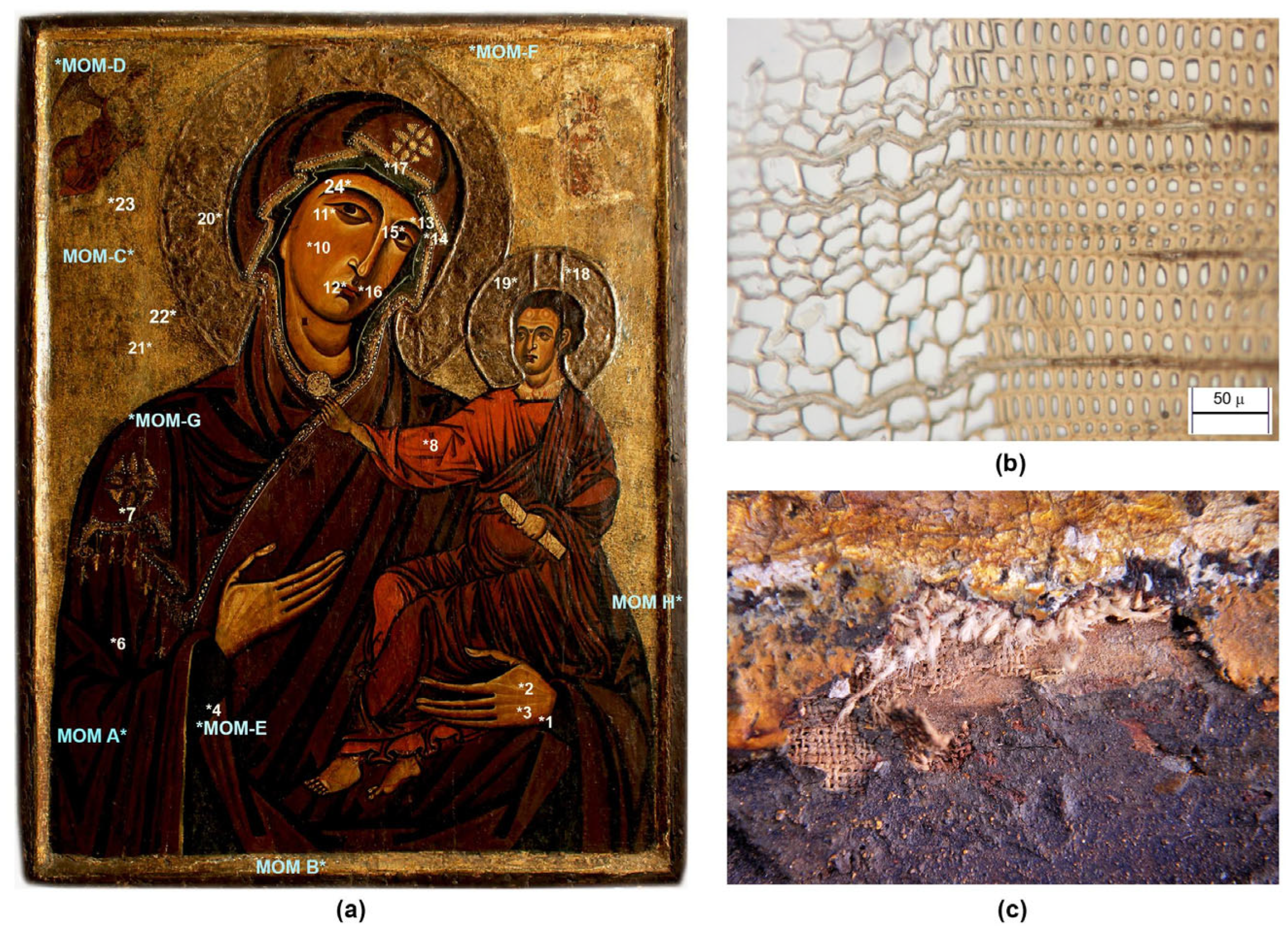
2.5. Sampling
2.6. Polarised Light Microscopy (PLM)
2.7. Environmental Scanning Electron Microscopy and Energy Dispersive X-ray Spectrometry (ESEM/EDX)
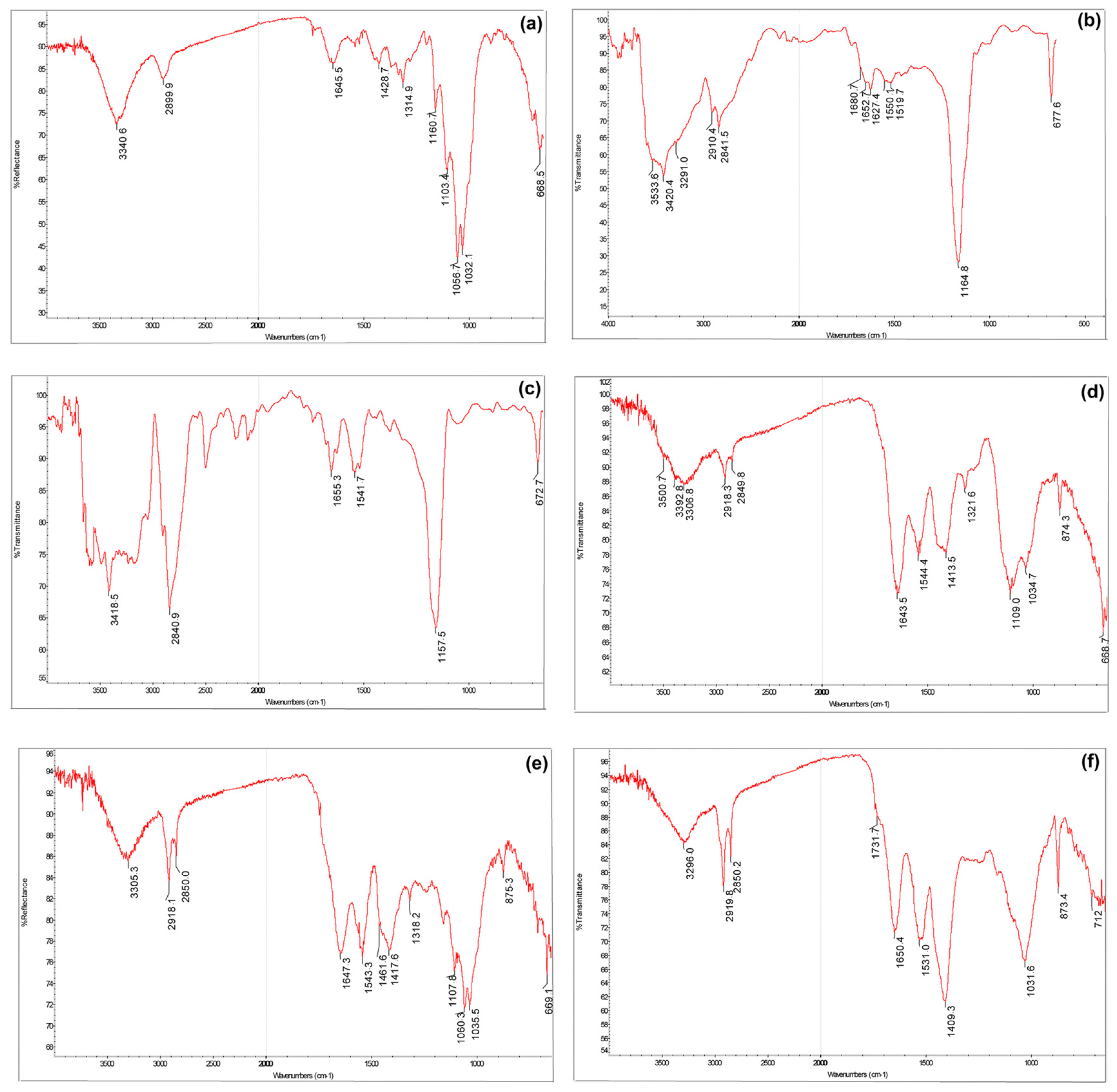
2.8. Fourier Transform Infrared Spectroscopy
2.9. Micro Raman Spectroscopy (µ-Raman)
3. Results
3.1. Supports and Ground Preparation
3.2. Preliminary Drawing
3.3. Decoration and Gilding
3.4. Painting Materials
3.5. Previous Restoration
4. Discussion
5. Conclusions
Supplementary Materials
Author Contributions
Funding
Acknowledgments
Conflicts of Interest
References
- Travagliato, G. La Madonna della Bruna di Monreale: Un testimone della maniera cypria nell’abbazia benedettina del re Guglielmo II. In L’Odigitria detta “di Guglielmo II” della Cattedrale di Monreale; Sebastianelli, M., Travagliato, G., Eds.; Palermo University Press: Palermo, Italy, 2019; pp. 18–41. [Google Scholar]
- Di Natale, M.C. Criteri di Museologia per il Museo Diocesano di Monreale. OADI 2015, 12, 15–16. [Google Scholar]
- Di Natale, M.C. L’icona di Monreale. In L’Odigitria detta “di Guglielmo II” della Cattedrale di Monreale; Sebastianelli, M., Travagliato, G., Eds.; Palermo University Press: Palermo, Italy, 2019; pp. 13–16. [Google Scholar]
- Travagliato, G. Icona graece, latine Imago dicitur: Culture figurative a confronto in Sicilia (secc. XII-XIX). In Tracce d’Oriente. La Tradizione Liturgica Greco-Albanese e Quella Latina in Sicilia, Catalogo Della Mostra (Palermo, Palazzo Bonocore, 26 Ottobre–25 Novembre 2007); Di Natale, M.C., Ed.; Edizioni Plaza Fondazione: Palermo, Italy, 2007; pp. 42–43. [Google Scholar]
- Chatzidakis, N. A Byzantine icon of the dexiokratousa Hodegetria from Crete at the Benaki Museum. In Images of the Mother of God, Perceptions of the Theotokos in Byzantium; Vassilaki, M., Ed.; Ashgate Publishing: Aldershot Hampshire, UK, 2005; pp. 337–406. [Google Scholar]
- Papageorghiou, A. Icons of Cyprus; Holy Archbishop of Cyprus: Nicosia, Cyprus, 1992; pp. 16–21. [Google Scholar]
- Sophocleus, S. Le peintre Theodoros Apsevidis et son entourage, Chypre 1183 et 1192. In Byzantinische Malerei. Bildprogramme- Ikonographie-Stil; Dr Ludwig Reichert Verlag: Wiesbaden, Germany, 2000; p. 319. [Google Scholar]
- Pace, V. La Puglia fra arte bizantina e maniera greca. In Ars auro gemmisque prior. Mélanges en hommage à Jean-Pierre Caillet; Blondeau, C., Boissavit-Camus, B., Boucherat, V., Volti, P., Eds.; University of Zagreb: Zagreb, Croatia, 2013; pp. 491–498. [Google Scholar]
- Frinta, M.S. Raised gilded adornment of the Cypriot icons, and occurrence of the technique in the west. Gesta 1981, 20, 333–347. [Google Scholar] [CrossRef]
- Mouriki, D. A Thirteen-Century Icon with a variant of the Hodegetria in Byzantine Museum of Athens. In Dumbarton Oaks Papers, Studies on Art and Archeology in Honor of ErnstKitzinger on His Seventy-Fifth Birthday; Dumbarton Oaks: Washington, WA, USA, 1987; Volume 41, pp. 403–414. [Google Scholar] [CrossRef]
- Sebastianelli, M. La Madonna Odigitria del Duomo di Monreale dalla leggenda alla realtà: Storia, tecnica e restauro critico di un’antica icona. In L’Odigitria detta “di Guglielmo II” della Cattedrale di Monreale; Sebastianelli, M., Travagliato, G., Eds.; Palermo University Press: Palermo, Italy, 2019; pp. 55–131. [Google Scholar]
- Pellerito, C.; Sebastianelli, M.; Raineri, R.; Megna, B.; Di Natale, M.C.; Pignataro, B.; Amadori, M.L.; Palla, F. Le indagini scientifiche per lo studio e la conservazione dei beni culturali: Un approccio analitico integrato, la conoscenza dei materiali costituitivi e della tecnica esecutiva della Madonna Odigitria di Monreale. In L’Odigitria detta “di Guglielmo II” della Cattedrale di Monreale; Sebastianelli, M., Travagliato, G., Eds.; Palermo University Press: Palermo, Italy, 2019; pp. 133–142. [Google Scholar]
- Weitzmann, K. Crusader icons and “La Maniera Greca”—Le Icone crociate e la “maniera greca”. In Il Medio Oriente e l’Occidente; Belting, H., Ed.; Atti del 24° Congresso Internazionale CIHA (Bologna 1979): Bologna, Italy, 1982; pp. 71–77. [Google Scholar]
- Belting, H. Likeness and Presence: A History of the Image before the Era of Art; University of Chicago Press: Chicago, IL, USA, 1994; pp. 4–5. [Google Scholar]
- Demus, O. L’arte Bizantina e l’Occidente; Einaudi: Torino, Italy, 2008. [Google Scholar]
- Bacci, M. San Luca: Il pittore dei pittori. In Artifex Bonus. Il Mondo Dell’artista Medievale; Castelnuovo, E., Ed.; Laterza: Bari, Italy, 2004; pp. 3–11. [Google Scholar]
- Lidov, A. The Miraculous Image in the Late Middle Ages and Renaissance; L’erma Di Bretschneider: Rome, Italy, 2004; pp. 273–304. [Google Scholar]
- Vasari, G. De la pittura. In Le vite de’ più eccellenti pittori, scultori, e architettori, Firenze 1550 (Torrentino) e 1568 (Giunti); Bellosi, L., Rossi, A., Eds.; Einaudi: Torino, Italy, 1991; Volume I, p. 66. [Google Scholar]
- Schmidt, V.M. Tavole dipinte. Tipologie, destinazioni e funzioni (secoli XII-XIV). In L’arte Medievale nel Contesto; Piva, P., Ed.; Jaka Book: Milano, Italy, 2006; pp. 211–212. [Google Scholar]
- Sandberg Vavalà, E. L’iconografia della Madonna col Bambino nella Pittura Italiana del Dugento; Multigrafica Editrice: Roma, Italy, 1983; p. 38. [Google Scholar]
- Teofilo. Translation and References by Tantalo, A. In Sulle Diverse Arti; Grandieri, M., Ed.; B.A. Graphics: Milano, Italy, 2005; pp. 18–19, 22–23. [Google Scholar]
- Thompson, D.V. The Materials and Techniques of Medieval Painting; Dover Publications: New York, NY, USA, 1956; pp. 105–117. [Google Scholar]
- Marette, J. Connaissance des Primitifs par l’étude du Bois, du XIIe au XVIe Siècle; A. & J. Picard: Paris, France, 1961; p. 285. [Google Scholar]
- Bomford, D.; Kirby, J. Art in the Making: Italian Painting before 1400: National Gallery London, 29 November 1989–28 February 1990; National Gallery: London, UK, 1990. [Google Scholar]
- Bellucci, R.; Castelli, C.; Ciani Passeri, F.; Ciatti, M.; Giovannini, C.; Parri, M.; Petrone, P.; Rossi Scarzanella, C.; Santacesaria, A. Tecniche pittoriche del XIII secolo: Il dossale di Meliore di Jacopo in San Leolino a Panzano. OPD Restauro 1990, 2, 186–211. [Google Scholar]
- Ramos-Poquí, G. The Technique of Icon Painting; Burns & Oates/Search Press: Tunbridge Wells, UK, 1999. [Google Scholar]
- Weissmann, G. Techniques of Traditional Icon Painting; Since Press LTD: Tunbridge Wells, UK, 2012. [Google Scholar]
- Ciatti, M. Some observations on panel painting technique in Tuscany from the twelfth to the thirteenth century. Stud. Conserv. 2013, 43, 1–4. [Google Scholar] [CrossRef]
- Mastrotheodoros, G.P.; Beltsios, K.G.; Bassiakos, Y.; Papadopoulou, V. On the metal-leaf decorations of post- Byzantine Greek icons. Archaeometry 2018, 60, 269–289. [Google Scholar] [CrossRef]
- Sotiropoulou, S.; Daniilia, S. Material aspect of icons. A review on physiochemical study of Greek icons. Acc. Chem. Res. 2010, 43, 877–887. [Google Scholar] [CrossRef]
- Lazidou, D.; Lampakis, D.; Karapanagiotis, I.; Panayiotou, C. Investigation of cross-section stratifications of icons using micro-Raman and micro-Fourier Transform Infrared (FT-IR) Spectroscopy. Appl. Spectrosc. 2018, 72, 1258–1271. [Google Scholar] [CrossRef]
- Daveri, A.; Doherty, B.; Moretti, P.; Grazia, C.; Romani, A.; Fiorin, E.; Brunetti, B.G.; Vagnini, M. An uncovered XIII century icon: Particular use of organic pigments and gilding techniques highlighted by analytical methods. Spectrochim Acta A Mol. 2015, 135, 398–404. [Google Scholar] [CrossRef]
- Salvadó, N.; Pradell, T.; Pantos, E.; Papiz, M.Z.; Molera, J.; Seco, M.; Vendrell-Saz, M. Identification of copper-based green pigments in Jaume Huguet’s Gothic altarpieces by Fourier transform infrared microspectroscopy and synchrotron radiation X-ray diffraction. J. Synchrotron Radiat. 2002, 9, 215–222. [Google Scholar] [CrossRef] [Green Version]
- Sandu, I.C.A.; Bracci, S.; Sandu, I.; Lobefaro, M. Integrated Analytical Study for the authentication of five Russian Icons (16th–17th centuries). Microsc. Res. Tech. 2009, 72, 755–765. [Google Scholar] [CrossRef]
- Miguel, C.; Lopes, J.A.; Clarke, M.; Melo, M.J. Combining infrared spectroscopy with chemometric analysis for the characterization of proteinaceous binders in medieval paints. Chemom. Intell. Lab. Syst. 2012, 19, 32–38. [Google Scholar] [CrossRef]
- Platania, E.; Streeton, N.L.W.; Lluveras-Tenorio, A.; Vila, A.; Buti, D.; Caruso, F.; Kutzke, H.; Karlsson, A.; Colombini, M.P.; Uggerud, E. Identification of green pigments and binders in late medieval painted wings from Norwegian churches. Microchem. J. 2020, 156, 104811. [Google Scholar] [CrossRef]
- Franceschi, E.; Nole, D.; Vassallo, S. Archaeometric Non-Invasive Study of a Byzantine Albanian Icon. J. Sci. Res. Rep. 2013, 2, 17–34. [Google Scholar] [CrossRef] [Green Version]
- Daniilia, S.; Bikiaris, D.; Burgio, L.; Gavala, P.; Clark, R.J.H.; Chryssoulakis, Y. An extensive non-destructive and micro-spectroscopic study of two post-Byzantine over-painted icons of the 16th century. J. Raman Spectrosc. 2002, 33, 807–814. [Google Scholar] [CrossRef]
- Karapanagiotis, I.; Minopoulou, E.; Valianou, L.; Daniilia, S.; Chryssoulakis, Y. Investigation of the colourants used in icons of the Cretan School of iconography. Anal. Chim. Acta 2009, 647, 231–242. [Google Scholar] [CrossRef]
- Valianou, L.; Wei, S.; Mubarak, M.S.; Farmakalidis, H.; Rosenberg, E.; Stassinopoulos, S.; Karapanagiotis, I. Identification of organic materials in icons of the Cretan School of iconography. J. Archeol. Sci. 2011, 38, 246–254. [Google Scholar] [CrossRef]
- Armetta, F.; Chirco, G.; Ciaramitaro, V.; Caponetti, E.; Midiri, M.; Saladino, M.L. Sicilian Byzantine Icons through the Use of Non-Invasive Imaging Techniques and Optical Spectroscopy: The Case of the Madonna dell’Elemosina. Molecules 2021, 26, 7595. [Google Scholar] [CrossRef]
- Sandu, I.C.A.; Afonso, L.U.; Murta, E.; De Sa, M.H. Gilding techniques in religious art between East and West, 14th–18th centuries. Int. J. Conserv. Sci. 2010, 1, 47–62. [Google Scholar]
- Conover, D.M.; Delaney, J.K.; Loew, M.H. Automatic Registration and Mosaicking of Conservation Images. In Optics for Arts, Architecture, and Archaeology IV; SPIE: Bellingham, WA, USA, 2013; Volume 87900A. [Google Scholar] [CrossRef]
- Dyer, J.; Verri, G.; Cupitt, J. Multispectral Imaging in Reflectance and Photo-Induced Luminescence Modes: A User Manual; Online: European CHARISMA Project; The British Museum: London, UK, 2013; pp. 122–151. [Google Scholar]
- Triolo, P.A.M. Manuale Pratico di Documentazione e Diagnostica per Immagine per i BB.CC; Il Prato: Saonara, Italy, 2019; pp. 222–223. [Google Scholar]
- Cartwright, C.R. The principles, procedures and pitfalls in identifying archaeological and historical wood samples. Ann. Bot. 2015, 116, 1–13. [Google Scholar] [CrossRef]
- Mazzeo, R.; Menu, M.; Amadori, M.L.; Bonacini, I.; Itié, E.; Eveno, M.; Joseph, E.; Lambert, E.; Laval, E.; Prati, S. Examination of the Uomini Illustri: Looking for the Origins of the portraits in the Studiolo of the Ducal Palace of Urbino. Part 2. In Studying Old Master Paintings: Technology and Practice; Spring, M., Ed.; Archetype Publications and the National Gallery: London, UK, 2011; pp. 44–51. [Google Scholar]
- Pellegrini, D.; Duce, C.; Bonaduce, I.; Biagi, S.; Ghezzi, L.; Colombini, M.P.; Tinè, M.R.; Bramanti, E. Fourier Transform Infrared Spectroscopic Study of Rabbit Glue/Inorganic Pigments Mixtures in Fresh and Aged Reference Paint Reconstructions. Microchem. J. 2015, 124, 31–32. [Google Scholar] [CrossRef]
- Conti, C.; Casati, M.; Colombo, C.; Realini, M.; Brambilla, L.; Zerbo, G. Phase transformation of calcium oxalate dihydrate–monohydrate: Effects of relative humidity and new spectroscopic data. Spectrochim. Acta A Mol. Biomol. Spectrosc. 2014, 128, 413–419. [Google Scholar] [CrossRef] [PubMed]
- Amadori, M.L.; Mengacci, V.; Vagnini, M.; Casoli, A.; Holakooei, P.; Eftekhari, N.; Kyi, L.; Maekawa, Y.; Germinario, G. Organic Matter and Pigments in the wall Paintings of Me-Taw-Ya Temple in Bagan Valley, Myanmar. Appl. Sci. 2021, 11, 11441. [Google Scholar] [CrossRef]
- Lane, M.D. Mid-infrared emission spectroscopy of sulphate and sulphate-bearing minerals. Am. Min. 2007, 92, 1–18. [Google Scholar] [CrossRef]
- Galassi, M.C. Il disegno svelato; Ilisso: Nuoro, Italy, 1998; p. 18. [Google Scholar]
- Bell, I.M.; Clark, R.J.H.; Gibbs, P.J. Raman spectroscopic library of natural and synthetic pigments (pre- ≈ 1850 AD). Spectrochim. Acta A. 1997, 53, 2159. [Google Scholar] [CrossRef] [PubMed]
- Kirby, J.; Spring, M.; Higgitt, C. The technology of red lake pigment manufacture: Study of the dyestuff substrate. Natl. Gallery Tech. Bull. 2005, 6, 71–87. [Google Scholar]
- Gliozzo, E. Pigments—Mercury-based red (cinnabar-vermilion) and white (calomel) and their degradation products. Archaeol. Anthropol. Sci. 2021, 13, 210. [Google Scholar] [CrossRef]
- Bonizzoni, L.; Bruni, S.; Gargano, M.; Guglielmi, V.; Zaffino, C.; Pezzotta, A.; Pilato, A.; Auricchio, T.; Delvaux, L.; Ludwig, N. Use of integrated non-invasive analyses for pigment characterization and indirect dating of old restorations on one Egyptian coffin of the XXI dynasty. Microchem. J. 2018, 138, 122–131. [Google Scholar] [CrossRef]
- Burgio, L.; Clark, R.J.H. Library of FT-Raman spectra of pigments, minerals, pigment media and varnishes, and supplement to existing library of Raman spectra of pigments with visible excitation. Spectrochim Acta A 2001, 57, 1491. [Google Scholar] [CrossRef]
- Kovala-Demertzi, D.; Papathanasis, L.; Mazzeo, R.; Demertzis, M.A.; Varella, E.A.; Prati, S. Pigment identification in a Greek icon by optical microscopy and infrared microspectroscopy. J. Cult. Herit. 2012, 13, 107–113. [Google Scholar] [CrossRef]
- Higgitt, C.; Spring, M.; Saunders, D. Pigment-medium interactions in oil paint films containing red lead or lead-tin yellow. Natl. Gallery Tech. Bull. 2003, 24, 75–95. [Google Scholar]
- Cardinali, M.; De Ruggieri, M.B.; Falcucci, C. Diagnostica Artistica. Tracce Materiali per la Storia Dell’arte e per la Conservazione; Palombi Editori: Roma, Italy, 2007; pp. 49–50, 115–118. [Google Scholar]
- Cosentino, A. Effects of different binders on technical photography and infrared reflectography of 54 historical pigments. Int. J. Conserv. Sci. 2015, 6, 290–291. [Google Scholar]
- Sebastianelli, M. Le strutture di sostegno dei dipinti su supporto tessile: I telai. Atti del VII Congresso Nazionale IGIIC (Napoli, Castel Dell’Ovo, 8–10 ottobre 2009). Lo Stato Dell’arte 2009, 7, 389–399. [Google Scholar]
- Guiggi, S. Da Bisanzio alla Russia e all’Italia: Il viaggio dell’icona. Web publication/site Porphyra. Arte e scienza a Bisanzio 2010, 1, 76. Available online: http://www.porphyra.it/numero-14-arte-e-scienza-a-bisanzio/ (accessed on 13 December 2022).
- Castelli, C. Tecniche di costruzione dei supporti lignei. In Dipinti su Tavola. La Tecnica e la Conservazione dei Supporti; Ciatti, M., Castelli, C., Santacesaria, A., Eds.; Edifir: Firenze, Italy, 1999; p. 60. [Google Scholar]
- Timar, M.C.; Gurău, L.; Porojan, M. Wood Species Identification. Int. J. Conserv. Sci. 2012, 3, 11–22. [Google Scholar]
- Uzielli, L. Historical overview of panel-making techniques in Central Italy. In ‘The Structural Conservation of Panel Paintings’ Proceedings of a Symposium at the J. Paul Getty Museum, 24–28 April 1995; Dardes, K., Rothe, A., Eds.; Getty Museum: Los Angeles, CA, USA, 1998; p. 114. [Google Scholar]
- Romagnoli, M.; Sarlatto, M.; Terranova, F.; Bizzarri, E.; Cesetti, S. Wood identification in the Cappella Palatina ceiling (12th century) in Palermo (Sicily, Italy). IAWA J. 2007, 28, 109–123. [Google Scholar] [CrossRef] [Green Version]
- Mastrotheodoros, G.P.; Beltsios, K.G. On the grounds of post-Byzantine Greek icons. Archaeometry 2016, 58, 830–847. [Google Scholar] [CrossRef]
- Burgio, L.; Clark, R.J.H.; Theodoraki, K. Raman microscopy of Greek icons: Identification of unusual pigments. Spectrochim. Acta A. 2003, 59, 2371–2389. [Google Scholar] [CrossRef]
- Melo, H.P.; Cruz, A.J.; Candeias, A.; Mirão, J.; Cardoso, A.M.; Oliveira, M.J.; Valadas, S. FTIR of calcium sulphate-based preparatory layers. Archaeometry 2014, 56, 513–526. [Google Scholar] [CrossRef]
- Gómez, S.S.; Moya, M.S.A.; Ródriguez, J.L.B.; Sastre, O.C.; Aglio, M.I.B.; Muñoz, A.R. Contribution to the study of grounds for panel painting of the Spanish school in the fifteenth and sixteenth centuries. In Painting Techniques: History, Materials and Studio Practice, Contributions to the Dublin Congress 7–11 September 1998; Roy, A., Smith, P., Eds.; International Institute for Conservation of Historic and Artistic Works: London, UK, 1998. [Google Scholar]
- Darque-Ceretti, E.; Felder, E.; Aucouturier, M. Foil and leaf gilding on cultural artefacts; forming and adhesion. Matéria (Rio J.) 2011, 16, 542. [Google Scholar] [CrossRef]
- Lobefaro, M. La tecnica esecutiva di base delle icone antiche. In Lo Stato dell’Arte 1, Atti del I Congresso, Nazionale IGIIC (Torino, Villa Gualino, 5–7 Giugno 2003); Il Prato: Saonara, Italy, 2003; pp. 299–304. [Google Scholar]
- Lazareff, V.N. Two newly-discovered pictures of the Lucca school. In The Burlington Magazine for Connoisseurs; Tatlock, R.R., Ed.; Savile Pub. Co.: London, UK, 1927; Volume 51, pp. 56–67. [Google Scholar]


| Sample Code | Sampling Area | Sample Description | Investigation |
|---|---|---|---|
| MOM-A | Maphorion of the Virgin | Red hue | PLM, ESEM-EDX, ATR-FTIR, µ-Raman |
| MOM-B | Table frame, front | Gilding | PLM, ESEM-EDX, ATR-FTIR |
| MOM-C | Background | Gilding | PLM, ESEM-EDX |
| MOM-D | Background near the angel, left side | Blue-red repainting | ESEM-EDX |
| MOM-E | Virgin’s sleeve | Blue hue | µ-Raman |
| MOM-F | Background near, right side | Blue repainting | µ-Raman |
| MOM-G | Right shoulder of the Virgin | Black underdrawing | µ-Raman |
| MOM-H | Back of the panel | Wood support | PLM |
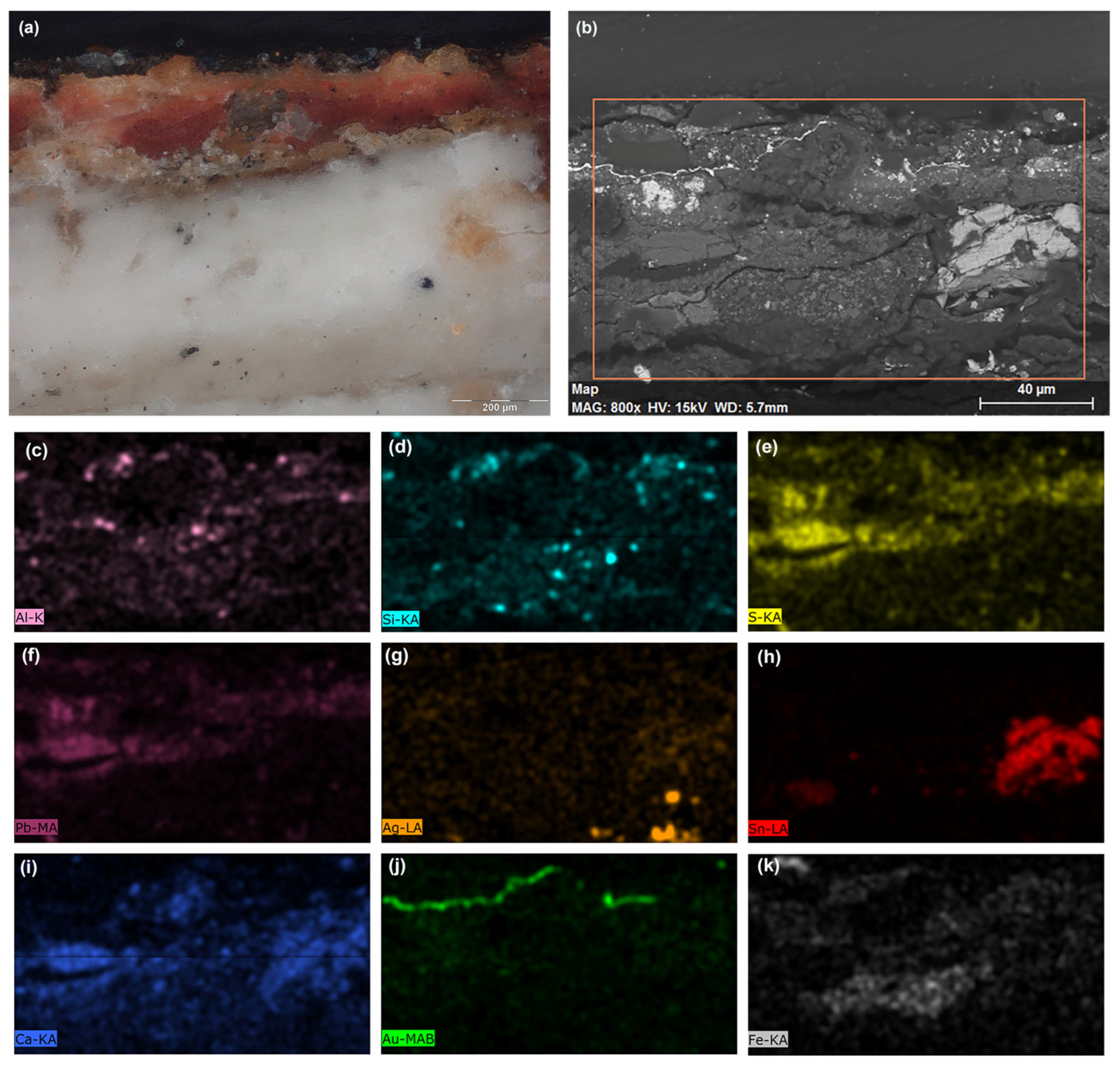
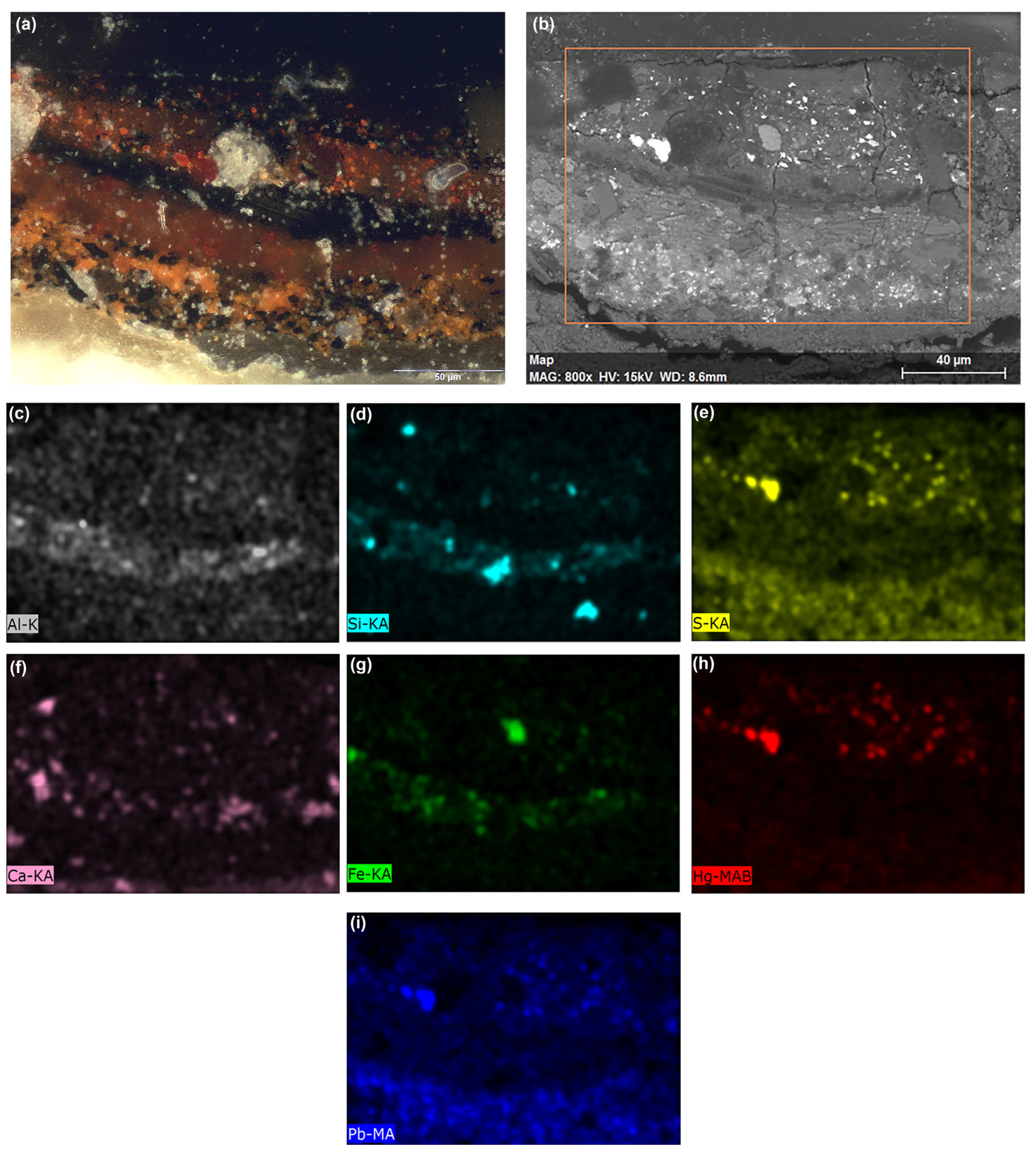
| Layer | Maximum Thickness (µm) | Typology | EDX, Detected Elements | Stratigraphic Identification (PLM, ESEM-EDX) | ATR-FTIR Identifications |
|---|---|---|---|---|---|
| 4 | 20 | repainting | C, Si, Al, Fe, K, Ca, Cu, P, Na, Ba, S | Fe-based pigments, Cu-based pigment, barite, organic matter | Proteins, silicates |
| 3 | 20 | S, Hg, Si, Al, Fe, Mg, Ca | Mercury-based red, red ochre, calcium carbonate, bone black | ||
| C | Film-forming substance | ||||
| 2 | 20 | original paint layer | Al, Fe, Ca, Si, K, Na, Mg | Red ochre, haematite, calcium carbonate, quartz | Calcium carbonate, silicates, proteins, esters of carboxylic acids, lead carbonate |
| 1 | 55 | Pb, C, Ca, P, Si, Al, K, Mg, Na | Minium, calcium carbonate, carbon black | ||
| underdrawing | |||||
| 0 | 1250 | ground layer | S, Ca, Si, Mg, Na, Al, K, Fe, P | Calcium sulphate, silicates, calcite, dolomite, microfossil, carbon black | Calcium sulphate dihydrate, proteins |
| canvas | Linen fibres | Cellulose, proteins | |||
| −1 | 1900 | ground layer | S, Ca, Fe, Si, Al, K, Na, Mg, P | Calcium sulphate, silicates, carbon black | Calcium sulphate dihydrate, proteins |
| canvas | Linen fibres | Cellulose, calcium oxalates, proteins |
| Layers | Maximum Thickness (µm) | Typology | EDX, Detected Elements | Stratigraphic Identification (PLM, SEM/ESEM-EDX) | ATR-FTIR Identification |
|---|---|---|---|---|---|
| 5 | 10 | gilding | Au | Gold leaf | |
| 4 | 60 | bole | Pb, Si, Ca, Al, Mg, Fe, K, Na, Cl | Lead white, calcium carbonate, red ochre | Lead white, kaolinite, proteins, esters of carboxylic acids |
| 3 | 23 | underlayer | Ca, S | Calcium sulphate | Calcium sulphate dihydrate, lipidic compound, esters of carboxylic acids |
| 2 | 20–80 | paint | Sn, Al, Ca, Si, Fe, Mg, K, P | Red lake, tin mordant residues, red ochre | Anthraquinone compounds—red lake, calcium carbonate, calcium oxalates |
| 1 | 10 | gilding | C, Ag | Silver leaf, film-forming substance | Silicates, proteins, calcium carbonate, calcium oxalates |
| 0 | 1250 | ground layer | Ca, S, Si, Al, K, Fe, Mg, Cl, P, Na, Ti | Calcium sulphate, silicates, carbon black, linen fibres | Anhydrite, calcium sulphate dihydrate, proteins |
| canvas | Linen fibres | Cellulose, calcium oxalates | |||
| −1 | 1600 | ground layer | Ca, S, Si, Al, K, Fe, Mg, Cl, P, Na, Ti | Calcium sulphate, calcium carbonate, kaolinite, silicates, microfossil | Anhydrite, proteins |
Disclaimer/Publisher’s Note: The statements, opinions and data contained in all publications are solely those of the individual author(s) and contributor(s) and not of MDPI and/or the editor(s). MDPI and/or the editor(s) disclaim responsibility for any injury to people or property resulting from any ideas, methods, instructions or products referred to in the content. |
© 2023 by the authors. Licensee MDPI, Basel, Switzerland. This article is an open access article distributed under the terms and conditions of the Creative Commons Attribution (CC BY) license (https://creativecommons.org/licenses/by/4.0/).
Share and Cite
Amadori, M.L.; Mengacci, V.; Sebastianelli, M.; Pignataro, B.; Agnello, S.; Triolo, P.; Pellerito, C. New Insight on Medieval Painting in Sicily: The Virgin Hodegetria Panel in Monreale Cathedral (Palermo, Italy). Heritage 2023, 6, 4692-4709. https://doi.org/10.3390/heritage6060249
Amadori ML, Mengacci V, Sebastianelli M, Pignataro B, Agnello S, Triolo P, Pellerito C. New Insight on Medieval Painting in Sicily: The Virgin Hodegetria Panel in Monreale Cathedral (Palermo, Italy). Heritage. 2023; 6(6):4692-4709. https://doi.org/10.3390/heritage6060249
Chicago/Turabian StyleAmadori, Maria Letizia, Valeria Mengacci, Mauro Sebastianelli, Bruno Pignataro, Simonpietro Agnello, Paolo Triolo, and Claudia Pellerito. 2023. "New Insight on Medieval Painting in Sicily: The Virgin Hodegetria Panel in Monreale Cathedral (Palermo, Italy)" Heritage 6, no. 6: 4692-4709. https://doi.org/10.3390/heritage6060249