
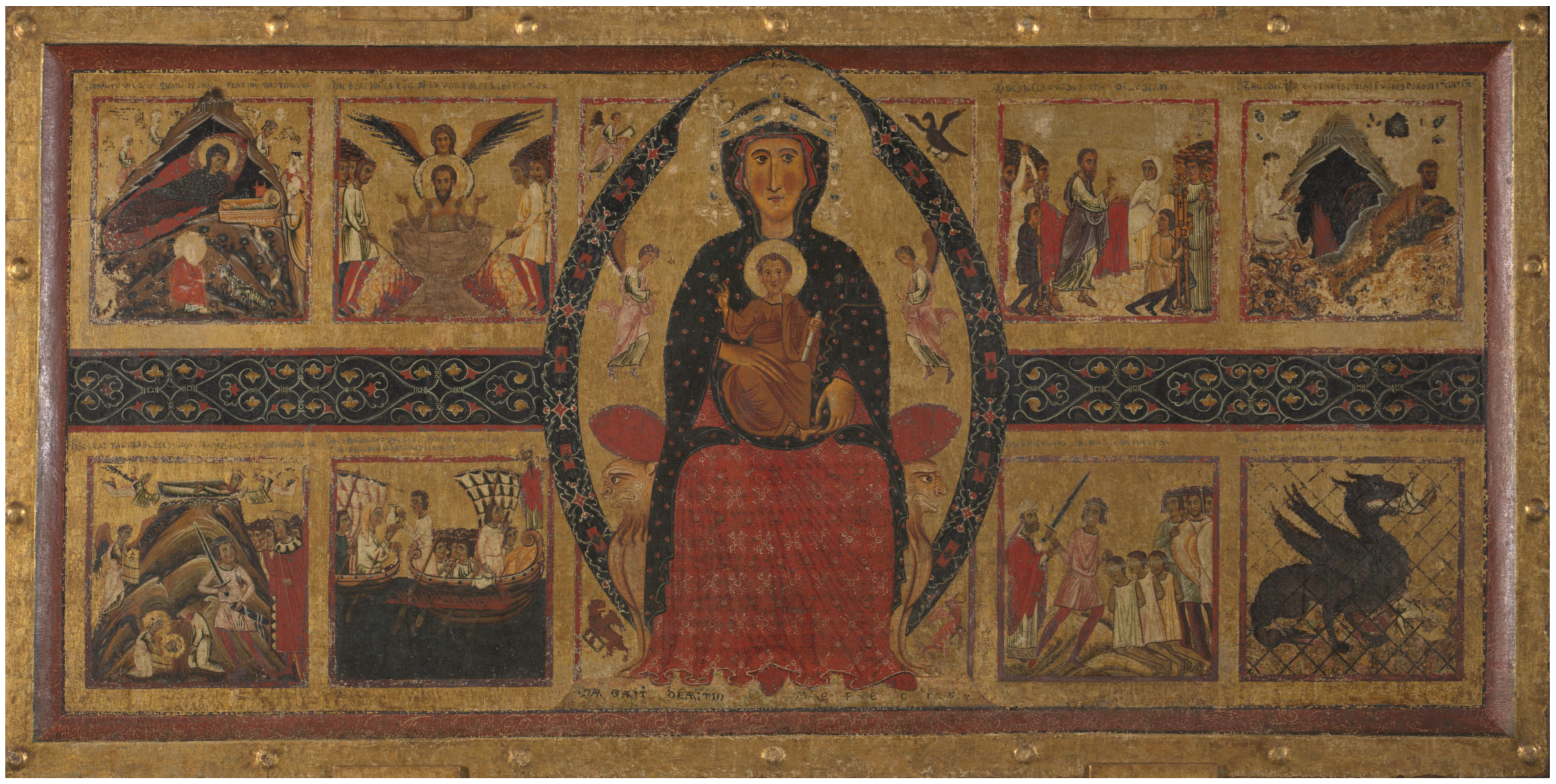
The Unexpected Discovery of Syngenite on Margarito d’Arezzo’s The Virgin and Child Enthroned, with Scenes of the Nativity and the Lives of the Saints (Probably 1263–4) and Its Possible Use as a Yellow Lake Substrate


Abstract
:1. Introduction
2. Materials and Methods
2.1. Analytical Techniques
2.1.1. HPLC
2.1.2. (ATR) FTIR
2.1.3. XRD
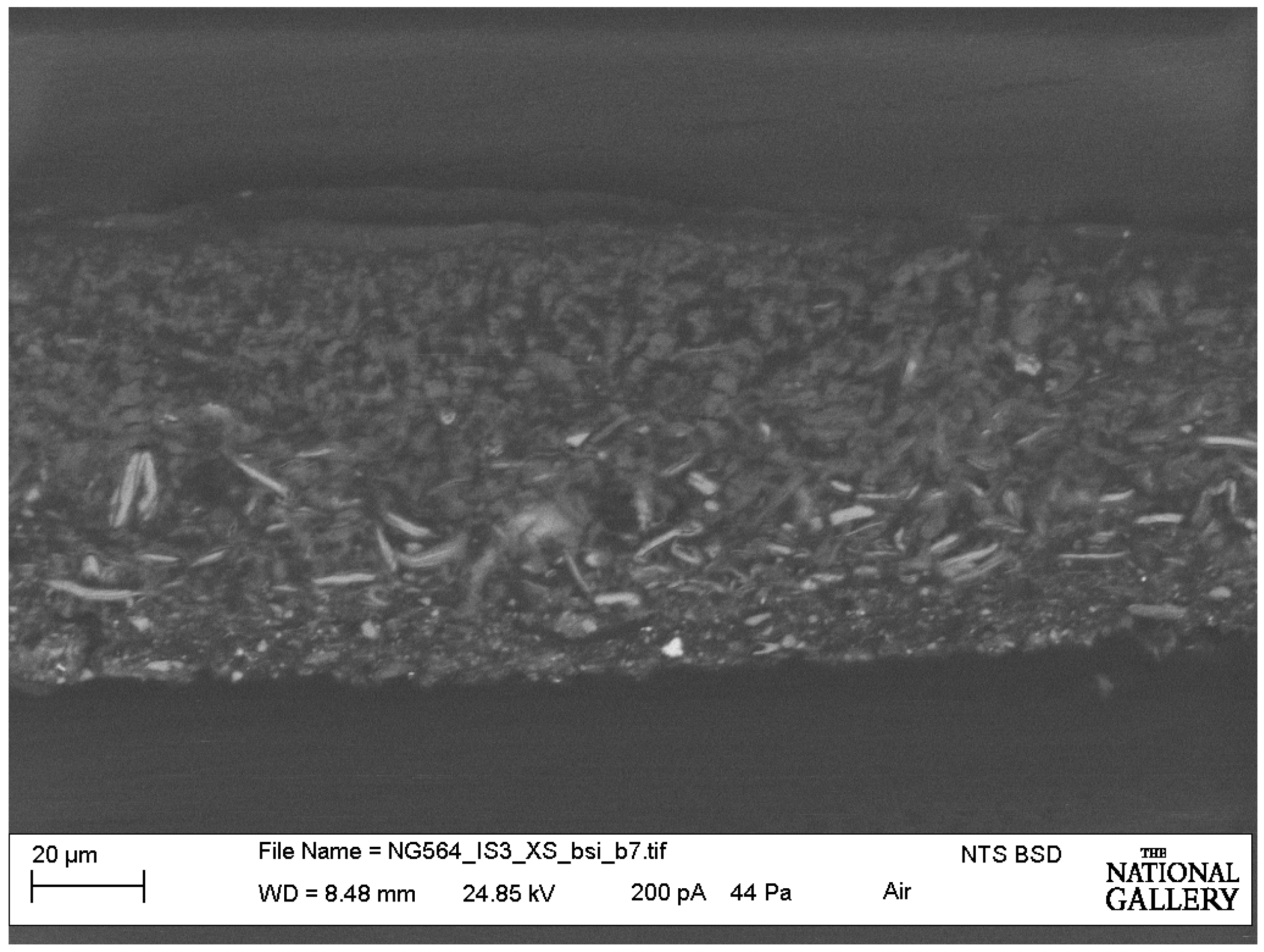
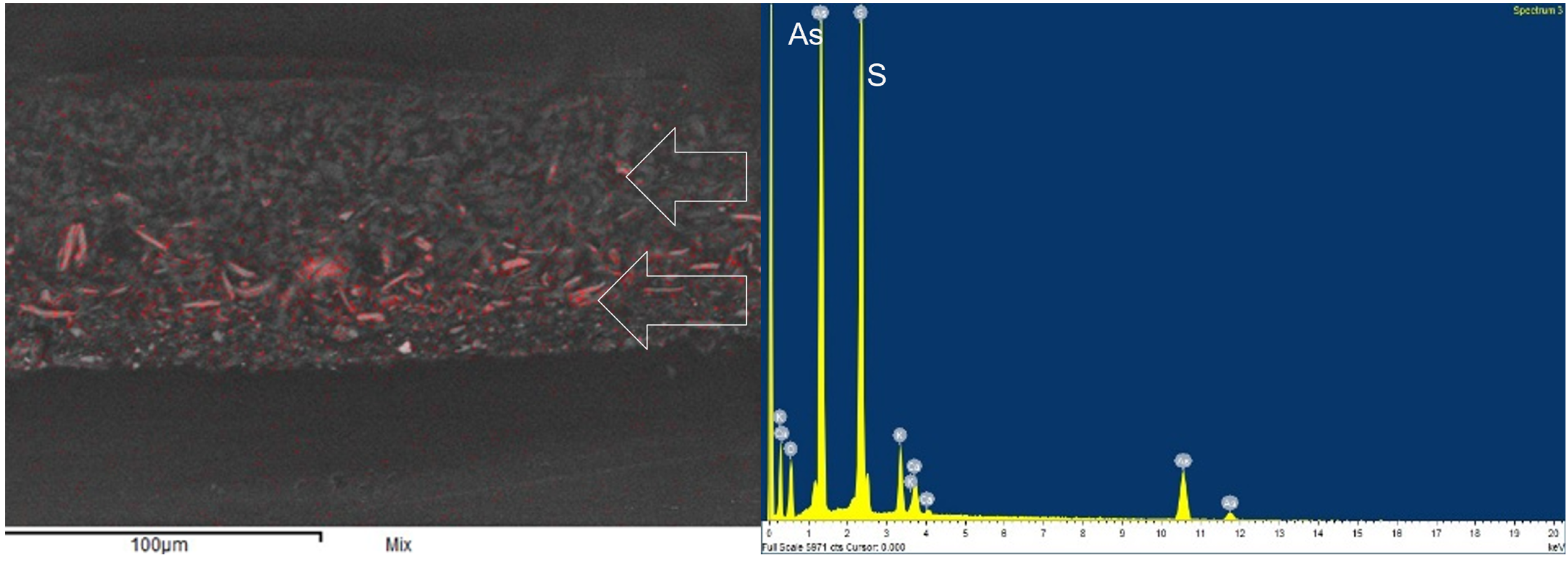
2.1.4. SEM-EDS
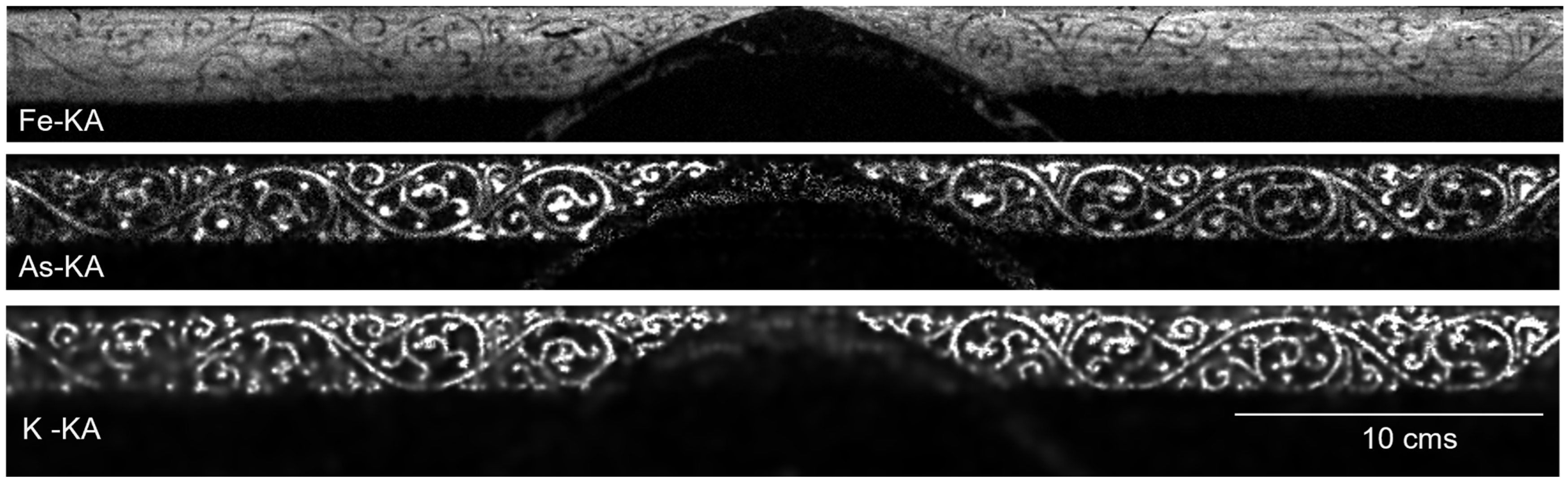
2.1.5. MA-XRF
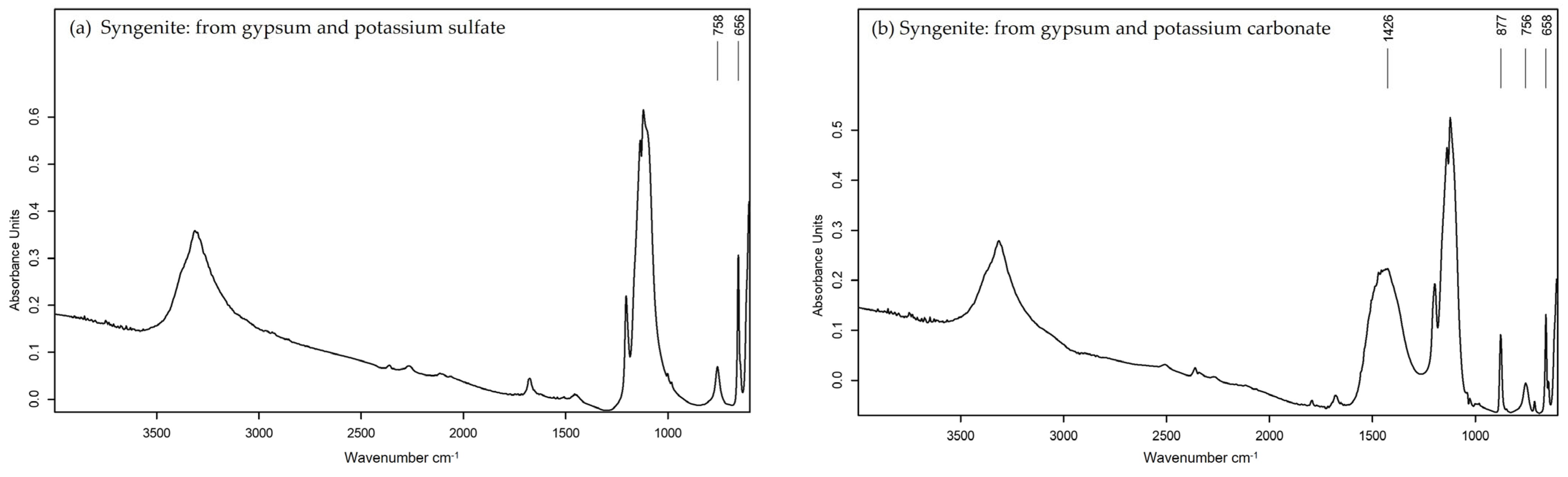
2.2. Synthesis of Syngenite
2.3. Preparation of Reference Organic Pigments
2.3.1. Weld on Gypsum Support
2.3.2. Weld on Syngenite Support
2.3.3. Safflower
3. Results
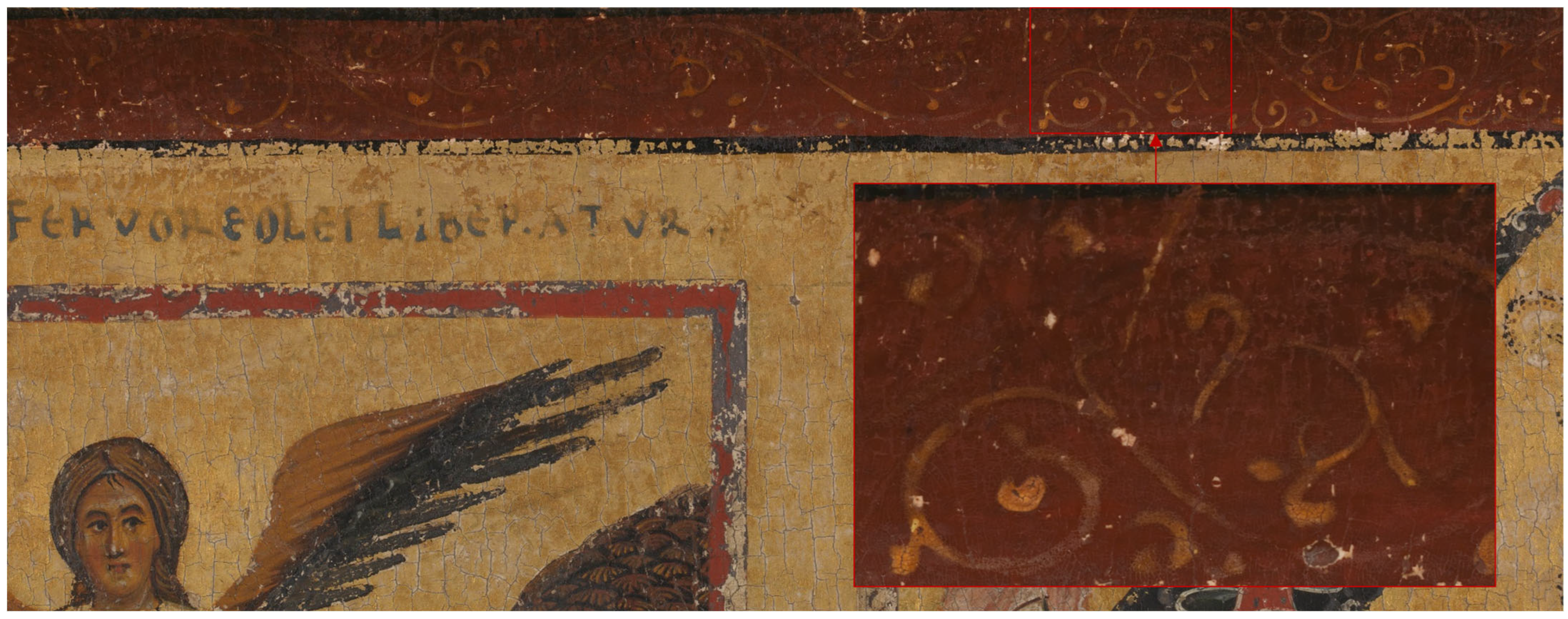
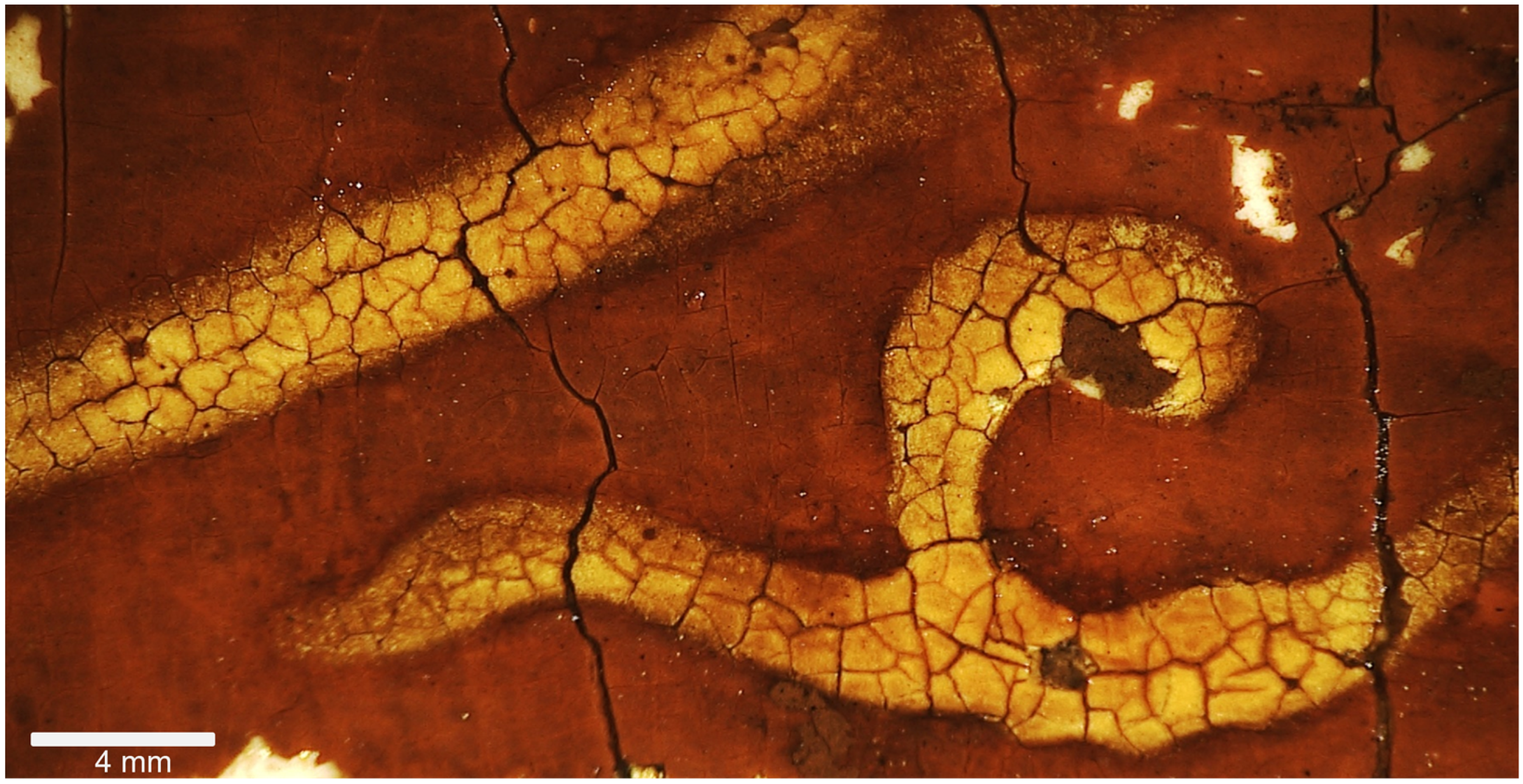
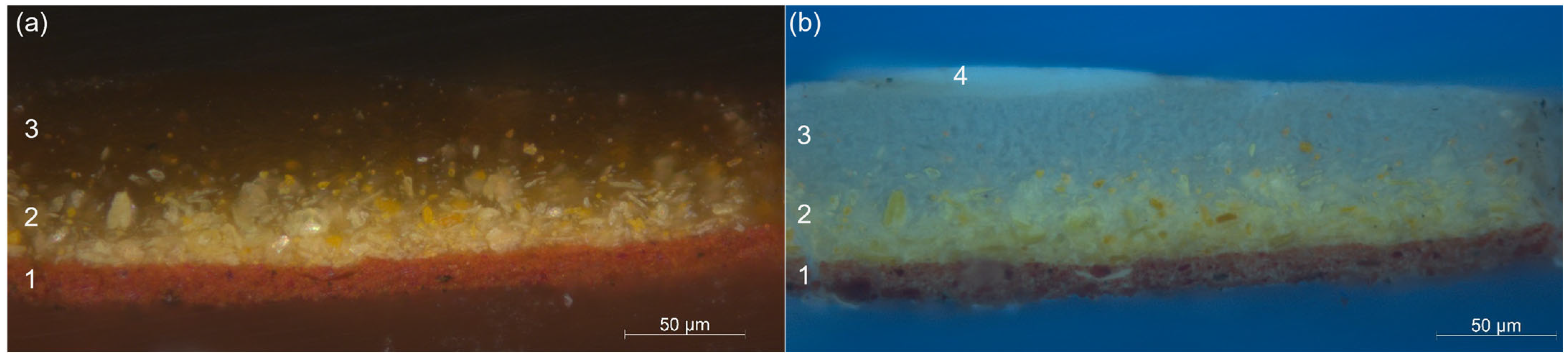
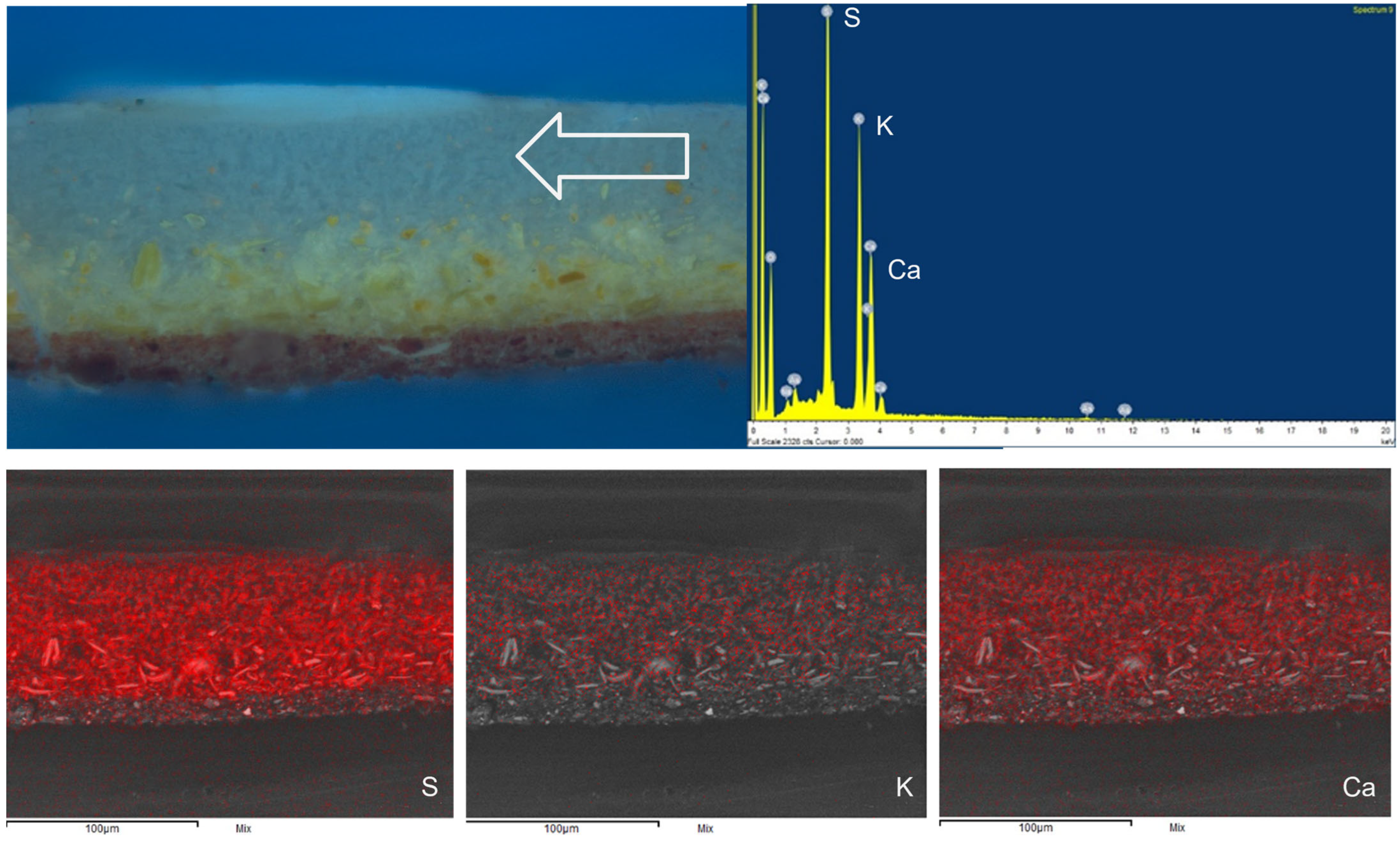
3.1. Analysis of Layer Structure of Yellow Foliate Pattern of Border
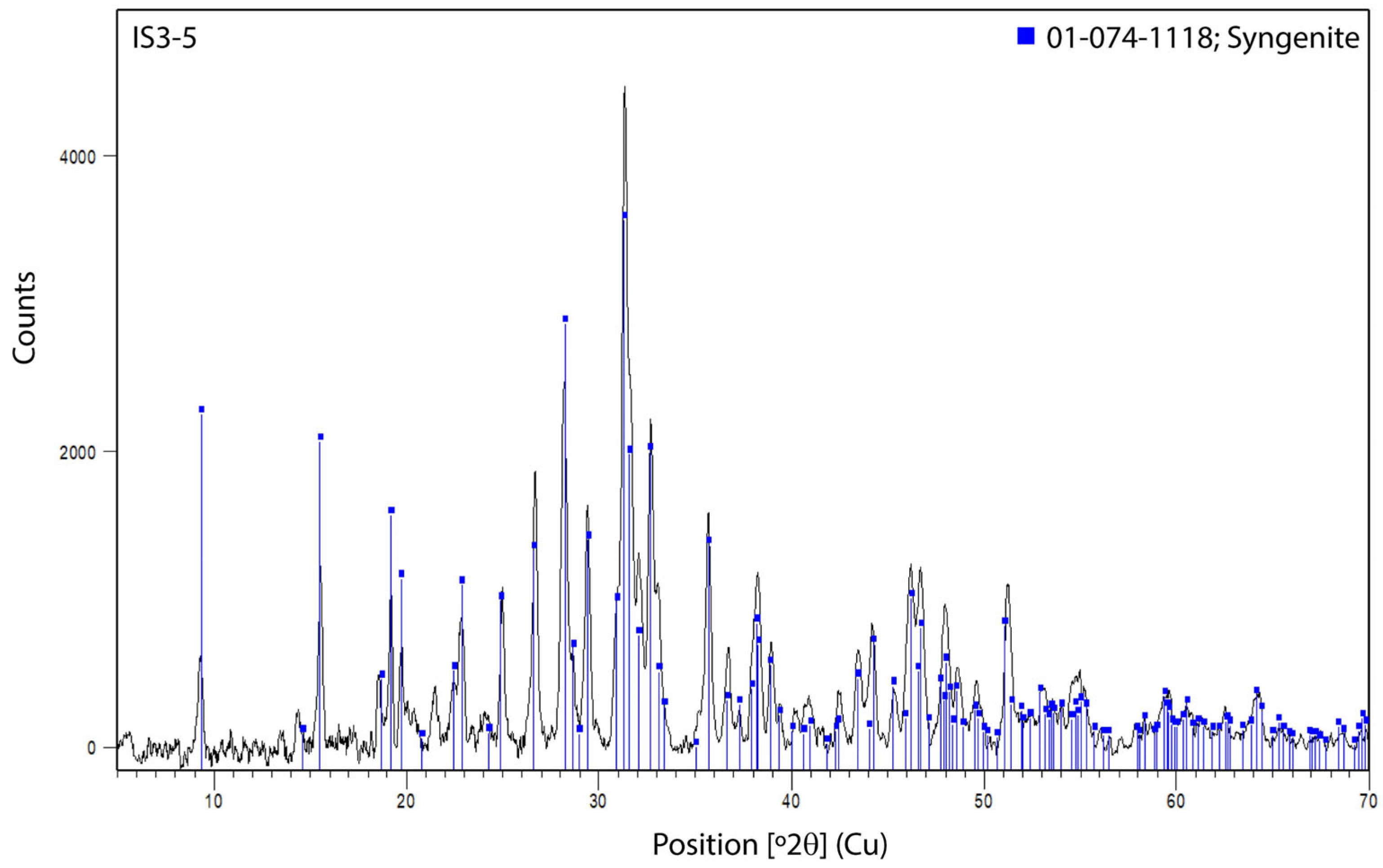
3.2. Identifcation of Syngenite (Micro-XRD Analysis)
4. Discussion
4.1. Organic Lake Pigments: Recipes and Possible Dye Sources
4.2. Syngenite and Its Possible Use as a Substrate for a Yellow Organic Lake Pigment
5. Conclusions
Author Contributions
Funding
Data Availability Statement
Conflicts of Interest
References
- Daveri, A.; Doherty, B.; Moretti, P.; Grazia, C.; Romani, A.; Fiorin, E.; Brunetti, B.G.; Vagnini, M. An uncovered XIII century icon: Particular use of organic pigments and gilding techniques highlighted by analytical methods. Spectrochem. Acta Part A Mol. Biomol. Spectrosc. 2015, 135, 398–404. [Google Scholar] [CrossRef] [PubMed]
- White, R.; Kirby, J. Some observations on the binder and dyestuff compositions of glaze paints in early European panel painting. In Medieval Painting in Northern Europe: Techniques, Analysis, Art History, Studies in Commemoration of the 70th Birthday of Unn Plahter; Nadolny, J., Kollandsrud, K., Sauerberg, M.L., Frøysaker, T., Eds.; Archetype Publications: London, UK, 2006; pp. 215–222. [Google Scholar]
- Ballirano, P.; Belardi, G.; Maras, A. Refinement of the structure of synthetic syngenite K2Ca(SO4)2.H2O from X-ray powder diffraction data. Neues Jahrb. Für Mineral. Abh. 2005, 182, 15–21. [Google Scholar]
- Ennaciri, Y.; Alaoui-Belghiti, H.E.; Bettach, M. Comparative Study of K2SO4 Production by Wet Conversion from Phosphogypsum and Synthetic Gypsum. J. Mater. Res. Technol. 2019, 8, 2586–2596. [Google Scholar] [CrossRef]
- Kloprogge, J.T.; Schuiling, R.D.; Ding, Z.; Hickey, L.; Wharton, D.; Frost, R.L. Vibrational Spectroscopic Study of Syngenite Formed during the Treatment of Liquid Manure with Sulphuric Acid. Vib. Spectrosc. 2002, 28, 209–221. [Google Scholar] [CrossRef] [Green Version]
- Smith, C.S.; Hawthorne, J.G. Mappae clavicula: A little key to the world of medieval techniques. Trans. Am. Philos. Soc. N.S. 1974, 64, 3–128. [Google Scholar] [CrossRef]
- Kirby, J. Some Aspects of Medieval and Renaissance Lake Pigment Technology. Dye. Hist. Archaeol. 2008, 21, 89–108. [Google Scholar]
- Bomford, D.; Dunkerton, J.; Gordon, D.; Roy, A.; Kirby, J. Art in the Making: Italian Painting before 1400; National Gallery Company: London, UK, 1989; rep. 2000; pp. 39, 123 and 181. [Google Scholar]
- Saunders, D.; Kirby, J. Light-induced Colour Changes in Red and Yellow Lake Pigments. Natl. Gallery Tech. Bull. 1994, 15, 79–97. [Google Scholar]
- Pollio, M.V. Vitruvius, The Ten Books on Architecture; Morgan, M.H., Translator; Harvard University Press: Cambridge, UK; Humphrey Milford, Oxford University Press: London, UK, 1914; (repr. Dover Publications Inc.: New York, NY, USA, 1960); Book VII, Chapter 14; p. 220. [Google Scholar]
- Merrifield, M.P. Original Treatises Dating from the XIIth to XVIIIth Centuries on the Arts of Painting, 2 Vols; John Murray: London, UK, 1849; (repr. Dover Publications Inc.: New York, NY, USA, 1967). [Google Scholar]
- Kirby, J.; van Bommel, M.; Verhecken, A. Natural Colorants for Dyeing and Lake Pigments: Practical Recipes and their Historical Sources; Archetype Publications: London, UK, 2014; pp. 64–65. [Google Scholar]
- Cavigli, R.; Refici, P. (Eds.) Una Madonna del XIII Secolo ad Arezzo: Restaurare per Conoscere; Istituto Nazionale Previdenza Sociale (INPS): Rome, Italy, 2015; pp. 48–49. [Google Scholar]
- Porter, C. The science of color: Color analysis and the roles of economics, geography, and tradition in the artist’s choice of colors for manuscript painting. In And Diverse Are Their Hues: Color in Islamic Art and Culture; Bloom, J., Blair, S., Eds.; The Qatar Foundation, Virginia Commonwealth University: Doha, Qatar; Virginia Commonwealth University School of the Arts in Qatar: Doha, Qatar; Yale University Press: New Haven, CT, USA, 2011; pp. 205–221. [Google Scholar]
- Barkeshli, M. Historical Persian recipes for paper dyes. Restaurator 2016, 37, 49–89. [Google Scholar] [CrossRef]
- Darmstaedter, E. Liber Misericordiae Geber. Eine lateinische Übersetzung des grösseren Kitâb alraḥma. Arch. Für Gesch. Der Med. 1925, 17, 181–197. [Google Scholar]
- Levey, M. Mediaeval Arabic bookmaking and its relation to early chemistry and pharmacology. Trans. Am. Philos. Soc. 1962, 52, 1–79. [Google Scholar] [CrossRef]
- Del Punta, I.; Rosati, M.L. Lucca, Una Città di Seta: Produzione, Commercio e Diffusione dei Tessuti Lucchesi net Tardo Medioevo; Maria Paccini Fazzi Editore: Lucca, Italy, 2017; pp. 169–170. [Google Scholar]
- Pegolotti, F.B. La Pratica Della Mercatura (c. 1340); Evans, A., Ed.; Mediaeval Academy of America: Cambridge, MA, USA, 1936; p. 372. [Google Scholar]
- Ploss, E.E. Ein Buch von alten Farben: Technologie der Textilfarben im Mittelalter mit einem Ausblick auf die festen Farben; Impuls Verlag Heinz Moos: Heidelberg/Berlin, Germany, 1962; the Nuremberg Kunstbuch, 2nd half 15th century: No. 55; pp. 115–116. [Google Scholar]
- Clarke, M. Tricks of the Medieval Trade. The Trinity Encyclopedia: A Collection of Fourteenth-Century English Craft Recipes; Archetype Publications: London, UK, 2018; pp. 46–48. [Google Scholar]
- Monico, L.; Prati, S.; Sciutto, G.; Catelli, E.; Romani, A.; Balbas, D.Q.; Li, Z.; De Meyer, S.; Nuyts, G.; Janssens, K.; et al. Development of a multi-method analytical approach based on the combination of synchrotron radiation X-ray micro-analytical techniques and vibrational micro-spectroscopy methods to unveil the causes and mechanism of darkening of ‘fake-gilded’ decorations in a Cimabue painting. J. Anal. At. Spectrom. 2022, 37, 114–129. [Google Scholar]
- Simoen, J.; De Meyer, S.; Vanmeert, F.; de Keyser, N.; Avranovich, E.; Van der Snickt, G.; van Loon, A.; Keune, K.; Janssens, K. Combined Micro- and Macro scale X-ray powder diffraction mapping of degraded Orpiment paint in a 17th century still life painting by Martinus Nellius. Heritage Sci. 2019, 7, 83. [Google Scholar] [CrossRef] [Green Version]
- Vanmeert, F.; de Keyser, N.; van Loon, A.; Klaassen, L.; Noble, P.; Janssens, K. Transmission and Reflection Mode Macroscopic X-ray Powder Diffraction Imaging for the Noninvasive Visualization of Paint Degradation in Still Life Paintings by Jan Davidsz. de Heem. Anal. Chem. 2019, 91, 7153–7161. [Google Scholar] [CrossRef] [PubMed]
- Handbook of Mineralogy. Available online: https://www.handbookofmineralogy.org/pdfs/syngenite.pdf (accessed on 7 December 2022).
- Mindat.org (Open Database of Minerals, Rocks, Meteorites and the Localities They Come from). Available online: https://www.mindat.org/min-3856.html#autoanchor19 (accessed on 7 December 2022).
- Abu-Eishah, S.I.; Bani-Kananeh, A.A.; Allawzi, M.A. K2SO4 production via the double decomposition reaction of KCl and phosphogypsum. Chem. Eng. J. 2000, 76, 197–207. [Google Scholar] [CrossRef]


| Sample ID | Major Phase | Minor Phase/s |
|---|---|---|
| IS3_1 | syngenite | weddellite; calomel or polyhalite |
| IS3_2 | syngenite | weddellite; calomel or polyhalite; anhydrite |
| IS3_3 | syngenite | calomel or polyhalite; possibly trace of weddellite |
| IS3_4 | very weak diffraction signal indicating the presence of amorphous material | |
| IS3_5 | syngenite | Calomel or polyhalite |
Disclaimer/Publisher’s Note: The statements, opinions and data contained in all publications are solely those of the individual author(s) and contributor(s) and not of MDPI and/or the editor(s). MDPI and/or the editor(s) disclaim responsibility for any injury to people or property resulting from any ideas, methods, instructions or products referred to in the content. |
© 2023 by the authors. Licensee MDPI, Basel, Switzerland. This article is an open access article distributed under the terms and conditions of the Creative Commons Attribution (CC BY) license (https://creativecommons.org/licenses/by/4.0/).
Share and Cite
Peggie, D.; Howard, H.; Kirby, J.; Najorka, J. The Unexpected Discovery of Syngenite on Margarito d’Arezzo’s The Virgin and Child Enthroned, with Scenes of the Nativity and the Lives of the Saints (Probably 1263–4) and Its Possible Use as a Yellow Lake Substrate. Heritage 2023, 6, 762-778. https://doi.org/10.3390/heritage6020041
Peggie D, Howard H, Kirby J, Najorka J. The Unexpected Discovery of Syngenite on Margarito d’Arezzo’s The Virgin and Child Enthroned, with Scenes of the Nativity and the Lives of the Saints (Probably 1263–4) and Its Possible Use as a Yellow Lake Substrate. Heritage. 2023; 6(2):762-778. https://doi.org/10.3390/heritage6020041
Chicago/Turabian StylePeggie, David, Helen Howard, Jo Kirby, and Jens Najorka. 2023. "The Unexpected Discovery of Syngenite on Margarito d’Arezzo’s The Virgin and Child Enthroned, with Scenes of the Nativity and the Lives of the Saints (Probably 1263–4) and Its Possible Use as a Yellow Lake Substrate" Heritage 6, no. 2: 762-778. https://doi.org/10.3390/heritage6020041