Review

Bioinspired Mechanical Materials—Development of High-Toughness Ceramics through Complexation of Calcium Phosphate and Organic Polymers

Department of Applied Chemistry, Faculty of Science and Engineering, Doshisha University, Kyotanabe 610-0394, Japan
*
Author to whom correspondence should be addressed.
Ceramics 2023, 6(4), 2117-2133; https://doi.org/10.3390/ceramics6040130
Submission received: 26 September 2023 / Revised: 17 October 2023 / Accepted: 27 October 2023 / Published: 30 October 2023
(This article belongs to the Special Issue Innovative Research on Calcium Phosphates Based Ceramics)

Abstract

:
Inspired by the process of bone formation in living organisms, many studies have been conducted to develop organic–inorganic composite materials by preparing calcium phosphate crystals within solutions or dispersions of polymers with appropriate functional groups. Bones are composite materials consisting of organic polymers (mainly type I collagen), carbonated apatite, and water, with volume fractions of 35–45%, 35–45%, and 15–25%, respectively. Carbonated apatite in bone contributes to rigidity, while organic polymers and water contribute to toughness. The inorganic crystal, carbonated apatite, is a plate-shaped crystal with dimensions of 50 nm × 25 nm × 1–4 nm, generating a significant organic–inorganic interface, due to its nanoscale size. This interface is believed to absorb externally applied forces to dissipate mechanical energy to thermal energy. Creating such nanometer-scale structures using top-down approaches is challenging, making bottom-up methods, such as the coprecipitation of polymer and inorganic crystals, more suitable. In this account, efforts to develop eco-friendly mechanical materials using biomass, such as cellulose and starch, based on the bottom-up approach to bone-like composites are described.

1. Introduction

Plastics are extensively used as mechanical materials in numerous products, due to their excellent mechanical properties, lightweight nature, affordability, and good processability [1]. Before the invention of plastics, materials like wood and bamboo were commonly used, but plastics have largely replaced them in many applications. However, there are several issues associated with the continued use of plastics. Firstly, plastics rely on fossil resources, such as petroleum, leading to concerns about resource depletion and carbon dioxide emissions during incineration. Additionally, many plastics lack biodegradability, causing marine pollution with microplastics, for example [2]. Another concern about the use of plastics as a structural material is the generation of poisonous gas when burning. Potential alternatives to plastics include cellulose and biodegradable plastics. However, the mass production of the parts of industrial products using bone and wood is challenging because their anisotropic mechanical properties would make a molding process difficult.
On the other hand, aside from cellulose, materials such as bones, teeth, shells, horns, and crustacean shells have been biosynthesized by living organisms, and they are integral parts of the biological structural materials. These materials are composites of organic polymers and inorganic crystals [3]. The inorganic crystals are calcium phosphates or calcium carbonates, while the organic polymers are proteins or polysaccharides, such as collagen and chitin. These composites form through the growth of inorganic crystals on the surface of organic polymers [4], resulting in composites with relatively lightweight and mechanically robust properties. If these materials can be industrially produced, they could serve as environmentally friendly alternatives to plastics [5,6]. Here, we review the structure and biosynthesis of bones, synthetic strategies of bioinspired organic–inorganic composites, and the synthesis and the mechanical properties of hydroxyapatite–polysaccharide composites. Although many studies on hydroxyapatite have been directed toward its medical applications, we focus on the mechanical properties and water resistances of the composites of hydroxyapatite and biopolymers. In particular, we focus on the composites consisting of polysaccharides, such as cellulose and starch, since the use of these biomasses will resolve the environmental issue of plastics.

2. The Structure of Bone and Related Tissues

Bone consists of three major components: carbonated apatite (nanometer-sized calcium phosphate crystals), collagen I fibers, and water in a volume ratio of approximately 56–60:30–40:10 [7,8,9]. Carbonated apatite crystals have dimensions of 50 nm in length, 25 nm in width, and 1–4 nm in thickness [10]. The small crystal size of carbonated apatite is advantageous in terms of mechanical strength [11]. When calculating the interface area between carbonated apatite and the organic phase based on the crystal size, it becomes ca. 210 m2/g. The brick-and-mortar structure (Figure 1) formed by carbonated apatite crystals and collagen fibers contributes to the toughness of bone [11,12,13,14,15]. One of the major properties of bone is stiffness, which can support and protect a body from deformation. The elastic moduli of bone range from 2 to 33 GPa, and the bending strengths range from 27 to 308 MPa [10,16]. Water plays an important role in the mechanical properties of bone [8,17,18,19,20]. Water removal causes increased stiffness but decreased toughness in bone [21]. To understand the mechanical function of bone, it is necessary to consider the interfaces and connections between bones and soft tissues. The elastic moduli of the soft tissues are 0.5–1 MPa (cartilage), 50–150 MPa (enthesis), and 0.45 GPa (tendon) [22]. The stiffness of the soft tissues varies depending on the degree of mineralization.

2.1. Carbonated Apatite and Its Interface

Hydroxyapatite (HAP) has a molecular formula of Ca10(PO4)6(OH)2. The crystals of HAP are hexagonal, with a 9.417 Å and c 6.875 Å [23]. HAP is the most stable phase formed when calcium ions and phosphate ions react under alkaline conditions. It is well known that isomorphous substitution occurs frequently: the calcium ion is substituted by Sr, Ba, Mg, Mn, K, Na, and Fe, and the phosphate ion is substituted by AsO43−, CO32− and VO43− [24]. Carbonated apatite (CAP) is formed by the substitution of OH or PO43− of hydroxyapatite by the carbonate ion, CO32−. We refer to hydroxyapatite in which the OH group has been replaced by carbonate ions as type A carbonated apatite, and when the PO43− is replaced by carbonate ions, it is referred to as type B carbonated apatite [25]. The most common crystal morphology is needle-like, and it grows along the c-axis direction. It is known that citric acid is adsorbed on the side surfaces of the needle-shaped crystals of the carbonated apatite of bone [26]. There is ongoing debate regarding the presence of organic materials on the surface of CAP, and NMR studies have revealed the binding of polysaccharides to the surface of CAP [27]. NMR studies also proved that the surface of mature bone mineral particles is made of hydrated amorphous calcium phosphate. The HPO42− ions are concentrated at the surface of bone mineral particles in the amorphous surface layer, where the thickness is estimated to be about 0.8 nm for a 4 nm thick particle of carbonated apatite [28].

2.2. Biosynthesis of Bone

Bone is formed by the precipitation of amorphous calcium phosphate on collagen fibers and its subsequent crystallization [29]. Acidic proteins containing many phosphates and/or carboxylate groups in the side chain play an important role in biomineralization [4,7,30]. These anionic groups can concentrate calcium ions, and a reaction with phosphate ions causes the precipitation of amorphous calcium phosphate. It crystallizes to carbonated apatite. Hierarchical structures of bone are described in several reviews [5,7,9,10,31,32,33,34].

2.3. Bioceramics and Woods as Biological Mechanical Materials

Bone and wood are lightweight, ductile, and biodegradable structural materials [35,36]. Although bone and wood are superior materials, there are disadvantages when it comes to their use as industrial materials. It is difficult to form the desired shape, due to their anisotropic properties, and they are not suitable for mass production because their molding process is inefficient. If we can make powder from these biomaterials and mold them into desired shapes, they can be employed as materials for various machine components.

3. Composite Materials Related to Bone

3.1. General Features of Organic–Inorganic Composites

Organic–inorganic composite materials can be broadly classified into two categories: ceramics-reinforced plastics and polymer-reinforced ceramics (Table 1). For the former, the low rigidity of the organic phase is enhanced through the addition of the inorganic phase. Glass fiber-reinforced plastics, clay–polyamide composites [37], and rubber, an important component of automobile tires, for example, are commonly used in daily life. If plastic is too soft to resist external stress, we can add inorganic fillers, such as glass fibers, to increase its stiffness. The major component of the composites of this category is an organic phase, and the minor component is an inorganic phase. For “polymer-reinforced ceramics”, the brittleness of ceramics is improved by incorporating organic polymers, as seen in laminated glass. For laminated glass, due to the sandwiched structure of glass plate–polymer film–glass plate, it would not be broken into pieces if a shock is given, owing to the organic polymer film. The major component of this category is an inorganic phase, and the minor component is an organic phase. Bone is polymer-reinforced ceramics and can be roughly described as nanometer-sized laminated glass with a brick-and-mortar structure (Figure 1). According to the brick-and-mortar model, ceramic in bone is a dispersed phase, and polymer is a continuous phase. When we attempt to prepare a bone-like composite, as the weight fraction of ceramics increases and is close to one, the ceramic phase becomes a continuous phase, resulting in fragile materials. Therefore, the fabrication of the brick-and-mortar structure becomes difficult, as the weight fraction of the ceramic phase is close to one.

3.2. Preparation of Organic–Inorganic Composites

For the preparation of organic–inorganic composites, the following three protocols have been reported:
  • Direct mixing of polymer and inorganic crystals.
  • Polymerization of the monomer in the presence of the inorganic crystal powder.
  • Crystallization of the inorganic phase in the presence of the organic polymer.
As an example of the first protocol, Bonfield and coworkers reported that a composite of polyethylene and apatite was prepared by mixing both components [38]. The elastic modulus of the composite was linearly related to the volume fraction of apatite. When the volume fraction was varied from 0 to 0.6, the elastic modulus ranged from 1 to 12 GPa, demonstrating that the filler enhanced the stiffness of the composites. The second protocol was employed to prepare the composite of poly(L-lactic acid-co-glycolic acid) and hydroxyapatite [39]. Polyamide–clay composites were prepared by the ring-open polymerization of ε-caprolactam in the presence of clay minerals and montmorillonite, which was ion-exchanged by ω-carboxyalkylammonium. Thus, the polyamide–clay composite was also prepared using the second protocol [37].
The third protocol is a biomimetic, bottom-up approach and is suitable for the preparation of nanometer-sized inorganic crystals that are bound to the polymer phase. Stupp et al. reported that precipitation occurred when an aqueous solution of polyglutamic acid was mixed with calcium hydroxide and phosphoric acid, and the resulting precipitate was the composite of the polymer and the apatite [40]. Polyglutamic acid sodium salt, polylysine hydrochloride, and polyacrylic acid aqueous solution (1 mM) were mixed with calcium hydroxide and phosphoric acid, and the pH of the mixture was adjusted to 7.4 at 37 °C to obtain polymer–HAP composites as precipitates. The composites were called organoapatite, and X-ray diffraction showed that the composites contained poorly crystalline hydroxyapatite. The ratio of Ca/P was close to 1.6, the theoretical value of hydroxyapatite in the absence of polymers, while the ratio was 1.4 to 1.6 in the presence of the polymers, showing that calcium-deficient hydroxyapatite formed.
Anionic functional groups of proteins play a crucial role in the biomineralization of bones and teeth [41,42,43,44,45,46,47,48,49,50,51,52,53,54]. Akkus and coworkers crystallized HAP in the presence of charged peptides and performed a systematic investigation on the effects of charged peptides on the HAP morphology [55]. They found that the amounts of anionic peptides, such as poly-L-Asp and poly-L-Glu bound to HAP, were larger than those of cationic peptides, such as poly-L-Lys and poly-L-Arg. Negatively charged peptides led to smaller crystals in HAP than positively charged ones. Research involving the complexation of hydroxyapatite with various polymers and polymer gels was carried out and reviewed [32,56,57,58,59,60,61,62,63,64,65,66,67,68,69,70,71,72,73,74,75,76,77,78].
Carbonated apatite in bone is a hexagonal crystal, and citrate anions are adsorbed onto the a and b faces [26]. Anions are adsorbed onto the a and b faces of hydroxyapatite, while cations are adsorbed onto the c face [48,79]. Thus, by adding calcium ions and phosphate ions to solutions of polymers with anionic functional groups, such as carboxylate, or phosphate groups, under an alkaline condition, the polymers are adsorbed onto the a and b faces of apatite through their anions, leading to crystal growth along the c-axis. As a result, it might be possible to control the crystal size in the nanometer range and achieve the composite formation of organic polymers and needle-like or plate-like inorganic crystals bonded with each other with aligned orientations. Formation of the brick-and-mortar structure is crucial for the toughness of the composites [3,9,12,15]. The polymers used here can include petroleum-derived synthetic polymers, proteins, and polysaccharides, but the use of biomass-derived polysaccharides synthesized from carbon dioxide through photosynthesis could address carbon dioxide emission and environmental issues.

3.3. Composite of Hydroxyapatite and Polysaccharide

The use of polysaccharides as an organic component of the composite is an attractive strategy because they are carbon neutral and degrade biologically. There have been several studies on the preparation of the composites of hydroxyapatite and polysaccharides [53,76,80,81,82]. Yao and coworkers crystallized HAP in the presence of polysaccharides and found that the crystal size of HAP decreased in the order of amylose (−OH) > chitosan (−NH2, −NHCOCH3, and −OH) > carrageenan (−OSO3 and −OH) > pectin (−COO and −OH) [53]. The polar functional groups of these polysaccharides are shown in the parentheses. It seems that the –COO group is the most effective in suppressing the crystal growth of HAP. We can avoid environmental issues, such as the microplastic pollution of sea water, by employing polysaccharide-based structural materials. The annual production of cellulose is 1.5 × 10¹² tons [83], while that of starch is 8 × 10⁸ tons (corn starch) [84]. Considering the large annual production of cellulose, the use of cellulose as an organic phase of the composite seems attractive. Yu and coworkers reported a composite of cellulose nanofibers and TiO2-coated mica. It showed a bending strength of 281 MPa and an elastic modulus of 20 GPa [5].

3.3.1. Creating Green Structural Materials in an Environmentally Friendly Process

By adding calcium ions and phosphate ions to a solution or a dispersion of polysaccharides, it is possible to synthesize nanometer-sized hydroxyapatite crystals that are bound to polysaccharides. In this process, the characteristics of the composites change based on factors such as the temperature during co-precipitation, the concentrations of polymers and inorganic ions, and the types of counter ions. The process can be carried out in water between room temperature and 90 °C, with optimal results often achieved around 50–70 °C. Conducting coprecipitation at a lower temperature leads to slow crystal growth in hydroxyapatite and the formation of amorphous calcium phosphate, resulting in weaker mechanical properties. On the other hand, coprecipitation at higher temperatures, such as 90 °C, tends to lead to the dissociation of bonds between organic polymers and inorganic crystal surfaces, also leading to a decrease in mechanical properties [85]. Sodium, potassium, or ammonium can be used as counter cations for phosphate ions, and the choice of counter cations also affects complexation. For instance, the composite of carboxymethyl cellulose (CMC) and hydroxyapatite with the highest elasticity was obtained when ammonium was used as the counter cation [86]. The obtained precipitate was separated by filtration, and then after drying, it was molded into shapes using uniaxial pressure molding. The molding temperatures ranged from room temperature to 120 °C, and the pressures ranged from 120 to 300 MPa. The molded compacts prepared in this manner, upon conducting three-point bending tests, exhibited bending strengths of 50–110 MPa and elastic moduli of 5–10 GPa. In comparison to common plastics, the bending strength was similar, while the elastic modulus exceeded that of plastics (Table 2) [85].

3.3.2. Starch–Hydroxyapatite Composites

Starch is, among polysaccharides, a widely distributed biopolymer produced by many plants. Starch consists of a mixture of amylose, which is composed of glucose units polymerized in a linear fashion, and amylopectin, which has a branched structure. Some naturally occurring starches have hydroxy groups partially esterified with phosphate. In addition to biologically phosphorylated starch, starch is phosphorylated chemically, and phosphorylated starch is used as an industrial product. When starch is heated with water, it undergoes gelation (gelatinization) and disperses in water. We gelatinized tapioca starch with and without phosphate esterification (containing 0.013% phosphorus by weight) and conducted co-precipitation by adding calcium ions and phosphate ions. The resulting white precipitate was filtered and dried in vacuo. The white powder was then molded into rectangular blocks using uniaxial pressure molding, and the mechanical strength was evaluated through three-point bending tests [88]. The bending strength, the elastic modulus, and the strain at failure of the molded compacts of phosphorylated tapioca starch (PTS), regular tapioca starch (TS), and HAP are shown in Figure 2. It was found that the phosphorylated starch–HAP composite showed superior mechanical properties, compared to the regular starch–HAP composite. This was ascribed to the contribution of the phosphate groups to crystal nucleation and the binding at the organic–inorganic interface.
Figure 3 shows the plot of the elastic modulus of the PTS–HAP composite compacts against the volume fraction of HAP in the composites. The elastic moduli linearly increased with the increasing HAP volume fractions, indicating that HAP contributes to the stiffness of the composites. On the other hand, non-phosphorylated starch–HAP composites did not show a meaningful correlation between them.

3.3.3. Cellulose Nanofibers–Hydroxyapatite Composites

TEMPO-oxidized cellulose nanofibers (TCNF) are obtained by oxidizing the C-6 carbon carrying a primary hydroxy group on the surface of the nanofibers into a carboxy group. The cross-section of the nanofibers consisted of 6 × 6 glucose units, with every other glucose on the surface oxidized to glucuronic acid (Figure 4) [90]. The width is around 3–4 nm, and the length is several micrometers. The carboxylate groups on the surface of nanofibers can bind with calcium ions, and it is expected that if hydroxyapatite crystal nucleation occurs, hydroxyapatite crystals will grow around TCNF fibers, aligning the c-axis of hydroxyapatite parallel with the cellulose nanofibers to form the brick-and-mortar structure. By adding phosphate ions and calcium ions into an aqueous dispersion of TCNF, we obtained white composite powder. It was subjected to uniaxial pressing to obtain rectangular compacts. The mechanical properties of the compacts were evaluated by the three-point bending tests. The compacts exhibited higher bending strengths and elastic moduli than those of the compacts of phosphorylated starch–hydroxyapatite composites (Figure 2). It is noteworthy that the fracture surface was not flat, implying non-brittle fracture (Figure 5). Fiber-like structures of lengths that were approximately 140 ± 30 mm were observed. The compacts of the TCNF–HAP composites bent and did not break into two pieces during the bending tests. This fracture surface is similar to that of the lamellar bone of rat tibia [91].
The fracture mode of the compacts of the TCNF–HAP composites was found to depend on the aggregation state of TCNF during coprecipitation. It was observed that the formation of a fiber structure formed by TCNF within the composite hindered crack propagation and suppressed brittle fracture under stress [89]. During the bending tests, the molded specimens only bent and did not separate into two parts. In contrast, the composites of starch–HAP and CMC–HAP exhibited brittle fractures [85,88]. Furthermore, it was discovered that this fracture pattern varied depending on the concentration of TCNF during co-precipitation. When coprecipitation was performed using a low concentration of TCNF, the formed material exhibited brittle fracture and separated into two pieces. Under dilute conditions, homogeneous nucleation of HAP is more likely to occur, making it less likely to form a brick-and-mortar structure. Figure 6 schematically illustrates the coprecipitation of TCNF–HAP composites at a low concentration and at an intermediate concentration of TCNF. At an intermediate concentration of TCNF, heterogeneous nucleation of HAP is the major process, while at a low concentration of TCNF, both homogeneous and heterogeneous nucleations could occur to prevent the formation of the brick-and-mortar structure.
Through this approach, the bending strength and the elastic modulus of the composites were comparable to those of plastics (Table 2). Particularly, the elastic modulus exceeded that of plastics, indicating greater rigidity. However, these composites were not water resistant. When they were immersed in water, the molded compacts easily disintegrated, resulting in a loss of structural material functionality. To address this issue, we investigated how the partial acylation of hydroxyl groups in polysaccharides would impact mechanical properties and water resistance [92,93].

3.4. Introduction of Hydrophobic Groups for Water-Resistance Enhancement

The surfaces of polysaccharides and hydroxyapatite are hydrophilic and readily hydrated. Thus, when molded composites are immersed in water, they often undergo substantial swelling, resulting in significant changes in mechanical properties. In bone’s apatite, the carboxylate groups of citric acid are adsorbed to the calcium ions on the a and b faces through ionic bonding. Accordingly, the methylene groups of citric acid face outward, contributing to the hydrophobicity of apatite crystals [26]. Bone contains around 10% water, and when it is dried, it becomes more elastic but also more brittle, compromising its toughness [21]. This implies that the water content plays a significant role in bone’s mechanical properties, especially toughness. The synthesized composites of cellulose or starch and hydroxyapatite demonstrated significant water absorption, leading to a near-complete loss of mechanical strength. Therefore, the acylation of the hydroxyl groups of polysaccharides was considered as a means to improve the water resistance of the composites.

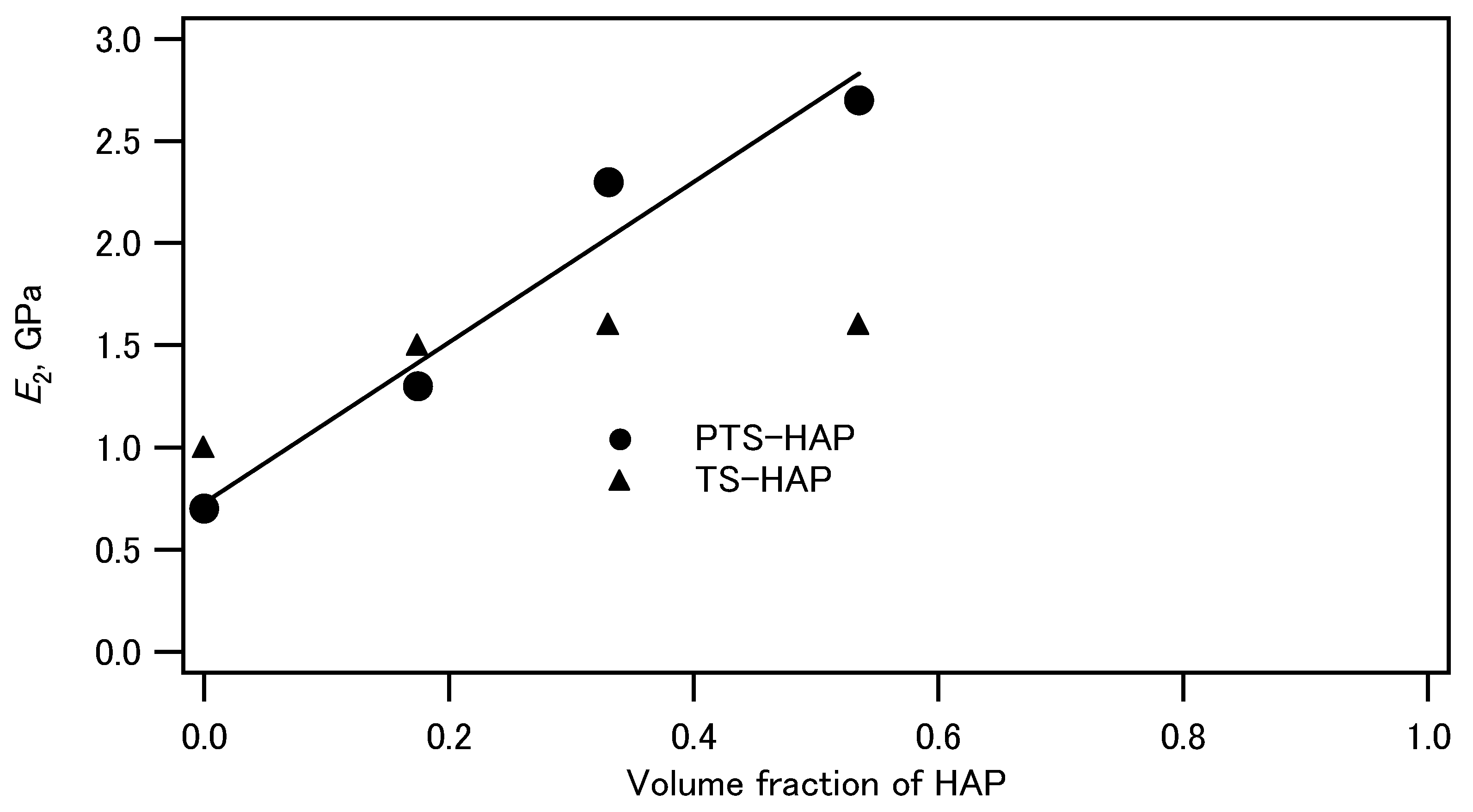
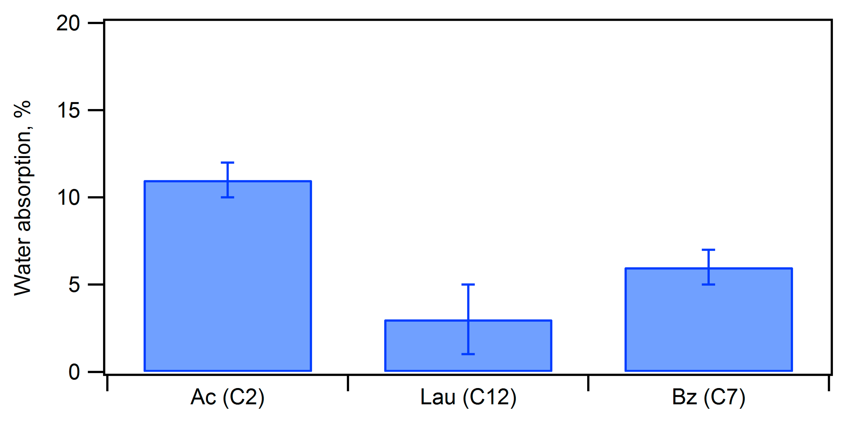
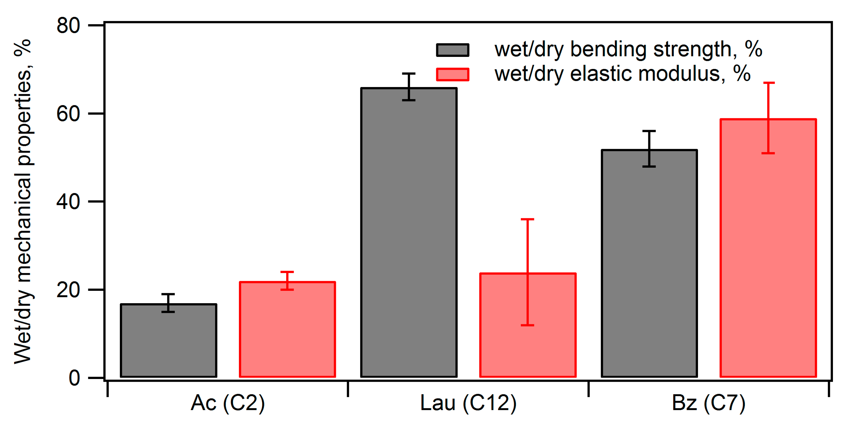
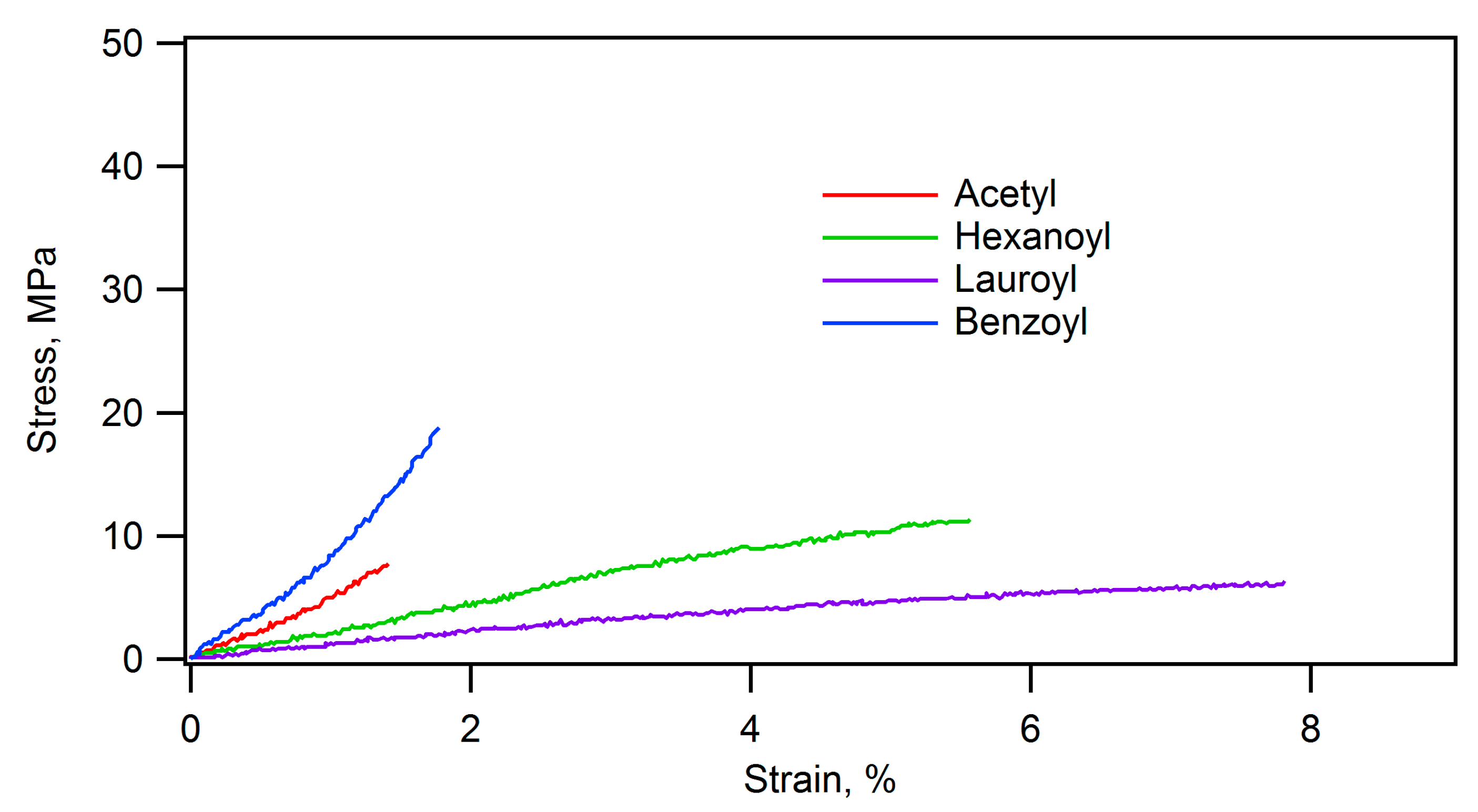
3.4.1. Acylation of Phosphorylated Starch–HAP Composites

The composite of starch and hydroxyapatite was dispersed in a polar solvent, such as dimethyl sulfoxide or N,N-dimethylformamide, and heated at 60–120 °C for 4–8 h after adding vinyl carboxylate, such as vinyl acetate, vinyl hexanoate, vinyl laurate, and vinyl benzoate, and potassium carbonate (Figure 7) [92]. The progress of acylation was confirmed by the infrared absorption spectra. The absorbance at 1750 cm−1 for aliphatic acyl groups and 1728 cm−1 for an aromatic acyl group increased, indicating that the acylation of the hydroxy groups of starch proceeded. The water absorption rates were evaluated by immersing the molded compacts before and after acylation in water for 24 h at room temperature, as shown in Figure 8. The water absorption rate before acylation was 136%, and acylation significantly reduced the water absorption rate. In particular, benzoyl and lauroyl groups were effective in reducing water absorption. Furthermore, the bending strengths of acetylated, lauroylated, and benzoylated compacts after immersion in water were 17, 66, and 52% of those of the dry compacts (Figure 9). The elastic moduli of acetylated, lauroylated, and benzoylated compacts after immersion in water were 22, 24, and 59% of those of the dry compacts. Therefore, benzoylation of the starch–hydroxyapatite composite effectively improved the water resistance.
The acylation of the starch and hydroxyapatite composite significantly influenced its mechanical and thermal properties. The stress–strain curves of the PTS–HAP composite and its acylated ones are shown in Figure 10, clearly demonstrating that the composite compacts were made softer by acylation, particularly with the long acyl groups. A comparison between Figure 10 and Figure 11 shows that the composite compacts became even more flexible when they were soaked in water. The bending strength, the elastic modulus, and the strain at failure of the composite before acylation were 47.3 MPa, 4.9 GPa, and 1.8%, respectively. After benzoylation, they became 28.7 MPa, 2.7 GPa, and 1.7%, and after lauroylation, they became 9.0 MPa, 0.2 GPa, and 5.8%. In particular, lauroylation resulted in increased flexibility. Additionally, the lauroylated composite demonstrated fluidity at 120 °C; that is, it exhibited thermoplasticity. The density of the molded form of the lauroylated composite was 1.2 g/cm3, demonstrating a lightweight characteristic similar to plastics.

3.4.2. Acylation of TCNF–HAP Composites

As described above, the TCNF–HAP composites showed a unique fracture mode similar to that of cardboard. The compacts also adsorbed much water when they were immersed in water, and their stiffness was lost in water. Because the TEMPO-oxidized cellulose nanofibers have a lot of carboxylate groups, the composites are hydrophilic. If the composites can be made hydrophobic with the non-brittle property unchanged, the composites can be used in many applications. We acylated the TCNF–HAP composites containing 62 wt% HAP in a similar fashion as that used for the acylation of the starch–HAP composites. The obtained composites were uniaxially pressed in a mold to prepare the compacts for bending tests. Water absorption of the acylated compacts, immersed in water at 25 °C for 24 h, are shown in Figure 12. The acetylated composite adsorbed 88% of the water, but the hexanoylated one adsorbed only 11%. The water absorption increased as the alkyl chain length increased further. The mechanical properties after immersion in water are shown in Figure 13. The acetylated composite showed almost no stiffness after immersion in water, while the hexanoylated and octanoylated ones showed ca. 30% of the bending strength and ca. 20% of the elastic modulus of the pristine compacts. Therefore, there is an appropriate length of the acyl groups for enhancing the water resistance.

3.4.3. Comparison of the Water Resistance of Starch–HAP, Cellulose–HAP, and Poly(DL-lactide)–HAP

Ritchie and coworkers reported that the composite of poly(DL-lactide) and HAP exhibited a bending strength of 23 MPa, while it decreased to 3 MPa when the compact was left at 37 °C for 30 h in a humid environment [94]. The ratio of the bending strength in the wet state to that in the dry state was 13% for the composite. Compared with this ratio, the ratio of lauroylated PTS–HAP was 66%, and the ratio of octanoylated TCNF–HAP was 30%. The number of anionic groups of the polymers decreased in the order of TCNF > PTS > poly(DL-lactide). The comparison suggests that the hydrophobicity of the polymer is not the dominant factor determining the water resistance of the composite, but there is an optimum hydrophilicity/hydrophobicity balance of the polymer to endow the composite with the water resistance (see Figure 14). Complementary arrangement of the polar groups of polymers and the polar sites on the apatite surface would prevent the hydration of the composite.

4. Future Outlook

The coprecipitation complexation, where calcium and phosphate ions are added to polymer solutions or dispersions, allowing the growth of calcium phosphate crystals using polymers as scaffolds, is an excellent method to uniformly composite nanometer-sized crystals under mild conditions via a simple process. In the case of hydroxyapatite, employing polymers with anionic functional groups leads to efficient coprecipitation, where the attractive interaction between the anionic groups of the polymer and the surface of the HAP crystals is expected to contribute to the stable interface between the organic phase and the inorganic phase and to good mechanical properties, such as high toughness and stiffness. On the other hand, polymers with polar groups and hydroxyapatite tend to readily be hydrated in water, leading to swelling and significant changes in the mechanical properties of the molded compacts in the presence of water. To enhance the water resistance of the composites, a balance between polar and nonpolar groups is essential, and the acylation reactions of the hydroxy groups of polysaccharides prove effective. Furthermore, the formation of fibrous structures through polymer aggregation can prevent brittle fracture in the composites, leading to more reliable structural materials. In particular, the use of polymers derived from cellulose makes it easier to induce such fibrous structures. Additionally, since cellulose is a type of biomass, it holds promise as an environmentally friendly structural material candidate in the future. As an alternative approach to the coprecipitation method, recent research has been exploring the mixing of inorganic crystals with a large aspect ratio and homogeneous sizes, which are prepared in advance, with polymers. The resultant composites exhibited excellent stiffness and strength, comparable to those of bone and teeth [5,95,96]. Therefore, the synthesis of inorganic crystals with a large aspect ratio can be crucial for the development of bioinspired mechanical materials. Although coprecipitation is a simple process under ambient conditions, it may be necessary to fine-tune the heterogeneous crystallization on the polymer to prepare crystals with a large aspect ratio.

Author Contributions

T.M. and Y.O. writing—original draft preparation, writing—review and editing. All authors have read and agreed to the published version of the manuscript.

Funding

This research received no external funding.

Conflicts of Interest

The authors declare no conflict of interest.

References

  1. Elias, H.-G. An Introduction to Polymer Science; VCH: Berlin, Germany, 1996. [Google Scholar]
  2. Eerkes-Medrano, D.; Thompson, R.C.; Aldridge, D.C. Microplastics in freshwater systems: A review of the emerging threats, identification of knowledge gaps and prioritisation of research needs. Water Res. 2015, 75, 63–82. [Google Scholar] [CrossRef] [PubMed]
  3. Stock, S.R. The Mineral-Collagen Interface in Bone. Calcif. Tissue Int. 2015, 97, 262–280. [Google Scholar] [CrossRef] [PubMed]
  4. Nudelman, F.; Lausch, A.J.; Sommerdijk, N.A.J.M.; Sone, E.D. In vitro models of collagen biomineralization. J. Struct. Biol. 2013, 183, 258–269. [Google Scholar] [CrossRef]
  5. Guan, Q.; Yang, H.; Han, Z.; Ling, Z.; Yu, S. An all-natural bioinspired structural material for plastic replacement. Nat. Commun. 2020, 11, 5401. [Google Scholar] [CrossRef]
  6. Curto, M.; Le Gall, M.; Catarino, A.I.; Niu, Z.; Davies, P.; Everaert, G.; Dhakal, H.N. Long-term durability and ecotoxicity of biocomposites in marine environments: A review. RSC Adv. 2021, 11, 32917–32941. [Google Scholar] [CrossRef]
  7. Olszta, M.J.; Cheng, X.; Jee, S.S.; Kumar, R.; Kim, Y.; Kaufman, M.J.; Douglas, E.P.; Gower, L.B. Bone structure and formation: A new perspective. Mater. Sci. Eng. R 2007, R58, 77–116. [Google Scholar] [CrossRef]
  8. Granke, M.; Does, M.D.; Nyman, J.S. The Role of Water Compartments in the Material Properties of Cortical Bone. Calcif. Tissue Int. 2015, 97, 292–307. [Google Scholar] [CrossRef]
  9. Dorozhkin, S.V.; Epple, M. Biological and Medical Significance of Calcium Phosphates. Angew. Chem. Int. Ed. 2002, 41, 3130–3146. [Google Scholar] [CrossRef]
  10. Weiner, S.; Wagner, H.D. The Material Bone: Structure-Mechanical Function Relations. Annu. Rev. Mater. Sci. 1998, 28, 271–298. [Google Scholar] [CrossRef]
  11. Gao, H.; Ji, B.; Jager, I.L.; Arzt, E.; Fratzl, P. Materials become insensitive to flaws at nanoscale: Lessons from nature. Proc. Nat. Acad. Sci. USA 2003, 100, 5597–5600. [Google Scholar] [CrossRef]
  12. Bouville, F.; Maire, E.; Meille, S.; Van de Moortele, B.; Stevenson, A.J.; Deville, S. Strong, tough and stiff bioinspired ceramics from brittle constituents. Nat. Mater. 2014, 13, 508–514. [Google Scholar] [CrossRef]
  13. Wilbrink, D.V.; Utz, M.; Ritchie, R.O.; Begley, M.R. Scaling of strength and ductility in bioinspired brick and mortar composites. Appl. Phys. Lett. 2010, 97, 193701. [Google Scholar] [CrossRef]
  14. Wang, R.; Gupta, H.S. Deformation and Fracture Mechanisms of Bone and Nacre. Annu. Rev. Mater. Res. 2011, 41, 41–73. [Google Scholar] [CrossRef]
  15. Sakhavand, N.; Shahsavari, R. Universal composition-structure property maps for natural and biomimetic platelet-matrix composites and stacked heterostructures. Nat. Commun. 2015, 6, 6523. [Google Scholar] [CrossRef]
  16. Currey, J.D. What determines the bending strength of compact bone? J. Exp. Biol. 1999, 202, 2495–2503. [Google Scholar] [CrossRef]
  17. Nalla, R.K.; Balooch, M.; Ager, J.W., III; Kruzic, J.J.; Kinney, J.H.; Ritchie, R.O. Effects of polar solvents on the fracture resistance of dentin: Role of water hydration. Acta Biomater. 2005, 1, 31–43. [Google Scholar] [CrossRef]
  18. Wilson, E.E.; Awonusi, A.; Morris, M.D.; Kohn, D.H.; Tecklenburg, M.M.J.; Beck, L.W. Three structural roles for water in bone observed by solid-state NMR. Biophys. J. 2006, 90, 3722–3731. [Google Scholar] [CrossRef] [PubMed]
  19. Samuel, J.; Sinha, D.; Zhao, J.C.; Wang, X. Water residing in small ultrastructural spaces plays a critical role in the mechanical behavior of bone. Bone 2014, 59, 199–206. [Google Scholar] [CrossRef]
  20. Gul-E-Noor, F.; Singh, C.; Papaioannou, A.; Sinha, N.; Boutis, G.S. Behavior of Water in Collagen and Hydroxyapatite Sites of Cortical Bone: Fracture, Mechanical Wear, and Load Bearing Studies. J. Phys. Chem. C 2015, 119, 21528–21537. [Google Scholar] [CrossRef] [PubMed]
  21. Nyman, J.S.; Roy, A.; Shen, X.; Acuna, R.L.; Tyler, J.H.; Wang, X. The influence of water removal on the strength and toughness of cortical bone. J. Biomech. 2006, 39, 931–938. [Google Scholar] [CrossRef]
  22. Pitta Kruize, C.; Panahkhahi, S.; Putra, N.E.; Diaz-Payno, P.; van Osch, G.; Zadpoor, A.A.; Mirzaali, M.J. Biomimetic Approaches for the Design and Fabrication of Bone-to-Soft Tissue Interfaces. ACS Biomater. Sci. Eng. 2023, 9, 3810–3831. [Google Scholar] [CrossRef] [PubMed]
  23. Hughes, J.M.; Cameron, M.; Crowley, K.D. Structural variations in natural F, OH, and Cl apatites. Am. Mineral. 1989, 74, 870–876. [Google Scholar]
  24. Dorozhkin, S.V. Calcium Orthophosphates in Nature, Biology and Medicine. Materials 2009, 2, 399–498. [Google Scholar] [CrossRef]
  25. Sato, K.; Kogure, T.; Kumagai, Y.; Tanaka, J. Crystal Orientation of Hydroxyapatite Induced by Ordered Carboxyl Groups. J. Colloid Interface Sci. 2001, 240, 133–138. [Google Scholar] [CrossRef] [PubMed]
  26. Hu, Y.-Y.; Rawal, A.; Schmidt-Rohr, K. Strongly bound citrate stabilizes the apatite nanocrystals in bone. Proc. Natl. Acad. Sci. USA 2010, 52, 22425–22429. [Google Scholar] [CrossRef] [PubMed]
  27. Wise, E.R.; Maltsev, S.; Davies, M.E.; Duer, M.J.; Jaeger, C.; Loveridge, N.; Murray, R.C.; Reid, D.G. The Organic-Mineral Interface in Bone Is Predominantly Polysaccharide. Chem. Mater. 2007, 19, 5055–5057. [Google Scholar] [CrossRef]
  28. Eden, M. Structure and formation of amorphous calcium phosphate and its role as surface layer of nanocrystalline apatite: Implications for bone mineralization. Materialia 2021, 17, 101107. [Google Scholar] [CrossRef]
  29. Gower, L.B. Biomimetic Model Systems for Investigating the Amorphous Precursor Pathway and Its Role in Biomineralization. Chem. Rev. 2008, 108, 4551–4627. [Google Scholar] [CrossRef]
  30. George, A.; Veis, A. Phosphorylated Proteins and Control over Apatite Nucleation, Crystal Growth, and Inhibition. Chem. Rev. 2008, 108, 4670–4693. [Google Scholar] [CrossRef]
  31. Rho, J.; Kuhn-Spearing, L.; Zioupos, P. Mechanical properties and the hierarchical structure of bone. Med. Eng. Phys. 1998, 20, 92–102. [Google Scholar] [CrossRef]
  32. Barthelat, F.; Yin, Z.; Buehler, M.J. Structure and mechanics of interfaces in biological materials. Nat. Rev. Mater. 2016, 1, 16007. [Google Scholar] [CrossRef]
  33. Fratzl, P.; Weinkamer, R. Nature’s hierarchical materials. Prog. Mater. Sci. 2007, 52, 1263–1334. [Google Scholar] [CrossRef]
  34. Nudelman, F.; Pieterse, K.; George, A.; Bomans, P.H.H.; Friedrich, H.; Brylka, L.J.; Hilbers, P.A.J.; de With, G.; Sommerdijk, N.A.J.M. The role of collagen in bone apatite formation in the presence of hydroxyapatite nucleation inhibitors. Nat. Mater. 2010, 9, 1004. [Google Scholar] [CrossRef]
  35. Espinosa, H.D.; Rim, J.E.; Barthelat, F.; Buehler, M.J. Merger of structure and material in nacre and bone? Perspectives on de novo biomimetic materials. Prog. Mater. Sci. 2009, 54, 1059–1100. [Google Scholar] [CrossRef]
  36. Wagermaier, W.; Klaushofer, K.; Fratzl, P. Fragility of Bone Material Controlled by Internal Interfaces. Calcif. Tissue Int. 2015, 97, 201–212. [Google Scholar] [CrossRef] [PubMed]
  37. Okada, A.; Usuki, A. Twenty Years of Polymer-Clay Nanocomposites. Macromol. Mater. Eng. 2006, 291, 1449–1476. [Google Scholar] [CrossRef]
  38. Bonfield, W.; Grynpas, M.D.; Tully, A.E.; Bowman, J.; Abram, J. Hydroxyapatite reinforced polyethylene—A mechanically compatible implant material for bone replacement. Biomaterials 1981, 2, 185–186. [Google Scholar] [CrossRef] [PubMed]
  39. Takeoka, Y.; Hayashi, M.; Sugiyama, N.; Yoshizawa-Fujita, M.; Aizawa, M.; Rikukawa, M. In situ preparation of poly(L-lactic acid-co-glycolic acid)/hydroxyapatite composites as artificial bone materials. Polym. J. 2015, 47, 164–170. [Google Scholar] [CrossRef]
  40. Stupp, S.I.; Ciegler, G.W. Organoapatite: Materials for artificial bone. I. Synthesis and microstructure. J. Biomed. Mater. Res. 1992, 26, 169–183. [Google Scholar] [CrossRef]
  41. Embery, G.; Rees, S.; Hall, R.; Rose, K.; Waddington, R.; Shellis, P. Calcium- and hydroxyapatite binding properties of glucuronic acid-rich and iduronic acid-rich glycosaminoglycans and proteoglycans. Eur. J. Oral Sci. 1998, 106, 267–273. [Google Scholar] [CrossRef] [PubMed]
  42. Kirkham, J.; Brookes, S.J.; Shore, R.C.; Wood, S.R.; Smith, D.A.; Zhang, J.; Chen, H.; Robinson, C. Physico-chemical properties of crystal surfaces in matrix-mineral interactions during mammalian biomineralisation. Current Opin. Colloid Interface Sci. 2002, 7, 124–132. [Google Scholar] [CrossRef]
  43. Bertoni, E.; Bigi, A.; Falini, G.; Panzavolta, S.; Roveri, N. Hydroxyapatite/polyacrylic acid nanocrystals, J. Mater. Chem. 1999, 9, 779–782. [Google Scholar] [CrossRef]
  44. Song, J.; Saiz, E.; Bertozzi, C.R. A New Approach to Mineralization of Biocompatible Hydrogel Scaffolds: An Efficient Process toward 3-Dimensional Bonelike Composites. J. Am. Chem. Soc. 2003, 125, 1236–1243. [Google Scholar] [CrossRef]
  45. Kikuchi, M.; Ikoma, T.; Itoh, S.; Matsumoto, H.N.; Koyama, Y.; Takakuda, K.; Shinomiya, K.; Tanaka, J. Biomimetic synthesis of bone-like nanocomposites using the self-organization mechanism of hydroxyapatite and collagen. Compos. Sci. Technol. 2004, 64, 819–825. [Google Scholar] [CrossRef]
  46. Pramanik, N.; Biswas, S.K.; Pramanik, P. Synthesis and Characterization of Hydroxyapatite/Poly(Vinyl Alcohol Phosphate) Nanocomposite Biomaterials. Int. J. Appl. Ceram. Technol. 2008, 5, 20–28. [Google Scholar] [CrossRef]
  47. Meldrum, F.C.; Coelfen, H. Controlling Mineral Morphologies and Structures in Biological and Synthetic Systems. Chem. Rev. 2008, 108, 4332–4432. [Google Scholar]
  48. Fujisawa, R.; Tamura, M. Acidic bone matrix proteins and their roles in calcification. Front. Biosci. 2012, 17, 1891–1903. [Google Scholar] [CrossRef]
  49. Coleman, R.J.; Jack, K.S.; Perrier, S.; Grondahl, L. Hydroxyapatite Mineralization in the Presence of Anionic Polymers. Cryst. Growth Des. 2013, 13, 4252–4259. [Google Scholar] [CrossRef]
  50. Veis, A.; Dorvee, J.R. Biomineralization Mechanisms: A New Paradigm for Crystal Nucleation in Organic Matrices. Calcif. Tissue Int. 2013, 93, 307–315. [Google Scholar] [CrossRef]
  51. Zhou, Y.; Liu, Q.; Zhao, Z.; Wang, W.; Zheng, L.; Li, Z. Preparation and Characterization of Phosphorylated Collagen and Hydroxyapatite Composite as a Potential Bone Substitute. Chem. Lett. 2013, 42, 83–85. [Google Scholar] [CrossRef]
  52. Morimune-Moriya, S.; Kondo, S.; Sugawara-Narutaki, A.; Nishimura, T.; Kato, T.; Ohtsuki, C. Hydroxyapatite formation on oxidized cellulose nanofibers in a solution mimicking body fluid. Polym. J. 2015, 47, 158–163. [Google Scholar] [CrossRef]
  53. Fang, W.; Zhang, H.; Yin, J.; Yang, B.; Zhang, Y.; Li, J.; Yao, F. Hydroxyapatite Crystal Formation in the Presence of Polysaccharide. Cryst. Growth Des. 2016, 16, 1247–1255. [Google Scholar] [CrossRef]
  54. Kusakabe, A.; Hirota, K.; Mizutani, T. Crystallisation of hydroxyapatite in phosphorylated poly(vinyl alcohol) as a synthetic route to tough mechanical hybrid materials. Mater. Sci. Eng. C 2017, 70, 487–493. [Google Scholar] [CrossRef]
  55. Diegmueller, J.J.; Cheng, X.; Akkus, O. Modulation of Hydroxyapatite Nanocrystal Size and Shape by Polyelectrolytic Peptides. Cryst. Growth Desig. 2009, 9, 5220–5226. [Google Scholar] [CrossRef]
  56. Mayer, G. Rigid Biological Systems as Models for Synthetic Composites. Science 2005, 310, 1144–1147. [Google Scholar] [CrossRef] [PubMed]
  57. Schweizer, S.; Taubert, A. Polymer-Controlled, Bio-Inspired Calcium Phosphate Mineralization from Aqueous Solution. Macromol. Biosci. 2007, 7, 1085–1099. [Google Scholar] [CrossRef]
  58. Palmer, L.C.; Newcomb, C.J.; Kaltz, S.R.; Spoerke, E.D.; Stupp, S.I. Biomimetic Systems for Hydroxyapatite Mineralization Inspired by Bone and Enamel. Chem. Rev. 2008, 108, 4754–4783. [Google Scholar] [CrossRef]
  59. Munch, E.; Launey, M.E.; Alsem, D.H.; Saiz, E.; Tomsia, A.P.; Ritchie, R.O. Tough, Bio-Inspired Hybrid Materials. Science 2008, 322, 1516–1520. [Google Scholar] [CrossRef]
  60. Roeder, R.K.; Converse, G.L.; Kane, R.J.; Yue, W. Hydroxyapatite-Reinforced Polymer Biocomposites for Synthetic Bone Substitutes. JOM 2008, 60, 38–45. [Google Scholar] [CrossRef]
  61. Li, B.; Wang, X.; Ma, J.; Huang, L. Preparation of phosphorylated chitosan/chitosan/hydroxyapatite composites by co-precipitation method. Adv. Mater. Res. 2009, 79–82, 401–404. [Google Scholar]
  62. Nudelman, F.; Sommerdijk, N.A.J.M. Biomineralization as an inspiration for materials chemistry. Angew. Chem. Int. Ed. 2012, 51, 6582–6596. [Google Scholar] [CrossRef] [PubMed]
  63. Zhang, C.Y.; Chen, J.; Zhuang, Z.; Zhang, T.; Wang, X.P.; Fang, Q.F. In situ hybridization and characterization of fibrous hydroxyapatite/chitosan nanocomposite. J. Appl. Polym. Sci. 2012, 124, 397–402. [Google Scholar] [CrossRef]
  64. Onoki, T.; Nakahira, A.; Tago, T.; Hasegawa, Y.; Kuno, T. Novel low temperature processing techniques for apatite ceramics and chitosan polymer composite bulk materials and its mechanical properties. Appl. Surf. Sci. 2012, 262, 263–266. [Google Scholar] [CrossRef]
  65. Yang, L.; Ning, X.; Bai, Y.; Jia, W. A scalable synthesis of non-agglomerated and low-aspect ratio hydroxyapatite nanocrystals using gelatinized starch matrix. Mater. Lett. 2013, 113, 142–145. [Google Scholar] [CrossRef]
  66. Bleek, K.; Taubert, A. New developments in polymer controlled, bioinspired calcium phosphate mineralization from aqueous solution. Acta Biomater. 2013, 9, 6283–6321. [Google Scholar] [CrossRef]
  67. Ma, J.; Wang, J.; Ai, X.; Zhang, S. Biomimetic self-assembly of apatite hybrid materials: From a single molecular template to bi-/multi-molecular templates. Biotechnol. Adv. 2014, 32, 744–760. [Google Scholar] [CrossRef]
  68. Won, J.-E.; El-Fiqi, A.; Jegal, S.-H.; Han, C.-M.; Lee, E.-J.; Knowles, J.C.; Kim, H.-W. Gelatin-apatite bone mimetic co-precipitates incorporated within biopolymer matrix to improve mechanical and biological properties useful for hard tissue repair. J. Biomater. Appl. 2014, 28, 1213–1225. [Google Scholar] [CrossRef]
  69. Li, J.; Baker, B.A.; Mou, X.; Ren, N.; Qiu, J.; Boughton, R.I.; Liu, H. Biopolymer/Calcium Phosphate Scaffolds for Bone Tissue Engineering. Adv. Healthc. Mater. 2014, 3, 469–484. [Google Scholar] [CrossRef]
  70. Shu, Y.; Yin, P.; Liang, B.; Wang, H.; Guo, L. Bioinspired Design and Assembly of Layered Double Hydroxide/Poly(vinyl alcohol) Film with High Mechanical Performance. ACS Appl. Mater. Interfaces 2014, 6, 15154–15161. [Google Scholar] [CrossRef]
  71. Cheng, Q.; Jiang, L.; Tang, Z. Bioinspired Layered Materials with Superior Mechanical Performance. Acc. Chem. Res. 2014, 47, 1256–1266. [Google Scholar] [CrossRef]
  72. Wegst, U.G.K.; Bai, H.; Saiz, E.; Tomsia, A.P.; Ritchie, R.O. Bioinspired structural materials. Nat. Mater. 2015, 14, 23–36. [Google Scholar] [PubMed]
  73. Le, H.; Natesan, K.; Pranti-Haran, S. Mechanical property and biocompatibility of co-precipitated nano hydroxyapatite-gelatine composites. J. Adv. Ceram. 2015, 4, 237–243. [Google Scholar] [CrossRef]
  74. Shakir, M.; Jolly, R.; Khan, M.S.; e Iram, N.; Khan, H.M. Nano-hydroxyapatite/chitosan starch nanocomposite as a novel bone construct: Synthesis and in vitro studies. Int. J. Biol. Macromol. 2015, 80, 282–292. [Google Scholar] [CrossRef]
  75. Zakharov, N.A.; Demina, L.I.; Aliev, A.D.; Kiselev, M.R.; Matveev, V.V.; Orlov, M.A.; Zakharova, T.V.; Kuznetsov, N.T. Synthesis and Properties of Calcium Hydroxyapatite/Silk Fibroin Organomineral Composites. Inorg. Mater. 2017, 53, 333–342. [Google Scholar] [CrossRef]
  76. Miculescu, F.; Maidaniuc, A.; Voicu, S.I.; Thakur, V.K.; Stan, G.E.; Ciocan, L.T. Progress in Hydroxyapatite. Starch Based Sustainable Biomaterials for Biomedical Bone Substitution Applications. ACS Sustain. Chem. Eng. 2017, 5, 8491–8512. [Google Scholar]
  77. Yang, R.L.; Zhu, Y.J.; Chen, F.F.; Qin, D.D.; Xiong, Z.C. Bioinspired Macroscopic Ribbon Fibers with a Nacre-Mimetic Architecture Based on Highly Ordered Alignment of Ultralong Hydroxyapatite Nanowires. ACS Nano 2018, 12, 12284–12295. [Google Scholar] [CrossRef]
  78. Zima, A. Hydroxyapatite-chitosan based bioactive hybrid biomaterials with improved mechanical strength. Spectrochim. Acta A 2018, 193, 175–184. [Google Scholar] [CrossRef]
  79. Kawasaki, T.; Takahashi, S.; Ikeda, K. Hydroxyapatite high-performance liquid chromatography: Column performance for proteins. Eur. J. Biochem. 1985, 152, 361–371. [Google Scholar] [CrossRef] [PubMed]
  80. Kim, M.; Yeo, M.; Kim, M.; Kim, G. Biomimetic cellulose/calcium-deficient hydroxyapatite composite scaffolds fabricated using an electric field for bone tissue engineering. RSC Adv. 2018, 8, 20637–20647. [Google Scholar]
  81. Malkaj, P.; Pierri, E.; Dalas, E. The crystallization of hydroxyapatite in the presence of sodium alginate. J. Mater. Sci. Mater. Med. 2005, 16, 733–737. [Google Scholar] [CrossRef]
  82. Ingole, V.H.; Vuherer, T.; Maver, U.; Vinchurkar, A.; Ghule, A.V.; Kokol, V. Mechanical Properties and Cytotoxicity of Differently Structured Nanocellulose-hydroxyapatite Based Composites for Bone Regeneration Application. Nanomaterials 2020, 10, 25. [Google Scholar] [CrossRef] [PubMed]
  83. Klemm, D.; Heublein, B.; Fink, H.; Bohn, A. Cellulose: Fascinating Biopolymer and Sustainable Raw Material. Angew. Chem. Int. Ed. 2005, 44, 3358–3393. [Google Scholar] [CrossRef]
  84. Schierbaum, F. The Current World Corn Situation: Production, Uses and Ending Stocks; Revised Figures for Fiscal Years 2006/07, 2007/08, Current Figures 2008/09, Production Profiles and Outlook. Starch—Stärke 2010, 62, 272–276. [Google Scholar] [CrossRef]
  85. Okuda, K.; Shigemasa, R.; Hirota, K.; Mizutani, T. In Situ Crystallization of Hydroxyapatite on Carboxymethyl Cellulose as a Biomimetic Approach to Biomass-Derived Composite Materials. ACS Omega 2022, 7, 12127–12137. [Google Scholar] [CrossRef]
  86. Okuda, K.; Mizutani, T. Hybridization of chemically modified cellulose and hydroxyapatite for development of biomass-derived tough and water resistant structural materials. In Proceedings of the JCCM-14, Tokyo, Japan, 14–16 March 2023. [Google Scholar]
  87. Brandrup, J.; Immergut, E.H.; Grulke, E.A. (Eds.) Polymer Handbook, 4th ed.; John Wiley & Sons, Inc.: New York, NY, USA, 1999. [Google Scholar]
  88. Okuda, K.; Hirota, K.; Mizutani, T.; Aoyama, Y. Co-precipitation of tapioca starch and hydroxyapatite. Effects of phosphorylation of starch on mechanical properties of the composites. Results Mater. 2019, 3, 100035. [Google Scholar]
  89. Okuda, K.; Mizutani, T.; Hirota, K.; Hayashi, T.; Zinno, K. Nonbrittle Nanocomposite Materials Prepared by Coprecipitation of TEMPO-Oxidized Cellulose Nanofibers and Hydroxyapatite. ACS Sustain. Chem. Eng. 2021, 9, 158–167. [Google Scholar]
  90. Okita, Y.; Saito, T.; Isogai, A. Entire Surface Oxidation of Various Cellulose Microfibrils by TEMPO-Mediated Oxidation. Biomacromolecules 2010, 11, 1696–1700. [Google Scholar] [CrossRef]
  91. Weiner, S.; Addadi, L. Design strategies in mineralized biological materials. J. Mater. Chem. 1997, 7, 689–702. [Google Scholar]
  92. Okuda, K.; Aoyama, Y.; Hirota, K.; Mizutani, T. Effects of Hydration on Mechanical Properties of Acylated Hydroxyapatite-Starch Composites. ACS Appl. Polym. Mater. 2022, 4, 1666–1674. [Google Scholar] [CrossRef]
  93. Okuda, K.; Kido, E.; Hirota, K.; Mizutani, T. Water-resistant Tough Composites of Cellulose Nanofibers and Hydroxyapatite. ACS Appl. Polym. Mater. 2023, 5, 8082–8088. [Google Scholar] [CrossRef]
  94. Neuendorf, R.E.; Saiz, E.; Tomsia, A.P.; Ritchie, R.O. Adhesion between biodegradable polymers and hydroxyapatite: Relevance to synthetic bone-like materials and tissue engineering scaffolds. Acta Biomater. 2008, 4, 1288–1296. [Google Scholar] [CrossRef] [PubMed]
  95. Das, P.; Malho, J.; Rahimi, K.; Schacher, F.H.; Wang, B.; Demco, D.E.; Walther, A. Nacre-mimetics with synthetic nanoclays up to ultrahigh aspect ratios. Nat. Commun. 2015, 6, 5967. [Google Scholar] [CrossRef]
  96. Yu, H.-C.; Zhu, Y.-J.; Xiong, Z.-C.; Lu, B.-Q. Bioinspired fiberboard-and-mortar structural nanocomposite based on ultralong hydroxyapatite nanowires with high mechanical performance. Chem. Eng. J. 2020, 399, 125666. [Google Scholar]
Figure 1. Brick-and-mortar structure of bone and polymer–HAP composites prevent fragile fracture.
Ceramics 06 00130 g001
Figure 2. Comparison of bending strength, elastic modulus, and strain at failure, evaluated by three-point bending tests of tapioca starch (TS)–hydroxyapatite, phosphorylated tapioca starch (PTS)–hydroxyapatite, and TEMPO-oxidized cellulose nanofibers (TCNF)–hydroxyapatite composites containing 70 wt% of hydroxyapatite [88,89].
Ceramics 06 00130 g002aCeramics 06 00130 g002b
Figure 3. Plot of elastic moduli of phosphorylated tapioca starch (PTS)–HAP composites and tapioca starch (TS)–HAP composites against the volume fraction of HAP.
Ceramics 06 00130 g003
Figure 4. Schematic representation of TEMPO-oxidized cellulose nanofibers [90].
Ceramics 06 00130 g004
Figure 5. Fracture surface of the compact of the TCNF-HAP composite, coprecipitated at 90 °C (left), and that of the compact of the phosphorylated starch–HAP composite, coprecipitated at 70 °C (right). In the three-point bending test of the TCNF-HAP compacts, the compacts only bent, so the fracture surface was produced by pulling the compact using bare hands.
Ceramics 06 00130 g005
Figure 6. Schematic representation of the coprecipitation of TCNF and HAP. At an intermediate concentration of TCNF, heterogeneous nucleation of HAP on TCNF is the major pathway to produce an aligned composite. At a low concentration of TCNF, both heterogeneous and homogeneous nucleations occur to prevent the formation of the fibrous structure.
Ceramics 06 00130 g006
Figure 7. Acylation of the composites of starch and hydroxyapatite.
Ceramics 06 00130 g007
Figure 8. Water absorption of the compacts of PTS-HAP composites containing 66 wt% HAP, acylated with acetyl (Ac), lauroyl (Lau), and benzoyl (Bz) groups.
Ceramics 06 00130 g008
Figure 9. The ratios of bending strength and elastic modulus after water immersion to those before water immersion of the acylated PTS-HAP composite compacts.
Ceramics 06 00130 g009
Figure 10. Stress–strain curves of the phosphorylated starch–HAP composite containing 50 wt% of the inorganic phase and its acylated composites.
Ceramics 06 00130 g010
Figure 11. Stress–strain curves of the acylated composites of phosphorylated starch–HAP after immersion in water at 25 °C for 24 h.
Ceramics 06 00130 g011
Figure 12. Water absorption of the compacts of TCNF-HAP composites containing 62 wt% HAP acylated with acetyl (Ac), hexanoyl (Hex), octanoyl (Oct), lauroyl (Lau), and benzoyl (Bz) groups.
Ceramics 06 00130 g012
Figure 13. The ratios of bending strength and elastic modulus after water immersion to those before water immersion of the acylated TCNF-HAP composite compacts.
Ceramics 06 00130 g013
Figure 14. Schematic representation of the water resistance of the composites of hydroxyapatite and hydrophilic and hydrophobic polymers. Appropriate hydrophilic/hydrophobic balance of the polymer is necessary for the water resistance of the composite.
Ceramics 06 00130 g014
Table 1. Classification of organic–inorganic composites.
Ceramic-Reinforced PolymerPolymer-Reinforced Ceramics
Major componentPolymer (matrix, continuous phase)Ceramics (filler, dispersed phase)
Minor componentCeramics (filler, dispersed phase)Polymers (matrix, continuous phase)
ExamplesGlass–fiber reinforced plasticsLaminated glass
Polyamide–clay compositesBones
TireTeeth
Table 2. Comparison of mechanical properties of the polysaccharide–HAP composites with those of plastics [87].
Bending Strength, MPaElastic Modulus, GPaDensity, g/cm3Uniaxial Press Pressure, MPa
PTS–HAP, HAP 70 wt%474.91.72120
CMC–HAP, HAP 70 wt%1137.71.8120
TCNF–HAP, HAP 62 wt%8011.61.94300
Poly(methyl methacrylate)1183.41.19
Polyamide-61182.81.14
Disclaimer/Publisher’s Note: The statements, opinions and data contained in all publications are solely those of the individual author(s) and contributor(s) and not of MDPI and/or the editor(s). MDPI and/or the editor(s) disclaim responsibility for any injury to people or property resulting from any ideas, methods, instructions or products referred to in the content.

