Article

Advancements in Healthcare: Development of a Comprehensive Medical Information System with Automated Classification for Ocular and Skin Pathologies—Structure, Functionalities, and Innovative Development Methods

1
Faculty of Electronics, Telecommunications and Information Technology, Politehnica Bucharest National University for Science and Technology, 061071 Bucharest, Romania
2
Independent Researcher, 021571 Bucharest, Romania
*
Author to whom correspondence should be addressed.
Appl. Syst. Innov. 2024, 7(2), 28; https://doi.org/10.3390/asi7020028
Submission received: 26 January 2024 / Revised: 11 March 2024 / Accepted: 20 March 2024 / Published: 27 March 2024
(This article belongs to the Section Information Systems)

Abstract

:
This article introduces a groundbreaking medical information system developed in Salesforce, featuring an automated classification module for ocular and skin pathologies using Google Teachable Machine. Integrating cutting-edge technology with Salesforce’s robust capabilities, the system provides a comprehensive solution for medical practitioners. The article explores the system’s structure, emphasizing innovative functionalities that enhance diagnostic precision and streamline medical workflows. Methods used in development are discussed, offering insights into the integration of Google Teachable Machine into the Salesforce framework. This collaborative approach is a significant stride in intelligent pathology classification, advancing the field of medical information systems and fostering efficient healthcare practices.

1. Introduction

The integration of an automated system within an Electronic Health Record (EHR) to detect pathologies with potential adverse outcomes not only enhances the efficiency and accuracy of screening but also empowers patients to actively engage in managing their health. Early detection through this system significantly influences patient outcomes, reduces healthcare costs, and promotes proactive preventive measures. This innovative approach adds substantial value to digital development through various means. It facilitates rapid diagnosis by swiftly identifying and categorizing medical images, aids in early disease detection, and ensures consistency and standardized analyses, saving healthcare professionals time and allowing them to focus more on patient care. The system enables quick access to labeled images within the EHR, supports valuable data collection for research and analysis, and contributes to population health management. With the capability to handle large data volumes, adapt to specific medical specialties, and automate image classification processes, the system optimizes workflows, enhances data security, and ensures compliance with regulations. Additionally, it enables remote access and collaboration on medical images, contributing to improved healthcare service accessibility, particularly in telemedicine. Automated image classification ensures a consistent high-quality standard in image analysis, reducing variations among healthcare professionals. Overall, EHR systems equipped with image classification capabilities pave the way for a cohesive and efficient medical ecosystem, aligning with regulations and standards in medical image management.
This paper will primarily explore the development of a computer system incorporating a module for the automated classification of medical images associated with two significant global causes of mortality: diabetes (resulting in 103,294 deaths) and cancer (accounting for 605,213 deaths) [1]. The emphasis of the paper will specifically revolve around the intelligent processing of retinal and skin medical images. In the realm of skin pathologies, the research will delve into the examination of two types of skin cancer (melanoma and basal cell carcinoma) as well as two other prevalent skin conditions that, if untreated, may progress to skin cancer (actinic keratosis and seborrheic keratosis). Concerning diabetes, the study will address complications related to ocular diabetes, encompassing diabetic retinopathy, glaucoma, and cataracts.
Moreover, the key points of this paper represent, on one hand a pioneering medical information system developed within Salesforce (SF), featuring an automated classification module for ocular and skin pathologies utilizing Google Teachable Machine (GT). This integration of cutting-edge technology with Salesforce’s robust capabilities offers a comprehensive solution for medical practitioners. The system’s structure is explored, highlighting innovative functionalities that enhance diagnostic precision and streamline medical workflows. Additionally, the discussion delves into the methods used in development, providing insights into the integration of GT in the SF framework. This collaborative approach holds significance in advancing the field of medical information systems and fostering efficient healthcare practices. Furthermore, the integration of the automated system within an HER enhances screening efficiency and accuracy, empowering patients to actively engage in managing their health through early detection. Early detection influences patient outcomes positively, reduces healthcare costs, and promotes proactive preventive measures. The system adds substantial value to digital development by facilitating rapid diagnosis, early disease detection, and standardized analyses. It also supports rapid access to labeled images within the EHR, valuable data collection for research and analysis, and contributes to population health management. Moreover, the system’s capability to handle large data volumes, adapt to specific medical specialties, automate image classification processes, optimize workflows, enhance data security, and ensure compliance with regulations underscores its importance. The enablement of remote access and collaboration on medical images, particularly in telemedicine, contributes to improved healthcare service accessibility. Automated image classification ensures a consistent high-quality standard in image analysis, thereby reducing variations among healthcare professionals. Overall, EHR systems equipped with image classification capabilities pave the way for a cohesive and efficient medical ecosystem, aligning with regulations and standards in medical image management.

2. Background

2.1. Ocular Pathologies

Detecting ocular diseases, such as glaucoma, in their early stages enables preventive measures against vascular accidents, another complication of diabetes that contributes to 162,890 deaths, making it a significant global cause of mortality.
Diabetes is associated with numerous complications, with the most severe being metabolic issues linked to mortality, such as ketoacidosis resulting from excessively high blood glucose levels (hyperglycemia) or coma due to excessively low blood glucose levels (hypoglycemia). Hyperglycemia leads to vascular complications (angiopathy) at both the macrovascular level (large vessels) and microvascular level (small vessels). Complications affecting small vessels impact the eyes, kidneys, and nerve pathways, while those affecting large vessels impact the heart and lungs, contributing to myocardial diseases and vascular accidents [2].
Diabetes is a chronic metabolic disorder characterized by elevated blood glucose levels resulting from the pancreas’s insufficient production of insulin, a hormone secreted by the pancreas that facilitates the body’s metabolism of carbohydrates found in food, preventing glucose levels from becoming excessively low (hypoglycemia) or high (hyperglycemia) [2,3]. Over time, diabetes leads to complications affecting organs such as the heart, kidneys, eyes, blood vessels, and nerves.
We will now delve into complications arising from the involvement of small vessels, specifically impacting vision. Various conditions, including diabetic retinopathy, macular edema, cataracts, and glaucoma, can manifest symptoms ranging from blurred vision to blindness.
a. Diabetic Retinopathy. Elevated glucose levels over time can lead to the blockage of tiny blood vessels nourishing the retina, reducing its blood supply. In the early stage (non-proliferative diabetic retinopathy), vessels do not proli: ferate, and no new vessels form. In advanced stages (proliferative diabetic retinopathy), affected vessels are destroyed, prompting the formation of new, leaky blood vessels affecting the vitreous body. Scar tissue can result in retinal detachment, creating intraocular pressure that damages the optic nerve, leading to glaucoma and eventual blindness [4].
b. Cataract. A cataract is a clouding of the lens in the eye that affects vision. The lens, which is typically clear, becomes cloudy over time, leading to a gradual decrease in vision. Cataracts are most commonly associated with aging, and they are a natural part of the aging process. However, cataracts can also develop due to other factors such as injury, certain medications, medical conditions (like diabetes), or prolonged exposure to ultraviolet light [5].
c. Glaucoma represents the degeneration of the optic nerve, progressively leading to blindness over time [6]. As per the World Health Organization (WHO), glaucoma stands as the second most prevalent cause of blindness globally, impacting over 60 million individuals. This condition is incurable and can advance without evident symptoms until the late stages. Early diagnosis is paramount, and effective management through appropriate treatment is vital. Eye alterations encompass a reduction in the number of retinal ganglion cells and the thinning of optic nerve fibers at the retinal nerve level.
The typical diagnostic procedures for ocular pathologies involve ophthalmoscopy or fundoscopy, enabling an in-depth examination of the eye’s interior, encompassing the retina, optic disc, and choroid. This process entails the application of dilating drops, followed by capturing internal eye images using an ophthalmoscope (Figure 1) [7].
The early detection of ocular diseases through AI holds significant importance for various reasons. Firstly, it contributes to vision preservation by identifying subtle changes indicative of conditions like glaucoma, diabetic retinopathy, and macular degeneration, which often progress unnoticed in their initial stages. This enables timely interventions, preventing or slowing vision loss and maintaining quality of life. Secondly, early detection enhances treatment outcomes, particularly for conditions such as diabetic retinopathy and macular degeneration, where prompt interventions can mitigate severe visual impairments or blindness. AI aids healthcare professionals in determining optimal treatment strategies based on disease progression and individual patient characteristics. Thirdly, the early identification and treatment of ocular diseases lead to substantial cost reductions associated with managing advanced stages. Addressing issues at an early stage results in less invasive treatments and fewer complications, reducing medical service utilization and associated expenses. Additionally, the implementation of AI-based screening tools enables accessible and scalable eye care, efficiently analyzing numerous images to identify potential cases for further examination by healthcare professionals. Furthermore, early detection through AI supports public health initiatives by identifying individuals at risk, allowing for proactive interventions and preventive measures, ultimately decreasing the overall incidence of ocular diseases in individuals, families, and healthcare systems. Lastly, AI-based screening tools empower patients to actively engage in their health, offering timely information about their condition, facilitating informed decisions on treatment options, and encouraging regular monitoring for optimal eye health. In conclusion, leveraging artificial intelligence for the early detection of ocular diseases holds the potential to safeguard visual acuity, enhance treatment outcomes, reduce medical costs, and overall improve individual well-being by addressing ocular conditions before they advance to critical stages [8,9,10,11,12,13,14,15,16,17,18].

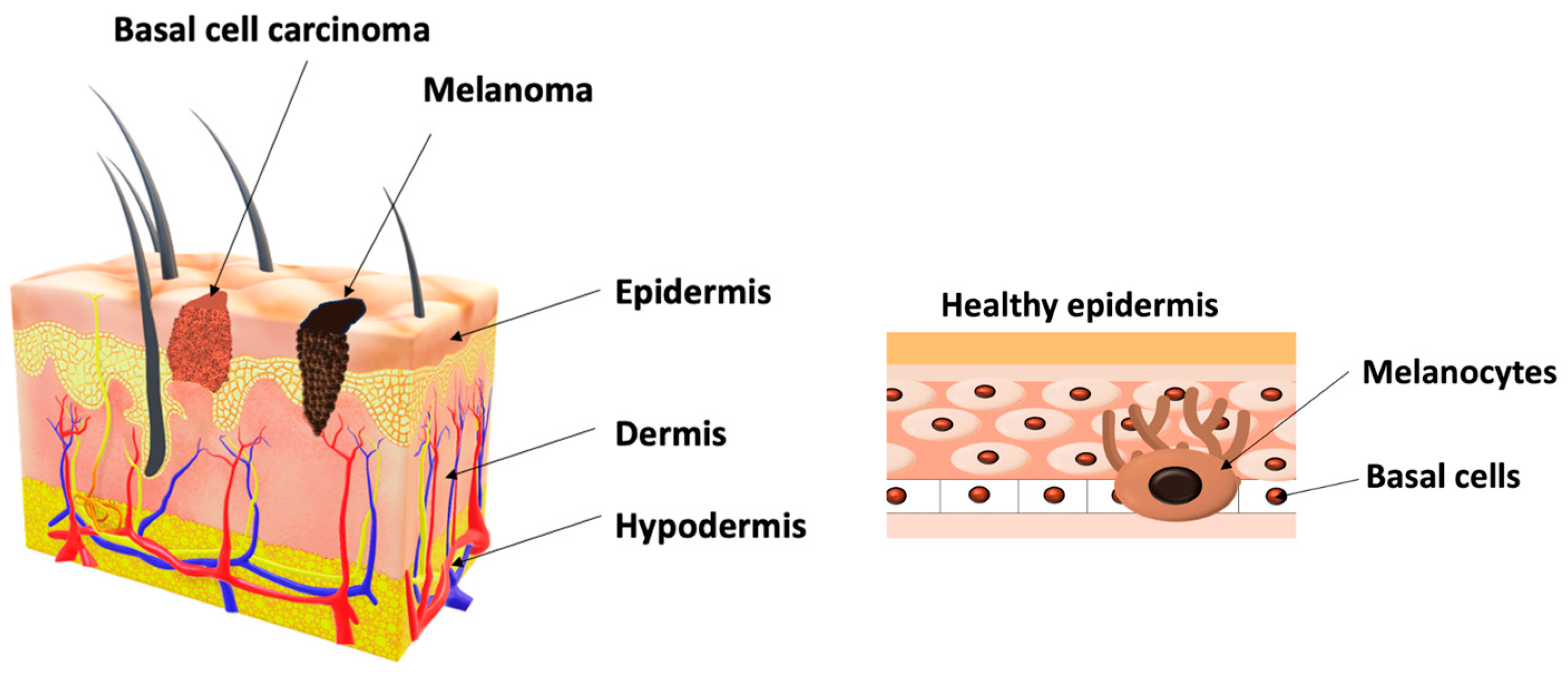
2.2. Skin Pathologies

Skin cancer is a type of cancer that originates in the skin cells. The skin is the body’s largest organ and is composed of several layers. Skin cancer develops when the deoxyribonucleic acid (DNA) of skin cells undergoes genetic mutations (Figure 2), typically due to exposure to ultraviolet (UV) radiation from the sun or tanning beds. These mutations cause the skin cells to grow uncontrollably and form malignant tumors [19].
The most common type of cancer is basal cell carcinoma (BCC): This is the most common type of skin cancer. It usually appears as a shiny or pearly bump, often with visible blood vessels. BCC rarely spreads to other parts of the body but can cause local damage if not treated.
The most aggressive type of skin cancer is melanoma and has the potential to spread rapidly to other organs, making early detection and treatment crucial. The exact causes of skin cancer remain unclear; however, exposure to ultraviolet radiation is identified as a risk factor for skin cancer. Factors that may heighten the risk of melanoma encompass:
  • Light skin color: Characterized by a reduced amount of pigment (melanin) in the skin, resulting in inadequate protection against UV rays.
  • Sunburns: The occurrence of one or more sunburns amplifies the risk of skin cancer.
  • Presence of multiple moles: an excess of 50 moles on the body can escalate the likelihood of developing melanoma.
  • Family history: A noteworthy likelihood of skin cancer exists if close relatives have a history of this type of cancer.
  • Weakened immune system: This factor has the potential to increase the risk of various types of cancer [19].
Preventive measures include practicing sun safety, such as using sunscreen, wearing protective clothing, and avoiding excessive sun exposure. Regular self-examinations and dermatological check-ups are essential for early detection and treatment. Early diagnosis and treatment significantly improve the prognosis for individuals with skin cancer.
Because early detection is crucial, we included in the research another two pathologies that untreated and not monitored closely could lead to skin cancer—actinic keratosis and seborrheic keratosis. Actinic keratosis manifests through skin lesions that develop as a result of sun exposure over the years and most commonly appears on the face, lips, ears, forearms, scalp, neck, and hands. The disease progresses slowly and typically occurs in individuals over 40 years old, but if left untreated, it can transform into skin cancer known as squamous cell carcinoma [20]. Actinic keratosis is a skin tumor within the epidermis that predominantly occurs in adults and the elderly. These lesions, often benign and not requiring treatment, result from the benign proliferation of immature keratinocytes, forming round, flat, well-defined growths that slowly increase in thickness over time [21]. Treatment involves eliminating these growths through cryotherapy (freezing), curettage, or electrocautery. The solution lies in the early detection and analysis of skin lesions, especially skin cancers. Currently, this requires a visit to a doctor and numerous tests for a diagnosis, which can be time-consuming and challenging due to a decrease in specialists capable of providing accurate diagnoses. The proposed solution in this study involves early detection of skin cancer using artificial intelligence, particularly neural networks, to assist doctors in making precise diagnoses in a simple and accessible manner for both patients and healthcare professionals. This is achieved by providing an image of the affected area (Figure 3) [22,23,24,25].
The proposed approach in this research entails leveraging artificial intelligence, particularly neural networks, for the early detection of skin cancer. This application aims to assist healthcare professionals in delivering accurate diagnoses in a straightforward and accessible manner for both patients and medical practitioners. The method involves presenting an image of the affected area.
Automated detection systems play a crucial role in advancing the prognosis of skin cancer by identifying suspicious lesions early on, allowing for timely interventions and ultimately improving patient outcomes. The impact extends to a reduction in mortality risks associated with skin cancer, particularly as early detection prevents the disease from progressing to critical stages that may affect vital organs. Moreover, the use of automated systems offers significant cost savings in treatment, as it enables less invasive and more cost-effective alternatives for addressing skin cancer, benefiting both patients and healthcare systems. The integration of these systems into routine health check-ups promotes regular screenings, fostering awareness about monitoring moles and other skin irregularities. Targeted screening for high-risk populations, such as those with a family history of skin cancer or excessive sun exposure, is facilitated by automated detection systems, leading to the early identification of suspicious lesions. The scalability and efficiency of these systems enable swift and accurate analysis of a large volume of images, making them ideal for screening extensive populations and thereby enhancing the chances of early skin cancer detection.
Healthcare professionals also benefit from the assistance provided by automated detection systems, as they aid in identifying potential melanoma cases, improving diagnostic accuracy, and reducing the likelihood of overlooking concerning lesions. Ultimately, patient empowerment is achieved through prompt feedback from automated systems, enabling individuals to take proactive measures for additional assessments and early treatment when necessary.
The proposed solution brings additional benefits from the perspective of having an automated pathology detection system within an EHR system that consolidates all workflows for medical team members in one place:
Improved patient outcomes: Early detection of pathologies allows for timely interventions and treatments, significantly improving patient outcomes. Skin and eye pathologies, if left undetected and untreated, can progress and lead to more severe health issues. By detecting these conditions in their early stages, healthcare professionals can implement effective treatment plans and prevent complications.
Disease progression prevention: Pathologies can progress rapidly if not identified in a timely manner. Integrating automated detection systems into an EHR allows healthcare providers to monitor patients continuously and detect any changes in their health status. This proactive approach helps prevent disease progression, reducing the risk of long-term deterioration and potentially saving lives.
Reduced healthcare costs: early detection and intervention can lead to less complex and invasive treatments, generally reducing healthcare costs by minimizing hospitalizations and resulting in significant savings for both patients and healthcare systems.
Increased access: An automated detection system integrated into an EHR provides patients with access to real-time medical information. This promotes patient engagement and encourages patients to play an active role in managing their health, making informed decisions, and adhering to treatment plans.
Population health management: EHR systems with integrated detection capabilities enable healthcare providers to analyze trends and patterns within patient populations. This data-driven approach allows public health authorities to identify high-risk groups, implement targeted preventive measures, and allocate resources efficiently.
Streamlined workflow: Integrating automated detection into EHR systems streamlines healthcare workflows. Clinicians can access patients’ medical history, investigation results, and treatment plans in a centralized platform, facilitating quick decision making and reducing the risk of omissions or communication errors.
Data-driven insights: Integrating automated detection systems generates data that can be analyzed to identify correlations, risk factors, and treatment outcomes. This data-driven approach can lead to the development of more effective treatment protocols and better-informed clinical decisions.
Research and development: Aggregated data from automated detection systems integrated into EHRs can be anonymized and used for medical research and the development of new treatment approaches. This contributes to advancing medical knowledge and the development of innovative therapies.
Essentially, early detection of pathologies through automated systems integrated into an EHR not only improves individual patient care but also has broader implications for public health, healthcare efficiency, cost reduction, and promotes medical research. This represents a significant step towards proactive and personalized healthcare management.

3. Materials and Methods

The implementation of the proposed solution required constructing a classification model designed to detect the specified pathologies. This involved utilizing various databases, developing a web-based system to create the classification model trained on an image database, and incorporating the automatic classification module into the informatics system as part of the overall global implementation of the information system. The number of images and classes are presented in Table 1.

3.1. Databases

The ORIGA (Online Retinal Fundus Image Dataset for Glaucoma Analysis and Research) is an accessible database containing 168 color fundus images illustrating the posterior pole with glaucoma and 482 images without any pathologies. These images were acquired as part of the Singapore Malay Eye Study, a screening program conducted by the Singapore Eye Research Institute [26].
Messidor, designed for diabetic retinopathy, encompasses 1200 retina images labeled by experts, both with and without glaucoma, with 800 of them acquired using dilated pupils. This collection was obtained during a screening program in Switzerland, focusing on diabetic patients aged 25–90, funded by the French Ministry under the Techno-Vison 2004 program [27].
EyePacs is a database available on Kaggle, containing 35,126 images of diabetic retinopathy obtained through screening programs [28].
The HRF (High-Resolution Fundus Image Database), provided by the CS5 Laboratory, consists of 15 normal fundus images, 15 images with glaucoma, and 15 images with diabetic retinopathy [29].
ISIC (International Skin Imaging Collaboration) includes 93,083 images of skin lesions for testing algorithms in melanoma detection, serving as the largest public database of dermatoscopic images. The database encourages the development of effective systems for detecting skin lesions and incorporates smaller databases like HAM 1000, BCN2000, and MSK [30].
A Kaggle-hosted database features 601 images showcasing various pathologies, including cataract, glaucoma, and diabetic retinopathy, divided into five classes [31].
RIM-ONE, a database of retinal images from ocular fundus images, comprises 200 images with glaucoma and 255 images without pathologies [32,33].
DermIS, the most comprehensive database, includes dermatological images representing nearly all types of skin conditions, acquired in collaboration with the University of Heidelberg and the Department of Dermatology at the University of Erlangen. It contains 500 images depicting melanoma and 500 non-melanoma images [34].
MED-NODE consists of 170 images, with 70 showcasing melanoma and 100 displaying normal pathology, acquired by the Department of Dermatology at the University Medical Center Groningen for training and testing the MED-NODE system for detecting cancer from macroscopic images [35].
PH2 encompasses 40 dermatoscopic images representing melanoma and 80 images without pathologies. Developed for testing algorithms in scientific research projects, this database was acquired at the dermatology department of Pedro Hispano Hospital in Matosinhos, Portugal, using the same method and having a resolution of 768 × 560 pixels [36,37,38,39].

3.2. Information System

The proposed solution in this work involves the use of an information system that integrates an application for automated classification of medical images. The chosen information system for this project is the SF platform, a leading global developer of cloud-based software products specializing in Customer Relationship Management (CRM) systems, providing a comprehensive view of customers and a global perspective on workflow processes and relevant information for all workflows. This system offers a significant advantage as software as a service (SaaS), allowing users to connect and utilize cloud-based applications through the internet via a browser. SF has been operational since 1999 and was founded by a former executive from Oracle [40].
Overall, the SF infrastructure forms the basis for delivering its CRM and cloud services. The combination of advanced technology, scalability, security, and redundancy ensures that customers can reliably and securely access and utilize SF capabilities. The platform will be customized as an EHR system. We chose EHR because it encompasses a broad domain, including the entire patient history, incorporating medical and clinical data, as well as information from other healthcare service providers and sources. EHRs are designed to be accessible to a variety of authorized users, including the patients themselves. Additionally, they are designed for interoperability, allowing the exchange of patient data between different healthcare organizations and systems, unlike Electronic Medical Records (EMR), for example, which are limited to a single institution and do not facilitate easy sharing with third parties. This is crucial for providing coordinated care, especially for patients consulting multiple specialists or requiring care in different settings. Moreover, patients’ access to their own data is facilitated through a portal, enabling appointment scheduling, information sharing, etc.

3.3. Classification Model

In order to select a model for the classification module, we conducted an evaluation of several web-based tool models. This evaluation involved comparing them in terms of accuracy, F1 score, and their suitability for integration into an EHR system. The comparison, as documented in [37], specifically targeted Google Teachable Machine, Google Vertex AI, Salesforce Einstein Vision, and Microsoft Azure Machine Learning, utilizing exclusively melanoma database images. The experimental results are presented comprehensively in Table 2.
Another comparison we conducted is detailed in [39], where we expanded the comparison to include more databases and pathologies. This comparison also involved GT, Google Vertex AI, Salesforce Einstein Vision, and Microsoft Azure Machine Learning, using 10 image databases (detailed in Section 3.1). These web-based tools were trained to detect multiple pathologies simultaneously. The classes and the number of images are provided in Table 1 and the results in Table 3.
The comparison covered aspects such as primary purpose, user skill level, model types supported, training ease, deployment options, integration with cloud services, customization and flexibility, ease of use for beginners, advanced features, and pricing models.
From both the experiments, we completed the primary objective of assessing various machine learning models and platforms like GT, Google Vertex AI, Salesforce Einstein Vision, and Microsoft Azure Machine Learning to identify the most suitable option for integration into an EHR system equipped with an automated pathology detection module. This evaluation is crucial for informed decision making during system development, focusing on accuracy, dependability, and real-world applicability in clinical settings. Decision fusion, leveraging diverse classifiers to reduce false positives and negatives, addresses uncertainties in diverse pathology detection. Evaluating speed, resource efficiency, scalability, and cost-effectiveness further guides the selection process. GT stands out as a versatile and cost-effective option, offering seamless integration and adjustable training parameters.
For creating the classification model, we used a web-based tool, GT, which functions as an online tool for creating machine learning models by utilizing TensorFlow through the TensorFlow.js library, an open-source tool for machine learning and artificial intelligence. These models are specifically designed for training and operation directly within a web browser, employing knowledge transfer learning techniques. The platform utilizes a technique called transfer learning, where a pre-trained model is fine-tuned using the user’s data. This allows users to create custom machine learning models for various tasks, such as image classification, sound recognition, or pose detection, without the need to build models from scratch.
GT is often used for educational purposes, enabling students and enthusiasts to experiment with machine learning concepts in a hands-on way. It has applications in a wide range of fields, including education, art, design, and prototyping, allowing users to explore the possibilities of machine learning in creative ways.
At its core, a pre-trained Convolutional Neural Network (CNN) forms the basis, with user-specific classes integrated as the final layer. MobileNet serves as the designated model for image classification, and the resulting models can seamlessly integrate into JavaScript-based applications. GT offers three distinct model types for various classifications: MobileNet for image classification, Speech Commands for audio classification, and PoseNet for real-time body position classification in video images [41].
In the realm of pathology classification, the utilization of CNNs offers notable advantages. Firstly, CNNs are adept at hierarchical feature extraction, autonomously acquiring hierarchical representations of features from raw input data. Specifically in the domain of eye-related issues, CNNs demonstrate proficiency in discerning intricate patterns and structures from medical images such as fundus images or retinal scans, enabling them to capture subtle details and variations associated with different eye conditions.
Secondly, CNNs excel in capturing spatial relationships within images, a critical aspect in identifying abnormalities or anomalies in the eye’s structure within eye-related conditions. Leveraging this capability, CNNs can discern spatial patterns indicative of various conditions, including optic nerve abnormalities, retinal lesions, or vascular changes.
Moreover, CNNs leverage local receptive fields, enabling them to focus on small image regions and extract features specific to those areas. This capability may be particularly advantageous in the analysis of medical images, where localized abnormalities may serve as indicators of particular conditions. By examining local features, CNNs provide insights into the spatial distribution and extent of abnormalities within the eye.
Additionally, CNNs can harness transfer learning, a technique where knowledge acquired from pre-trained models on extensive datasets can be adapted and fine-tuned for specific tasks. Pre-trained CNN models trained on diverse datasets of natural images capture general visual representations, which can be effectively applied to the analysis of medical images. This approach streamlines model development and augments performance, especially in scenarios where labeled medical image datasets are limited.
While specifics may vary across versions and implementations, here, a general overview is that the input layer receives the input data, usually images or visual data. Convolutional layers, comprising multiple filters, extract features by scanning for patterns at different spatial scales. Activation functions introduce non-linearity for learning complex data relationships. Pooling layers downsample feature maps while retaining crucial information. Fully connected layers transform extracted features for prediction, often with numerous parameters to learn intricate patterns. The output layer generates final predictions or classifications, such as image categorization in GT.
Regarding the training process, GT comprises several stages. Firstly, users gather and prepare their dataset, which contains examples of the classes or categories the model will recognize. Next, the dataset is uploaded onto the GT platform, which offers the option to upload various types of data, such as images, sounds, or poses, depending on the model’s requirements. Following that, each example in the dataset is labeled with its corresponding class or category.
Then, the GT platform utilizes transfer learning techniques to train a pre-existing machine learning model with the provided dataset. This involves fine-tuning a pre-trained model with the user’s data to tailor it to the specific task at hand. After the training is complete, users can validate the model to evaluate its performance. This typically entails testing the model on a separate validation dataset to assess its accuracy and performance metrics.
Finally, upon validation, the trained model can be deployed for use in various applications. Users have the option to export the model for integration into their own projects, such as websites, apps, or other software applications. Regarding the real-world application of GT for detecting pathologies depends on several key factors. Accessibility is paramount; GT offers an intuitive interface, making it accessible to a wide audience, including healthcare professionals, researchers, educators, and students, regardless of their level of expertise in machine learning. Customization plays a crucial role; users can train GT models with their datasets, allowing for tailored solutions to address specific healthcare challenges and needs. Versatility is another aspect; GT supports various data inputs such as images, sounds, and poses, enabling pathology detection across different modalities like medical imaging, audio analysis, or movement recognition. Educational use is common; GT serves as a valuable tool for educational purposes, providing students and healthcare professionals with hands-on experience in pathology detection and machine learning concepts, fostering familiarity with these techniques in healthcare applications. Rapid prototyping capabilities are essential; GT facilitates quick iteration and refinement of pathology detection models, enabling experimentation with different approaches and adjustments based on feedback and performance.
Integration potential is a key feature; trained models can be exported from GT and seamlessly integrated into various applications, including websites, apps, or other software systems, allowing for the deployment of pathology detection solutions in real-world healthcare settings. One of the advantages of GT is its user-friendly interface, allowing individuals to create and train machine learning models without requiring programming skills, ensuring accessibility for a diverse user base. It facilitates rapid prototyping, enabling experimentation with different ideas, crucial for testing concepts in research projects. Supporting various inputs such as images, audio, and posture, GT allows the creation of models catering to diverse needs. The use of transfer learning expedites the training process, facilitating efficient model creation with relatively small amounts of data. Models created in GT can be exported as TensorFlow.js models, seamlessly incorporated into web applications without the need for a complex infrastructure.
However, when considering integration with another platform, certain drawbacks should be taken into account. While GT is designed for accessibility, it may have limitations in customization compared to more advanced machine learning frameworks. Its simplified workflows, while advantageous for rapid prototyping, may lack the same level of control as more complex frameworks, necessitating extensive development outside GT for advanced customization or model optimization. Integrating GT into another platform may create a dependency on Google’s infrastructure and ecosystem, potentially limiting flexibility for future migrations or changes. Additionally, GT is tailored for simple, small-scale machine learning projects and may not be suitable for larger applications with high computational requirements. While Google provides documentation and resources for Teachable Machine, the level of support and resources may be limited compared to more widely used machine learning frameworks. As GT primarily operates as a web-based tool, offline implementation or integration with non-web-based platforms may require additional development effort to adapt the GT model. Despite these considerations, in the context of the project’s objective, which involves integrating an image classification model, GT remains versatile, free, and easily integrable into platforms using APEX as the programming language.
In our pursuit of developing a robust and adaptable classification model for accurately detecting eye and skin pathologies, we adopted a meticulous approach to curate our training datasets. Recognizing the importance of ensuring that our training data accurately represents real-life scenarios, we implemented several techniques to achieve this goal.
First and foremost, we prioritized the utilization of diverse and representative training data. By assembling a dataset that captures the variability and complexity of real-world situations, we provided our model with the opportunity to learn robust patterns and relationships that can be effectively generalized to unseen data. This foundational approach lays the groundwork for the model’s ability to navigate the intricacies of diagnosing eye and skin conditions with precision and accuracy.
To further bolster the adaptability of our classification model, we meticulously sourced data from various databases, strategically targeting images afflicted by a myriad of challenges. This included images affected by noise, poor resolution, varying lighting conditions, equipment disparities, and the presence of artifacts. For example, within our skin conditions dataset, we ensured inclusion of images boasting diverse textures, colors, and body locations. Similarly, our eye pathologies dataset encompassed fundus images spanning individuals of varying ages, severity levels, ethnicities, and other pertinent factors. This deliberate selection process enabled our model to familiarize itself with the diverse array of real-world scenarios it may encounter in clinical practice.
In addition, we took proactive measures to balance the distribution of data across different classes or categories within our datasets. By mitigating biases and promoting effective generalization of the model, we sought to prevent skewed predictions and to mitigate the risk of inferior performance on underrepresented classes. This meticulous attention to dataset balance enhances the reliability and robustness of our classification model’s predictions.
Lastly, to validate the efficacy of our trained model, we subjected it to rigorous evaluation using separate validation datasets. These validation datasets were thoughtfully crafted to closely resemble the distribution of real-world data encountered by the system in practical settings. Through this validation process, we meticulously assessed the model’s generalization capabilities and diligently identified any disparities between the training and real-world data distributions. This critical step ensures that our model is primed for deployment in real-world healthcare settings, equipped with the adaptability and accuracy necessary to make informed diagnostic decisions.
Regarding the accuracy and the stability of the model over time, it is necessary to regularly assess the model’s performance and closely monitor its behavior to gauge its stability over time. If significant deviations are detected or there are signs of declines in performance, recalibration or retraining the model may be needed to maintain its effectiveness.
Our observations during the development phase suggest that the algorithm is robust, largely due to its utilization of transfer learning. By leveraging knowledge from a pre-trained model, which has likely been exposed to a diverse dataset, the algorithm is better equipped to handle variations in new datasets. This robustness to data variability enhances the algorithm’s stability as it encounters different instances of the task. Currently, the prototype that underwent evaluation is based on multiple factors. The next phase of the system aims to improve accuracy and ensure the system’s adaptability as an automated classification module. Looking ahead, the model will undergo regular retraining and expansion of the datasets to maintain higher levels of accuracy and adaptability.

4. Medical Information System Development

The development and implementation of a medical informatics system, specifically an EHR with an integrated module for automatic pathology detection, involve several crucial stages to ensure a successful implementation of the solution. Firstly, in the needs identification and planning phase, specific goals are identified for the medical informatics system, including integration objectives for the pathology detection module in the EHR and its limitations. This phase also includes the development of a comprehensive project plan outlining scope, timeline, resources, and budget, as well as the definition of pathology types to be detected and identification of target user groups and their requirements.
In the system design phase, the EHR system architecture is meticulously designed with considerations for scalability, databases, data security, and potential interoperability with other healthcare systems. Data flow and integration points between the EHR and the pathology detection module are established, and decisions are made regarding technologies, programming languages, and frameworks for development.
Subsequently, the data integration phase focuses on setting up mechanisms to seamlessly transfer patient data, medical images, and relevant clinical information between the EHR and the pathology detection module. The development of the information system involves creating functionalities and specifications based on the system design, implementing data structures, algorithms, and logic, designing an intuitive interface for healthcare providers, and creating visualizations and interactive tools to display detected pathologies and clinical data.
The development of the machine learning model is a critical step, involving the development and selection of suitable machine learning algorithms and models for automatic pathology detection. This includes the collection and preparation of a comprehensive dataset for training and validation and the training and optimization of the machine learning model to achieve precise detection results.
The integration and testing phase focus on seamlessly integrating the machine learning model and pathology detection module into the EHR system. Tests are conducted to ensure the accurate functioning of the automatic detection module, including integration tests, system tests, and user acceptance testing (UAT). Global tests are performed to validate data flow, integration, and user interface functionality.
Ensuring data confidentiality and security is a key aspect, involving the implementation of measures to protect patient information and medical images, along with compliance with relevant data protection regulations such as HIPAA or GDPR.
Finally, the system implementation phase involves the full implementation of the integrated EHR system with the automatic pathology detection module within the healthcare organization. This includes the configuration of databases and infrastructure components, insertion and modification of data in the system, and ongoing monitoring of system performance.

4.1. Developing Functionalities

The infrastructure for implementing new functionalities is built using:
APEX Programming Language: APEX is a programming language developed by SF specifically for building applications on its platform. It is based on the Java language, both in syntax and libraries. Being Java-based, APEX is an object-oriented programming language that handles data manipulation and integrates with the SF database. APEX is used to create custom logic, triggers, and web services within SF. It is primarily employed for server-side programming in the SF ecosystem. Developers utilize APEX to craft custom logic, automate processes, build triggers, and extend SF core functionality with custom extensions. It allows developers to interact with SF data and services, making it a central part of SF application development. While APEX is the primary programming language for customizing and extending SF, it also offers various integration options to work with other programming languages, databases, and external systems through APIs and web services. This enables interoperability between SF and other technologies, including languages like Java, Python, and JavaScript, depending on specific integration requirements.
Visualforce: It is a markup language that enables the creation of custom user interfaces for Salesforce applications. Using tag-based syntax similar to HTML, Visualforce can be utilized to build pages, components, and email templates.
Lightning Component Framework: This is a modern framework for developing dynamic and responsive user interfaces in SF. It utilizes JavaScript on the client side and APEX on the server side to create reusable components easily integrated into applications. One of the programming models in this framework used in the current project is Lightning Web Components (LWC), a modern, standards-based approach introduced by Salesforce for building dynamic and performant web components within the Salesforce platform. LWCs are part of the Lightning Component framework, used for creating custom user interfaces and experiences in Salesforce applications. An advantage of LWC is that it utilizes the resources of the web browser rather than the server, making web page loading faster and more stable. These components cannot be edited or viewed in the application; a metadata retrieval application is required.
APIs: SF provides various Application Programming Interfaces (APIs) allowing interaction with and extension of the platform. These APIs include REST API, SOAP API, Bulk API, and Streaming API, enabling data integration, automation, and custom application development.
Classes: These are components of the APEX programming language. They are code blocks containing the logic and functionality required to perform various operations on the platform. Salesforce classes can be used to manipulate and process data, interact with the database, perform operations, and create custom logic for applications and processes. They are essential for developing custom applications on the SF platform.
Controller: This is an APEX class that provides logic and data for a Visualforce page, LWC. A controller enables the interaction and manipulation of data from the database.
Static Resources: These are files (images, CSS styles, JavaScript files, and other web resources) that can be uploaded and used in Salesforce applications. These resources are considered “static” because their content does not change during application runtime and is directly delivered from the server to the user’s browser. Using static resources is useful for incorporating design and interactivity elements into Salesforce application pages and components [40,42].

4.2. Database Management

A crucial element of information systems is the database, which is established by generating records within the objects of the information system. To facilitate the management of extensive data inputs, modifications, or deletions, SF offers various options for mass data handling. One such tool is the Data Loader, capable of managing larger data volumes. Additionally, it can update records based on the unique ID associated with each individual record. In the creation of the database containing information about patients, equipment, care plans, tasks, etc., we employed a mass data insertion using Data Loader.

4.3. Notifications

A big part of developing the functionalities was the notifications framework. We created a framework for sending notifications to patients and medical teams when a test or procedure was started or completed and for receiving the results. For sending notifications, a code-based solution was chosen. Specifically, APEX Triggers were used to trigger the logic, and APEX Handlers were used for the logic content. The email sent to users is stored in the “Classic Email Templates” metadata.
APEX Trigger: This is metadata that invokes complex logic (code) based on events related to a record. These events can be before or after the creation, update, or deletion of a record (before insert, after update). The before event is typically used for validations and/or updates of the respective record. The after event is generally used for updates to other records in different tables or objects. In this case, the after event is used for both updates and sending notifications, as in the present case.
APEX Handler: It represents a design pattern that separates the logic from the trigger that invokes it. It is represented by an APEX class containing encapsulated logic in a method, making the resulting code easily reusable and readable. In this case, the method in the handler retrieves all necessary records according to the desired conditions (record creation and completion), saves patient information in a MAP (name, SSN, id), saves information about each member of the patient’s team in another MAP, and sends the corresponding notification using the standard APEX methods of the Messaging class.
Classic Email Templates: It represents the email template that can be used for multiple types of notifications. It is used to standardize and reuse an email sent by the organization. Field placeholders or alias-type texts can be used to fill in information at the notification level. In this work, both field placeholders ({!Patient_Procedure__c.Name}) and aliases ({!CustomTextBody}) are used. These are filled directly with the necessary information at the method level of the APEX Handler that sends the notification. Example in Figure 4.

4.4. Developing System Automations

Salesforce offers a variety of automation tools to simplify workflows and increase productivity. Some of the main types of Salesforce automations include:
  • Process Builder: A visual tool that allows users to create automated processes through an interface. It extends the functionality of workflows by supporting more complex processes with multiple actions and decisions.
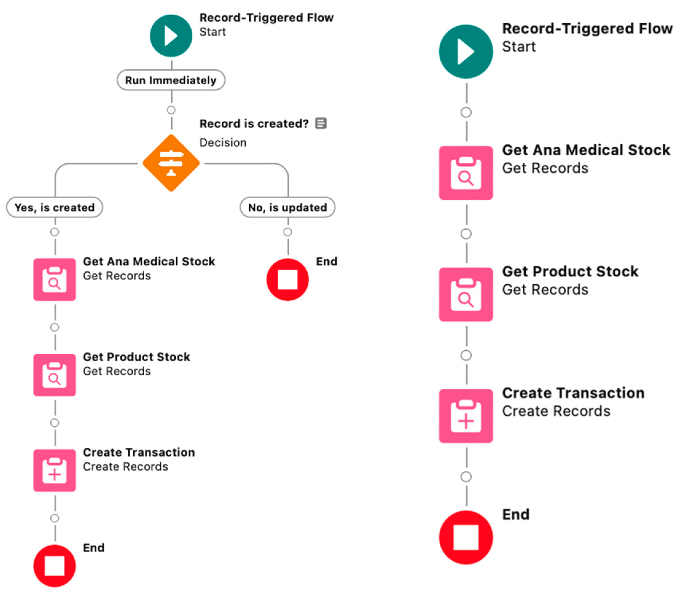
  • Flows: Powerful automation tools that allow users to create complex processes with multiple steps without writing code. SF Flow enables the design and execution of complex flows, guiding users through screens for data input and decision-making. It is part of SF’s low-code development tools, providing administrators and developers the ability to create powerful applications and automations with a visual interface. Key aspects:
    Visual Design: Flows are visually designed using Flow Builder, a drag-and-drop interface that allows users to create flows by adding elements and connecting them on a canvas. This visual approach simplifies the process of building complex logic.
    Declarative Logic: Flows provide a way to express flow logic declaratively, meaning steps can be defined without writing code. This makes it accessible to a wider audience, including administrators and analysts.
    Process Automation: Flows can automate processes by guiding users through a series of screens, collecting data, making decisions, and performing actions based on user inputs or predefined criteria.
    Record Updates: Flows can be used to update or create records in Salesforce.
    Integration Capabilities: flows can be integrated with external systems and services, allowing the incorporation of data and functionality from other platforms into SF processes.
    Reusable Elements: Flow components can be reused in different flows, making it easier to build and maintain consistent processes [40,42].
Flows can be initiated by user interaction (Screen Flows), system events (Auto-Launched Flows), or record changes (Record-Triggered Flows). In this thesis, Record-Triggered Flows were used, and they come in various types:
  • Triggering Event: Initiated by specific events on records, such as creating a new record, updating an existing record, or deleting a record.
  • Object-Specific: Record-Triggered Flows are associated with a specific Salesforce object (e.g., Account, Contact, Opportunity) and designed to respond to changes in records of that particular object.
  • Visual Design: Like other flows in SF, Record-Triggered Flows are built using Flow Builder, a low-code visual development tool. Users can design the flow by dragging and dropping elements on the canvas.
  • Automated Actions: Record-Triggered Flows allow users to define automated actions that should occur when a record meets specific criteria. These actions can include updating related records, sending emails, creating new records, and more.
  • Before or After Save: Record-Triggered Flows can be configured to run either before or after a record is saved to the database, providing flexibility in defining when the flow should be executed in relation to the record’s lifecycle.
  • Access to Record Data: Flows have the ability to access and manipulate record data during their execution, empowering them as tools for implementing custom logic without relying on APEX code.
  • Approval Processes: Automate the approval of records by defining approval criteria, configuring approval steps, and designating roles for approval.
  • Triggers: Snippets of APEX code that run before or after the insertion, updating, deleting, or querying of records. They provide developers with a way to implement custom logic and respond to events in the SF platform.
  • Batch Processes: Allow developers to process large sets of records asynchronously. They are commonly used for tasks such as data cleaning, data migration, or other operations involving massive record manipulation.
  • Scheduled Flows: Allow users to schedule the execution of a flow at a specific time or on a recurring basis. They are useful for automating repetitive tasks, such as sending periodic email notifications.
  • Scheduled Actions: Represent scheduled actions within processes or flows, allowing users to define actions that should occur at a specific time or in relation to a date field. They can be used for sending notifications, updating records, or triggering other automated tasks.
  • Event-Driven Automation: Event-based automation involves responding to specific events or changes in the system. This may include the use of triggers, platform events, or other mechanisms to initiate event-based automated processes [40,42].
Each type of automation in SF serves different purposes and is suitable for specific scenarios. Choosing the right automation tool depends on the complexity of the process to be automated and the type of events that should trigger the automation. In the elaboration of this paper, Record-Triggered Flows were used for managing medicine inventory and order management.
We used Flow Builder for updating the inventory upon completion of an order and automating the stock update when administering medication to a patient. For Creation, firstly, it checks whether the respective record has been created or not using an SF method (ISNEW) to control the creation event.
For Update, the stock is updated only if that order has been activated. It checks the activation by examining if the status value has changed. The flow allows manipulation of the old values of fields (Record Prior—this variable saves all old values from all fields). It checks if the status has been modified and if the status value is “Activated”. The activation of the order is verified. In a single flow, multiple events can be managed based on the order’s status.
The inventory is taken over. To maintain stock, a “Product Item” must be created for each. It maintains the quantity that first comes into stock. Using “Product Item Transactions”, the consumption of products from stock is controlled. In the flow, new records are automatically created to manage the stock.
The inventory is taken over, all products from the order are taken over, and product IDs are taken to retrieve the “Product Item”. It goes through each product in that order and through each “Product Item”. It goes through all “Product Items” until it reaches the one that contains the respective product for quantity update. It checks that the product is the same as the one on the “Product Item”.
The transaction list is created—the record in which all necessary information is taken: the found “Product Item”, the quantity from the order product, the transaction type, and the automatically calculated description to know where the transaction record was created.
To avoid reaching Flow limits, the newly created record is not inserted into the loop within Loop; instead, it is added to a list that was initially empty (before the first loop). Later, after exiting the loop within Loop, a single insertion event is performed, inserting the previously created list.
For administering medication to the patient, the medication is deducted from stock, and the stock is updated. The inventory is taken over, the “Product Item” is taken, whether it has been created is checked, and the medication stock update transaction is created. The flow is presented in Figure 5.

4.5. Forms

The solution for generating object-level forms in SF was implemented using Visualforce Page, which is the only way to build files in SF (Word, PDF, Excel, CSV) Exmple in Figure 6. The functionality of VFP is as follows:
  • Customization: VFP serves as a user interface model in SF, allowing the creation of custom interfaces for applications. It is a markup language with HTML and is used to design and display user interface components in an SF application. Visualforce pages can be used to create custom forms, lists, tables, charts, and other user interface elements.
  • Integration: VFP can be seamlessly integrated with standard SF objects and data, allowing the presentation and manipulation of data from different sources.
  • Controller Logic: VFP can be associated with a controller, which is essentially APEX code that provides page logic. This enables the generation of dynamic content and interaction with the data behind the application.
  • Visualforce Tags: VFP provides a variety of custom tags that define the structure and behavior of the page. These tags are used to create forms, tables, buttons, input fields, and more.
  • JavaScript: VFP can include JavaScript to enhance interactivity and client-side behavior. JavaScript can be used for validation, user interface enhancements, and communication with the server.
  • Styling: VFP allows access to CSS to control the appearance of Visualforce pages, ensuring a consistent and visually appealing user experience.
  • Mobile Device Support: VFP can be designed to be responsive and accessible on various devices, including desktops, tablets, and smartphones.
  • Security: VFP inherits the security and access controls of the Salesforce platform, ensuring data protection and functionality. Visualforce has been a fundamental technology for customizing user interfaces in SF applications [40,42].
Considering that the application forms are based on the same template, CSS (Cascading Style Sheets) settings are stored in a static resource. At the same time, a specific VFP component for the PDF file header was created, which is called for each form to avoid the need to rewrite it every time.
Another advantage of this approach is that when the CSS file in the static resource is modified, all new documents generated from all forms will adopt the new settings. This results in easier management of the solution. Generating such forms (any documents that need to be generated from the information system) allows the creation of templates that can be displayed in any desired manner. We generate a PDF with information gathered from the system for GDPR Consent Form, Medical Electronic Chart and Medical Procedure Consent Form.

4.6. Automatic Classification Module

Models for classification have been created in GT for detecting skin pathologies (melanoma, basal cell carcinoma, actinic keratosis, and seborrheic keratosis) and for detecting ocular pathologies (glaucoma, cataract, diabetic retinopathy). Subsequently, the models were uploaded to the Google Teachable cloud. Later, the JavaScript source code will be used to embed it in the integration with the SF platform. To call the model in SF, VFP (Visualforce Page) was used. The code arguments are:
  • Model (the file containing the model’s topology (json)).
  • Weights (the file containing the weights).
  • Metadata (the file containing the model’s labels and other additional information) [38].
i. 
Image
Within VFP, the front end of the application was implemented, and GT (Google Teachable) libraries were attached to access the classification model. Specifically, HTML code for loading an image, for the background of the classification module, and the button triggering the JavaScript controller with integration to Google Teachable can be found. The JavaScript custom controller is kept in a static resource and is called at the VFP level, thus maintaining the reuse characteristic and easier management of the controller. The button calls a “initPicture()” method at the controller level, which retrieves the model, metadata from GT and checks the predictions from the classifier. At the same time, an error message is returned if any type of error occurs while reading the image. Integration was achieved by accessing public URLs from GT.
ii. 
Video
For the video solution, the same working method was implemented as in the image loading solution. The difference lies in the use of the computer’s webcam. Specifically, when the button is pressed, the camera opens, utilizing the model and metadata from GT to run the classifier in real time and make predictions for the acquired video images. This classification module displays the result in real time with the acquisition of video images.

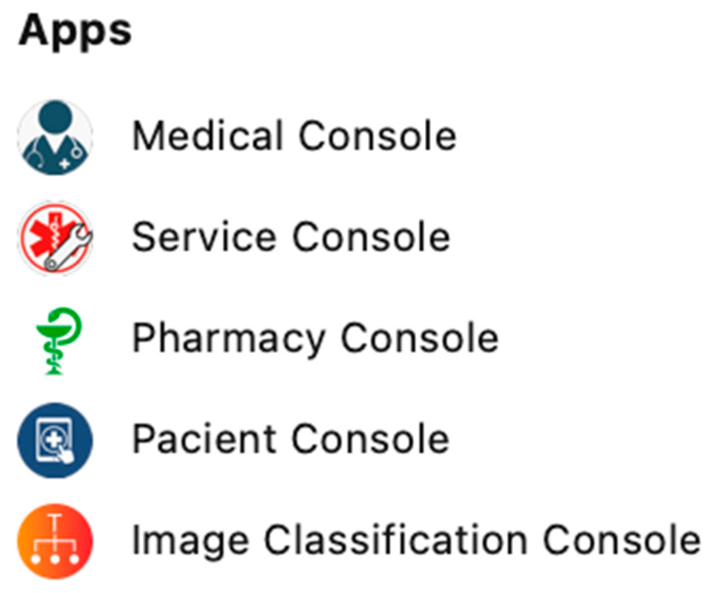
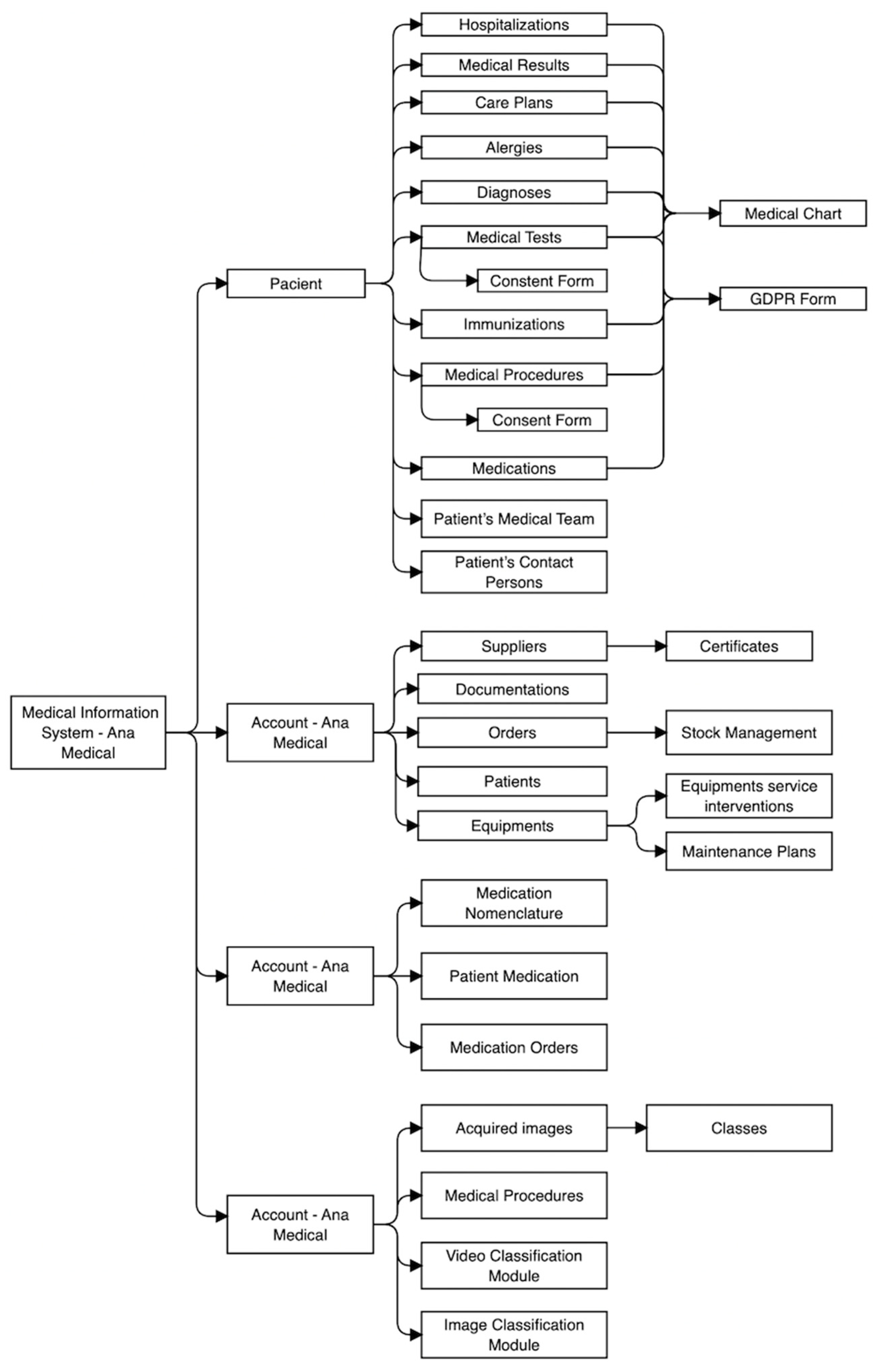
5. Medical Information System Architecture

To achieve the proposed objectives, we started with a CRM-type information system that ensures tracking the basic workflow and processing data in an institution interacting with clients. Subsequently, the platform was modified and developed both vertically and horizontally, resulting in a new her-type information system that integrates solutions for automatic classification of medical images using artificial intelligence, as well as newly developed applications and functionalities that respond to the tracking of a complex workflow in a medical institution. The new information system obtained, called “Ana Medical”, developed by the author of the thesis, fulfills the proposed objectives and ensures the functioning and integration of the application modules that compose it, as well as newly created functionalities to optimize the workflow or enhance the end user experience. In developing the architecture for the medical information system, a range of components such as objects, fields, consoles, layouts, reports, and charts were devised to enhance the efficiency of the workflow. Objects were implemented to store essential information related to patients, equipment, results, etc., at the database level. Within SF, data are structured using elements like objects, records, fields, instances, consoles, and applications. The structure of the information system is structured into several modules (consoles) as presented in Figure 7. The architecture of the system is exemplified in Figure 8.

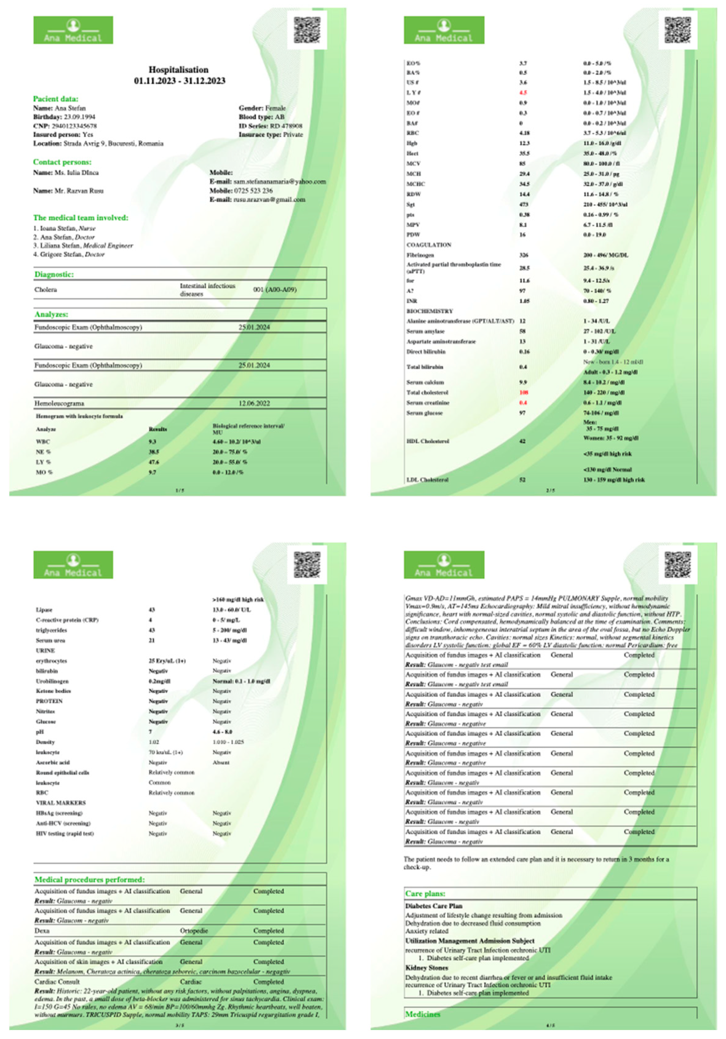
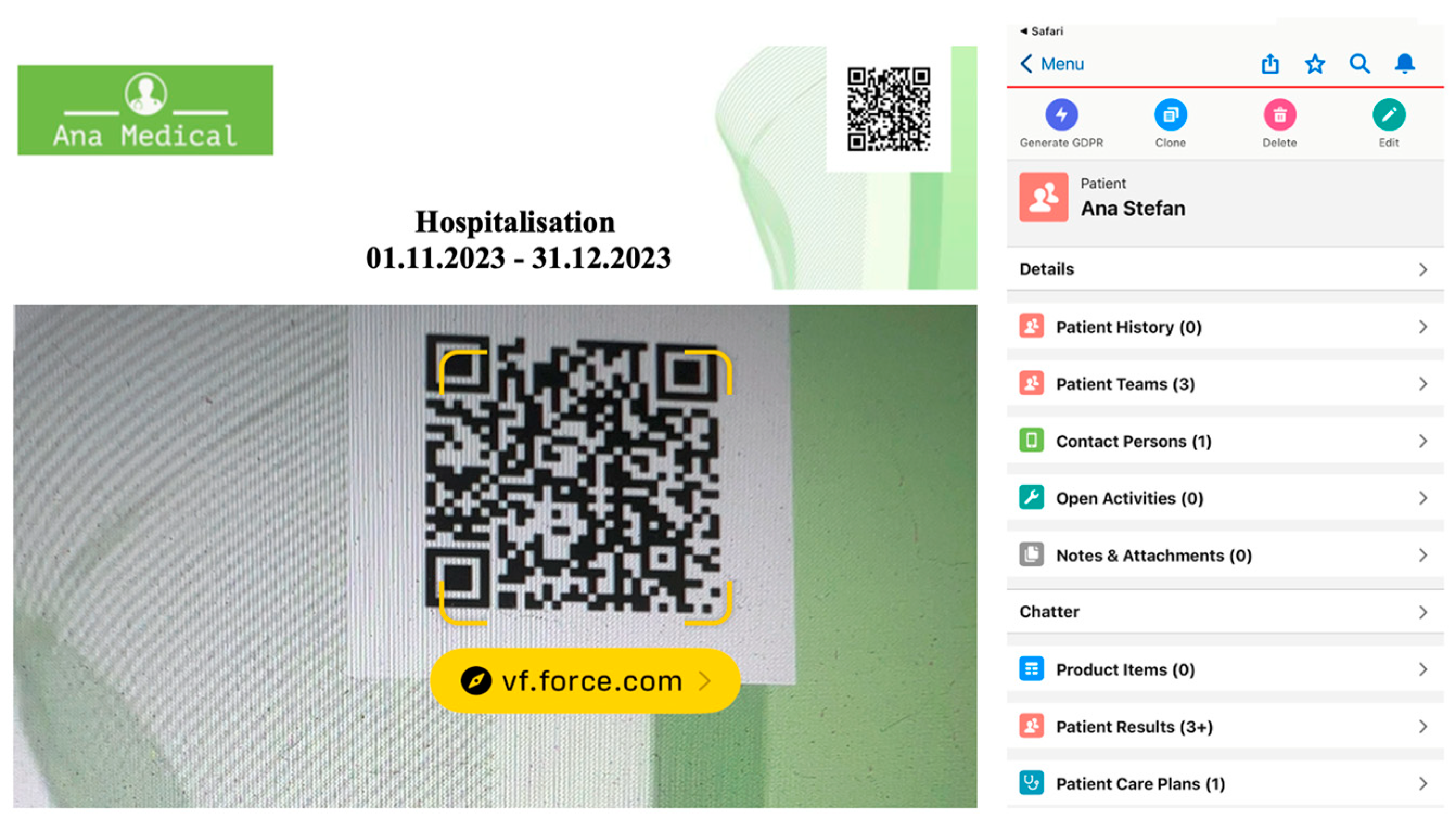
5.1. Medical Console

The console is dedicated to the medical team responsible for patient flow and medical interactions and consists of multiple objects. Patients are patients recorded by the medical institution. The information found at the level of this object includes the following (as seen in Figure 9):
  • Medical record number.
  • Patient’s name.
  • Personal Identification Number.
  • Medical insurance status.
  • Type of medical insurance.
  • Date of death.
  • Address.
  • Gender.
  • Date of birth.
  • Disability status.
  • Height.
  • Weight.
  • Blood type.
  • RH factor.
Dependent on this object are the following client-related pieces of information:
  • Patient encounters—Hospitalizations of the patient.
  • Patient results—Results of medical analyses and procedures.
  • Patient care plans—Care plans for patients.
  • Patient allergies—Patient allergies.
  • Patient diagnosis—Patient diagnoses.
  • Patient medical tests—Medical tests for the patient.
  • Patient immunizations—Patient vaccinations.
  • Patient procedures—Patient procedures.
  • Patient medication list—Medications the patient is on.
  • Patient teams—Patient’s medical team.
  • Contact persons—Patient’s contact persons.
These patient-related pieces of information are grouped in Related Lists (forming a link between the patient and the specific nomenclature). A related list is a component that displays a list of related records based on a specific object. At the patient record level, various pieces of information about them can be found. At the level of this object, multiple forms are generated, such as GDPR (General Data Protection Regulation), to process personal data in the system. For medical procedures and tests, a patient’s informed consent form is generated, as well as the patient’s medical record containing information generated during their hospitalization in the medical institution.

5.2. Service Console

The console is dedicated to the technical team of the hospital (medical engineers) responsible for medical equipment—maintenance, calibration, settings, usage, etc. The service console consists of multiple objects:
  • Account: Contains information about the medical institution—“Ana Medical.” This object includes related lists linked to other relevant objects:
  • Business Licenses: An object containing information about the institution’s tax certificates and more.
  • Providers: Information about the institution’s providers (address, website, contact, tax information).
  • Assets: An asset is equipment owned by the medical institution. It contains information with images about the product, clinical images, videos about the equipment, hands-on guides, etc., as well as documents about the equipment (product tree, service manuals, equipment conformity declaration, etc.).
  • Maintenance Plans: Are created for the installed equipment base, and interventions on specific equipment can be automatically generated based on certain criteria.
  • Work Orders: Medical engineers responsible for equipment reviews, perform interventions, and generate reports on the respective interventions.
  • Work Types: Types of interventions that can be performed on medical equipment. Based on these, automated action lists can be generated for a specific equipment model.

5.3. Pharmacy Console

The console is dedicated to the team managing the hospital’s pharmacy and deals with the medication procurement flow. The Pharmacy Console consists of multiple objects:
  • Orders: Manages medication orders.
  • Product Item: The institution’s equipment stock.
  • Medication List: The list of medications.
  • Patient Medication List: Links between the medication list and medications administered to patients, helping manage medication circulation.

5.4. Patient Console

The console is dedicated to patients. This is the portal through which patients can view results, treatment plans, medical history, etc. The patient console consists of a single object—patients. Depending on the logged-in patient, it contains a single record about that specific patient with relevant information:
  • Patient Encounters: Hospitalizations of the patient.
  • Patient Results: Results of medical analyses and procedures.
  • Patient Care Plans: Care plans for the patient.
  • Patient Allergies: Patient allergies.
  • Patient Diagnosis: Patient diagnoses.
  • Patient Medical Tests: Medical tests for the patient.
  • Patient Immunizations: Patient vaccinations.
  • Patient Procedures: Patient procedures.
  • Patient Medication List: Medications the patient is on.
  • Patient Teams: Patient’s medical team.
  • Contact Persons: Patient’s contact persons.
Scanning the QR code on any medical document generated by the institution directly refers to this patient console in a browser and on a mobile device—as seen in Figure 10.

5.5. Image Classification Console

The console is dedicated to the team of medical engineers specialized in Artificial Intelligence. The console contains the necessary objects for the acquisition and processing of medical images:
  • Patient Procedures: Procedures that contain AI processing.
  • Acquired Images: Each record contains:
    Classes: Classes of acquired images (pathology/pathologies, normal). Each class contains either the acquired images or an archive containing the acquired images. Both variants can be uploaded simultaneously.
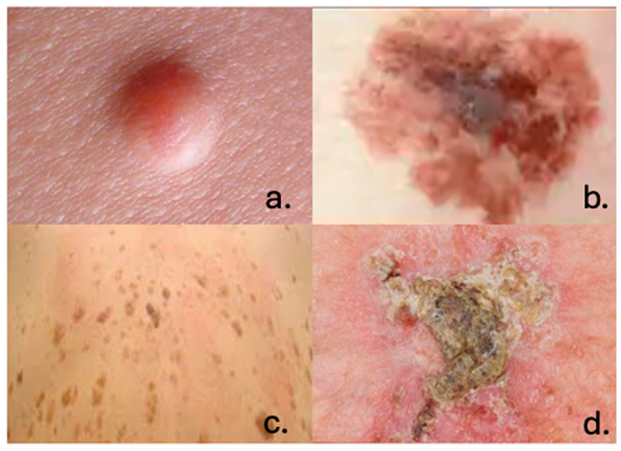
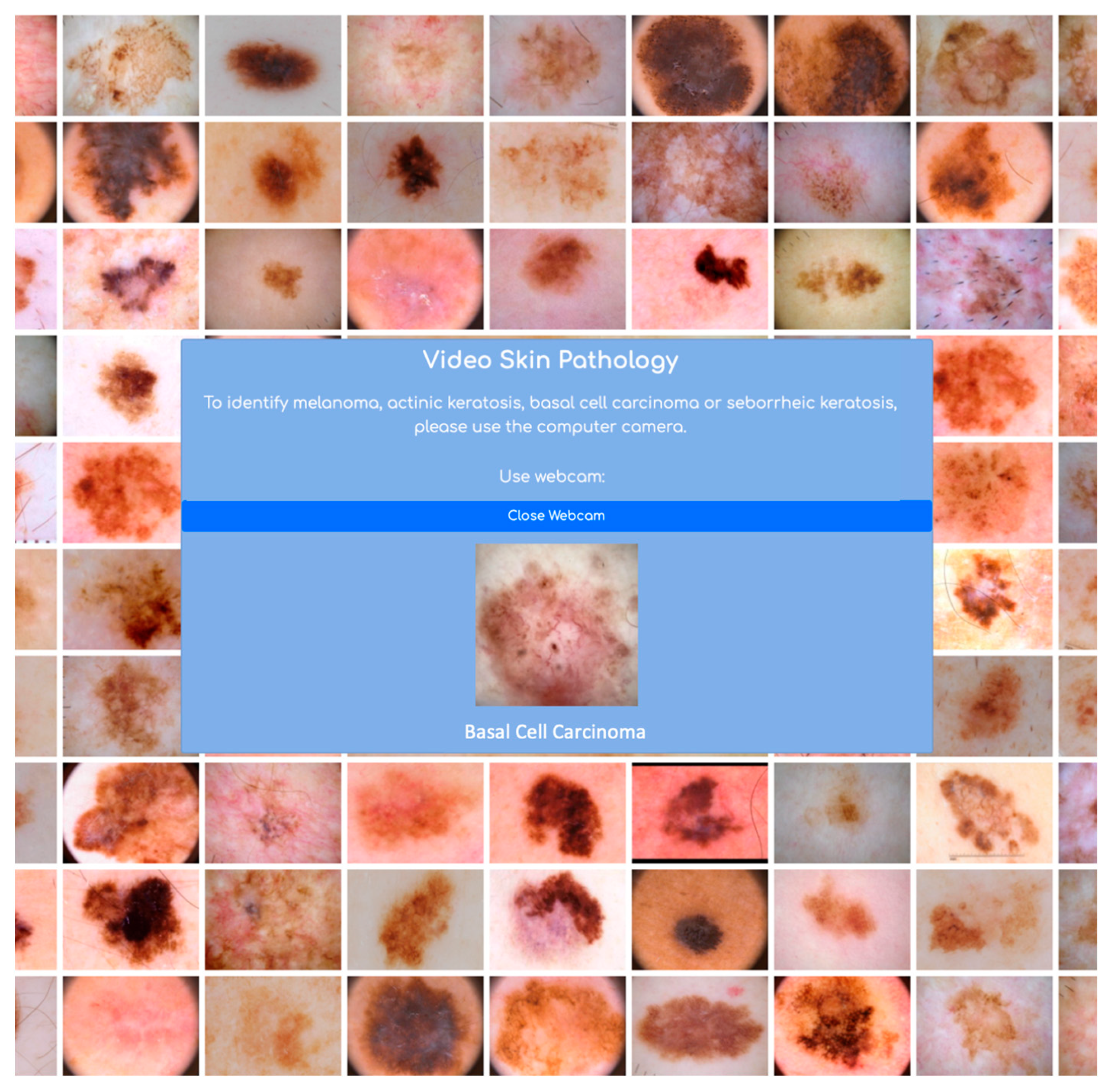
  • Skin Pathology Detection Module: Similar to the example in [38], but using multiple image classes: Melanoma, Actinic Keratosis, Seborrheic Keratosis, Basal Cell Carcinoma, and a class for images without pathologies. Video solution—a custom classification application for real-time classification of skin images. The module operates based on the camera of a computer or mobile device where the open platform of the information system is running. It encompasses the area with a lesion and displays, in real time, one of the four classes for which the classifier was trained (Figure 11).
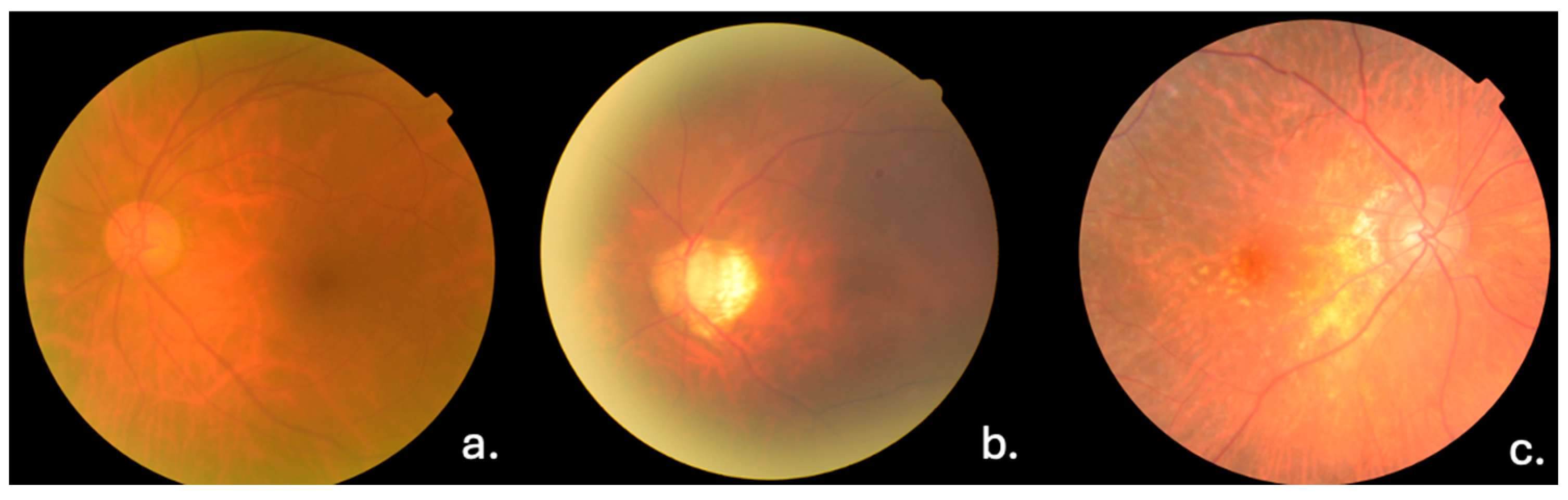
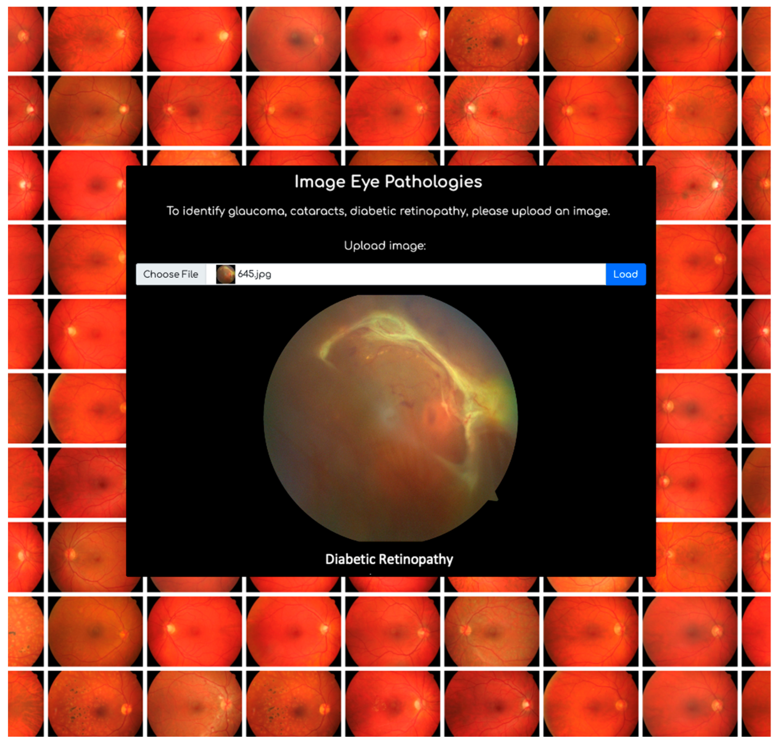
  • Ocular Pathology Detection Model: This model was created for classes such as Glaucoma, Cataract, and Diabetic Retinopathy. Image solution is a custom classification application for classifying fundus images acquired and stored in the database. The module operates by uploading an image and receiving a response based on the three pathologies for which the classification model is trained (Figure 12).
All these objects contribute to the management of workflows in the medical institution, such as medication ordering, sending notifications to teams with different roles in patient care, creating records when a team member needs to perform a medical procedure or take specific analyses. These activities are scheduled for easy management, and there is a notification flow upon completion of certain activities or upon receiving medical results, etc.

6. Ethical Correlates and Limitations

In the integration of automated screening systems within EHRs, various bioethical and social/psychological considerations arise. This section addresses key ethical concerns surrounding privacy, informed consent, accuracy, equity, psychological impact, patient autonomy, trust in healthcare providers, and societal implications. During this stage in our research, we first addressed ethical correlates of the system.
  • Privacy and Confidentiality: Implementing this kind of system raises concerns about patient privacy and the security of medical data. Safeguarding patient confidentiality and ensuring data protection are paramount. One of the reasons for implementing the system in SF was the security of the platform. SF security refers to the measures and protocols put in place to protect data and ensure the privacy, integrity, and availability of information within the SF platform. Salesforce employs a multi-layered approach to security, incorporating various features and functionalities to safeguard data from unauthorized access, breaches, and other threats. Some key aspects of Salesforce security include:
    User Authentication and Access Control: SF provides robust authentication mechanisms, such as username/password, multi-factor authentication (MFA), and single sign-on (SSO), to verify user identities. Access controls, including role-based access control (RBAC) and permission sets, enable administrators to define and enforce granular access permissions based on user roles and responsibilities.
    Data Encryption: SF encrypts data both at rest and in transit to protect it from unauthorized access. Encryption ensures that sensitive information remains confidential even if accessed by unauthorized parties.
    Network Security: SF employs network security measures, such as firewalls, intrusion detection and prevention systems (IDPS), and distributed denial-of-service (DDoS) protection, to safeguard its infrastructure and prevent unauthorized access or attacks.
    Application Security: SF follows secure coding practices and regularly conducts security assessments and audits to identify and address vulnerabilities in its applications. Features like SF Shield provide additional security controls, such as event monitoring, data loss prevention (DLP), and encryption of sensitive data within Salesforce.
    Compliance and Governance: SF adheres to industry standards and regulatory requirements, such as GDPR, HIPAA, and SOC 2, to ensure compliance with data protection and privacy regulations. The platform also offers features like audit trails, compliance dashboards, and security certifications to help organizations demonstrate compliance and governance.
    Threat Detection and Monitoring: SF continuously monitors its platform for suspicious activities, unauthorized access attempts, and other security threats. Automated alerts, security logs, and real-time monitoring tools enable administrators to detect and respond to security incidents promptly.
    Secure Development Lifecycle: SF follows a secure development lifecycle (SDLC), incorporating security best practices into every stage of the software development process. This approach helps minimize security vulnerabilities and ensures that security is prioritized from the initial design phase through deployment and maintenance.
  • Informed Consent: Patients should be adequately informed about the use of automated screening systems during routine check-ups. Transparent communication regarding data collection, storage, and usage is essential to respect patients’ autonomy and decision-making. We developed and included forms like GDPR and Consent that the patients would be asked to sign before any medical procedures.
  • Accuracy and Reliability: Ethical considerations include ensuring the accuracy and reliability of automated screening systems to minimize the risk of false positives or false negatives, which could lead to unnecessary anxiety or missed diagnoses. We took into consideration both accuracy and F1 score to ensure that the system is both reliable and accurate.
In the subsequent phase of our research, implementing this prototype within a medical institution entails addressing social and psychological factors. This encompasses both educating and providing support to patients throughout the process.
  • Psychological Impact: Early detection of potentially harmful pathologies through automated screening may have psychological implications for patients. This includes managing anxiety, coping with uncertainty, and providing appropriate support for individuals undergoing further diagnostic procedures.
  • Patient Autonomy: While automated screening systems may improve early detection rates, patients should retain autonomy in healthcare decision-making. Respect for patient preferences and values should guide discussions about screening, diagnosis, and treatment.
  • Trust in Healthcare Providers: The introduction of automated screening systems may impact patient-provider relationships and trust. Open communication, shared decision-making, and transparency about the limitations of technology can help maintain trust and confidence in healthcare providers.
  • Societal Impact: Implementation of automated screening systems may have broader societal implications, including changes in healthcare delivery, resource allocation, and health policy. Ethical considerations should address potential societal benefits and risks associated with widespread adoption of these technologies.
Introducing a digital system, particularly a medical information system, into a healthcare institution involves recognizing and addressing limitations early in the project, with a focus on finding solutions to overcome these challenges. The limitations of the proposed system are as follows:
  • Limited Scope: Automated classification modules may be limited in their scope and may not cover all possible diagnoses or conditions. Certain rare or complex conditions may not be adequately captured by the system, requiring manual intervention by healthcare providers.
  • User Adoption and Training: The success of an information system depends on user adoption and proficiency. If users are not adequately trained or if the system is not user-friendly, it may lead to low adoption rates and underutilization of the system’s capabilities.
  • Maintenance and Updates: Automated classification algorithms require regular maintenance and updates to remain effective and up to date with the latest medical knowledge and technological advancements. This ongoing maintenance can be resource-intensive and time-consuming.
  • Integration Challenges: Integrating an automated classification module into an existing EHR system can be complex and may require significant changes to the system’s infrastructure and workflow. Ensuring seamless integration and compatibility with existing processes is crucial for successful implementation.
  • Interpretability: Deep learning algorithms used for automated classification often lack interpretability, making it challenging to understand why a particular classification decision was made. This lack of transparency can undermine trust in the system among healthcare providers and patients.
  • Dependence on Technology: Information systems rely on technology infrastructure, software, and networks. Any disruptions or failures in these components can impact the availability and functionality of the system.
  • Cost: Developing, implementing, and maintaining an information system can be expensive. Organizations need to budget for initial development costs as well as ongoing operational expenses, including licensing fees, hardware upgrades, and personnel costs.
  • Maintenance: Information systems require regular maintenance, updates, and patches to ensure optimal performance and security. Neglecting maintenance tasks can result in system downtime, vulnerabilities, and performance issues.
  • Scalability: As organizations grow and evolve, their information systems need to scale accordingly to accommodate increased data volume, user load, and business requirements. Scaling up an information system can be challenging and may require significant investment in infrastructure and resources.
Addressing these limitations requires a holistic approach that involves careful planning, investment in technology infrastructure and resources, ongoing training and support for users, and collaboration with healthcare stakeholders to ensure that the medical information system meets the needs of patients and providers while adhering to regulatory requirements.

7. Future Enhancements and Considerations

The next steps for improving the solution of the automated pathology detection module presented in this work include replacing VFP with LWC, which poses a more challenging development solution. However, the advantage of LWC lies in the fact that the application utilizes browser resources rather than server resources. Another direction for enhancing the solution involves integrating various platforms with the information system to harness computational power available on platforms built for implementing complex solutions. This includes integration with frameworks enabling the construction of custom classifiers or systems based on the fusion of decisions from multiple convolutional neural networks, as presented in [28]. The expansion of diagnostics for diverse pathologies using different types of data beyond medical images is also a consideration.
Additionally, to employ this solution in the medical industry, another useful tool would be a telemedicine module that can connect patients, medical service providers, and emergency services for real-time data collection in the information system. Another useful tool could be integration with the SF Tableau native analytics system for analyzing collected data to identify trends, patterns, and changes in a patient’s condition over time. Predictive analysis can forecast potential pathology risks based on historical data and patient profiles, coupled with the integration of genetic databases to enhance pathology detection by considering genetic predispositions and mutations.
Furthermore, an opportunity lies in increasing patient awareness and education by providing resources to enhance understanding of the importance of early detection, potential risks associated with late detection of various pathologies, and recommended actions for their treatment. These ideas can contribute to improving the capabilities and usability of an EHR system that integrates automated pathology detection, ultimately enhancing patient care, diagnostic accuracy, and medical workflows.
For the expansion of the information system’s usage, the development of a financial-accounting module is necessary, along with the integration of medical imaging acquisition equipment or analysis equipment. Implementing quotation modules for medical packages and health insurance, a module for managing employee data, and a marketing console to promote the institution and the built information system are also crucial aspects.

8. Conclusions

In summary, this article’s findings and contributions offer fresh perspectives on healthcare technology through the introduction of a medical information system developed within the SF environment. The standout feature of this system is its automated classification module, which addresses both ocular and skin pathologies. By harnessing the capabilities of GT, our integration combines state-of-the-art technology with the robust functionalities of SF, offering a comprehensive solution tailored for medical practitioners.
The article meticulously explores the intricate structure of the system, shedding light on its innovative functionalities meticulously crafted to augment diagnostic precision and streamline medical workflows. The methods employed in the development of these functionalities are dissected, offering valuable insights into the seamless integration of GT within the SF framework. This collaborative approach not only propels the landscape of medical information systems forward but also signifies a momentous stride towards intelligent and efficient pathology classification.
The article’s contributions extend beyond the technological intricacies, emphasizing the system’s ability to classify diverse pathologies, including ocular complications of diabetes (cataract, diabetic retinopathy, glaucoma), skin cancers like melanoma and basal cell carcinoma, and other potential precancerous conditions such as actinic keratosis and seborrheic keratosis. By addressing these specific pathologies, the system stands poised to significantly impact the healthcare domain, offering a comprehensive and intelligent solution for practitioners.
In essence, this article marks a transformative chapter in medical information systems, blending advanced technology with practical applications to elevate the standards of pathology classification. The amalgamation of GT and SF not only presents an innovative solution for current medical challenges but also sets a precedent for future developments in intelligent healthcare systems.
The integration of the automated detection system into an EHR as a concept holds promise for facilitating more frequent assessments by including screenings during patient visits, thereby potentially enhancing the chance of early detection of suspicious changes. This would also translate into numerous advantages for healthcare providers. The automated detection system streamlines the screening process, enabling healthcare professionals to analyze images efficiently and flag potentially concerning cases for further examination. Conceptually, this approach alleviates the workload of healthcare providers, enabling them to concentrate on crucial aspects of patient care. It positions this implementation as a support tool for clinical personnel.
The system provides real-time alerts, promptly notifying healthcare professionals of suspicious changes during routine visits or medical record reviews, facilitating timely monitoring and intervention. Utilizing advanced algorithms and machine learning, the automated detection system enhances diagnostic accuracy, contributing to the reduction in human errors (like exhaustion, misinterpretation of symptoms, incomplete examination, diagnostic bias, limited experience, diagnostic uncertainty and so on).
Patient engagement and education are fostered through EHR systems, allowing patients to comprehend screening results, understand risk factors, and take a proactive approach to their health. High-risk patients benefit from efficient monitoring, as the integrated system allows healthcare providers to track them more effectively, ensuring regular screenings and follow-up evaluations. The accumulation of data from multiple screenings and patient histories enables data-driven decision-making and trend analysis, leading to targeted prevention and intervention strategies. Aggregated data from the automated skin pathology detection system supports population health management, helping healthcare institutions identify trends in skin cancer prevalence and allocate resources more efficiently. Interoperability within the EHR ensures accessibility of skin cancer screening results to authorized healthcare providers, promoting communication and healthcare continuity while avoiding redundant screenings.
Early detection of ocular diseases through the automated system allows for prompt intervention and treatment initiation, which is crucial in preserving vision and preventing irreversible damage. Integrating automated detection systems into EHRs streamlines the diagnostic process, providing healthcare professionals with effortless access to complete medical records for quick decision making and personalized treatment planning. Continuous monitoring of patients’ ocular health over time allows for the prompt identification of subtle changes, leading to early diagnosis and management. The early detection of ocular diseases contributes to cost reduction by minimizing resource requirements and preventing complications that necessitate extensive treatments. Additionally, automated detection systems support remote monitoring and telemedicine, facilitating healthcare consultations even for patients in remote or underserved areas. The EHR’s capability to capture and store longitudinal data enables retrospective analysis, offering insights into disease progression, treatment effectiveness, and outcomes. Aggregated and anonymized data from integrated automated detection systems can significantly contribute to research and population health studies, unveiling trends, risk factors, and treatment outcomes, ultimately aiding in the development of more effective strategies for early detection and treatment. The implementation of an automated classification module for diagnosis is not designed to replace the role of doctors or supplant medical opinion. Rather, it serves as a complementary tool aimed at enhancing diagnostic capabilities and supporting healthcare professionals in their decision-making processes. The module’s purpose is to streamline data analysis and provide additional insights to aid in accurate diagnosis, while ultimately empowering physicians to make informed decisions based on their expertise and clinical judgment.

Author Contributions

Conceptualization, methodology, software, validation and formal analysis, A.-M.Ș. and N.-R.R.; investigation, resources, data curation, writing—original draft preparation, writing—review and editing, visualization, project administration A.-M.Ș.; supervision E.O. and M.C.; funding acquisition, M.C. All authors have read and agreed to the published version of the manuscript.

Funding

This research was funded by POLITEHNICA Bucharest National University of Science and Technology through PubArt doctoral program.

Data Availability Statement

The data presented in this study are available on request from the corresponding author due to privacy reasons.

Conflicts of Interest

Author Nicu-Răzvan Rusu was an independent researcher. The remaining authors declare that the research was conducted in the absence of any commercial or financial relationships that could be construed as a potential conflict of interest.

Abbreviations

APIApplication Programming Interface
CNNConvolutional Neural Network
CRMCustomer Relationship Management
CSSCascading Style Sheets
DNADeoxyribonucleic acid
EMRElectronic Medical Records
EHRElectronic Health Record
GDPRGeneral Data Protection Regulation
GTGoogle Teachable Machine
HIPAAHealth Insurance Portability and Accountability
HTMLHyperText Markup Language
LWCLightning Web Components
SFSalesforce
SaaSSoftware as a Service
UVUltraviolet
UATUser Acceptance Testing
VFPVisualforce Page
WHOWorld Health Organization

References

  1. Available online: https://www.who.int (accessed on 20 February 2022).
  2. Nouwen, A.; Nefs, G.; Caramlau, I.; Connock, M.; Winkley, K.; Lloyd, C.E.; Peyrot, M.; Pouwer, F. Prevalence of Depression in Individuals with Impaired Glucose Metabolism or Undiagnosed Diabetes: A systematic review and meta-analysis of the European Depression in Diabetes (EDID) Research Consortium. Diabetes Care 2011, 34, 752–762. [Google Scholar] [CrossRef]
  3. Diabetes. Available online: https://www.who.int/health-topics/diabetes#tab=tab_1 (accessed on 20 February 2022).
  4. Diabetic Retinopathy—Symptoms & Causes—Mayo Clinic. (21 February 2022). Available online: https://www.mayoclinic.org/diseases-conditions/diabetic-retinopathy/symptoms-causes/syc-20371611 (accessed on 20 February 2022).
  5. Nizami, A.A.; Gulani, A.C. Cataract. In StatPearls; StatPearls Publishing: Treasure Island, FL, USA, 2024; [Updated 5 July 2022]. Available online: https://www.ncbi.nlm.nih.gov/books/NBK539699/ (accessed on 20 February 2023).
  6. Schuster, A.K.; Erb, C.; Hoffmann, E.M.; Dietlein, T.; Pfeiffer, N. The Diagnosis and Treatment of Glaucoma. Dtsch. Ärzteblatt Int. 2020, 117, 225–234. [Google Scholar] [CrossRef]
  7. Kamińska, A.; Pinkas, J.; Wrześniewska-Wal, I.; Ostrowski, J.; Jankowski, M. Awareness of Common Eye Diseases and Their Risk Factors—A Nationwide Cross-Sectional Survey among Adults in Poland. Int. J. Environ. Res. Public Health 2023, 20, 3594. [Google Scholar] [CrossRef]
  8. Han, J. Artificial Intelligence in Eye Disease: Recent Developments, Applications, and Surveys. Diagnostics 2022, 12, 1927. [Google Scholar] [CrossRef]
  9. Tan, Y.; Sun, X. Ocular images-based artificial intelligence on systemic diseases. BioMed. Eng. OnLine 2023, 22, 49. [Google Scholar] [CrossRef]
  10. Xiao, X.; Long-Yi, X.; Lin, Y.F.; Li, W.; He, Y. Health care cost and benefits of artificial intelligence-assisted population-based glaucoma screening for the elderly in remote areas of China: A cost-offset analysis. BMC Public Health 2021, 21, 1065. [Google Scholar] [CrossRef]
  11. Gomez Rossi, J.; Rojas-Perilla, N.; Krois, J.; Schwendicke, F. Cost-effectiveness of Artificial Intelligence as a Decision-Support System Applied to the Detection and Grading of Melanoma, Dental Caries, and Diabetic Retinopathy. JAMA Netw. Open 2022, 5, e220269. [Google Scholar] [CrossRef]
  12. Javaid, M.; Haleem, A.; Pratap Singh, R.; Suman, R.; Rab, S. Significance of machine learning in healthcare: Features, pillars and applications. Int. J. Intell. Netw. 2022, 3, 58–73. [Google Scholar] [CrossRef]
  13. Chen, X.; Xu, Y.; Wong, D.W.K.; Wong, T.Y.; Liu, J. Glaucoma detection based on deep convolutional neural network. In Proceedings of the 2015 37th Annual International Conference of the IEEE Engineering in Medicine and Biology Society (EMBC), Milan, Italy, 25–29 August 2015; pp. 715–718. [Google Scholar] [CrossRef]
  14. Parashar, D.; Agrawal, D.K. Classification of Glaucoma Stages Using Image Empirical Mode Decomposition from Fundus Images. J. Digit. Imaging 2022, 35, 1283–1292. [Google Scholar] [CrossRef]
  15. Chakrabarty, N.; Chatterjee, S. A Novel Approach to Glaucoma Screening using Computer Vision. In Proceedings of the 2019 International Conference on Smart Systems and Inventive Technology (ICSSIT), Tirunelveli, India, 27–29 November 2019. [Google Scholar] [CrossRef]
  16. Zhang, Z.; Lee, B.H.; Liu, J.; Wong, D.W.K.; Tan, N.M.; Lim, J.H.; Yin, F.; Huang, W.; Li, H.; Wong, T.Y. Optic disc region of interest localization in fundus image for Glaucoma detection in ARGALI. In Proceedings of the 2010 5th IEEE Conference on Industrial Electronics and Applications, Taichung, Taiwan, 15–17 June 2010. [Google Scholar] [CrossRef]
  17. Norouzifard, M.; Nemati, A.; GholamHosseini, H.; Klette, R.; Nouri-Mahdavi, K.; Yousefi, S. Automated Glaucoma Diagnosis Using Deep and Transfer Learning: Proposal of a System for Clinical Testing. In Proceedings of the 2018 International Conference on Image and Vision Computing New Zealand (IVCNZ), Auckland, New Zealand, 19–21 November 2018. [Google Scholar] [CrossRef]
  18. Serener, A.; Serte, S. Transfer Learning for Early and Advanced Glaucoma Detection with Convolutional Neural Networks. In Proceedings of the 2019 Medical Technologies Congress (TIPTEKNO), Izmir, Turkey, 3–5 October 2019. [Google Scholar] [CrossRef]
  19. Hasan, N.; Nadaf, A.; Imran, M.; Jiba, U.; Sheikh, A.; Almalki, W.H.; Almujri, S.S.; Mohammed, Y.H.; Kesharwani, P. Skin cancer: Understanding the journey of transformation from conventional to advanced treatment approaches. Mol. Cancer 2023, 22, 168. [Google Scholar] [CrossRef]
  20. Reinehr, C.P.H.; Bakos, R.M. Actinic keratoses: Review of clinical, dermoscopic, and therapeutic aspects. An. Bras. Dermatol. 2019, 94, 637–657. [Google Scholar] [CrossRef]
  21. Greco, M.J.; Mahabadi, N.; Gossman, W. Seborrheic Keratosis. PubMed. StatPearls Publishing. Available online: https://www.ncbi.nlm.nih.gov/books/NBK545285/ (accessed on 20 June 2023).
  22. Saginala, K.; Barsouk, A.; Aluru, J.S.; Rawla, P.; Barsouk, A. Epidemiology of Melanoma. Med. Sci. 2021, 9, 63. [Google Scholar] [CrossRef]
  23. Scolyer, R.A.; Rawson, R.V.; Gershenwald, J.E.; Ferguson, P.M.; Prieto, V.G. Melanoma pathology reporting and staging. Mod. Pathol. 2019, 33 (Suppl. S1), 15–24. [Google Scholar] [CrossRef]
  24. Mazhar, T.; Haq, I.; Ditta, A.; Syed Rehman, F.; Zafar, I.; Gansau, J.A.; Goh, L.P.W. The Role of Machine Learning and Deep Learning Approaches for the Detection of Skin Cancer. Healthcare 2023, 11, 415. [Google Scholar] [CrossRef]
  25. Antohe, M.; Coman, A.; Turcu, G.; Nedelcu, R.I.; Brinzea, A.; Balaban, M.; Moroianu, A.; Manea, L.; Hulea, I.; Balasescu, E.; et al. The prognostic significance of the clinical and histological parameters in primary cutaneous melanoma patients. Med. Pharm. Rep. 2022, 95, 229. [Google Scholar] [CrossRef]
  26. Zhang, Z.; Yin, F.S.; Liu, J.; Wong, W.K.; Tan, N.M.; Lee, B.H.; Cheng, J.; Wong, T.Y. ORIGA-light: An online retinal fundus image database for glaucoma analysis and research. In Proceedings of the 2010 Annual International Conference of the IEEE Engineering in Medicine and Biology, Buenos Aires, Argentina, 31 August–4 September 2010. [Google Scholar] [CrossRef]
  27. Messidor. Available online: http://www.adcis.net/en/third-party/messidor/ (accessed on 20 February 2022).
  28. Data Analysis. Available online: http://www.eyepacs.com/data-analysis (accessed on 20 February 2022).
  29. High-Resolution Fundus (HRF) Image Database. Available online: https://www5.cs.fau.de/research/data/fundus-images/ (accessed on 20 February 2023).
  30. The International Skin Imaging Collaboration. Available online: https://www.isic-archive.com (accessed on 20 February 2022).
  31. Diabetic Retinopathy Detection. Available online: https://www.kaggle.com/c/diabetic-retinopathy-detection/data (accessed on 20 February 2023).
  32. Available online: https://github.com/miag-ull/rim-one-dl?tab=readme-ov-file (accessed on 21 March 2024).
  33. Fumero, F.; Alayon, S.; Sanchez, J.L.; Sigut, J.; Gonzalez-Hernandez, M. RIM-ONE: An open retinal image database for optic nerve evaluation. In Proceedings of the 2011 24th International Symposium on Computer-Based Medical Systems (CBMS), Bristol, UK, 27–30 June 2011. [Google Scholar] [CrossRef]
  34. Dermatology Information System. Available online: https://www.dermis.net/dermisroot/en/home/index.htm (accessed on 20 February 2022).
  35. Giotis, I.; Molders, N.; Land, S.; Biehl, M.; Jonkman, M.F.; Petkov, N. MED-NODE: A computer-assisted melanoma diagnosis system using non-dermoscopic images. Expert Syst. Appl. 2015, 42, 6578–6585. [Google Scholar] [CrossRef]
  36. Mendonca, T.; Ferreira, P.M.; Marques, J.S.; Marcal, A.R.S.; Rozeira, J. PH2—A dermoscopic image database for research and benchmarking. In Proceedings of the 2013 35th Annual International Conference of the IEEE Engineering in Medicine and Biology Society (EMBC), Osaka, Japan, 3–7 July 2013. [Google Scholar] [CrossRef]
  37. El-khatib, H.; Ștefan, A.-M.; Popescu, D. Performance Improvement of Melanoma Detection Using a Multi-Network System Based on Decision Fusion. Appl. Sci. 2023, 13, 10536. [Google Scholar] [CrossRef]
  38. Ștefan, A.-M.; El-khatib, H.; Popescu, D. Melanoma Automated Detection System Integrated with an EHR Platform. UPB Sci. Bull. Ser. C Electr. Eng. Comput. Sci. 2024, 1. [Google Scholar]
  39. Ștefan, A.-M.; Ovreiu, E.; Ciuc, M. Comparative Analysis of Web-Based Machine Learning Models. Rom. J. Inf. Technol. Autom. Control. 2024, 34. [Google Scholar]
  40. Salesforce. Available online: https://www.salesforce.com (accessed on 20 February 2022).
  41. Teachable Machine. Available online: https://teachablemachine.withgoogle.com (accessed on 20 February 2022).
  42. Malmqvist, L. Architecting AI Solutions on Salesforce; Packt Publishing Ltd.: Birmingham, UK, 2021; ISBN 978-1-80107-229-8. [Google Scholar]
Figure 1. Cataract (a), Glaucoma (b), Diabetic Retinopathy (c).
Asi 07 00028 g001
Figure 2. Skin cancer evolution.
Asi 07 00028 g002
Figure 3. Basal Cell Carcinoma (a), Melanoma (b), Seborrheic keratosis (c), Actinic keratosis (d).
Asi 07 00028 g003
Figure 4. Email notification example.
Asi 07 00028 g004
Figure 5. Automation of updating the stock during medication administration.
Asi 07 00028 g005
Figure 6. Medical Electronic Chart Form.
Asi 07 00028 g006
Figure 7. “AnaMedical” information system console structure.
Asi 07 00028 g007
Figure 8. “AnaMedical” information system arhitecture.
Asi 07 00028 g008
Figure 9. Patient object and its information.
Asi 07 00028 g009
Figure 10. Scanning the QR code on the Medical Chart.
Asi 07 00028 g010
Figure 11. Automatic classification module for skin pathologies.
Asi 07 00028 g011
Figure 12. Automatic classification module for ocular pathologies.
Asi 07 00028 g012
Table 1. Classes and the number of images based on pathology.
ClassImage Number
Ocular pathologies
Glaucoma1150
Cataract1176
Diabetic retinopathy1006
Normal1299
Skin pathologies
Melanoma2463
Normal2463
Actinic keratosis2166
Seborrheic keratosis2764
Basal cell carcinoma2764
Table 2. Experimental results for web-based models on melanoma.
Web-Based ModelDatabaseAccuracyF1 Score
Google Teachable MachineDermIS0.780.77
ISIC0.760.75
Google Vertex AIDermIS0.830.83
ISIC0.740.73
Microsoft Azure Machine LearningDermIS0.790.79
ISIC0.820.82
SalesForce
Einstein
DermIS0.800.80
ISIC0.790.79
Table 3. Experimental results for web-based models on various pathology.
Web-Based ModelPathologyAccuracyF1 Score
Azure Machine LearningMelanoma0.900.90
Actinic keratosis0.930.93
Seborrheic keratosis0.950.95
Basal cell carcinoma0.920.92
Google TeachableMelanoma0.940.94
Actinic keratosis0.940.94
Seborrheic keratosis0.980.98
Basal cell carcinoma0.980.98
Google Cloud—Vision AIMelanoma0.970.97
Actinic keratosis0.950.95
Seborrheic keratosis0.970.97
Basal cell carcinoma0.960.96
Einstein VisionMelanoma0.910.91
Actinic keratosis0.910.91
Seborrheic keratosis0.910.91
Basal cell carcinoma0.900.90
Azure Machine LearningGlaucoma0.960.96
Cataract0.900.90
Diabetic retinopathy0.960.96
Google TeachableGlaucoma0.970.97
Cataract0.960.96
Diabetic retinopathy0.940.94
Google Cloud—Vision AIGlaucoma0.980.98
Cataract0.980.98
Diabetic retinopathy0.980.98
Einstein VisionGlaucoma0.910.91
Cataract0.870.87
Diabetic retinopathy0.900.90
Disclaimer/Publisher’s Note: The statements, opinions and data contained in all publications are solely those of the individual author(s) and contributor(s) and not of MDPI and/or the editor(s). MDPI and/or the editor(s) disclaim responsibility for any injury to people or property resulting from any ideas, methods, instructions or products referred to in the content.

