Article

Stability Studies and the In Vitro Leishmanicidal Activity of Hyaluronic Acid-Based Nanoemulsion Containing Pterodon pubescens Benth. Oil

by
Sirlene Adriana Kleinubing
1,
Priscila Miyuki Outuki
1,
Éverton da Silva Santos
1,
Jaqueline Hoscheid
2,3,*,†,
Getulio Capello Tominc
2,
Mariana Dalmagro
3,
Edson Antônio da Silva
4,
Marli Miriam de Souza Lima
1,
Celso Vataru Nakamura
1 and
Mara Lane Carvalho Cardoso
1
1
Programa de Pós-Graduação em Ciências Farmacêuticas, Universidade Estadual de Maringá, Maringá 87020-900, Brazil
2
Programa de Mestrado Profissional em Plantas Medicinais e Fitoterápicos na Atenção Básica, Universidade Paranaense, Umuarama 87502-210, Brazil
3
Programa de Pós-Graduação em Biotecnologia Aplicada à Agricultura, Universidade Paranaense, Umuarama 87502-210, Brazil
4
Programa de Pós-Graduação em Engenharia Química, Centro de Engenharias e Ciências Exatas, Universidade Estadual do Oeste do Paraná, Toledo 85903-000, Brazil
*
Author to whom correspondence should be addressed.
Current address: Universidade Paranaense, Umuarama 87502-210, Brazil.
Colloids Interfaces 2022, 6(4), 64; https://doi.org/10.3390/colloids6040064
Submission received: 14 September 2022 / Revised: 3 October 2022 / Accepted: 1 November 2022 / Published: 3 November 2022
(This article belongs to the Special Issue Recent Advances in Emulsions and Applications)

Abstract

:
The physicochemical and microbiological stability of a hyaluronic acid-based nanostructured topical delivery system containing P. pubescens fruit oil was evaluated, and the in vitro antileishmanial activity of the nanoemulsion against Leishmania amazonensis and the cytotoxicity on macrophages was investigated. The formulation stored at 5 ± 2 °C, compared with the formulation stored at 30 and 40 ± 2 °C, showed a higher chemical and physical stability during the period analyzed and in the accelerated physical stability study. The formulation stored at 40 °C presented a significant change in droplet diameter, polydispersity index, zeta potential, pH, active compound, and consistency index and was considered unstable. The microbiological stability of the formulations was confirmed. The leishmanicidal activity of the selected system against intracellular amastigotes was significantly superior to that observed for the free oil. However, further research is needed to explore the use of the hyaluronic acid-based nanostructured system containing P. pubescens fruit oil for the treatment of cutaneous leishmaniasis.

Graphical Abstract

1. Introduction

Leishmaniasis is one of the most important parasitic diseases, caused by the protozoa of the genus Leishmania, with a great impact on global public health [1,2]. This infection manifests itself in several clinical forms, mainly visceral, mucocutaneous, and cutaneous leishmaniasis (CL). The cutaneous form is worldwide the most prevalent clinical form of leishmaniasis, characterized by chronic lesions and permanent scars on the skin, with deformations in the infected areas [3,4,5]; it is caused by several species of Leishmania, including Leishmania tropica, Leishmania major, Leishmania amazonensis, and Leishmania braziliensis. The chemotherapy of leishmaniasis includes pentavalent antimonials, miltefosine, amphotericin B, and paromomycin [2]. However, the high cost, the side effects, and the development of resistance are the main disadvantages of these drugs that compromise the efficacy of the treatment [6]. Therefore, many efforts have been made to develop new drug therapies [7,8].
Recent studies focused on antileishmanial activities of medicinal plants showed their potential to inhibit the growth of several species of Leishmania [6,8,9]. Pterodon pubescens Benth. is a native plant of the central region of Brazil, popularly known as “sucupira branca”. The P. pubescens fruit oil is used in popular medicine to treat various diseases for its anti-rheumatic, analgesic, anti-inflammatory, and antileishmanial properties [10,11,12,13,14,15,16,17]. According to some studies, the fruit oil is rich in derivatives of geranylgeraniol and vouacapan diterpenes, which are related to the leishmanicidal activity of this plant [7,18,19].
Nanoemulsions have attracted great attention as vehicles for the delivery of hydrophobic active substances, due to the stability conferred by these systems and to the reduced droplet size, which provides a greater surface area and a high bioavailability of the active substance [20,21]. Targeted drug delivery systems have been considered an effective strategy for the prevention and treatment of leishmaniasis [22]. Since macrophages are the main phagocytic cells involved in leishmaniasis infection, drug delivery systems that can target the antileishmanial agent to these cells can represent a promising approach to increasing the therapeutic efficacy for this disease, and to reducing the toxic effects in the normal cells [2,23].
The potential of hyaluronic acid (HA) for use in drug delivery systems is related to its biological, rheological, and physicochemical properties, in addition to it being biocompatible, non-toxic, and completely biodegradable [24,25,26,27]. Macrophages are known to express CD44 receptors (HA receptors). HA is considered the main ligand of the CD44 receptor. The CD44-HA binding allows for the targeting of nanocarriers with HA to cells that contain this receptor. Furthermore, HA can cross the cell membrane, an extremely useful strategy for intracellular therapeutic delivery [28]. Thus, HA-based nanocarriers may be used for targeting active substances into macrophages, aiming to treat diseases associated with this cell [28,29,30].
In a previous study, we reported the development of HA-based nanoemulsion containing P. pubescens fruit oil [31]. In the present study, the physicochemical stability of the previously selected system was investigated under different storage conditions. In addition, considering the therapeutic properties of P. pubescens oil, the in vitro leishmanicidal activity of the nanoemulsion against the intracellular amastigotes forms of Leishmania amazonensis and its cytotoxicity in macrophages was also investigated.

2. Materials and Methods

2.1. Materials

Soybean phospholipids (Lipoid S100—soybean lecithin, ≥94% phosphatidylcholine) and polyethylene glycol hydrogenated castor oil/sorbitan oleate (PEG-40H) were kindly provided by Lipoid GMBH (Ludwigshaffen, Germany) and Oxiteno (São Paulo, Brazil), respectively. Hyaluronic acid (HA) was obtained from Via Farma (São Paulo, Brazil). Ultra-purified water was obtained using a Milli-Q Plus (Millipore Corporation, Billerica, MA, USA) system. All other chemicals and reagents were of analytical grade.

2.2. Oil Extraction of P. pubescens Fruit

The P. pubescens fruit was collected in the city of Nossa Senhora do Livramento, Mato Grosso state, Brazil (15°89′ S; longitude 56°41′ W). The taxonomic identification was performed, and voucher specimens were deposited at the herbarium of the Federal University of Mato Grosso (number 39551) and State University of Maringá (number 20502). The oil extraction from the P. pubescens fruit was performed as previously reported [32], by turbo extraction (Ultra-Turrax UTC115KT, IKA® Works, Wilmington, NC, USA).

2.3. Nanoemulsion Preparation

The oil phase consisting of the P. pubescens oil (3.0%, w/w) and the lipoid S100 surfactant (1.0%, w/w) was heated to 70 °C and added to the aqueous phase constituted of PEG-40H (10.0%, w/w) and water (at 70 °C) and under constant stirring, using an Ultra-Turrax T-25 (IKA® Works, Wilmington, NC, USA), at 18,000 rpm for 15 min. Afterward, HA (0.2%, w/w) was added to the formulation and stirred for 2 h at room temperature, using a magnetic stirrer. The formulation was kept at rest for 24 h at 25 ± 1 °C before further characterization [31].

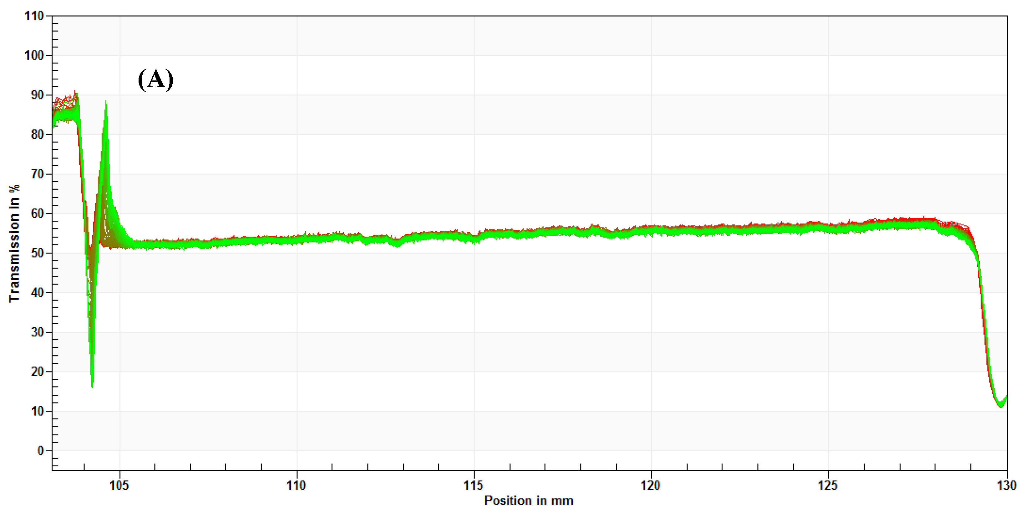
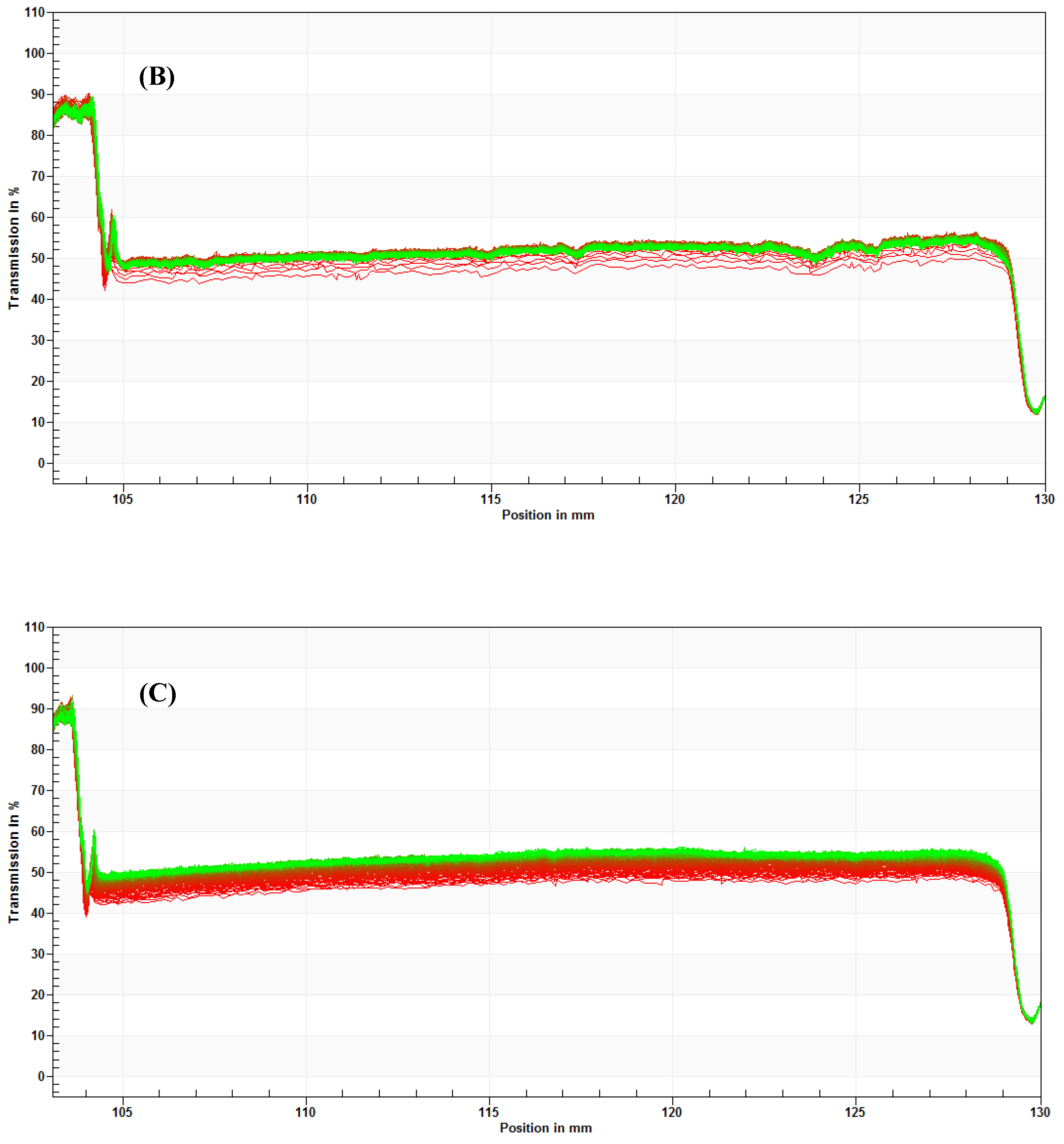
2.4. Accelerated Physical Stability

The physical stability of the nanoemulsion was determined by centrifugal force using a multisampling analytical centrifuge (LUMiSizer®; LUM GmbH, Berlin, Germany). This study was performed 24 h after preparation of the formulations. Samples were placed in rectangular cuvettes with an optical path of 10.0 mm, and exposed to a rotational speed of 4000 rpm for 8 h, at temperatures of 5, 30, and 40 °C (the same temperatures tested during the stability study for 180 d). The physical stability of the formulations was analyzed by the transmission profiles and the instability index, calculated using the SepView 6.0 software (LUM, Berlin, Germany).

2.5. Physicochemical Stability Study

The study of the physicochemical properties of a nanostructured system can reveal valuable information about its stability [21]. This stability study was performed according to a guide for stability studies [33].
The nanoemulsion was added to glass containers (5 mL) with a bung and screw cap and incubated at different storage conditions: 5 ± 2 °C, 30 ± 2 °C, and 40 ± 2 °C (75% relative humidity). After pre-determined intervals (24 h and 30, 60, 90, and 180 d), the samples were evaluated with respect to macroscopic analysis, droplet diameter, PDI, zeta potential, pH, vouacapan content, and consistency index. Free P. pubescens oil samples were also kept under the same storage conditions, to compare the chemical stability of the free oil with the oil present in the nanoemulsion.

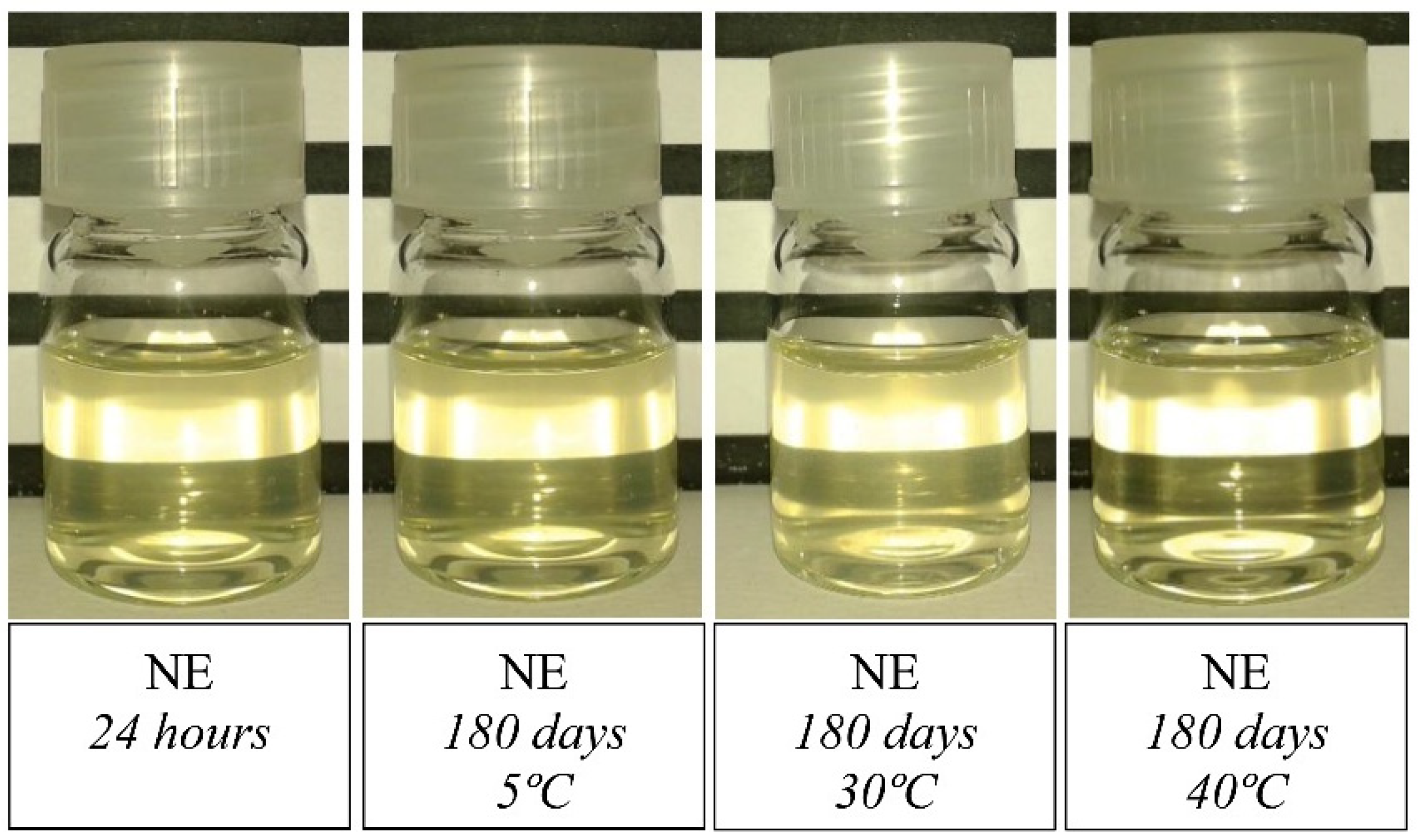
2.5.1. Macroscopic Analysis

The samples were visually evaluated in relation to color, appearance, and phase separation.

2.5.2. Droplet Diameter and Polydispersity Index (PDI)

The determination of average droplet diameter and PDI formulation were performed using a particle analyzer (NanoPlus-3 zeta/nano particle analyzer, Micromeritics Instrument Corporation, Norcross, GA, USA) by dynamic light scattering (DLS). All measurements were taken at a fixed angle of 90° and at a temperature of 25 ± 0.1 °C. The samples were diluted 1:10 in ultra-purified water before measurement, to avoid multiple scattering effects [34].

2.5.3. Zeta Potential

The zeta potential was determined by electrostatic mobility using a NanoPlus/zeta particle analyzer (Micromeritics Instrument Corporation, GA, USA) at 25 ± 0.1 °C. Before measurements, the samples were diluted 1:10 in ultra-purified water, pH 6.8.

2.5.4. pH

The pH of the samples was measured at 25 ± 1 °C, using a calibrated pH meter (TECNAL, São Paulo, Brazil).

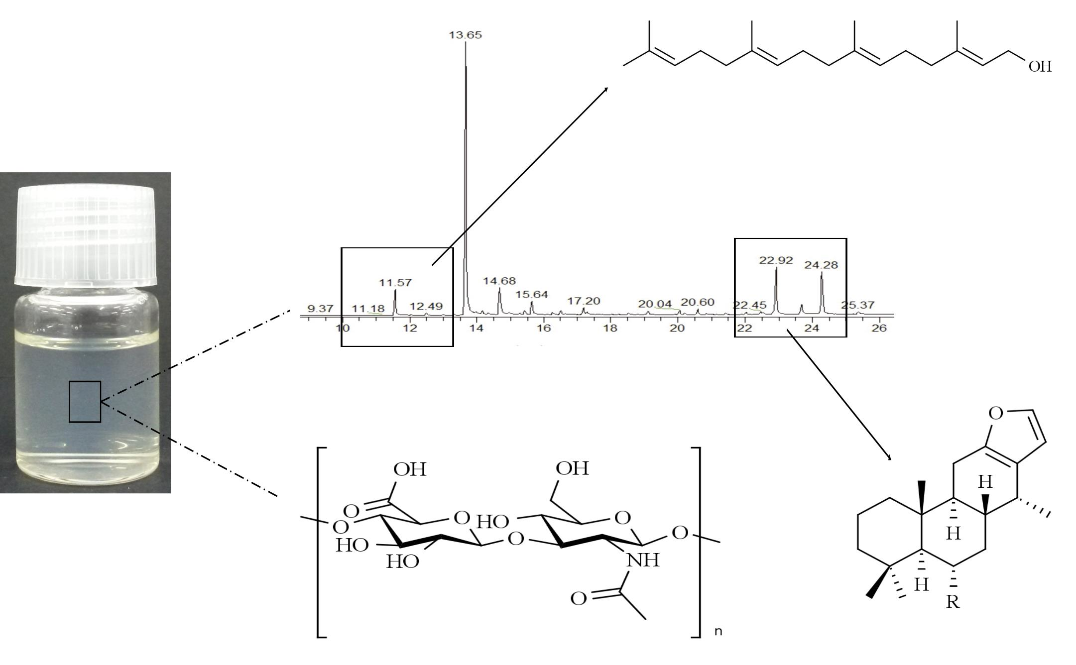
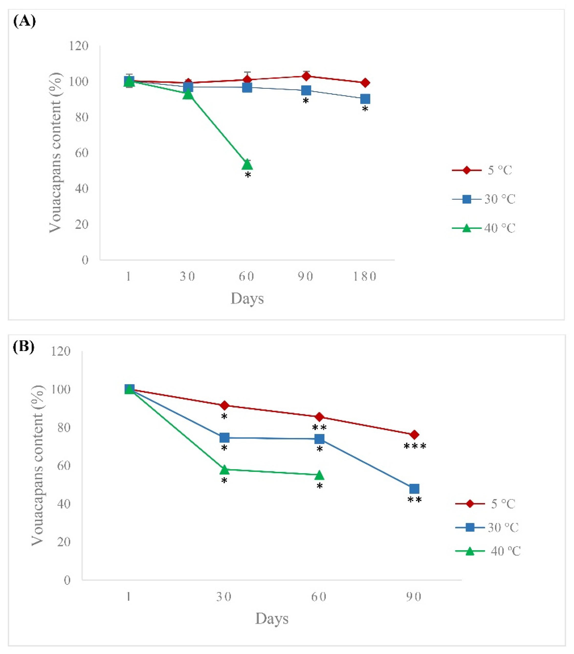
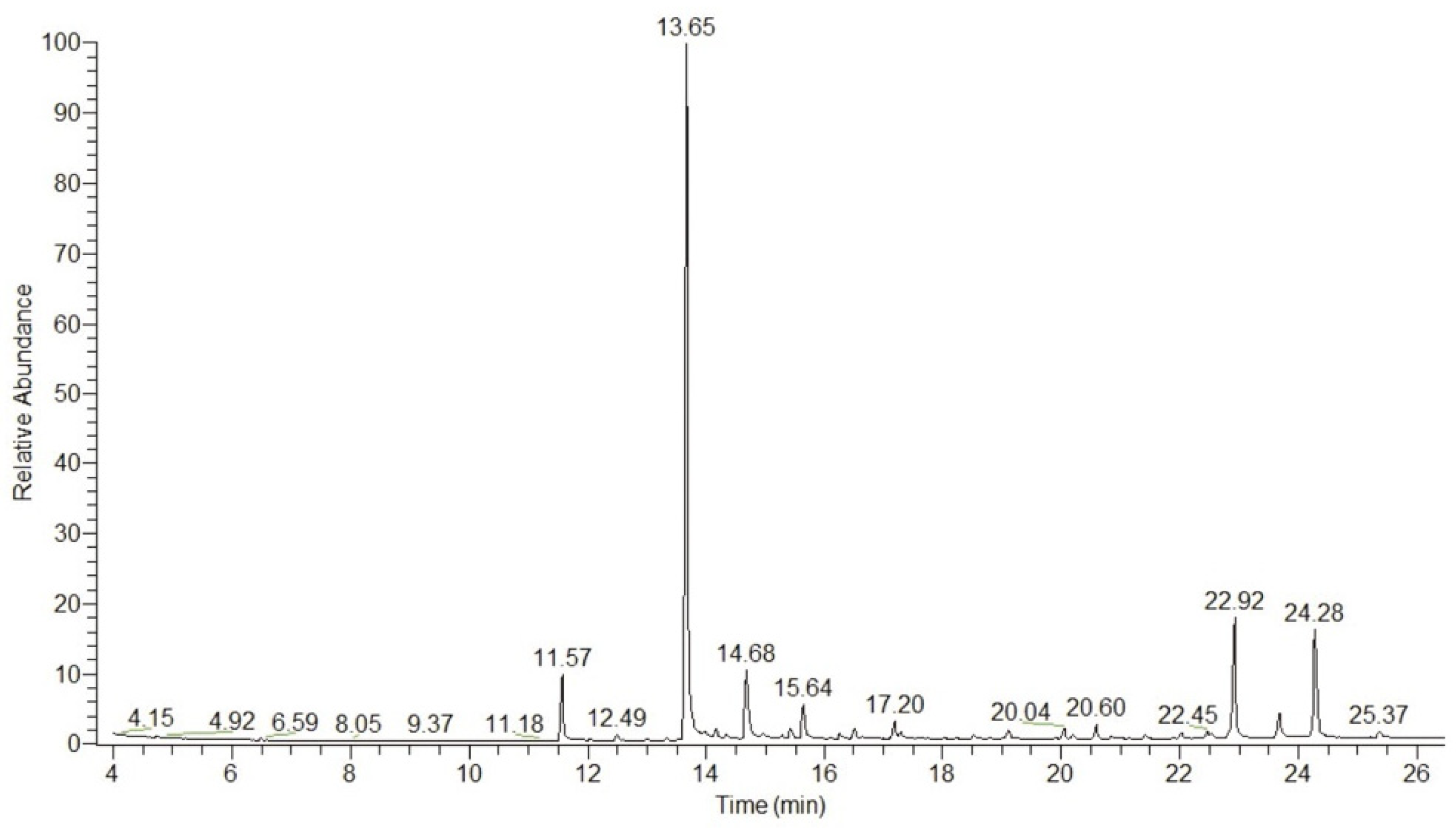
2.5.5. Chemical Analysis of P. pubescens Oil by GC-MS

The chromatographic profiles of P. pubescens oil present in the formulation were obtained by gas chromatography coupled with mass spectrometry (GC-MS) (Thermo Electron Corporation DSQ II; TLC, Thermo Fisher Scientific Inc., Waltham, MA, USA), equipped with an HP-5 capillary column (30 m × 0.25 mm). The vouacapan, used as a marker, was quantified by the monitoring of selected ions using a single ion monitoring system [34]. Lyophilized aliquots of the formulation were resuspended in 1 mL of chloroform and injected for analysis. The percentage of recovery was calculated as the ratio of the experimental concentration of vouacapan present in the samples to the theoretical concentration multiplied by 100.

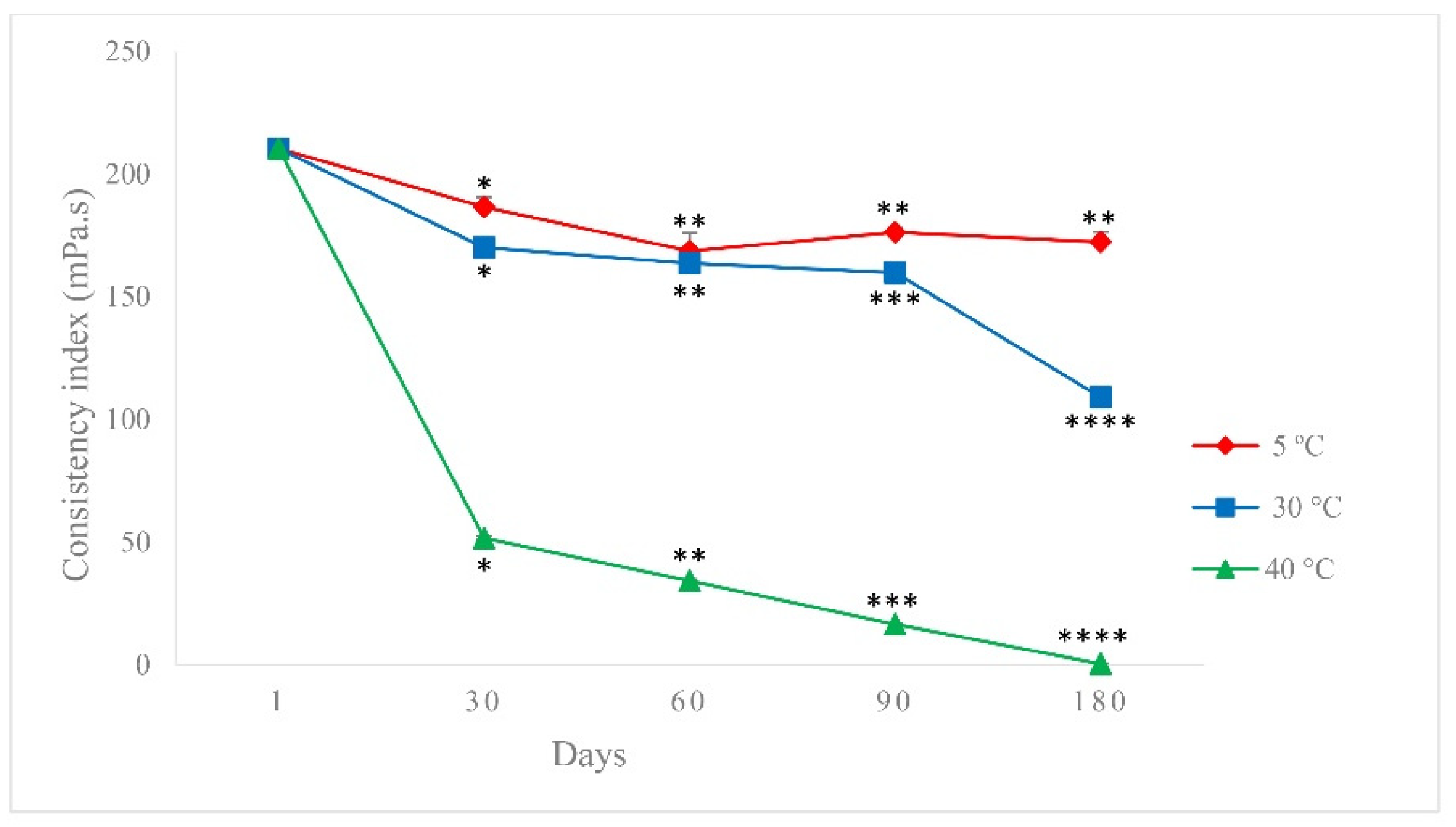
2.5.6. Consistency Index

The consistency index of the formulation was determined by continuous shear, using a MARS II (Haake®) controlled stress rheometer (Thermo Fisher Scientific Inc., Newington, Germany), inflow mode, at 25 ± 0.1 °C, with a controlled shear rate (CR), equipped with a cone-plate geometry of 35 mm and a 2° angle, separated by a fixed distance of 0.052 mm [31]. The Ostwald–de Waele equation (Power Law) was used to obtain the consistency index.

2.6. Microbiological Stability

The microbiological stability of nanoemulsion was determined according to the Brazilian Pharmacopoeia [35]. The test was carried out with fresh samples (after 24 h of preparation) and with the samples kept at 30 °C for 180 d. The analysis was performed using the pour plate technique for the following microorganisms: heterotrophic bacteria, fungi, and molds. Aliquots of nanoemulsion diluted in sterile saline were placed in Petri dishes containing 20 mL of a TSA medium (for heterotrophic bacteria) and Sabouraud dextrose agar (for fungi and molds), previously melted and stabilized at 45 °C. After homogenization and solidification of the medium, the samples were incubated in BOD incubators (Biogenic Oxygen Demand, TE-390 model, TECNAL, Piracicaba, Brazil) under the following conditions: 72 h at 35 °C to determine the presence of total heterotrophic bacteria, and 7 d at 28 °C to determine the presence of fungi and molds. After this period, the number of colony-forming units (CFU)/mL was determined. All analyses were performed in triplicate.

2.7. In Vitro Antileishmanial Activity

Peritoneal macrophages were obtained from BALB/c mice (age 3–8 weeks). Syringes were filled aseptically with 0.01 M cold phosphate buffer (PBS), and the content was injected into the peritoneal cavity of mice previously euthanized with lidocaine (10 mg/kg) and thiopental (200 mg/kg). The contents were then removed from the cavity, and maintained at 4 °C. This suspension was centrifuged for 10 min at 1500 rpm and 4 °C, promoting the formation of a cell pellet. This cell pellet was resuspended in an RPMI 1640 medium supplemented with 10% inactivated fetal bovine serum (FBS, Gibco), to form a suspension with a concentration of 5 × 10⁵ macrophages/mL. These cells were added to glass coverslips in 24-well plates. The plates were incubated at 37 °C and 5% CO2 for 2 h, to promote the adhesion of macrophages to the coverslips. The cells that did not adhere were removed by rinsing the well with a culture medium. Afterward, macrophages were infected with L. amazonensis promastigotes cultured for 5–6 d at a 7:1 parasite/macrophage ratio, and then incubated at 34 °C and 5% CO2 for 4 h. The wells were again washed to remove non-internalized protozoa.
Afterwards, the free P. pubescens oil and the nanoemulsion, at different concentrations, were diluted in an RPMI medium supplemented with 10% FBS, and added to the wells. The plates were then incubated for 48 h at 34 °C and 5% CO2. At the end of this period, the supernatant was removed, and the coverslips were fixed with methanol for 10 min and stained with 10% Giemsa in 0.01 M PBS for 40 min. Slides were analyzed under an optical microscope by counting the total number of macrophages, the number of infected macrophages, and the number of amastigotes per cell. The concentration capable of inhibiting 50% of the parasite growth (IC50) was calculated by linear regression analysis. The results were compared with the results for positive (Miltefosine) and negative (culture medium alone) controls.

2.8. Cytotoxicity Assays

Macrophages (J774A1) were cultured in an RPMI 1640 medium (Sigma-Aldrich Corporation, St. Louis, MO, USA) at pH 7.6 supplemented with 10% FBS, penicillin (5000 U/mL), and streptomycin (5 mg/mL). Cytotoxic assays were performed in sterile 96-well plates, with an initial inoculum of 5 × 10⁵ cells/mL in an RPMI 1640 medium incubated at 37 °C and 5% CO2 for 24 h. Afterwards, the free P. pubescens oil and the nanoemulsion were added to the cell monolayers and incubated for 48 h. The culture medium was then removed, the cells were washed with PBS, and 50 μL of MTT (3-[4,5-dimethylthiazol-2-yl]-2,5-diphenyltetrazolium bromide formazan; 2 mg/mL in PBS) were added. The cells were incubated while protected from light for 4 h at 37 °C and 5% CO2. To solubilize the formazan crystals, 150 μL of dimethylsulfoxide DMSO were added to the plates. The absorbance of the resulting solutions was read at 570 nm in spectrophotometer plates (Bio-Tek—Power Wave XS) to measure cell metabolic viability [36]. The concentration capable of inhibiting 50% of the cell growth was expressed as the cytotoxic concentration (CC50). The selectivity index was determined by the ratio of cytotoxicity (CC50) on J774A1 cells to leishmanicidal activity (IC50) against L. amazonensis.

2.9. Statistical Analysis

All experiments were performed in triplicate, and were presented as mean ± standard deviation. ANOVA and a post hoc Tukey’s test were used for statistical comparison of the results. The results were considered statistically significant for values of p ≤ 0.05.

3. Results

3.1. Physical Stability Accelerated

The transmission profile result obtained at different temperatures is shown in Figure 1. Overlapped transmission profiles indicate a stable system, and the best result was found at 5 °C, indicating a greater stability of the formulation.
Table 1 shows the results of the instability indexes. It was observed that the instability index increased by around 65% when centrifuged at 40 °C, as compared with 30 °C, demonstrating the instability of the nanoemulsion at this temperature. Comparing the results obtained at 5 and 30 °C, the instability index was approximately 10% lower at 5 °C, suggesting that the formulation would be more stable when stored at this temperature.

3.2. Physicochemical Stability Study

This study was carried out to test the best storage conditions for the developed system. The formulation developed for the stability study was characterized 24 h after preparation. It was translucent, homogeneous, and without any signal of phase separation.
The pictures in Figure 2 reveal no change in the macroscopic aspects of the formulation stored for 180 d, at the different temperatures. The formulation presented no signal of phase separation, and had the same transparent and homogeneous appearance as the freshly prepared formulation.
The average droplet diameter of the formulation at temperatures of 5, 30, and 40 °C, at intervals of 24 h and 30, 60, 90, and 180 d, are presented in Table 2. When stored at 5 °C, there was an increase in diameter from 60 to 180 d, which was statistically significant (p < 0.05), in comparison with 24 h and 30 d.
At 30 °C, an increase in droplet diameter occurred at 90 d and remained constant until the end of the study period. Regarding the temperature of 40 °C, the droplet diameter did not change until 90 d, but a decrease was observed at 180 d.
In general, a slight increase in the PDI as a function of time was observed when stored at 5 and 30 °C. However, the formulation had a homogeneous droplet distribution, since the PDI of the samples remained at <0.3. The zeta potential values remained stable for the formulation stored at 5 °C and 30 °C until 180 d.
On the other hand, the formulation stored at 40 °C showed a decrease in zeta potential, suggesting that this system may present less physical stability compared with the formulation stored at 5 and 30 °C. In the accelerated stability study, the instability index was higher for the formulation evaluated at 40 °C. Thus, both studies suggest that this temperature may decrease the stability of the formulation.
It is important to monitor the pH values of the formulation, since pH change indicates chemical reactions, and may compromise system quality. It was observed that the pH of the nanoemulsion at temperatures of 5 and 30 °C decreased during storage, but remained around 6.0 at the end of the study. However, when the formulation was stored at 40 °C, there was a more pronounced decrease in the pH at 180 d, with a value slightly above 4.0.
The total content of the P. pubescens oil was determined by the quantification of the derivatives of the vouacapans. Values close to 100% were found, demonstrating the efficacy of the method used to prepare the nanoemulsion. Figure 3 presents the percentage recovery of vouacapans (used as markers) from the nanoemulsion (Figure 3A) and from the free P. pubescens oil (Figure 3B), stored at temperatures at 5, 30, and 40 °C for 180 d.
The percentage of recovery of the oil present in the nanoemulsion showed a small, non-significant change during the analyzed period, when maintained at 5 °C. However, when stored at 30 °C, a small decrease occurred in the content of vouacapans from 90 d. For the free oil stored at temperatures of 5 and 30 °C, it can be seen that the content of the active compound was much lower, compared with the formulation.
The oil content in the nanoemulsion stored at 40 °C had a pronounced decrease from the second month of analysis (approximately 50%). For the free oil, at 40 °C, a marked reduction of the active compound was observed in the first month of analysis.
Figure 4 presents the chromatographic profile of the P. pubescens oil present in the nanoemulsion stored at 5 °C for 180 d. It is possible to observe the presence of characteristic peaks of the derivatives of geranylgeraniol (between 11 and 15 min) and vouacapan diterpenes (between 22 and 25 min) in the P. pubescens oil.
The consistency index obtained for the nanoemulsion during the stability study is presented in Figure 5.
The initial consistency index was 210.37 ± 1.34 mPa·s. At all temperatures analyzed, the consistency index decreased over time. However, comparing the formulation maintained at 5 and 30 °C, it was observed that the consistency index of the formulation maintained at 5 °C showed a less pronounced decrease (around 18%) than that of the formulation stored at 30 °C, which showed a decrease of approximately 50%.
For the formulation maintained at 40 °C, a marked decrease in the consistency index was observed in the first month of study, and this decrease was almost 100% until 180 d, demonstrating the physical instability of the system at this temperature. Thus, based on these results, it is suggested that the formulation is more stable when stored at 5 °C. These results are in agreement with the results obtained in the accelerated stability study using centrifugal force, in which the formulation evaluated at 5 °C was also more stable.

3.3. Microbiological Stability

The results showed that there was no growth of the microorganisms tested.

3.4. In Vitro Antileishmanial Activity and Cytotoxicity Assays

The leishmanicidal activity (IC50) for the free P. pubescens oil and the formulation is shown in Table 3. The IC50 values demonstrated an expressive leishmanicidal activity of the nanoemulsion, compared with the free P. pubescens oil, which represents a decrease in IC50 of approximately 95%, suggesting the great potential of the formulation in the treatment of CL. The assays of cytotoxicity showed that the free oil was cytotoxic to macrophages with CC50 values of approximately 36 µg/mL.

4. Discussion

The droplet diameter and the PDI are representative parameters that indicate the stability of nanoemulsions [37]. The small initial droplet diameter contributed to the transparency of the nanoemulsion [38]. Despite the increase in the diameter of the droplets for the system at 5 and 30 °C, there was no evidence of creaming or coalescence of the droplets during storage, since the diameters remained around 30 nm, contributing to the kinetic stability of the formulation [38].
The PDI is a representation of the distribution of droplet diameter within a given sample, which can range from 0 to 1. PDI values < 0.2 indicate the existence of a homogeneous particle size, while values close to 1 indicate heterogeneity and physical instability [39,40]. In drug delivery applications using lipid-based carriers such as nanoemulsions, a PDI < 0.3 is considered acceptable [39,41,42], indicating a homogenous population of droplets and a high physical stability during 180 d of storage [43,44,45].
Additionally, the zeta potential may be an indicator, but it is not a unique criterion to predict the stability of a nanoemulsion [46]. Zeta potential values above 30 mV (absolute value) provide a high energy barrier that causes repulsion of the adjacent droplets, resulting in the formation of stabilized emulsions [47] and the high physical stability of the system [48]. The negative charge observed in this study is related to the anionic nature of HA, which is absorbed in the oil–water interface [47].
Furthermore, high temperatures can change the solubility of nonionic surfactant (PEG-40H), due to the breakdown of hydrogen bonds at the surfactant surface, facilitating aggregation and coalescence by reducing the mechanical barrier, and resulting in a loss of stability [45]. The formulation stored at 40 °C showed a decrease in zeta potential, compared with those stored at 5 and 30 °C, and in the accelerated stability study, the instability index was higher for the formulation evaluated at 40 °C; thus, it is suggested that this temperature may decrease the stability of the formulation, by a reduction of electrical and mechanical barrier energies [44,45].
The pH values close to 6.0 observed for the formulation stored at temperatures of 5 and 30 °C are acceptable, since these values are within the favorable pH range for an emulsified system that remains stable, and are safe values for topical application. On the other hand, the pH values close to 4.0 found for the formulation stored at 40 °C could be the reason for the decrease of zeta potential. pH change indicates the occurrence of chemical reactions that may compromise the quality of the final product [45]. A pH decrease may have occurred due to oxidation of the oil phase, leading to hydroperoxide formations, or due to hydrolysis of triglycerides with the formation of free fatty acids [49]. Additionally, the chromatographic profile of the P. pubescens oil present in the nanoemulsion stored at 40 °C for 180 d showed a high chemical degradation of the constituents of the oil present in the formulation, demonstrating the instability of the P. pubescens oil at this temperature.
However, when stored at 5 °C, the developed system presented a good recovery content. Comparing the formulation with the free oil, it was observed that the degradation of P. pubescens oil present in the nanoemulsion was lower than that of the free oil, indicating that the nanoemulsion protected the vegetable oil from degradation, mainly at temperatures of 5 and 30 °C. These results confirm the results obtained in the thermal analysis [31,50], which demonstrated the ability of the nanoemulsion to protect the P. pubescens oil from thermal degradation. According to Bajerski et al. [51], one of the advantages of using nanoemulsion to encapsulate vegetable oils is the ability of these systems to protect the vegetable oil from photo-, thermal, and volatilization instability, improving the chemical stability of the oil.
The viscosity of the system is of great importance for the formation and stability of emulsions. A decrease in the viscosity over time may indicate a kinetic instability of the system [52]. Instabilities arising from variation in droplet size, particle number, and emulsifier orientation or migration over time, can be detected by changes in the viscosity of the product [49].
In this study, we observed that the PDI increased during storage, which may be attributed, in part, to the reduction in consistency index [45]. The maintenance of the consistency index is an important factor to consider, since viscous emulsions are more stable compared with less viscous emulsions, due to the delay of the phenomena of physical instability. According to Ali et al. [52], the factors that govern creaming are the dispersed-phase globule size and the viscosity of the external phase. The viscosity reduction in a nanoemulsified system increases the probability of droplets moving freely and colliding with each other, favoring the phenomenon of coalescence. In addition, for a topical formulation, maintenance of the viscosity is necessary [49].
The stability of the nanoemulsion is associated with physical integrity, chemical stability, and protection against microbial contamination [53]. Direct sources of contamination (raw materials, packaging, and production environment) and indirect sources (resulting from cleaning equipment and/or training operators) can affect the microbial quality of the formulation [54]. Microbial contamination is associated with the loss of therapeutic efficacy due to the chemical degradation of the constituents of the oil or changes in chemical and physical parameters [45]. The microbiological stability showed that there was no growth of the microorganisms tested. According to some studies [11,55], P. pubescens extracts present antimicrobial activity, which may favor the maintenance of the microbiological stability of the developed system.
The results obtained in the biological assay against the intracellular amastigotes of L. amazonensis indicate that the nanoscale droplets (≈25 nm) and the presence of HA in the formulation may have contributed to the internalization of P. pubescens oil in the infected macrophages, increasing leishmanicidal activity in approximately 95%, compared with the free oil. Typically, delivery using nanocarriers to the target cell increases the bioavailability of active components and decreases toxicity in normal cells [2].
These results suggest that the P. pubescens oil was successfully internalized in the infected macrophages using the formulation, suggesting that a site-specific drug delivery system is promising for the treatment of CL. However, further investigations regarding the internalization of the P. pubescens oil present in the nanoemulsion by HA receptors, are necessary.
An important aspect of active compounds with potential therapeutic application is the absence of toxic effects on the host cells, which can be evaluated via the selectivity index. This index reveals whether the compounds act preferentially on the parasite or on the host defense cells. The greater the selectivity index, the more selective the formulation is in inhibiting the parasite [56].
Evaluation of the formulation showed that the CC50 value decreased, but the selectivity index was approximately double that of the free P. pubescens oil, demonstrating the potential of the nanoemulsion to improve the selectivity of the active substance over cells containing the parasite. According to the literature, furanoditerpenes with a vouacapan skeleton are the main compounds in Pterodon species capable of causing cytotoxicity in cancer cells [57,58]. Considering the potential of P. pubescens oil in the treatment of leishmaniasis [18,19], and the results obtained in this study, the development of a drug release system capable of increasing the therapeutic efficacy of P. pubescens oil is encouraged; however, additional studies on the compound responsible for its cytotoxicity are needed.

5. Conclusions

The HA-based nanoemulsion containing P. pubescens oil was more stable when stored at 5 °C, in which the parameters evaluated remained practically the same during the study period. The degradation of the P. pubescens oil present in the nanoemulsion was lower than that of the free oil, showing that the chemical stability of the oil increased when it was encapsulated in the nanoemulsion. The microbiological stability test confirmed the absence of bacteria or fungi in the formulation.
The biological assay against the intracellular amastigotes of L. amazonensis indicates that the P. pubescens oil was successfully internalized in the infected macrophages using the formulation, suggesting that a site-specific drug delivery system is promising for the treatment of CL. The development of a drug release system capable of increasing the therapeutic efficacy of P. pubescens oil is encouraged, but further investigations on the internalization of the oil present in the nanoemulsion by HA receptors, and on the increase in selectivity index, together with in vivo studies, are needed.

Author Contributions

We declare that this work was carried out by the author(s) named in this article and that all liabilities pertaining to claims relating to the content of this article will be borne by the authors. S.A.K., P.M.O. and J.H. contributed to laboratory work management, data analysis, and manuscript drafting. G.C.T. and M.D. contributed to microbiological stability and to critical reading of the manuscript. M.M.d.S.L. contributed to rheometry analysis, accelerated physical stability, and manuscript drafting. É.d.S.S. and C.V.N. contributed to biological studies (in vitro antileishmanial activity and cytotoxicity assays) and to manuscript drafting. E.A.d.S. and M.L.C.C. designed the study, supervised the laboratory work, and contributed to critical reading of the manuscript. All authors have read and agreed to the published version of the manuscript.

Funding

This research received no external funding.

Data Availability Statement

Not applicable.

Acknowledgments

The authors are grateful to Financiadora de Estudos e Projetos (FINEP), Fundação Araucária, Coordenadoria de Aperfeiçoamento de Pessoal do Ensino Superior (CAPES), and Conselho Nacional de Desenvolvimento Científico e Tecnológico (CNPq) for their financial support.

Conflicts of Interest

The authors declare that they have no conflict of interest.

References

  1. Souza, T.M.; Morais-Braga, M.F.B.; Saraiva, A.A.F.; Rolón, M.; Veja, C.; Arias, A.R.; Costa, J.G.M.; Menezes, I.R.A. Evaluation of the anti-Leishmania activity of ethanol extract and fractions of the leaves from Pityrogramma calomelanos (L.) link. Nat. Prod. Res. 2013, 27, 992–996. [Google Scholar] [CrossRef] [PubMed]
  2. Akbari, M.; Oryan, A.; Hatam, G. Application of nanotechnology in treatment of leishmaniasis: A Review. Acta Trop. 2017, 172, 86–90. [Google Scholar] [CrossRef] [PubMed]
  3. Kheirandish, F.; Delfan, B.; Mahmoudvand, H.; Moradi, N.; Ezatpour, B.; Ebrahimzadeh, F.; Rashidipour, M. Antileishmanial, antioxidant, and cytotoxic activities of Quercus infectoria Olivier extract. Biomed. Pharm. 2016, 82, 208–215. [Google Scholar] [CrossRef]
  4. Ammar, A.A.; Nasereddin, A.; Ereqat, S.; Dan-Goor, M.; Jaffe, C.L.; Zussman, E.; Abdeen, Z. Amphotericin B-loaded nanoparticles for local treatment of cutaneous leishmaniasis. Drug Deliv. Transl. Res. 2019, 9, 76–84. [Google Scholar] [CrossRef]
  5. Galvão, E.L.; Pedras, M.J.; Cota, G.F.; Rabello, A.; Simões, T.C. How cutaneous leishmaniasis and treatment impacts in the patients’ lives: A cross-sectional study. PLoS ONE 2019, 14, e0211374. [Google Scholar] [CrossRef] [PubMed] [Green Version]
  6. Essid, R.; Rahali, F.Z.; Msaada, K.; Sghair, I.; Hammami, M.; Bouratbine, A.; Aoun, K.; Limam, F. Antileishmanial and cytotoxic potential of essential oils from medicinal plants in Northern Tunisia. Ind. Crop. Prod. 2015, 77, 795–802. [Google Scholar] [CrossRef]
  7. Oliveira, L.A.R.; Oliveira, G.A.R.; Borges, L.L.; Bara, M.T.F.; Silveira, D. Vouacapane diterpenoids isolated from Pterodon and their biological activities. Braz. J. Pharm. 2017, 27, 663–672. [Google Scholar] [CrossRef]
  8. Moraes, A.R.D.P.; Tavares, G.D.; Rocha, F.J.S.; Paula, E.; Giorgio, S. Effects of nanoemulsions prepared with essential oils of copaiba- and andiroba against Leishmania infantum and Leishmania amazonensis infections. Exp. Parasitol. 2018, 187, 12–21. [Google Scholar] [CrossRef]
  9. Bouyahya, A.; Et-Touys, A.; Dakka, N.; Fellah, H.; Abrini, J.; Bakri, Y. Antileishmanial potential of medicinal plant extracts from the North-West of Morocco. Beni.-Suef. Univ. J. Basic. Appl. Sci. 2018, 7, 50–54. [Google Scholar] [CrossRef]
  10. Coelho, L.P.; Reis, P.A.; Castro, F.L.; Gayer, C.R.M.; Lopes, C.S.; e Silva, M.C.C.; Sabino, K.C.C.; Todeschini, A.R.; Coelho, M.G.P. Antinociceptive properties of ethanolic extract and fractions of Pterodon pubescens Benth. Seeds. J. Ethnopharmacol. 2005, 98, 109–116. [Google Scholar] [CrossRef]
  11. Bustamante, K.G.L.; Lima, A.D.F.; Soares, M.L.; Fiuza, T.S.; Tresvenzol, L.M.F.; Bara, M.T.F.; Pimenta, F.C.; Paula, J.R. Avaliação da atividade antimicrobiana do extrato etanólico bruto da casca da sucupira branca (Pterodon emarginatus Vogel)—Fabaceae. Rev. Bras. Plantas Med. 2010, 12, 341–345. [Google Scholar] [CrossRef]
  12. Spindola, H.M.; Servat, L.; Rodrigues, R.A.F.; Sousa, I.M.O.; Carvalho, J.E.; Foglio, M.A. Geranylgeraniol and 6α,7β-dihydroxyvouacapan-17β-oate methyl ester isolated from Pterodon pubescens Benth: Further investigation on the antinociceptive mechanisms of action. Eur. J. Pharm. 2011, 656, 45–51. [Google Scholar] [CrossRef] [Green Version]
  13. Nucci, C.; Mazzardo-Martins, L.; Stramosk, J.; Brethanha, L.C.; Pizzolatti, M.G.; Santos, A.R.S.; Martins, D.F. Oleaginous extract from the fruits Pterodon pubescens Benth induces antinociceptionin animal models of acute and chronic pain. J. Ethnopharmacol. 2012, 143, 170–178. [Google Scholar] [CrossRef] [Green Version]
  14. Alves, S.F.; Borges, L.L.; de Paula, J.A.M.; Vieira, R.F.; Ferri, P.H.; de Couto, R.O.; de Paula, J.R.; Bara, M.T.F. Chemical variability of the essential oils from fruits of Pterodon emarginatus in the Brazilian Cerrado. Braz. J. Pharm. 2013, 23, 224–229. [Google Scholar] [CrossRef] [Green Version]
  15. Hoscheid, J.; Bersani-Amado, C.A.; da Rocha, B.A.; Outuki, P.M.; da Silva, M.A.R.C.P.; Froehlich, D.L.; Cardoso, M.L.C. Inhibitory Effect of the Hexane Fraction of the Ethanolic Extract of the Fruits of Pterodon pubescens Benth. Acute Chronic Inflamm. ECAM 2013, 2013, 272795. [Google Scholar] [CrossRef] [Green Version]
  16. Hoscheid, J.; Cardoso, M.L.C. Sucupira as a potential plant for arthritis treatment and other diseases. Arthritis 2015, 2015, 379459. [Google Scholar] [CrossRef] [Green Version]
  17. Goes, P.R.N.; Hoscheid, J.; Silva-Filho, S.E.; Froehlich, D.L.; Pelegrini, B.L.; Canoff, J.R.A.; Lima, M.M.S.; Cuman, R.K.N.; Cardoso, M.L.C. Rheological behavior and antiarthritic activity of Pterodon pubescens nanoemulsion. Res. Soc. Dev. 2020, 9, e179108119. [Google Scholar] [CrossRef]
  18. Santos, E.S.; Garcia, F.P.; Outuki, P.M.; Hoscheid, J.; de Goes, P.R.N.; Cardozo-Filho, L.; Nakamura, C.V.; Cardoso, M.L.C. Optimization of extraction method and evaluation of antileishmanial activity of oil and nanoemulsions of Pterodon pubescens Benth. Fruit Extracts. Exp. Parasitol. 2016, 170, 252–260. [Google Scholar] [CrossRef]
  19. Oliveira, L.A.R.; Oliveira, G.A.R.; Lemes, G.F.; Romão, W.; Vaz, B.G.; Albuquerque, S.; Gonçalez, C.; Lião, L.M.; Bara, M.T.F. Isolation and Structural Characterization of Two New Furanoditerpenes from Pterodon emarginatus (Fabaceae). J. Braz. Chem. Soc. 2017, 28, 1911–1916. [Google Scholar] [CrossRef]
  20. Wulff-Pérez, M.; Torcello-Gómez, A.; Gálvez-Ruíz, M.J.; Martín-Rodríguez, A. Stability of emulsions for parenteral feeding: Preparation and characterization of o/w nanoemulsions with natural oils and Pluronic f68 as surfactant. Food Hydrocoll. 2009, 23, 1096–1102. [Google Scholar] [CrossRef]
  21. Bangia, J.K.; Singh, M.; Om, H.; Behera, K. A comparative study on the effect of temperature on density, sound velocity and refractive index of nanoemulsions formed by castor, olive and linseed oils in aqueous cellulose acetate propionate and butyrate and Tween80. Thermochim. Acta 2016, 641, 43–48. [Google Scholar] [CrossRef]
  22. Want, M.Y.; Islamuddin, M.; Chouhan, G.; Dasgupta, A.K.; Chattopadhyay, A.P.; Afrin, F. A new approach for the delivery of artemisinin: Formulation, characterization, and ex-vivo antileishmanial studies. J. Colloid Interface Sci. 2014, 432, 258–269. [Google Scholar] [CrossRef] [PubMed]
  23. Souza, A.; Martins, D.S.S.; Mathias, S.L.; Monteiro, L.M.; Yukuyama, M.N.; Scarim, C.B.; Löberberg, R. Promising nanotherapy in treating leishmaniasis. Int. J. Pharm. 2018, 547, 421–431. [Google Scholar] [CrossRef] [Green Version]
  24. Vasi, A.M.; Popa, M.I.; Butnaru, M.; Dodi, G.; Verestiuc, L. Chemical functionalization of hyaluronic acid for drug delivery applications. Mater. Sci. Eng. C 2014, 38, 177–185. [Google Scholar] [CrossRef]
  25. Kutlusoy, T.; Oktay, B.; Apohan, N.K.; Süleymanoglu, M.; Kuruca, S.E. Chitosan-co-Hyaluronic acid porous cryogels and their application intissue engineering. Int. J. Biol. Macromol. 2017, 103, 366–378. [Google Scholar] [CrossRef] [PubMed]
  26. Pedrosa, S.S.; Pereira, P.; Correia, A.; Gama, F.M. Targetability of hyaluronic acid nanogel to cancer cells: In Vitro and in vivo studies. Eur. J. Pharm. Sci. 2017, 104, 102–113. [Google Scholar] [CrossRef] [Green Version]
  27. Quinones, J.P.; Jokinen, J.; Keinänen, S.; Covas, C.P.; Brüggemann, O.; Ossipov, D. Self-assembled hyaluronic acid-testosterone nanocarriers for delivery of anticancer drugs. Eur. Pol. J. 2018, 99, 384–393. [Google Scholar] [CrossRef]
  28. Montanari, E.; Di Meo, C.; Oates, A.; Coviello, T.; Matricardi, P. Pursuing Intracellular Pathogens with Hyaluronan. From a ‘Pro-Infection’ Polymer to a Biomaterial for ‘Trojan Horse’ Systems. Molecules 2018, 23, 939. [Google Scholar] [CrossRef] [Green Version]
  29. Stefanello, T.F.; Szarpak-Jankowska, A.; Appaix, F.; Louage, B.; Hamard, L.; De Geest, B.G.; Sander, B.V.D.; Nakamura, C.V.; Auzély-Velty, R. Thermoresponsive hyaluronic acid nanogels as hydrophobic drug carrier to macrophages. Acta Biomater. 2014, 10, 750–4758. [Google Scholar] [CrossRef]
  30. Micale, N.; Piperno, A.; Mahfoudh, N.; Schurigt, U.; Schultheis, M.; Mineo, P.G.; Schirmeister, T.; Scala, A.; Grassi, G. A hyaluronic acid–pentamidine bioconjugate as a macrophage mediated drug targeting delivery system for the treatment of leishmaniasis. RSC Adv. 2015, 5, 95545–95550. [Google Scholar] [CrossRef]
  31. Kleinubing, S.A.; Outuki, P.M.; Hoscheid, J.; Pelegrini, B.L.; da Silva, E.A.; Canoff, J.R.A.; Lima, M.M.S.; Cardoso, M.L.C. Hyaluronic acid incorporation into nanoemulsions containing Pterodon pubescens Benth. fruit oil for topical drug delivery. Biocatal. Agric. Biotechnol. 2021, 32, 101939. [Google Scholar] [CrossRef]
  32. Hoscheid, J.; Reinas, A.; Cortez, D.A.G.; da Costa, W.F.; Cardoso, M.L.C. Determination by GC–MS-SIM of furanoditerpenes in Pterodon pubescens Benth.: Development and validation. Talanta 2012, 100, 372–376. [Google Scholar] [CrossRef] [PubMed] [Green Version]
  33. Brasil. Resolução No 01, de 29 de Julho de 2005. Guia Para a Realização de Estudos de Estabilidade. Brasília: Agência Nacional de Vigilância Sanitária. 2005. Available online: http://bvsms.saude.gov.br/bvs/saudelegis/anvisa/2005/res0001_29_07_2005.html (accessed on 7 July 2022).
  34. Hoscheid, J.; Outuki, P.M.; Kleinubing, S.A.; Silva, M.F.; Bruschi, M.L.; Cardoso, M.L.C. Development and characterization of Pterodon pubescens oil nanoemulsions as a possible delivery system for the treatment of rheumatoid arthritis. Colloids Surf. A Physicochem. Eng. Asp. 2015, 484, 19–27. [Google Scholar] [CrossRef]
  35. Brasil. Farmacopeia Brasileira. Diário Of. União 2010, 1, 236–253. [Google Scholar]
  36. Mosmann, T. Rapid colorimetric assay for cellular growth and survival: Application to proliferation and cytotoxicity assays. J. Immunol. Methods 1983, 65, 55–63. [Google Scholar] [CrossRef]
  37. Kelmann, R.G.; Kuminek, G.; Teixeira, H.F.; Koester, L.S. Carbamazepine parenteral nanoemulsions prepared by spontaneous emulsification process. Int. J. Pharm. 2007, 342, 231–239. [Google Scholar] [CrossRef]
  38. Teixeira, M.C.; Severino, P.; Andreani, T.; Boonme, P.; Santini, A.; Silva, A.M.; Souto, E.B. D-α-tocopherol nanoemulsions: Size properties, rheological behavior, surface tension, osmolarity and cytotoxicity. Saudi Pharm. J. 2017, 25, 231–235. [Google Scholar] [CrossRef] [Green Version]
  39. Danaei, M.; Dehghankhold, M.; Ataei, S.; Hasanzadeh Davarani, F.; Javanmard, R.; Dokhani, A.; Khorasani, S.; Mozafari, M.R. Impact of Particle Size and Polydispersity Index on the Clinical Applications of Lipidic Nanocarrier Systems. Pharmaceutics 2018, 10, 57. [Google Scholar] [CrossRef] [Green Version]
  40. Sato, T.S.; De Medeiros, T.M.; Hoscheid, J.; Prochnau, I.S. Proposta de formulação contendo extrato de folhas de Eugenia involucrata e análise da atividade antimicrobiana. Proposal of a formulation containing leaves extract of Eugenia involucrata. Rev. Fitos 2018, 12, 68–82. [Google Scholar] [CrossRef]
  41. Vighi, E.; Ruozi, B.; Montanari, M.; Battini, R.; Leo, E. pDNA condensation capacity and in vitro gene delivery properties of cationic solid lipid nanoparticles. Int. J. Pharm. 2010, 389, 254–261. [Google Scholar] [CrossRef] [PubMed]
  42. Outuki, P.M.; Kleinubing, S.A.; Hoscheid, J.; Montanha, M.C.; da Silva, E.A.; do Couto, R.O.; Kimura, E.; Cardoso, M.L.C. The incorporation of Pterodon pubescens fruit oil into optimized nanostructured lipid carriers improves its effectiveness in colorectal cancer. Ind. Crops Prod. 2018, 123, 719–730. [Google Scholar] [CrossRef]
  43. Galindo-Alvarez, J.; Le, K.-A.; Sadtler, V.; Marchal, P.; Perrin, P.; Tribet, C.; Marie, E.; Durand, A. Enhanced stability of nanoemulsions using mixtures of nonionic surfactant and amphiphilic polyelectrolyte. Colloids Surf. A Physicochem. Eng. Asp. 2011, 389, 237–245. [Google Scholar] [CrossRef]
  44. Li, B.; Ge, Z.Q. Nanostructured lipid carriers improve skin permeation and chemical stability of idebenone. AAPS PharmSciTech 2012, 13, 276–283. [Google Scholar] [CrossRef] [Green Version]
  45. Hoscheid, J.; Outuki, P.M.; Kleinubing, S.A.; de Goes, P.R.N.; Lima, M.M.S.; Cuman, R.K.N.; Cardoso, M.L.C. Pterodon pubescens oil nanoemulsions: Physiochemical and microbiological characterization and in vivo anti-inflammatory efficacy studies. Braz. J. Pharmacog. 2017, 27, 375–383. [Google Scholar] [CrossRef]
  46. Kong, M.; Park, H.J. Stability investigation of hyaluronic acid based nanoemulsion and its potential as transdermal carrier. Carbohydr. Polym. 2011, 83, 1303–1310. [Google Scholar] [CrossRef]
  47. Yang, S.C.; Benita, S. Enhanced Absorption and Drug Targeting by Positively Charged Submicron Emulsions. Drug Dev. Res. 2000, 50, 476–486. [Google Scholar] [CrossRef]
  48. Luo, X.; Zhou, Y.; Bai, L.; Liu, F.; Deng, Y.; McClements, D.J. Fabrication of b-carotene nanoemulsion-based delivery systems using dual-channel microfluidization: Physical and chemical stability. J. Colloid Interf. Sci. 2017, 490, 328–335. [Google Scholar] [CrossRef] [Green Version]
  49. Barradas, T.N.; Senna, J.P.; Cardoso, S.A.; Nicoli, A.; Padula, C.; Santi, P.; Rossi, F.; e Silva, K.G.H.; Mansur, C.R.E. Hydrogel-thickened nanoemulsions based on essential oils for topical delivery of psoralen: Permeation and stability studies. Eur. J. Pharm. Biopharm. 2017, 116, 38–50. [Google Scholar] [CrossRef]
  50. Reinas, A.E.; Hoscheid, J.; Outuki, P.M.; Cardoso, M.L.C. Preparation and characterization of microcapsules of Pterodon pubescens Benth. by using natural polymers. Braz. J. Pharm. Sci. 2014, 50, 920–930. [Google Scholar] [CrossRef] [Green Version]
  51. Bajerski, L.; Michels, L.R.; Colomé, L.M.; Bender, E.A.; Freddo, R.J.; Bruxel, F.; Haas, S.E. The use of Brazilian vegetable oils in nanoemulsions: An update on preparation and biological applications. Braz. J. Pharm. Sci. 2016, 52, 348–363. [Google Scholar] [CrossRef] [Green Version]
  52. Ali, M.S.; Alam, M.S.; Alam, N.; Anwer, T.; Safhi, M.M.A. Accelerated Stability Testing of a Clobetasol Propionate-Loaded Nanoemulsion as per ICH Guidelines. Sci. Pharm. 2013, 81, 1089–1100. [Google Scholar] [CrossRef] [PubMed]
  53. Alam, M.S.; Ali, M.S.; Alam, M.I.; Anwer, T.; Safhi, M.M.A. Stability Testing of Beclomethasone Dipropionate Nanoemulsion. Trop. J. Pharm. Res. 2015, 14, 15–20. [Google Scholar] [CrossRef] [Green Version]
  54. Ferreira, B.C.A.; Novais, E.B.; Ribeiro, R.B.C.; Fernandes, C.K.C. Estudo de estabilidade físico-química e microbiológica de dipirona em gotas armazenadas em residências do municipio de São Luis de Montes Belos-GO. Rev. Facul Montes Belos. 2014, 7, 109–120. [Google Scholar]
  55. Dutra, R.C.; Braga, F.G.; Coimbra, E.S.; Silva, A.D.; Barbosa, N.R. Antimicrobial and leishmanicidal activities of seeds of Pterodon emarginatus. Braz. J. Pharm. 2009, 19, 429–435. [Google Scholar] [CrossRef] [Green Version]
  56. Caldeira, L.R.; Fernandes, F.R.; Costa, D.F.; Frézard, F.; Afonso, L.C.C.; Ferreira, L.A.M. Nanoemulsions loaded with amphotericin B: A new approach for the treatment of leishmaniasis. Eur. J. Pharm. Sci. 2015, 70, 125–131. [Google Scholar] [CrossRef] [PubMed] [Green Version]
  57. Vieira, C.R.; Marques, M.F.; Soares, P.R.; Matuda, L.; Oliveira, C.M.A.; Kato, L.; Silva, C.C.; Grillo, L.A. Antiproliferative activity of Pterodon pubescens Benth. seed oil and its active principle on human melanoma cells. Phytomedicine 2008, 15, 528–532. [Google Scholar] [CrossRef]
  58. Spindola, H.M.; Carvalho, J.E.; Ruiz, A.L.T.G.; Rodrigues, R.A.F.; Denny, C.; Sousa, I.M.O.; Tamashiro, J.Y.; Foglio, M.A. Furanoditerpenes from Pterodon pubescens Benth. with selective in vitro anticancer activity for prostate cell line. J. Braz. Chem. Soc. 2009, 20, 569–575. [Google Scholar] [CrossRef]
Figure 1. Evolution of the transmission profiles of the HA-based nanoemulsion containing P. pubescens oil at (A) 5 °C, (B) 30 °C, and (C) 40 °C.
Colloids 06 00064 g001aColloids 06 00064 g001b
Figure 2. Pictures of the HA-based nanoemulsion containing P. pubescens oil stored for 180 d at temperatures of 5, 30, and 40 °C.
Colloids 06 00064 g002
Figure 3. Percentage of recovery of vouacapans of the nanoemulsion (A) and free P. pubescens oil (B), stored at temperatures of 5, 30, and 40 °C for 180 d. Mean ± SD (n = 3). One-way ANOVA followed by post hoc Tukey’s test; different specimens (*, **, and ***) are considered statistically different at p < 0.05.
Colloids 06 00064 g003
Figure 4. Chromatographic profile of P. pubescens oil present in the nanoemulsion stored at 5 °C for 180 d.
Colloids 06 00064 g004
Figure 5. Consistency index of HA-based nanoemulsion containing P. pubescens oil stored at temperatures of 5, 30, and 40 °C for 180 d. Mean ± SD (n = 3). One-way ANOVA followed by post hoc Tukey’s test; different specimens (*, **, ***, and ****) are considered statistically different at p < 0.05.
Colloids 06 00064 g005
Table 1. Instability index of HA-based nanoemulsion containing P. pubescens oil at temperatures of 5, 30, and 40 °C.
Temperature
5 °C30 °C40 °C
Instability index0.13 ± 0.06 b0.15 ± 0.00 b0.42 ± 0.12 a
Notes: The instability index was calculated using the SepView 6.0 software (LUM, Berlin, Germany). Different letters are considered to be statistically different (p < 0.05).
Table 2. Droplet diameter, PDI, zeta potential, and pH of the HA-based nanoemulsion containing P. pubescens oil stored at temperatures of 5, 30, and 40 °C for 180 d.
Time
24 h30 d60 d90 d180 d
Droplet diameter (nm) 5 °C24.86 ± 0.64 b24.66 ± 0.85 b29.50 ± 1.20 a31.33 ± 1.50 a32.36 ± 1.45 a
30 °C24.86 ± 0.64 b24.83 ± 0.68 b25.06 ± 0.49 b27.30 ± 0.46 a27.13 ± 1.10 a
40 °C24.86 ± 0.64 a23.70 ± 0.75 a23.90 ± 0.11 a23.66 ± 0.75 a18.83 ± 0.64 b
5 °C0.24 ± 0.04 c0.26 ± 0.07 b0.26 ± 0.09 b0.26 ± 0.01 b0.29 ± 0.06 a
PDI30 °C0.24 ± 0.04 c0.24 ± 0.04 cd0.23 ± 0.03 d0.29 ± 0.09 b0.30 ± 0.04 a
40 °C0.24 ± 0.04 a0.23 ± 0.05 b0.23 ± 0.02 b0.22 ± 0.06 c0.19 ± 0.03 d
Zeta potential (mV)5 °C−31.45 ± 0.26 aNDNDND−32.44 ± 0.20 a
30 °C−31.45 ± 0.26 aNDNDND−29.67 ± 0.09 a
40 °C−31.45 ± 0.26 aNDNDND−18.47 ± 1.50 b
pH5 °C6.80 ± 0.03 a6.70 ± 0.05 b6.56 ± 0.03 ab6.33 ± 0.05 bc6.00 ± 0.01 c
30 °C6.80 ± 0.03 a6.50 ± 0.01 b6.47 ± 0.04 b6.15 ± 0.07 c6.09 ± 0.14 c
40 °C6.80 ± 0.03 a5.22 ± 0.07 b4.79 ± 0.04 c4.55 ± 0.01 d4.22 ± 0.00 e
Notes: Non-determined (ND). Mean ± SD (n = 3). One-way ANOVA followed by post hoc Tukey’s test; different letters (a to e) are statistically different (p < 0.05).
Table 3. Leishmanicidal activity (IC50), cytotoxicity (CC50) and selectivity index.
SamplesIC50 (µg/mL)CC50 (µg/mL)Selectivity Index
Nanoemulsion2.00 ± 0.043.50 ± 1.001.75
Free P. pubescens oil 41.50 ± 3.5036.00 ± 1.400.87
Miltefosine0.70 ± 0.0222.40 ± 0.8032.00
Notes: IC50: concentration capable of inhibiting 50% of the parasite growth; CC50: concentration capable of inhibiting 50% of the cell growth.
Publisher’s Note: MDPI stays neutral with regard to jurisdictional claims in published maps and institutional affiliations.

