Review

3D Printing of Dental Prostheses: Current and Emerging Applications

1
Department of Endodontic, Faculty of Dentistry, Tabriz University of Medical Sciences, Tabriz P.O. Box 5163639888, Iran
2
Department of Medical Nanotechnology, Faculty of Advanced Medical Sciences, Tabriz University of Medical Sciences, Tabriz P.O. Box 5163639888, Iran
3
Research Center for Pharmaceutical Nanotechnology, Biomedicine Institute, Tabriz University of Medical Sciences, Tabriz P.O. Box 5163639888, Iran
4
Department of Pharmaceutics, School of Pharmacy, Shiraz University of Medical Sciences, Shiraz P.O. Box 6468571468, Iran
5
Department of Chemical Technologies, Iranian Research Organization for Science and Technology (IROST), Tehran P.O. Box 33535111, Iran
6
School of Dentistry, Shahed University of Medical Sciences, Tehran P.O. Box 5163639888, Iran
7
School of Dentistry, Marquette University, Milwaukee, WI 53233, USA
8
Hamdan Bin Mohammed College of Dental Medicine (HBMCDM), Mohammed Bin Rashid University of Medicine and Health Sciences (MBRU), Dubai P.O. Box 505055, United Arab Emirates
9
Center for Dental Medicine/Cranio-Maxillofacial and Oral Surgery, Oral Biotechnology and Bioengineering, University of Zurich, Plattenstrasse 11, CH-8032 Zurich, Switzerland
*
Author to whom correspondence should be addressed.
J. Compos. Sci. 2023, 7(2), 80; https://doi.org/10.3390/jcs7020080
Submission received: 5 December 2022 / Revised: 6 January 2023 / Accepted: 31 January 2023 / Published: 15 February 2023
(This article belongs to the Special Issue Composites: Biomaterials in Dental Fields, Volume II)

Abstract

:
Revolutionary fabrication technologies such as three-dimensional (3D) printing to develop dental structures are expected to replace traditional methods due to their ability to establish constructs with the required mechanical properties and detailed structures. Three-dimensional printing, as an additive manufacturing approach, has the potential to rapidly fabricate complex dental prostheses by employing a bottom-up strategy in a layer-by-layer fashion. This new technology allows dentists to extend their degree of freedom in selecting, creating, and performing the required treatments. Three-dimensional printing has been narrowly employed in the fabrication of various kinds of prostheses and implants. There is still an on-demand production procedure that offers a reasonable method with superior efficiency to engineer multifaceted dental constructs. This review article aims to cover the most recent applications of 3D printing techniques in the manufacturing of dental prosthetics. More specifically, after describing various 3D printing techniques and their advantages/disadvantages, the applications of 3D printing in dental prostheses are elaborated in various examples in the literature. Different 3D printing techniques have the capability to use different materials, including thermoplastic polymers, ceramics, and metals with distinctive suitability for dental applications, which are discussed in this article. The relevant limitations and challenges that currently limit the efficacy of 3D printing in this field are also reviewed. This review article has employed five major scientific databases, including Google Scholar, PubMed, ScienceDirect, Web of Science, and Scopus, with appropriate keywords to find the most relevant literature in the subject of dental prostheses 3D printing.

1. Introduction

Shifting from a traditional and manual workflow to a digital one is one of the major tasks of the dental community in today’s world. The integration of new technologies, techniques, and instruments, which can be known as a routine practice, is the backbone of such shifting. In the last decade, the field of restorative dentistry has been significantly impacted by the emergence of novel, fully automated, and rapid prototyping techniques to design and fabricate dental prostheses in a three-dimensional (3D) manner [1]. Digital dentistry has witnessed enormous progress, especially with regard to computer-aided design (CAD)/computer-aided manufacturing (CAM) imaging and milling systems, which addressed many challenges in clinical dentistry [2]. Three-dimensional printing is an additive manufacturing procedure included in the most recent wave of technological progress [3]. Nowadays, a wide range of dental treatment trials, including orthodontics, dental implants, mandibular reconstructions, prosthodontic rehabilitation, surgical, and nonsurgical endodontics, have extensively exploited 3D printing technology [4].
This technique utilizes CAD to produce complex 3D constructs with desired geometries, allowing for a highly efficient, low-cost, and patient-specific design approach with the potential for rapid prototyping. Indeed, the advent of novel 3D printing methods for dental application and the availability of related products in the market has attracted attention to this technology, due to its utility and burgeoning research [5,6]. Benefiting from 3D printing, clinicians may repair and replace damaged dental structures using specific biomaterials [7].
Unlike the 3D printing of complex and detailed structures, such as dental casts [8,9], orthodontics [10,11], and surgical guides [12], the process of fabricating single-unit crowns is relatively simple, allowing clinicians to perform same-day fabrication. It only takes a few minutes to scan a 3D model and send it to a chair-side 3D printer to print a unit crown. The clinician can then easily remove the supports and instantly cement the printed prosthesis. This new procedure can enhance clinical productivity and efficiency by providing an alternative to the analog methods of fabricating provisional restorations. Despite the availability of this technology, it is still limited by issues with material compatibility, availability, cost-efficiency, and operator calibration [13]. Despite the existence of the technology that allows clinicians to carry out these procedures, there are still some bottlenecks, such as a lack of experience in applying the appropriate dental 3D printers and materials, in relation to compatibility, availability, and cost-efficiency, which should be addressed. In recent studies, dental prostheses have been obtained with precise 3D printing systems using a variety of materials, including zirconia [14], pure titanium [15], and polymer-based composites [16].
In general, 3D printing is becoming increasingly popular in dentistry because it allows practitioners to create highly customized and precise dental prosthetics, such as crowns, bridges, and implants. This can be particularly beneficial for patients who have unique dental anatomy or who require customized solutions, due to injury or other conditions.
Using 3D printing technology, dentists can create models of a patient’s teeth and jaw that can be used to plan and prepare for various dental procedures. They can also create physical models of the finished product, which can be used to ensure a proper fit and make any necessary adjustments before the final product is produced.
In addition to improving patient care, 3D printing can also be more efficient and cost-effective than traditional manufacturing techniques, as it allows for the production of complex and customized products on demand, without the need for large quantities of inventory or specialized equipment.
Although 3D printing is known as a storm in the world of technology and manufacturing for different fields, it is still at the beginning of its growth in dentistry. The importance of the present study is providing a review of the recent progress regarding the use of 3D printing in dentistry and, more specifically, in dental prostheses, which can include the crowns, bridges, caps, dentures, or surgical tools. Employing different groups of materials that can fundamentally be handled by different methods of 3D printing has special significance in dental applications, which is discussed in this manuscript. There are various limitations and challenges regarding the use of 3D printing for the manufacturing of dental prostheses, which are elaborated in this article.
Five key scientific databases, including Google Scholar, PubMed, ScienceDirect, Web of Science, and Scopus, were employed in order to search the articles with the most relevant subject and scope to the title of the manuscript. The keywords used for the search include 3D printing AND dental prostheses OR dental crown OR dental bridges OR dental implants. This review covers the articles published from 1999 to 2022.

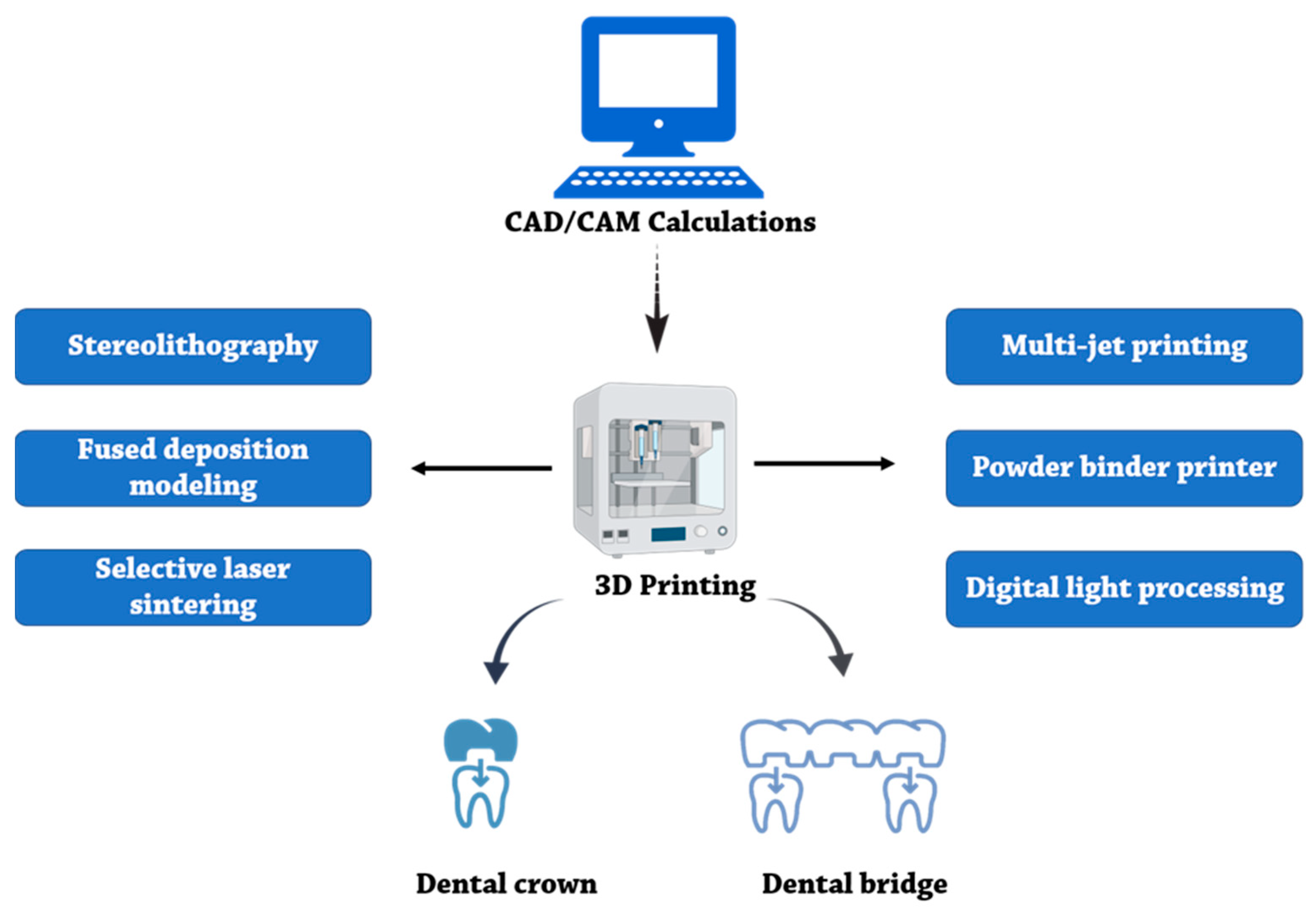
2. General Overview of 3D Printing Techniques, Advantages, and Disadvantages

Herein, the results of the review works have been mentioned by details. The details of 3D printing technics and their pros and cons are documented. Investigators developed the first 3D printer at the Massachusetts Institute of Technology (MIT) in 1993 by expanding the techniques of 2D inkjet printing [17]. A few other commonly used printing methods have been developed since then. Nowadays, with recent advances in inventing 3D printing machines with different mechanisms, from extrusion to sintering, there are various 3D printers, both in dentistry and industry. Among the 3D printing techniques, stereolithography (SLA/SLG), fused deposition modeling (FDM), selective laser sintering (SLS), selective laser melting (SLM), powder binder printers (PBP), and digital light processing (DLP) are the most commonly used techniques [5,18].
  • Briefly, the extrusion-based methods employ a nozzle with a particular diameter to dispense the desired material in three axes, controlled by a computer. These methods rely on a continuous ejection of extruded material driven out of the nozzle, mechanically or pneumatically, to form a 3D structure at the centimeter scale [19]. In the FDM method, which is an extrusion-based technique, different materials, such as thermoplastic polymers, are melted and then driven out from the nozzle. The melting material deposition on the instrument support is concomitant with the cooling down of melted material, which is the main step for fabricating favorable 3D structures [20]. Extrusion-based techniques with low costs can quickly construct basic and less complex models. Recently, the use of microfluidics to develop the FDM method has been progressed. This gives them advantages, such as affordability and low cost. The application of microfluidic in the fabricating of dental prostheses development has attracted much attention [21].
  • In the laser melting/sintering method, high-power pulsed laser light increases the temperature of specific areas to weld or sinter the added material on a three-axis moving stage. Using the SLS technique, a wide range of thermoplastic materials, including thermoplastic polymers, glass, ceramics, and metals, can be fused. Then, new surface layers can be created by refreshing the surfaces with a roller or blade. Finally, a powder form of material is applied to each sintered layer. One of the most important advantages of sintering techniques is that they lead to an autoclavable product that can be handled safely through common dental treatments [22]. The prostheses are easily duplicated at the dentistry office by applying computer-aided software and a desktop SLA 3D printer. This increases the rate and quality of manufacturing at a low moment [23].
  • In the digital light processing (DLP) method, a projector light source cures the liquid resin layer-by-layer, and each layer is created upside down. To overcome problems during the DLP method, such as shrinkage or increasing the error when the size of the point out varies, the combination of DLP and FDM models has been proposed. The FDM accuracy for full-arched dental models and FDM inaccuracy for crown prostheses have been considered. So, a hybrid method of DLP (for special die) and FDM (for full dental model) is suggested [24].
  • In the powder binder printers (PBP) method, the apparatus infiltrates pigmented liquid droplets layer-by-layer using an inkjet head [5]. However, utilizing biocompatible powders in tissue engineering is important. Calcium phosphate-based materials, as a reactive component, will be a good material (because of their similarity to dental sources) for implant applications [25].
  • Lastly, lithography-based techniques employ photopolymers as the printing material. These photopolymers are directly exposed to the laser or a UV light beam or via the lithography-based ceramic manufacturing technique, while the stage moves in different directions to obtain the 3D structure. In these techniques, motorizing mirrors help concentrate the light beam on the surfaces containing the photoreactive liquid resin meant to be fused. Then, the curved surface is recoated by a wiper. This process is followed by another fusion step to infiltrate or stain the particular areas of the printed material [22]. These photopolymers have various properties, such as elemental composition microstructure and fracture mechanics. Ucar et al. compared these features in three products. The lithography ceramic-based technique was the most promising [26].
The pros and cons of the most commonly used 3D printing techniques are presented in Table 1. Additionally, the various methods are mentions at Figure 1.

2.1. Three-Dimensional Printing and Modeling

Three-dimensional printing has the ability to be concomitantly exploited with other computer-aided technologies, such as core-beam computed tomography (CBCT) and computer-aided design (CAD)/manufacturing (CAM) [4]. Digital oral scanners make it possible to perform a reliable 3D modeling of teeth by using a computer [32]. Additionally, fixed prostheses can be fabricated by these scanners without any need for conventional working models [33].
Today, numerous kinds of fabricating methods have been developed that are capable of shaping the most typical materials. Different types of 3D printing technologies, including stereolithography apparatus, SLS, FFF [34,35], digital light processing, and MJP, can be used to perform 3D modeling.
According to ISO 5725-1:1994/Cor 1:1998, an accurate 3D printing method has trueness and precision concomitantly. In a recent study, using three different technologies, including SLA, DLP, and MJP, they were able to design and manufacture three models of dental prostheses with various resins. Analysis showed that MJP presented meaningfully higher accuracy and trueness than DLP and SLA techniques if the presence of 3D color map analysis for MJP shows surface roughness at the lowest level [36].
Two methods for producing working dental models are obtained by CAD/CAM modeling: milling and 3D printing. Using patient data from oral scanning is the input of the dental modeling. (1) The milling method has more disadvantages (e.g., unnecessary missing over milling, high cost, and time loss over the fabricating process). (2) On the other hand, the 3D printing method has more advantages (e.g., fabricating favorite prostheses, prostheses models with a minimum amount of materials, and the capability to create multiple products at a time) [37]. CAD/CAM has different software choices (e.g., Slic3R and Geomagic, respectively). The most important step in manufacturing the prototype is the progress of a suitable CAD solid model. Newly, hybrid manufacturing (HM) combines various fabricating processes methods of additive and subtractive technics and has important attention [38]. Additionally, using the CAD/CAM methods will promote the yield of prostheses manufacturing [39]. These techniques are employed widely in the fabrication of dental crowns and bridges, with an effective role in clinical settings [40]. For example, Joo et al. reported a clinical case that described a complete ceramic crown and interim fabrication for a 44-year-old man using CAD/CAM modeling and 3D printing techniques [41].
One CAD/CAM modeling capability is determining prostheses’ marginal and internal fits, such as crowns or bridges. A recent study evaluated the marginal and internal fit of feldspathic ceramic crowns by utilizing a tomography analysis [42]. They measured 2D marginal-internal fit and 3D volumetric fit and compared them between crown and reference dies. They found that the mean marginal fit between three different methods was 113.2 µm. These scanning methods will help clinicians to fabricate better prostheses. In another study, Abdullah et al. compared the CAD/CAM technique vs. conventional methods. They investigated the effect of CAD/CAM techniques on the marginal and internal fit. They evaluated a marginal gap in provisional crowns. These were fabricated by utilizing four kinds of resin by applying low-viscosity silicone impression material. They informed a mean marginal gap of 47–193 µm [43]. Additionally, Yao et al. documented a marginal cleft of 150–280 µm. They fabricated provisional crowns utilizing four different resin types [44].
The clinical digital workflow can be divided into five parts, which are shown in Figure 2 [45].
There are several software programs that are commonly used for the fabrication of dental prosthetics, surgical guides, and aligners [46].
For prosthetics, CAD/CAM (computer-aided design/computer-aided manufacturing) software is often used to design and mill crowns, bridges, and other dental restorations. Examples of CAD/CAM software used in dentistry include CEREC, 3Shape, and Dental Wings [47,48,49].
For surgical guides, specialized software is used to plan and fabricate guides that help surgeons accurately place dental implants. Examples of surgical guide software include Simplant, NobelGuide, and X-Guide [50,51,52,53].
For aligners, software is used to create treatment plans and generate 3D models of the teeth that can be used to fabricate the aligners. Examples of aligner software include Invisalign, ClearCorrect, and OrthoPlan [54,55].

2.2. Three-Dimensional Materials Characteristics

In prosthodontics, common clinical procedures, including the fabrication of crowns and bridges, have gained attention to be performed by additive manufacturing techniques. The reliability and precision of traditionally fabricated dental restorations can be affected by human errors, due to their labor intensiveness [56]. Regarding the marginal gap, occlusal fit, and internal discrepancies of dental restorations, it has been found that both milling and additive manufacturing techniques are more accurate and precise, compared to manual techniques [57]. Similar results were also obtained by other scientists regarding marginal fit [58]. In addition to these results, a systematic review performed by Papadiochou et al. has shown that the identity and quality of restorative 3D printable materials can affect the performance of CAD/CAM processes [59]. In other research, Yoo-Geum Jeong et al. used the root mean square (RMS) value in their study, and according to the results, less value of the RMS in 3D-printed models showed that they were more accurate than those manufactured by the milling method [37].
If the provisional restoration is utilized for a long time, visual concerns of patients will be amplified, particularly in anterior restorations. Clinicians should carefully consider the optical properties, as well as their stability over time [60].
Revilla-León et al. evaluated the color of the 3D printable materials and then compared it with conventional acrylic resin-based interim materials [34]. They comprised five various 3D printable dental materials; however, they did not measure the translucency and color stability. Moreover, Shin et al. measured three CAD/CAM blocks and two 3D printable materials’ color stability [61]. Thus, studies on the color and translucency of 3D printable dental materials, as well as their stability over an extended period, are needed. According to CIELAB (International Commission on Illumination) color space, color is used to obtain these visual criteria. It is concluded based on the difference of colors at different times during one hour [62].
The threshold values for clinically apprehensible color and translucency differences are defined as ΔE ≥ 3.7 and |ΔTP| ≥ 2.0, respectively [4,63]. In a recent study, five substances of different resin types were evaluated [4]. These results showed that the color and translucent dental materials for crown and bridge restorations changed over time. These products are fabricated by the DLP technique. One of the most important limitations related to 3D printable dental materials is that the color of materials that are used for 3D printing can be changed over time [4]. Moreover, the colors of 3D printable materials are basically different. In contrast to the color, the translucency of the material undergoes a relatively minor change. Accordingly, the materials used in the 3D printing of dental materials become opaque, darker, and more yellowish during the 6-month storage in the aqueous medium [4].
Karatas et al. [64] investigated the effects of staining on the resin surface properties. More specifically, they studied the effect of bleaching and staining on the surface microhardness, roughness, and color changes (ΔE). After bleaching, a significant decrease of microhardness was observed in the microhybrid composite specimens.
Surface roughness is also one of the important factors used to evaluate the performance of dental materials [65]. In a study, Sobbah et al. [66] were able to study the effect of different layers and thicknesses on surface roughness. The results showed that changing layer thickness and storage time does not affect surface roughness. On the other hand, in their study, Arnold et al. found that roughness parameter and printing angles are the sources of variations in roughness [67].
Jain et al. fabricated 3D-printed resins to evaluate the color stability of dental prostheses. They found that, after diving into denture cleanser solutions, all of the denture base resins had a substantial variation in color [68].
In addition, some novel methods have been carried out to assess the property changes. For example, Warnecki et al. [69] utilized fractal dimension analysis, texture analysis, and wetting angle assessment to evaluate the mechanical characterizes and behavior of the appliances.
Three-dimensional printing constructs have been influenced by various factors during preparation, printing, and post-printing procedures, with direct effects on the mechanical behavior and quality of fabricated components [70]. For example, raster orientation and printing speed are two key printing parameters that impact the specimens feature [70,71].

3. Application of 3D Printing in Dental Prostheses

Three-dimensional printing in dentistry, mainly when applied to the fabrication of dental provisional restorations (bridges and crowns), is new, with related studies dating as recently as 2013. Most of these studies focused on the fit of 3D-printed restorations and compared their characteristics with those fabricated by conventional methods (Table 2). Marginal and internal fit is identified factors that have a critical impression on the long-term achievement of dental restorations [72]. The marginal fit of restorations is highly related to their manufacturing technique [73]. Some parameters can influence the marginal fit of prostheses manufactured by 3D printing, such as type of resin, type of printing machine, device calibration, in terms of environmental temperature and moisture, and restoration shape complexity [74]. Table 2 shows the methods and materials used for 3D printing applications in dental prostheses and crowns [75].
In a recent study, Lee et al. investigated the internal fit of dental crowns obtained by 3D printing and the CAD/CAM milling method using stainless steel and vinyl-polysiloxane [76]. As a result, the mean discrepancy values were measured to be 141.1 and 91.1 µm for the crowns fabricated by two brands of 3D printing systems and 171.6 µm for those fabricated by the milling system. Furthermore, the internal and marginal fit of the fabricated crowns obtained from 3D printing systems were significantly improved, compared to those fabricated by CAD/CAM milling system. Tahayeri et al. optimized the 3D printing of dental materials for bridge restorations and provisional crowns using a low-cost stereolithography 3D printer and compared their mechanical properties to conventionally cured provisional dental materials [77]. Fourier-transform infrared spectroscopy (FT-IR) analysis and three-point bending methods were employed to evaluate the degree of conversion of the resin and the peak stress and the elastic modulus of 3D-printed bars, respectively. They also compared the obtained results with two conventionally cured provisional materials, Jet®, Lang Dental Inc., Wheeling, IL, USA, and Integrity®, Dentsply, Charlotte, NC, USA. As reported, there was no direct correlation between the printing layer thickness and peak stress or elastic modulus. The 3D-printed models showed similar and significantly higher peak stress compared to Integrity® and Jet®, respectively (Figure 3). Furthermore, compared to Jet® and Integrity®, the 3D-printed samples had comparable and significantly lower elastic modulus. Interestingly, the 3D-printed samples also showed an enhanced degree of conversion than those of Jet® and Integrity®.
Similarly, Alharbi et al. compared the effects of 3D printing and milling methods and different finish line designs on the marginal and internal fit of interim restorations [58]. As reported, the mean internal gaps for milled restorations were 89, 177, 185, and 154 µm, fabricated on a knife-edge (KE), chamfer (C), rounded shoulder (RS), and rounded shoulder with bevel (RSB) finish line designs, respectively. On the other hand, these values for 3D-printed restorations were reported to be 66, 149, 130, and 95 µm, respectively, for KE, C, RS, and RSB, indicating the significantly lower internal gap of 3D-printed restorations, compared to those obtained from milling methods (Figure 4). Furthermore, the 3D-printed restorations showed lower mean absolute marginal discrepancy (30, 41, 30, 28 µm) than the milled restorations (56, 54, 52, 38 µm) for KE, C, RS, and RSB, respectively. The aforementioned results designate that the finish-line design techniques have a lower impact on the fit than the fabrication methods [58].
In another study, Chaturvedi et al. evaluated the marginal and internal fit, using finish line chamfer, rounded shoulder, and rounded shoulder with bevel, of the provisional crowns obtained by three-dimensional (3D) printing, compression molding, and milling methods [81]. The result of that fabrication method and finish line design has a substantial effect on the internal and marginal gap. The minimal marginal gap and the best internal fit in all various finish lines belonged to the 3D printing methods, in comparison with compression molding and milling methods [27].
Despite providing a rapid technique for the fabrication of zirconia dental restorations, CAD/CAM milling systems present a few disadvantages, such as material waste, processing defects, such as microscopic fractures, and inadequate accuracy [98]. Ebert et al. employed direct inkjet 3D printing to fabricate all-ceramic dental restorations using zirconia-based ceramic suspension as the source material [85]. The obtained 3D-printed dental crowns possessed high mean fracture toughness (KIc = 6.7 MPam0.5) and characteristic strength of the ground bars (σ0 = 763 MPa) with a 90% confidence interval of [678;859]. Revealed by scanning electron microscopy (SEM), homogeneous cross-sections were apparent, with no significant defects (except for a few process-related ones) on the surface of the specimens. In view of cost-efficiency, this promising method consumes a minimum number of materials.
To further evidence the superior efficiency of 3D printing procedures for the manufacturing of dental prostheses compared to conventional techniques, Eftekhar Ashtiani et al. employed intraoral scanning and 3D printing of the pattern for the fabrication of intracoronal restorations and compared their dimensional accuracy to those obtained from the conventional fabrication of a resin pattern [79]. Interestingly, the conventional method resulted in more accuracy than the 3D printing, regarding impression making and the fabrication of intracoronal restorations. However, the fabricated restorations with both methods yielded a clinically acceptable fit.
Similarly, Homsy et al. compared the internal and marginal fit accuracy of lithium disilicate-based inlays manufactured with conventional milling and 3D printing methods [80]. Here, the marginal and internal fit accuracy of the inlays fabricated by digital impressions and subtractive milling of wax patterns were significantly better than those of the conventional impression/fabrication and 3D printing techniques. It is worth noting that the fit values measured for 3D-printed wax patterns were similar to those of the conventionally waxed inlays.
Mai et al. examined the fit of interim crowns (proximal, marginal, internal axial, and internal occlusal regions) made by photopolymer jetting 3D printing and compared it to compression molding and milling techniques [57]. According to this study, in the marginal and proximal regions, the milling and 3D printing methods exhibited more accurate results than the molding method. Furthermore, 3D printing resulted in the most accurate values in the occlusal region. The internal discrepancies were the highest for the milling method, meaning that the fit of temporary crowns, particularly in the occlusal region, can be efficiently enhanced by taking advantage of polymer-jet 3D printing.
Fathi et al. reported similar results, stating that the 3D-printed wax crowns were more accurate, in terms of internal and marginal fit, than milled and manually fabricated wax crowns [82]. Furthermore, conventional hand carving and milling of wax crowns both resulted in significant discrepancies in occlusal gap points, when compared to additive methods, demonstrating 3D designing and manufacturing of wax patterns for complete crowns as a more accurate procedure [99].
As the fracture load is a critical characteristic for dental restorations, it is crucial to assess this index for structures that novel approaches, such 3D printing, have manufactured [100]. To this end, Zimmermann et al. determined and compared the fracture load of crowns made of three particle-filled composite CAD/CAM materials, including Cerasmart (GC Corporation), Brilliant Crios (Coltène AG; Altstätten), and Lava Ultimate (3M ESPE), and one 3D-printed composite (els-3D Harz; Saremco Dental AG) as a function of three different material thicknesses (0.5, 1, and 1.5 mm) [83]. Amongst all groups, Brilliant Crios and els-3D Harz, respectively, showed the highest mean loading forces before fracture, which were measured to be 1580.4 N and 1478.7 N, with 1.5 mm thickness. The fact is that the fracture loading force mainly depends on the respective material and thickness; therefore, none of the 0.5 mm ceramic crowns (as group control) survived the fatigue testing, while all the resin-based crowns did. Consequently, regardless of the fabrication mode (CAD/CAM or 3D printing), the particle-filled composite resins may play an essential role in manufacturing minimally invasive restorations with good mechanical properties [83]. A wide range of complications can be developed as consequences of the marginal discrepancy between the abutment and the restoration material [101].
Similar studies on the fabrication of CAD/CAM-based temporary crowns and evaluation of the internal matching were conducted by Lee et al. First, they scanned the ready-made stainless model using digital scanners and designed it with software CAD/CAM. Then, they used 3D printers to fabricate dental crowns. Zirkonzahn (3D milling system), Stratasys, and Dentis (3D printing system) technology were used to fabricate the crowns [76]. Vipi block, VeroGlaze MED620, and ZMD-1000B resin were applied in the fabrication. For each group, ten files were made. The mean ± standard deviation (SD) values of marginal discrepancy were found to be 171.6 ± 97.4, 149.1 ± 65.9, and 91.1 ± 36.4 for the CAD/CAM milling group, Stratasys group, and Dentis group, respectively. p-values for the 3D printing groups (consisting of Stratasys and Dentis groups) were lower, compared to that for the CAD/CAM milling group. According to the significant level of the p-value (a p-value less than 0.05 was considered significant), the mean discrepancy values were found to have statistically significant differences. Accordingly, a high level of completion is an advantage that makes the 3D printing method a suitable method to be applied in the production of dental prostheses, in addition to the interim restoration production [76].
The solidification of liquid polymer resins, including resins, photopolymers, and transparent resins, is a process in additive manufacturing performed by UV light in digital light processing (DLP) printing technology [102]. Applications of DLP in dentistry include the fabrication of dental models, dental implants, cochlear implants, and dental restorations. Moraru et al. used this technique to fabricate dental prostheses [92]. The advantages of this method are its high accuracy in printing different parts, as well as the appropriate surface of the final product [103]. Son et al. fabricated three 3D printing Interim dental crowns by SLA and DLP and milling methods. They focused on the comparison of intaglio surface trueness at each implant. They used CAD/CAM to design and model the crowns, as in previous studies. In the fabricating procedure of interim dental crowns, 3D printing technologies presented higher accuracy than milling [91].
Firlej et al. investigated five materials for 3D printing applications. These resins were UV-curable form. Isopropyl alcohol was sprayed on all of them. This removed the rest of the resins on the implants. They evaluated the effect of artificial aging on the quality of materials in various implants [88]. In some cases, both milling and 3D printing methods are clinically acceptable. For example, H Galeva et al. compared the internal and external accuracy fit of metal-ceramic fixed prosthetic constructions. Additionally, the temperature influence evaluations showed that the differences were not noticeable [93].
Temporary restoration modeling and fabrication are important in various dental applications. Mohajeri et al. investigated the effect of conventional fabricating methods against 3D printing on the marginal fit [94]. By utilizing these three fabrication methods, the restorations were generated via clinically suitable marginal fit.
In a recent study, Ryu et al. investigated the effect of different directions, including 120°, 135°, 150°, 180°, 210°, and 225 °C [95]. The best fit was obtained with the shape angles of 180° and 150°. Figure 5 shows the marginal gap from a different view. The orientation of the build is important in the fabrication techniques [104]. For example, Lee et al. examined the accuracy (trueness and precision) of the interim crowns. They utilized a DLP printer and investigated post-curing time. The build angle was 135° [96]. Yu et al. [97] examined the intaglio surface and build angle on the interim crown. Additionally, they determined the effect of build angle, including 0, 120, 135, 150, 180, 210, 225, 240, and 270 degrees, on dental crown fabricates’ mechanical properties. The results showed the build angle effect on the intaglio surface trueness and marginal gap. The recommended angle was between 150 and 210 degrees [97].
A surgical guide in dentistry is a template that is used to accurately position dental implants in the jawbone [105]. Three-dimensional printing can be used to fabricate surgical guides by creating a physical model of the patient’s jaw and teeth based on a digital 3D scan [106,107]. The guide is then created by slicing the digital model and exporting the slice data to a 3D printer [52,108]. Here is a step-by-step overview of the process:
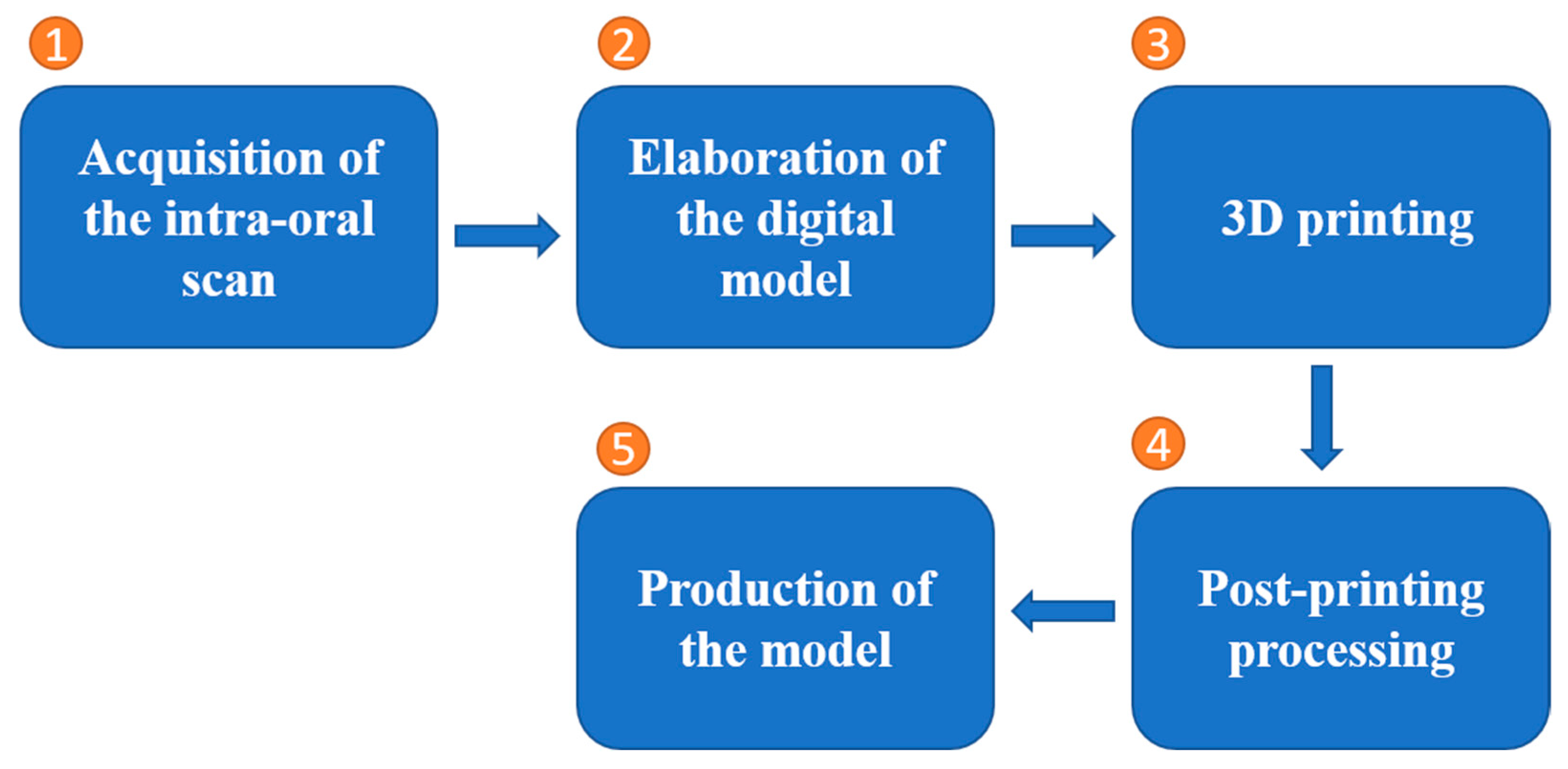
  • Digital 3D scan of the patient’s jaw and teeth: The first step in fabricating a surgical guide using 3D printing is to obtain a digital 3D scan of the patient’s jaw and teeth. This can be achieved using a variety of technologies, including CBCT and intraoral scanners.
  • Design of the surgical guide: Once a digital 3D scan has been obtained, the next step is to design the surgical guide using specialized software. This involves creating a virtual model of the patient’s jaw and teeth and then planning the placement of the dental implants based on the specific needs of the patient.
  • Slicing the digital model: The next step is to slice the digital model of the surgical guide into layers, which can then be exported to the 3D printer. This process involves specifying the thickness of the layers and the type of 3D printing technology to be used.
  • 3D printing: The slice data is then sent to the 3D printer, which creates a physical model of the surgical guide using a variety of materials, such as plastic or metal.
  • Post-processing: Once the surgical guide has been printed, it may need to be post-processed, in order to smooth out any rough edges and ensure that it is accurate and ready for use. This may involve sanding, polishing, and sterilizing the guide.
  • Use in surgery: The surgical guide is then used during the actual implant surgery to accurately position the implants in the jawbone. The guide helps the surgeon to place the implants in the correct location, ensuring that they are properly aligned and positioned for optimal function.
Three-dimensional printing can also be used to fabricate aligners, which are employed in orthodontic treatment to straighten teeth [54,109]. The process begins by creating a 3D model of the patient’s teeth using a digital impression or a physical impression that is scanned into a computer [54]. The 3D model is then used to design the aligners, which are customized to fit the patient’s teeth and apply the necessary forces to move the teeth into their desired positions [110]. The aligners are typically made of a clear, biocompatible plastic material that is suitable for long-term wear in the mouth. Once the aligners have been designed, they can be fabricated using a 3D printer. The finished aligners are then sent to the orthodontist, who gives them to the patient to wear, according to a prescribed treatment plan [54,111].
There are a few limitations to the use of 3D printing for the fabrication of dental surgical guides and aligners in dentistry [52,105,106,108,112,113]:
Material properties: The properties of 3D-printed materials may not be suitable for all dental surgical applications. For example, 3D-printed surgical guides may not be as rigid as those made from other materials, such as stainless steel.
Accuracy: While 3D printing can produce objects with high levels of accuracy, the accuracy of the final printed product may be affected by factors such as the resolution of the printer and the quality of the 3D model used as a reference.
Time: Three-dimensional printing can be a time-consuming process, especially for large or complex objects. This may not be practical in cases where time is of the essence, such as in emergency surgery.
Cost: Three-dimensional printing can be an expensive option, compared to other manufacturing methods, especially for large volume production.
Regulatory considerations: There may be regulatory hurdles to overcome, in order to use 3D-printed dental surgical guides and aligners in clinical practice, as they may be considered medical devices. This may require additional testing and documentation to demonstrate their safety and effectiveness.
There are a variety of clinical case reports regarding the use of 3D printing for dental prosthetics. Three-dimensional printing can be clinically used to create crowns, bridges, and dentures that are custom fit to a patient’s teeth, which can improve the comfort and aesthetic appearance of the restoration [114,115]. For example, Srinivasan et al. performed a double-blind, randomized, and crossover clinical study to show the differences between 3D-printed and milled complete removable dental dentures [115]. They revealed that the cost and time of the workflow are likely same for two groups of patients [115].
One of the important clinical applications of 3D printing in dentistry is the use of this technology for the fabrication of a surgical guide and aligner. Surgical guides created by 3D printing can be used to accurately place dental implants in the jawbone, which can help to reduce the risk of complications and improve the overall success of the surgery. For example, there is a case report by Zoran et al. in which a guide was created by 3D printing technique for implant placement [116]. Its results showed that, considering both surgical and prosthetic aspects, the 3D-printed surgical guide could facilitate having an optimal positioning of the implant [116].
The fabrication of custom dental implants is another clinical application of 3D printing in dentistry. Printing can be used to create custom dental implants that are tailored to the specific shape and size of a patient’s jawbone, which can improve the fit and function of the implant. Par et al. reported a case study on 3D-printed titanium implant [117].
At the clinical stage, 3D printing has various pros and cons [118]. The typical advantages can be attributed to the customizability, flexible and fast design and production, long-term cost, ease of access, minimizing waste, and being environmentally friendly [118,119]. Similar to other fabrication methods, 3D printing has its own drawbacks for clinical applications, including limitations in the material selections, possible post-processing requirements and probable design inaccuracies [118,120].

4. Dental 3D Printing Materials

As discussed, there are various 3D printing techniques in dentistry and along with these techniques, a wide range of biomaterials, including hydrogels, ceramics, metals, resins, and thermoplastic polymers, have heavily been explored.
More specifically, there are a number of materials that are commonly used in 3D printing for dental prosthetics, including acrylonitrile butadiene styrene (ABS), polylactic acid (PLA), and a variety of resins. Several synthetic polymers, including poly(ethylene glycol) (PEG) and poly(vinyl alcohol) (PVA), have also been exploited in the 3D printing of dental biomaterials, due to their tailorable mechanical and degradational properties [27,121]. Some newer materials are being developed and used in 3D printing for dental prosthetics. One example of a new material that has been used in the 3D printing of dental prosthetics is bioceramic. Bioceramic materials are ceramic materials that are biocompatible, meaning they are safe to use in the human body [122]. They have been used in a variety of medical applications, including dental prosthetics, due to their strength, durability, and ability to bond with living tissue. Bioceramic materials are also resistant to wear and corrosion, making them a good choice for use in dental prosthetics that will be subjected to high levels of stress and exposure to oral fluids. Some examples of bioceramic materials that have been used in the 3D printing of dental prosthetics include zirconia, alumina, and hydroxyapatite [123,124].
Composite materials that combine ceramics with other materials, such as plastic or metal, are also good options for the 3D printing of dental prosthetics [125]. These materials offer improved strength, durability, and aesthetics, making them suitable for a wide range of dental applications. Additionally, some of the newer 3D printing technologies, such as SLS and electron beam melting (EBM), allow for the use of metal materials, such as titanium and cobalt-chrome, which can be used to create more durable and biocompatible dental prosthetics.
In addition to different fabricating technologies, different materials present other mechanical properties, such as thickness. For example, in a recent study, three resins, including bisacrylic, acrylic, and PMMA, were used for microcomputed tomography of 3D-printed dental crowns. Utilizing them with 3D printing technology presents higher film thickness in dental crowns [126].

4.1. Thermoplastic Polymers

Among the various options available for the fabrication of dental 3D printable substances, polymer-based materials are the most commonly utilized materials. Photopolymerization is the most feasible technique for the fabrication of dental resins or polymeric 3D-printed materials [27]. A smoother surface, strong chemical bonding, suitable mechanical strength, and high-quality build resolution can be provided using photopolymerization [6]. Thermoplastic polymers are the most frequently used polymer-based biomaterials for dental 3D printing [127]. The filaments that make the main backbone structural compartments of thermoplastic polymers can flow through the nozzles by applying heat [128,129]. Among these polymers, polyethylene (PE), polypropylene (PP), PLA, and ABS are the most frequently used in the field of 3D printing of polymeric dental biomaterials [130]. Due to the non-toxic properties of PLA against the oral cavity, it is assumed to be more favorable for utilizing in 3D printing, compared to ABS [129,131]. Thermoplastic filament polymers with higher glass transition temperatures, such as polymethyl methacrylate (PMMA) and polyether ether ketone (PEEK), have been recently studied for the fabrication of dental 3D-printed materials [27]. Schönhoff et al. compared the amorphous polyphenylene sulfone (PPSU) with established semi-crystalline polyetheretherketone (PEEK). They found that PPSU can be a suitable material, instead of PEEK. They evaluated the mechanical properties that confirmed recently proposed research [132].
Wieckiewicz et al. [133] investigated the surface roughness, color stability, and elasticity of polyamide-12 (PA) versus polymethyl methacrylate (PMMA) denture-based material as a control. The results suggested that PA showed a higher susceptibility than PMMA to discoloration.

4.2. Ceramics

Compared to polymers, ceramics are less frequently used in 3D printing [134]. However, due to their unique properties, ceramics are good candidates to be utilized in stereolithography (SLA) and SLS, in which ceramic powder or a pre-sintered ceramic material are processed to form a structure with strong bonding [1,129,135,136]. A biocompatible microenvironment can be formed in ceramic structure by adding mineral substances, such as hydroxyapatite and β-tricalcium phosphate, which provide calcium and phosphate ions [137]. Moreover, the incorporation of calcium and phosphate mineral phases has been shown to improve cell-to-cell interactions and induce cell differentiation and proliferation, which makes these types of ceramics good candidates for craniofacial applications [138,139]. However, due to the challenges associated with the processing of ceramic powders to high-density structures, the products of the selective laser sintering of these powders are porous structures. Additionally, anisotropic shrinkage and stair-step effects can occur upon additive manufacturing approaches. The above-mentioned challenges have limited the ceramic utilization of ceramics for the development and fabrication of ceramic 3D-printed restorations [129,140,141].

4.3. Metals

Metals have been mainly used in selective laser sintering to fabricate dental 3D-printed materials [142]. In dentistry, metallic alloys, including nickel, cobalt-chromium, and titanium alloys have been vastly studied and used [143]. However, due to the possible allergic reactions in the oral cavity, scientists no longer suggest the utilization of nickel alloys in dental metallic materials [144]. Similar to ceramics, the fabrication of metallic biomaterials by selective laser sintering leads to porous products [145]. Due to the negative effect of porosity formation on the strength and resistance of 3D-printed materials, some solutions, such as equipping a vacuum pump with a sintering instrument, have been considered, in order to improve the quality of metallic dental prostheses [7,129,146,147]. Among metallic materials, cobalt-chromium and titanium are the most commonly used alloys for the fabrication of 3D-printed metallic prostheses, due to their strength and ductility [148]. Furthermore, various clinical trials in the field of maxillofacial prostheses have been performed on titanium alloys, specifically Ti6Al4V [1,129,149].

4.4. Others

According to SLM and SLS methods, there are some limitations in utilizing ceramics or metals materials [150]. This matter is because of the thermal shock conditions and high melting temperature in metals and ceramics, respectively [151]. Thus, researchers developed novel approaches to overcome these limitations. For example, in a recent study [152], a combination-producing method, called additive manufacturing, was utilized to make high-density zirconia dental crowns. The authors of this study could fabricate high-density ceramic materials, which are difficult to make with conventional methods.

5. Prospects and Conclusions

As a cutting-edge additive technology, 3D printing has played a revolutionary role in most of the scientific fields, to which dentistry is no exception, especially regarding dental prostheses with complex structures [153]. Along with an expanding range of utility for 3D printing technology in dentistry, private office manufacturing with 3D printing seems to be a progressively more realistic prospect [154,155]. More specifically, 3D printing is being employed in dental offices to create a variety of objects, including crowns, bridges, and surgical guides. Three-dimensional printing allows dentists to create customized objects that fit a patient’s mouth perfectly, which can improve the accuracy and effectiveness of dental procedures. It can also help to reduce the turnaround time for certain procedures, as objects can be printed on-demand, rather than having to be ordered and shipped from a lab. Demonstrably, a wide variety of materials can be used to fabricate prostheses with clinically sound results and with similar, and sometimes improved, results, compared to analog and subtractive manufacturing methods [156]. There are a variety of materials that can be used for 3D printing crowns, bridges, surgical guides, aligners, and other dental prosthetics, including ABS, PLA, and various types of resins. The most commonly used material for 3D printing these types of dental prosthetics is a type of resin called a photopolymer. This material is capable of producing highly detailed and accurate objects and is suitable for a wide range of applications. Other materials that may be used for 3D printing of dental prosthetics include stainless steel, titanium, and cobalt-chrome alloys. As research progresses, more materials are being implemented with this technology, providing an expanded repertoire of services. The addition of metal 3D printing is currently being investigated; while more evidence is required, the results could allow for expanding 3D printing services to a convenient in-office framework fabrication for fixed and removable prosthetics [157]. Metal 3D printing involves using a laser to fuse together metal powders into the desired shape. This process allows for the production of highly accurate and complex objects, including dental prosthetics [121]. Some of the advantages of using metal 3D printing for dental prosthetics include the ability to create customized and anatomically accurate objects, the ability to produce objects with complex internal structures, and the ability to produce objects with a high level of detail [158,159]. Additionally, metal 3D printing can be used to produce dental prosthetics that are stronger and more durable than those made with other methods. How this technology will be integrated with commercially available polymer and ceramic-based printers is yet to be seen. However, it is important to note that metal 3D printing can be a more expensive option, compared to other methods of fabrication, and it may not be suitable for all types of dental prosthetics. It is important to note that the choice of material and fabrication method will depend on the specific application, the requirements of the patient, and the clinical indications of the case.
There are several challenges attributed to the 3D printing of dental prostheses [160]. The most important one is accuracy. It is vital for dental prostheses to fit accurately in the mouth, in order to function properly. Three-dimensional printing allows for the creation of highly precise prostheses, but achieving the necessary level of accuracy can be challenging [158,161]. Material selection is another challenge. Dental prostheses need to be made from materials that are biocompatible, durable, and able to withstand the stresses of everyday use. There are a variety of materials that can be used for 3D printing dental prostheses, but choosing the right one for a specific application can be difficult [162,163]. The surface finish is also an important matter to consider in the 3D printing of dental prostheses. They need to have a smooth, polished surface finish, in order to be comfortable in the mouth. Achieving this finish can be challenging when using 3D printing technologies [164,165]. Three-dimensional printing dental prostheses can also be expensive, due to the cost of the equipment and materials [160,166]. This can make it difficult for some patients to afford these types of prostheses. Time can also be an issue in the fabrication of some unique 3D printing dental prostheses. It can be time-consuming, especially when compared to traditional manufacturing techniques [167,168]. This can be a challenge for dental practices that need to produce multiple prostheses in a short amount of time.
In conclusion, 3D printing technology has the potential to revolutionize the way dental prostheses are made. Three-dimensional printing allows for the creation of more comfortable and natural-looking dental prostheses, which can improve the patient’s experience. The use of 3D printing in dentistry has increased in recent years, and it is expected to continue to grow in the coming years by increasing the accuracy, reducing the production time, and lowering the cost. Overall, the prospects for the use of 3D printing in the production of dental prostheses are very promising. It is expected that 3D printing will become increasingly prevalent in dentistry in the coming years, as the technology continues to advance and become more widely adopted.

Author Contributions

Conceptualization, L.T., F.R. and M.F.; investigation, F.R., M.M., F.E.W. and L.T.; resources, L.T.; writing—original draft preparation, F.R., M.M., M.D., R.M., F.A., J.W., K.M.; writing—review and editing, L.T., K.M. and F.E.W.; supervision, L.T.; funding acquisition, L.T. All authors have read and agreed to the published version of the manuscript.

Funding

Part of the research reported in this paper was supported by National Institute of Dental and Craniofacial Research of the National Institutes of Health, under award numbers R15DE027533, R56DE029191, and 3R15DE027533-01A1W1.

Informed Consent Statement

The content is solely the responsibility of the authors and does not necessarily represent the official views of the National Institutes of Health.

Data Availability Statement

Not applicable.

Conflicts of Interest

The authors declare no conflict of interest.

References

  1. Abduo, J.; Lyons, K.; Bennamoun, M. Trends in computer-aided manufacturing in prosthodontics: A review of the available streams. Int. J. Dent. 2014, 2014, 783948. [Google Scholar] [CrossRef] [Green Version]
  2. Mainjot, A.; Dupont, N.; Oudkerk, J.; Dewael, T.; Sadoun, M. From artisanal to CAD-CAM blocks: State of the art of indirect composites. J. Dent. Res. 2016, 95, 487–495. [Google Scholar] [CrossRef]
  3. Cicciù, M.; Fiorillo, L.; Herford, A.S.; Crimi, S.; Bianchi, A.; D’Amico, C.; Laino, L.; Cervino, G. Bioactive Titanium Surfaces: Interactions of Eukaryotic and Prokaryotic Cells of Nano Devices Applied to Dental Practice. Biomedicines 2019, 7, 12. [Google Scholar] [CrossRef] [Green Version]
  4. Kim, J.-E.; Choi, W.-H.; Lee, D.; Shin, Y.; Park, S.-H.; Roh, B.-D.; Kim, D. Color and translucency stability of three-Dimensional printable dental materials for crown and bridge restorations. Materials 2021, 14, 650. [Google Scholar] [CrossRef]
  5. Dawood, A.; Marti, B.M.; Sauret-Jackson, V.; Darwood, A. 3D printing in dentistry. Br. Dent. J. 2015, 219, 521. [Google Scholar] [CrossRef]
  6. Stansbury, J.W.; Idacavage, M.J. 3D printing with polymers: Challenges among expanding options and opportunities. Dent. Mater. 2016, 32, 54–64. [Google Scholar] [CrossRef]
  7. Guilak, F.; Butler, D.L.; Goldstein, S.A.; Baaijens, F.P. Biomechanics and mechanobiology in functional tissue engineering. J. Biomech. 2014, 47, 1933–1940. [Google Scholar] [CrossRef] [Green Version]
  8. Nayar, S.; Bhuminathan, S.; Bhat, W.M. Rapid prototyping and stereolithography in dentistry. J. Pharm. Bioallied Sci. 2015, 7, S216. [Google Scholar] [CrossRef]
  9. Tasaka, A.; Shimizu, T.; Kato, Y.; Okano, H.; Ida, Y.; Higuchi, S.; Yamashita, S. Accuracy of removable partial denture framework fabricated by casting with a 3D printed pattern and selective laser sintering. J. Prosthodont. Res. 2019, 64, 224–230. [Google Scholar] [CrossRef]
  10. Salmi, M.; Paloheimo, K.-S.; Tuomi, J.; Ingman, T.; Mäkitie, A. A digital process for additive manufacturing of occlusal splints: A clinical pilot study. J. R. Soc. Interface 2013, 10, 20130203. [Google Scholar] [CrossRef]
  11. Kalberer, N.; Mehl, A.; Schimmel, M.; Müller, F.; Srinivasan, M. CAD-CAM milled versus rapidly prototyped (3D-printed) complete dentures: An in vitro evaluation of trueness. J. Prosthet. Dent. 2019, 121, 637–643. [Google Scholar] [CrossRef] [PubMed]
  12. Di Giacomo, G.d.A.; Cury, P.R.; da Silva, A.M.; da Silva, J.V.; Ajzen, S.A. A selective laser sintering prototype guide used to fabricate immediate interim fixed complete arch prostheses in flapless dental implant surgery: Technique description and clinical results. J. Prosthet. Dent. 2016, 116, 874–879. [Google Scholar] [CrossRef] [PubMed]
  13. Tserovski, S.; Georgieva, S.; Simeonov, R.; Bigdeli, A.; Röttinger, H.; Kinov, P. Advantages and disadvantages of 3D printing for pre-operative planning of revision hip surgery. J. Surg. Case Rep. 2019, 2019, rjz214. [Google Scholar] [CrossRef] [PubMed]
  14. Nakamura, K.; Harada, A.; Inagaki, R.; Kanno, T.; Niwano, Y.; Milleding, P.; Örtengren, U. Fracture resistance of monolithic zirconia molar crowns with reduced thickness. Acta Odontol. Scand. 2015, 73, 602–608. [Google Scholar] [CrossRef] [Green Version]
  15. Taira, Y.; Odatsu, T.; Sawase, T. Effects of a fluoride etchant and a phosphate primer on bonding of veneering composite to Ti–6Al–4V alloy for CAD/CAM restorations. J. Prosthodont. Res. 2013, 57, 30–35. [Google Scholar] [CrossRef] [Green Version]
  16. Lauvahutanon, S.; Takahashi, H.; Oki, M.; Arksornnukit, M.; Kanehira, M.; Finger, W.J. In vitro evaluation of the wear resistance of composite resin blocks for CAD/CAM. Dent. Mater. J. 2015, 34, 495–502. [Google Scholar] [CrossRef] [Green Version]
  17. Sachs, E.M.; Haggerty, J.S.; Cima, M.J.; Williams, P.A. Three-Dimensional Printing Techniques. Google Patents US5204055A, 20 April 1993. [Google Scholar]
  18. Etemad-Shahidi, Y.; Qallandar, O.B.; Evenden, J.; Alifui-Segbaya, F.; Ahmed, K.E. Accuracy of 3-Dimensionally Printed Full-Arch Dental Models: A Systematic Review. J. Clin. Med. 2020, 9, 3357. [Google Scholar] [CrossRef]
  19. Chameettachal, S.; Yeleswarapu, S.; Sasikumar, S.; Shukla, P.; Hibare, P.; Bera, A.K.; Bojedla, S.S.R.; Pati, F. 3D Bioprinting: Recent Trends and Challenges. J. Indian Inst. Sci. 2019, 99, 375–403. [Google Scholar] [CrossRef]
  20. Carneiro, O.S.; Silva, A.; Gomes, R. Fused deposition modeling with polypropylene. Mater. Des. 2015, 83, 768–776. [Google Scholar] [CrossRef]
  21. Tiboni, M.; Tiboni, M.; Pierro, A.; Del Papa, M.; Sparaventi, S.; Cespi, M.; Casettari, L. Microfluidics for nanomedicines manufacturing: An affordable and low-cost 3D printing approach. Int. J. Pharm. 2021, 599, 120464. [Google Scholar] [CrossRef]
  22. Shah, P.; Chong, B. 3D imaging, 3D printing and 3D virtual planning in endodontics. Clin. Oral Investig. 2018, 22, 641–654. [Google Scholar] [CrossRef] [PubMed]
  23. Lin, W.-S.; Harris, B.T.; Morton, D. Use of CBCT Imaging, Open-Source Modeling Software, and Desktop Stereolithography 3D Printing to Duplicate a Removable Dental Prosthesis-A Proof of Concept. Compend. Contin. Educ. Dent. 2017, 38, e5–e8. [Google Scholar] [PubMed]
  24. Lee, D.; Lee, S.Y.; Kim, H.; Park, C. A Hybrid Dental Model Concept Utilizing Fused Deposition Modeling and Digital Light Processing 3D Printing. Int. J. Prosthodont. 2020, 33, 229–231. [Google Scholar] [CrossRef] [PubMed]
  25. Khalyfa, A.; Vogt, S.; Weisser, J.; Grimm, G.; Rechtenbach, A.; Meyer, W.; Schnabelrauch, M. Development of a new calcium phosphate powder-binder system for the 3D printing of patient specific implants. J. Mater. Sci. Mater. Med. 2007, 18, 909–916. [Google Scholar] [CrossRef]
  26. Uçar, Y.; Aysan Meriç, İ.; Ekren, O. Layered manufacturing of dental ceramics: Fracture mechanics, microstructure, and elemental composition of lithography-sintered ceramic. J. Prosthodont. 2019, 28, e310–e318. [Google Scholar] [CrossRef] [PubMed] [Green Version]
  27. Pillai, S.; Upadhyay, A.; Khayambashi, P.; Farooq, I.; Sabri, H.; Tarar, M.; Lee, K.T.; Harb, I.; Zhou, S.; Wang, Y. Dental 3D-printing: Transferring art from the laboratories to the clinics. Polymers 2021, 13, 157. [Google Scholar] [CrossRef]
  28. Ackerman, S.; Aguilera, F.C.; Buie, J.M.; Glickman, G.N.; Umorin, M.; Wang, Q.; Jalali, P. Accuracy of 3-dimensional–printed endodontic surgical guide: A human cadaver study. J. Endod. 2019, 45, 615–618. [Google Scholar] [CrossRef]
  29. Buda, M.; Bratos, M.; Sorensen, J.A. Accuracy of 3-dimensional computer-aided manufactured single-tooth implant definitive casts. J. Prosthet. Dent. 2018, 120, 913–918. [Google Scholar] [CrossRef]
  30. Zhang, Z.-c.; Li, P.-l.; Chu, F.-t.; Shen, G. Influence of the three-dimensional printing technique and printing layer thickness on model accuracy. J. Orofac. Orthop. 2019, 80, 194–204. [Google Scholar] [CrossRef]
  31. Yang, W.-f.; Choi, W.S.; Leung, Y.Y.; Curtin, J.P.; Du, R.; Zhang, C.-y.; Chen, X.-s.; Su, Y.-x. Three-dimensional printing of patient-specific surgical plates in head and neck reconstruction: A prospective pilot study. Oral Oncol. 2018, 78, 31–36. [Google Scholar] [CrossRef]
  32. Lavorgna, L.; Cervino, G.; Fiorillo, L.; Di Leo, G.; Troiano, G.; Ortensi, M.; Galantucci, L.; Cicciù, M. Reliability of a virtual prosthodontic project realized through a 2d and 3d photographic acquisition: An experimental study on the accuracy of different digital systems. Int. J. Environ. Res. Public Health 2019, 16, 5139. [Google Scholar] [CrossRef] [Green Version]
  33. Takeuchi, Y.; Koizumi, H.; Furuchi, M.; Sato, Y.; Ohkubo, C.; Matsumura, H. Use of digital impression systems with intraoral scanners for fabricating restorations and fixed dental prostheses. J. Oral Sci. 2018, 60, 1–7. [Google Scholar] [CrossRef] [PubMed] [Green Version]
  34. Revilla-León, M.; Umorin, M.; Özcan, M.; Piedra-Cascón, W. Color dimensions of additive manufactured interim restorative dental material. J. Prosthet. Dent. 2020, 123, 754–760. [Google Scholar] [CrossRef] [PubMed]
  35. Lüchtenborg, J.; Burkhardt, F.; Nold, J.; Rothlauf, S.; Wesemann, C.; Pieralli, S.; Wemken, G.; Witkowski, S.; Spies, B.C. Implementation of Fused Filament Fabrication in Dentistry. Appl. Sci. 2021, 11, 6444. [Google Scholar] [CrossRef]
  36. Yoo, S.-Y.; Kim, S.-K.; Heo, S.-J.; Koak, J.-Y.; Kim, J.-G. Dimensional Accuracy of Dental Models for Three-Unit Prostheses Fabricated by Various 3D Printing Technologies. Materials 2021, 14, 1550. [Google Scholar] [CrossRef]
  37. Jeong, Y.-G.; Lee, W.-S.; Lee, K.-B. Accuracy evaluation of dental models manufactured by CAD/CAM milling method and 3D printing method. J. Adv. Prosthodont. 2018, 10, 245–251. [Google Scholar] [CrossRef] [Green Version]
  38. Xie, Z.; Gao, M.; Lobo, A.O.; Webster, T.J. 3D bioprinting in tissue engineering for medical applications: The classic and the hybrid. Polymers 2020, 12, 1717. [Google Scholar] [CrossRef]
  39. Mikolajczyk, T.; Malinowski, T.; Moldovan, L.; Fuwen, H.; Paczkowski, T.; Ciobanu, I. CAD CAM system for manufacturing innovative hybrid design using 3D printing. Procedia Manuf. 2019, 32, 22–28. [Google Scholar] [CrossRef]
  40. Harris, B.T.; Montero, D.; Grant, G.T.; Morton, D.; Llop, D.R.; Lin, W.-S. Creation of a 3-dimensional virtual dental patient for computer-guided surgery and CAD-CAM interim complete removable and fixed dental prostheses: A clinical report. J. Prosthet. Dent. 2017, 117, 197–204. [Google Scholar] [CrossRef]
  41. Joo, H.-S.; Park, S.-W.; Yun, K.-D.; Lim, H.-P. Complete-mouth rehabilitation using a 3D printing technique and the CAD/CAM double scanning method: A clinical report. J. Prosthet. Dent. 2016, 116, 3–7. [Google Scholar] [CrossRef]
  42. Oğuz, E.İ.; Kılıçarslan, M.A.; Ocak, M.; Bilecenoğlu, B.; Ekici, Z. Marginal and internal fit of feldspathic ceramic CAD/CAM crowns fabricated via different extraoral digitization methods: A micro-computed tomography analysis. Odontology 2021, 109, 440–447. [Google Scholar] [CrossRef] [PubMed]
  43. Abdullah, A.O.; Tsitrou, E.A.; Pollington, S. Comparative in vitro evaluation of CAD/CAM vs conventional provisional crowns. J. Appl. Oral Sci. 2016, 24, 258–263. [Google Scholar] [CrossRef] [PubMed] [Green Version]
  44. Yao, J.; Li, J.; Wang, Y.; Huang, H. Comparison of the flexural strength and marginal accuracy of traditional and CAD/CAM interim materials before and after thermal cycling. J. Prosthet. Dent. 2014, 112, 649–657. [Google Scholar] [CrossRef]
  45. Cousley, R.R. Introducing 3D printing in your orthodontic practice. J. Orthod. 2020, 47, 265–272. [Google Scholar] [CrossRef] [PubMed]
  46. Straub, J. Initial work on the characterization of additive manufacturing (3D printing) using software image analysis. Machines 2015, 3, 55–71. [Google Scholar] [CrossRef] [Green Version]
  47. Sheela, U.B.; Usha, P.G.; Joseph, M.M.; Melo, J.S.; Nair, S.T.T.; Tripathi, A. 3D printing in dental implants. In 3D Printing in Medicine and Surgery; Elsevier: Amsterdam, The Netherlands, 2021; pp. 83–104. [Google Scholar]
  48. Londono, J.; Tadros, M.; Salgueiro, M.; Baker, P.S. Digital design and 3D printing of an implant-supported prosthetic stent for protecting complete arch soft tissue grafts around dental implants: A dental technique. J. Prosthet. Dent. 2018, 120, 801–804. [Google Scholar] [CrossRef]
  49. Ellakany, P.; Al-Harbi, F.; El Tantawi, M.; Mohsen, C. Evaluation of the accuracy of digital and 3D-printed casts compared with conventional stone casts. J. Prosthet. Dent. 2022, 127, 438–444. [Google Scholar] [CrossRef]
  50. Marei, H.F.; Alshaia, A.; Alarifi, S.; Almasoud, N.; Abdelhady, A. Effect of steam heat sterilization on the accuracy of 3D printed surgical guides. Implant. Dent. 2019, 28, 372–377. [Google Scholar] [CrossRef]
  51. Kalaivani, G.; Balaji, V.R.; Manikandan, D.; Rohini, G. Expectation and reality of guided implant surgery protocol using computer-assisted static and dynamic navigation system at present scenario: Evidence-based literature review. J. Indian Soc. Periodontol. 2020, 24, 398. [Google Scholar] [CrossRef]
  52. Bell, C.K.; Sahl, E.F.; Kim, Y.J.; Rice, D.D. Accuracy of Implants Placed with Surgical Guides: Thermoplastic Versus 3D Printed. Int. J. Periodontics Restor. Dent. 2018, 38, 113–119. [Google Scholar] [CrossRef]
  53. Block, M.S.; Emery, R.W.; Lank, K.; Ryan, J. Implant Placement Accuracy Using Dynamic Navigation. Int. J. Oral Maxillofac. Implant. 2017, 32. [Google Scholar] [CrossRef] [PubMed]
  54. Tartaglia, G.M.; Mapelli, A.; Maspero, C.; Santaniello, T.; Serafin, M.; Farronato, M.; Caprioglio, A. Direct 3D printing of clear orthodontic aligners: Current state and future possibilities. Materials 2021, 14, 1799. [Google Scholar] [CrossRef] [PubMed]
  55. Martorelli, M.; Gerbino, S.; Giudice, M.; Ausiello, P. A comparison between customized clear and removable orthodontic appliances manufactured using RP and CNC techniques. Dent. Mater. 2013, 29, e1–e10. [Google Scholar] [CrossRef] [PubMed]
  56. Schweiger, J.; Edelhoff, D.; Güth, J.-F. 3D printing in digital prosthetic dentistry: An overview of recent developments in additive manufacturing. J. Clin. Med. 2021, 10, 2010. [Google Scholar] [CrossRef]
  57. Mai, H.-N.; Lee, K.-B.; Lee, D.-H. Fit of interim crowns fabricated using photopolymer-jetting 3D printing. J. Prosthet. Dent. 2017, 118, 208–215. [Google Scholar] [CrossRef]
  58. Alharbi, N.; Alharbi, S.; Cuijpers, V.M.; Osman, R.B.; Wismeijer, D. Three-dimensional evaluation of marginal and internal fit of 3D-printed interim restorations fabricated on different finish line designs. J. Prosthodont. Res. 2018, 62, 218–226. [Google Scholar] [CrossRef]
  59. Papadiochou, S.; Pissiotis, A.L. Marginal adaptation and CAD-CAM technology: A systematic review of restorative material and fabrication techniques. J. Prosthet. Dent. 2018, 119, 545–551. [Google Scholar] [CrossRef]
  60. Joshi, N. Physical and Optical Properties of Provisional Crown and Bridge Materials Fabricated Using CAD/CAM Milling or 3D Printing Technology; Nova Southeastern University: Fort Lauderdale, FL, USA, 2019. [Google Scholar]
  61. Shin, J.-W.; Kim, J.-E.; Choi, Y.-J.; Shin, S.-H.; Nam, N.-E.; Shim, J.-S.; Lee, K.-W. Evaluation of the Color Stability of 3D-Printed Crown and Bridge Materials against Various Sources of Discoloration: An In Vitro Study. Materials 2020, 13, 5359. [Google Scholar] [CrossRef]
  62. De Oliveira, D.C.R.S.; Ayres, A.P.A.; Rocha, M.G.; Giannini, M.; Puppin Rontani, R.M.; Ferracane, J.L.; Sinhoreti, M.A.C. Effect of different in vitro aging methods on color stability of a dental resin-based composite using CIELAB and CIEDE 2000 color-difference formulas. J. Esthet. Dent. 2015, 27, 322–330. [Google Scholar] [CrossRef]
  63. Dozic, A.; Voit, N.F.; Zwartser, R.; Khashayar, G.; Aartman, I. Color coverage of a newly developed system for color determination and reproduction in dentistry. J. Dent. 2010, 38, e50–e56. [Google Scholar] [CrossRef]
  64. Karatas, O.; Gul, P.; Akgul, N.; Celik, N.; Gundogdu, M.; Duymus, Z.Y.; Seven, N. Effect of staining and bleaching on the microhardness, surface roughness and color of different composite resins. Dent. Med. Probl. 2021, 58, 369–376. [Google Scholar] [CrossRef] [PubMed]
  65. Schestatsky, R.; Zucuni, C.P.; Dapieve, K.S.; Burgo, T.A.L.; Spazzin, A.O.; Bacchi, A.; Valandro, L.F.; Pereira, G.K.R. Microstructure, topography, surface roughness, fractal dimension, internal and marginal adaptation of pressed and milled lithium-disilicate monolithic restorations. J. Prosthodont. Res. 2019, 64, 12–19. [Google Scholar] [CrossRef] [PubMed]
  66. Sabbah, A.; Romanos, G.; Delgado-Ruiz, R. Impact of Layer Thickness and Storage Time on the Properties of 3D-Printed Dental Dies. Materials 2021, 14, 509. [Google Scholar] [CrossRef] [PubMed]
  67. Arnold, C.; Monsees, D.; Hey, J.; Schweyen, R. Surface quality of 3D-printed models as a function of various printing parameters. Materials 2019, 12, 1970. [Google Scholar] [CrossRef] [PubMed] [Green Version]
  68. Jain, S.; Sayed, M.; Ahmed, W.M.; Halawi, A.H.A.; Najmi, N.M.A.; Aggarwal, A.; Bhandi, S.; Patil, S. An In-Vitro Study to Evaluate the Effect of Denture Cleansing Agents on Color Stability of Denture Bases Fabricated Using CAD/CAM Milling, 3D-Printing and Conventional Techniques. Coatings 2021, 11, 962. [Google Scholar] [CrossRef]
  69. Warnecki, M.; Sarul, M.; Kozakiewicz, M.; Zięty, A.; Babiarczuk, B.; Kawala, B.; Jurczyszyn, K. Surface Evaluation of Aligners after Immersion in Coca-Cola and Orange Juice. Materials 2022, 15, 6341. [Google Scholar] [CrossRef]
  70. Kovacev, N.; Li, S.; Essa, K. Effect of the preparation techniques of photopolymerizable ceramic slurry and printing parameters on the accuracy of 3D printed lattice structures. J. Eur. Ceram. Soc. 2021, 41, 7734–7743. [Google Scholar] [CrossRef]
  71. Khosravani, M.R.; Berto, F.; Ayatollahi, M.R.; Reinicke, T. Characterization of 3D-printed PLA parts with different raster orientations and printing speeds. Sci. Rep. 2022, 12, 1016. [Google Scholar] [CrossRef]
  72. Huang, Z.; Zhang, L.; Zhu, J.; Zhao, Y.; Zhang, X. Clinical marginal and internal fit of crowns fabricated using different CAD/CAM technologies. J. Prosthodont. 2015, 24, 291–295. [Google Scholar] [CrossRef]
  73. Al Deeb, L.; Al Ahdal, K.; Alotaibi, G.; Alshehri, A.; Alotaibi, B.; Alabdulwahab, F.; Al Deeb, M.; AlFawaz, Y.F.; Vohra, F.; Abduljabbar, T. Marginal Integrity, Internal Adaptation and Compressive Strength of 3D Printed, Computer Aided Design and Computer Aided Manufacture and Conventional Interim Fixed Partial Dentures. J. Biomater. Tissue Eng. 2019, 9, 1745–1750. [Google Scholar] [CrossRef]
  74. Tian, Y.; Chen, C.; Xu, X.; Wang, J.; Hou, X.; Li, K.; Lu, X.; Shi, H.; Lee, E.-S.; Jiang, H.B. A review of 3D printing in dentistry: Technologies, affecting factors, and applications. Scanning 2021, 2021, 9950131. [Google Scholar] [CrossRef] [PubMed]
  75. Vasiliu, R.-D.; Porojan, S.D.; Porojan, L. In Vitro Study of Comparative Evaluation of Marginal and Internal Fit between Heat-Pressed and CAD-CAM Monolithic Glass-Ceramic Restorations after Thermal Aging. Materials 2020, 13, 4239. [Google Scholar] [CrossRef]
  76. Lee, W.-S.; Lee, D.-H.; Lee, K.-B. Evaluation of internal fit of interim crown fabricated with CAD/CAM milling and 3D printing system. J. Adv. Prosthodont. 2017, 9, 265. [Google Scholar] [CrossRef] [PubMed] [Green Version]
  77. Tahayeri, A.; Morgan, M.; Fugolin, A.P.; Bompolaki, D.; Athirasala, A.; Pfeifer, C.S.; Ferracane, J.L.; Bertassoni, L.E. 3D printed versus conventionally cured provisional crown and bridge dental materials. Dent. Mater. 2018, 34, 192–200. [Google Scholar] [CrossRef] [PubMed]
  78. Yang, M.; Zeng, Q.; Vieira, M.P.; Olsson, B. Three-Dimensional Printing in Dentistry: An Advanced Technology for Craniofacial Regeneration. In Mesenchymal Stem Cells and Craniofacial Regeneration; Bentham Science Publishers: Sharjah, United Arab Emirates, 2016; pp. 33–59. [Google Scholar]
  79. Ashtiani, R.E.; Khanlar, L.N.; Mahshid, M.; Moshaverinia, A. Comparison of dimensional accuracy of conventionally and digitally manufactured intracoronal restorations. J. Prosthet. Dent. 2018, 119, 233–238. [Google Scholar] [CrossRef]
  80. Homsy, F.R.; Özcan, M.; Khoury, M.; Majzoub, Z.A. Marginal and internal fit of pressed lithium disilicate inlays fabricated with milling, 3D printing, and conventional technologies. J. Prosthet. Dent. 2018, 119, 783–790. [Google Scholar] [CrossRef]
  81. Chaturvedi, S.; Alqahtani, N.M.; Addas, M.K.; Alfarsi, M.A. Marginal and internal fit of provisional crowns fabricated using 3D printing technology. Technol. Health Care 2020, 28, 635–642. [Google Scholar] [CrossRef]
  82. Fathi, H.M.; Al-Masoody, A.H.; El-Ghezawi, N.; Johnson, A. The Accuracy of Fit of Crowns Made From Wax Patterns Produced Conventionally (Hand Formed) and Via CAD/CAM Technology. Eur. J. Prosthodont. Restor. Dent. 2016, 24, 10–17. [Google Scholar]
  83. Zimmermann, M.; Ender, A.; Egli, G.; Özcan, M.; Mehl, A. Fracture load of CAD/CAM-fabricated and 3D-printed composite crowns as a function of material thickness. Clin. Oral Investig. 2019, 23, 2777–2784. [Google Scholar] [CrossRef] [Green Version]
  84. Çakmak, G.; Cuellar, A.R.; Donmez, M.B.; Schimmel, M.; Abou-Ayash, S.; Lu, W.-E.; Yilmaz, B. Effect of Printing Layer Thickness on the Trueness and Margin Quality of 3D-Printed Interim Dental Crowns. Appl. Sci. 2021, 11, 9246. [Google Scholar] [CrossRef]
  85. Ebert, J.; Özkol, E.; Zeichner, A.; Uibel, K.; Weiss, Ö.; Koops, U.; Telle, R.; Fischer, H. Direct inkjet printing of dental prostheses made of zirconia. J. Dent. Res. 2009, 88, 673–676. [Google Scholar] [CrossRef] [PubMed]
  86. Zhao, M.; Geng, Y.; Fan, S.; Yao, X.; Zhu, M.; Zhang, Y. 3D-Printed Strong Hybrid Materials with Low Shrinkage for Dental Restoration. Compos. Sci. Technol. 2021, 213, 108902. [Google Scholar] [CrossRef]
  87. Wesemann, C.; Spies, B.C.; Sterzenbach, G.; Beuer, F.; Kohal, R.; Wemken, G.; Krügel, M.; Pieralli, S. Polymers for conventional, subtractive, and additive manufacturing of occlusal devices differ in hardness and flexural properties but not in wear resistance. Dent. Mater. 2021, 37, 432–442. [Google Scholar] [CrossRef] [PubMed]
  88. Firlej, M.; Pieniak, D.; Niewczas, A.M.; Walczak, A.; Domagała, I.; Borucka, A.; Przystupa, K.; Igielska-Kalwat, J.; Jarosz, W.; Biedziak, B. Effect of Artificial Aging on Mechanical and Tribological Properties of CAD/CAM Composite Materials Used in Dentistry. Materials 2021, 14, 4678. [Google Scholar] [CrossRef]
  89. Oliver, W.C.; Pharr, G.M. Measurement of hardness and elastic modulus by instrumented indentation: Advances in understanding and refinements to methodology. J. Mater. Res. 2004, 19, 3–20. [Google Scholar] [CrossRef]
  90. Hardiman, M.; Vaughan, T.J.; McCarthy, C.T. The effects of pile-up, viscoelasticity and hydrostatic stress on polymer matrix nanoindentation. Polym. Test. 2016, 52, 157–166. [Google Scholar] [CrossRef] [Green Version]
  91. Son, K.; Lee, J.-H.; Lee, K.-B. Comparison of intaglio surface trueness of interim dental crowns fabricated with SLA 3D printing, DLP 3D printing, and milling technologies. Healthcare 2021, 9, 983. [Google Scholar] [CrossRef]
  92. Moraru, E.; Besnea, D.; Dontu, O.; Gheorghe, G.I.; Constantin, V. Applications of additive technologies in realization of customized dental prosthesesr. Int. J. Mechatron. Appl. Mech. 2018, 3, 66–71. [Google Scholar]
  93. Galeva, H.; Uzunov, T.; Sofronov, Y.; Todorov, G. Accuracy evaluation of fixed prosthetic constructions made by milling and printing technologies and the influence of temperature changes. J. Phys. Conf. Ser. 2021, 1859, 012064. [Google Scholar] [CrossRef]
  94. Mohajeri, M.; Khazaei, S.; Vafaee, F.; Firouz, F.; Gholiabad, S.G.; Shisheian, A. Marginal Fit of Temporary Restorations Fabricated by the Conventional Chairside Method, 3D Printing, and Milling. Front. Dent. 2021, 18, 31. [Google Scholar] [CrossRef]
  95. Ryu, J.-E.; Kim, Y.-L.; Kong, H.-J.; Chang, H.-S.; Jung, J.-H. Marginal and internal fit of 3D printed provisional crowns according to build directions. J. Adv. Prosthodont. 2020, 12, 225. [Google Scholar] [CrossRef] [PubMed]
  96. Lee, B.-I.; You, S.-G.; You, S.-M.; Kim, D.-Y.; Kim, J.-H. Evaluating the accuracy (trueness and precision) of interim crowns manufactured using digital light processing according to post-curing time: An in vitro study. J. Adv. Prosthodont. 2021, 13, 89. [Google Scholar] [CrossRef] [PubMed]
  97. Yu, B.-Y.; Son, K.; Lee, K.-B. Evaluation of intaglio surface trueness and margin quality of interim crowns in accordance with the build angle of stereolithography apparatus 3-dimensional printing. J. Prosthet. Dent. 2021, 126, 231–237. [Google Scholar] [CrossRef]
  98. Galante, R.; Figueiredo-Pina, C.G.; Serro, A.P. Additive manufacturing of ceramics for dental applications: A review. Dent. Mater. 2019, 35, 825–846. [Google Scholar] [CrossRef] [PubMed]
  99. Alqahtani, A.S.; AlFadda, A.M.; Eldesouky, M.; Alnuwaiser, M.K.; Al-Saleh, S.; Alresayes, S.; Alshahrani, A.; Vohra, F.; Abduljabbar, T. Comparison of Marginal Integrity and Surface Roughness of Selective Laser Melting, CAD-CAM and Digital Light Processing Manufactured Co-Cr Alloy Copings. Appl. Sci. 2021, 11, 8328. [Google Scholar] [CrossRef]
  100. Veeman, D.; Sai, M.S.; Sureshkumar, P.; Jagadeesha, T.; Natrayan, L.; Ravichandran, M.; Mammo, W.D. Additive manufacturing of biopolymers for tissue engineering and regenerative medicine: An overview, potential applications, advancements, and trends. Int. J. Polym. Sci. 2021, 2021, 4907027. [Google Scholar] [CrossRef]
  101. Karamifar, K.; Tondari, A.; Saghiri, M.A. Endodontic periapical lesion: An overview on the etiology, diagnosis and current treatment Modalities. Eur. Endod. J. 2020, 5, 54. [Google Scholar] [CrossRef]
  102. Kowsari, K.; Akbari, S.; Wang, D.; Fang, N.X.; Ge, Q. High-efficiency high-resolution multimaterial fabrication for digital light processing-based three-dimensional printing. 3D Print. Addit. Manuf. 2018, 5, 185–193. [Google Scholar] [CrossRef]
  103. Javaid, M.; Haleem, A. Current status and applications of additive manufacturing in dentistry: A literature-based review. J. Oral Biol. Craniofac. Res. 2019, 9, 179–185. [Google Scholar] [CrossRef]
  104. Jin, M.-C.; Yoon, H.-I.; Yeo, I.-S.; Kim, S.-H.; Han, J.-S. The effect of build angle on the tissue surface adaptation of maxillary and mandibular complete denture bases manufactured by digital light processing. J. Prosthet. Dent. 2020, 123, 473–482. [Google Scholar] [CrossRef]
  105. D’Souza, K.M.; Aras, M.A. Types of implant surgical guides in dentistry: A review. J. Oral Implantol. 2012, 38, 643–652. [Google Scholar] [CrossRef] [PubMed]
  106. Kim, T.; Lee, S.; Kim, G.B.; Hong, D.; Kwon, J.; Park, J.-w.; Kim, N. Accuracy of a simplified 3D-printed implant surgical guide. J. Prosthet. Dent. 2020, 124, 195–201.e2. [Google Scholar] [CrossRef] [PubMed]
  107. Miljanovic, D.; Seyedmahmoudian, M.; Horan, B.; Stojcevski, A. Novel and accurate 3D-Printed surgical guide for mandibular reconstruction with integrated dental implants. Comput. Biol. Med. 2022, 151, 106327. [Google Scholar] [CrossRef] [PubMed]
  108. Yeung, M.; Abdulmajeed, A.; Carrico, C.K.; Deeb, G.R.; Bencharit, S. Accuracy and precision of 3D-printed implant surgical guides with different implant systems: An in vitro study. J. Prosthet. Dent. 2020, 123, 821–828. [Google Scholar] [CrossRef] [PubMed]
  109. Jindal, P.; Juneja, M.; Siena, F.L.; Bajaj, D.; Breedon, P. Mechanical and geometric properties of thermoformed and 3D printed clear dental aligners. Am. J. Orthod. Dentofac. Orthop. 2019, 156, 694–701. [Google Scholar] [CrossRef] [Green Version]
  110. Venezia, P.; Ronsivalle, V.; Rustico, L.; Barbato, E.; Leonardi, R.; Giudice, A.L. Accuracy of orthodontic models prototyped for clear aligners therapy: A 3D imaging analysis comparing different market segments 3D printing protocols. J. Dent. 2022, 124, 104212. [Google Scholar] [CrossRef]
  111. Zinelis, S.; Panayi, N.; Polychronis, G.; Papageorgiou, S.N.; Eliades, T. Comparative analysis of mechanical properties of orthodontic aligners produced by different contemporary 3D printers. Orthod. Craniofacial Res. 2022, 25, 336–341. [Google Scholar] [CrossRef]
  112. Mukai, S.; Mukai, E.; Santos-Junior, J.A.; Shibli, J.A.; Faveri, M.; Giro, G. Assessment of the reproducibility and precision of milling and 3D printing surgical guides. BMC Oral Health 2021, 21, 1. [Google Scholar] [CrossRef]
  113. Oh, K.C.; Park, J.-M.; Shim, J.-S.; Kim, J.-H.; Kim, J.-E.; Kim, J.-H. Assessment of metal sleeve-free 3D-printed implant surgical guides. Dent. Mater. 2019, 35, 468–476. [Google Scholar] [CrossRef]
  114. Kohal, R.J.; Klaus, G. A zirconia implant-crown system: A case report. Int. J. Periodontics Restor. Dent. 2004, 24, 147–153. [Google Scholar]
  115. Srinivasan, M.; Kalberer, N.; Fankhauser, N.; Naharro, M.; Maniewicz, S.; Müller, F. CAD-CAM complete removable dental prostheses: A double-blind, randomized, crossover clinical trial evaluating milled and 3D-printed dentures. J. Dent. 2021, 115, 103842. [Google Scholar] [CrossRef] [PubMed]
  116. Vlahović, Z.; Mikić, M. 3D printing guide implant placement: A case report. Balk. J. Dent. Med. 2017, 21, 65–68. [Google Scholar] [CrossRef] [Green Version]
  117. Park, J.-H.; Odkhuu, M.; Cho, S.; Li, J.; Park, B.-Y.; Kim, J.-W. 3D-printed titanium implant with pre-mounted dental implants for mandible reconstruction: A case report. Maxillofac. Plast. Reconstr. Surg. 2020, 42, 28. [Google Scholar] [CrossRef] [PubMed]
  118. Tsioukas, V.; Pikridas, C.; Karolos, I.-A. Challenges, opportunities, and limitations in 3D printing. 3d Print. Appl. Med. Surg. 2020, 1, 151–155. [Google Scholar]
  119. Martelli, N.; Serrano, C.; van den Brink, H.; Pineau, J.; Prognon, P.; Borget, I.; El Batti, S. Advantages and disadvantages of 3-dimensional printing in surgery: A systematic review. Surgery 2016, 159, 1485–1500. [Google Scholar] [CrossRef]
  120. Son, K.; Lee, K.-B. A novel method for precise guided hole fabrication of dental implant surgical guide fabricated with 3d printing technology. Appl. Sci. 2020, 11, 49. [Google Scholar] [CrossRef]
  121. Li, C.-H.; Wu, C.-H.; Lin, C.-L. Design of a patient-specific mandible reconstruction implant with dental prosthesis for metal 3D printing using integrated weighted topology optimization and finite element analysis. J. Mech. Behav. Biomed. Mater. 2020, 105, 103700. [Google Scholar] [CrossRef]
  122. Tamimi, F.; Torres, J.; Al-Abedalla, K.; Lopez-Cabarcos, E.; Alkhraisat, M.H.; Bassett, D.C.; Gbureck, U.; Barralet, J.E. Osseointegration of dental implants in 3D-printed synthetic onlay grafts customized according to bone metabolic activity in recipient site. Biomaterials 2014, 35, 5436–5445. [Google Scholar] [CrossRef]
  123. Zhang, F.; Spies, B.C.; Willems, E.; Inokoshi, M.; Wesemann, C.; Cokic, S.M.; Hache, B.; Kohal, R.J.; Altmann, B.; Vleugels, J.; et al. 3D printed zirconia dental implants with integrated directional surface pores combine mechanical strength with favorable osteoblast response. Acta Biomater. 2022, 150, 427–441. [Google Scholar] [CrossRef]
  124. Zandinejad, A.; Revilla-León, M.; Methani, M.M.; Nasiry Khanlar, L.; Morton, D. The Fracture Resistance of Additively Manufactured Monolithic Zirconia vs. Bi-Layered Alumina Toughened Zirconia Crowns When Cemented to Zirconia Abutments. Evaluating the Potential of 3D Printing of Ceramic Crowns: An In Vitro Study. Dent. J. 2021, 9, 115. [Google Scholar] [CrossRef]
  125. Zhang, L.; Liu, H.; Yao, H.; Zeng, Y.; Chen, J. Preparation, microstructure, and properties of ZrO2 (3Y)/Al2O3 bioceramics for 3D printing of all-ceramic dental implants by vat photopolymerization. Chin. J. Mech. Eng. Addit. Manuf. Front. 2022, 1, 100023. [Google Scholar] [CrossRef]
  126. Sampaio, C.S.; Niemann, K.D.; Schweitzer, D.D.; Hirata, R.; Atria, P.J. Microcomputed tomography evaluation of cement film thickness of veneers and crowns made with conventional and 3D printed provisional materials. J. Esthet. Restor. Dent. 2021, 33, 487–495. [Google Scholar] [CrossRef] [PubMed]
  127. Pugliese, R.; Beltrami, B.; Regondi, S.; Lunetta, C. Polymeric biomaterials for 3D printing in medicine: An overview. Ann. 3d Print. Med. 2021, 2, 100011. [Google Scholar] [CrossRef]
  128. Van Noort, R. The future of dental devices is digital. Dent. Mater. 2012, 28, 3–12. [Google Scholar] [CrossRef]
  129. Barazanchi, A.; Li, K.C.; Al-Amleh, B.; Lyons, K.; Waddell, J.N. Additive technology: Update on current materials and applications in dentistry. J. Prosthodont. 2017, 26, 156–163. [Google Scholar] [CrossRef]
  130. Pal, A.K.; Mohanty, A.K.; Misra, M. Additive manufacturing technology of polymeric materials for customized products: Recent developments and future prospective. RSC Adv. 2021, 11, 36398–36438. [Google Scholar] [CrossRef]
  131. Turner, B.N.; Strong, R.; Gold, S.A. A review of melt extrusion additive manufacturing processes: I. Process design and modeling. Rapid Prototyp. J. 2014, 20, 192–204. [Google Scholar] [CrossRef]
  132. Schönhoff, L.M.; Mayinger, F.; Eichberger, M.; Reznikova, E.; Stawarczyk, B. 3D printing of dental restorations: Mechanical properties of thermoplastic polymer materials. J. Mech. Behav. Biomed. Mater. 2021, 119, 104544. [Google Scholar] [CrossRef]
  133. Wieckiewicz, M.; Opitz, V.; Richter, G.; Boening, K.W. Physical properties of polyamide-12 versus PMMA denture base material. BioMed Res. Int. 2014, 2014, 150298. [Google Scholar] [CrossRef] [Green Version]
  134. Chen, Z.; Li, Z.; Li, J.; Liu, C.; Lao, C.; Fu, Y.; Liu, C.; Li, Y.; Wang, P.; He, Y. 3D printing of ceramics: A review. J. Eur. Ceram. Soc. 2019, 39, 661–687. [Google Scholar] [CrossRef]
  135. Malik, H.H.; Darwood, A.R.; Shaunak, S.; Kulatilake, P.; Abdulrahman, A.; Mulki, O.; Baskaradas, A. Three-dimensional printing in surgery: A review of current surgical applications. J. Surg. Res. 2015, 199, 512–522. [Google Scholar] [CrossRef] [PubMed]
  136. Bukhari, S.; Goodacre, B.J.; AlHelal, A.; Kattadiyil, M.T.; Richardson, P.M. Three-dimensional printing in contemporary fixed prosthodontics: A technique article. J. Prosthet. Dent. 2018, 119, 530–534. [Google Scholar] [CrossRef]
  137. Tang, Z.; Li, X.; Tan, Y.; Fan, H.; Zhang, X. The material and biological characteristics of osteoinductive calcium phosphate ceramics. Regen. Biomater. 2018, 5, 43–59. [Google Scholar] [CrossRef] [PubMed] [Green Version]
  138. Michna, S.; Wu, W.; Lewis, J.A. Concentrated hydroxyapatite inks for direct-write assembly of 3-D periodic scaffolds. Biomaterials 2005, 26, 5632–5639. [Google Scholar] [CrossRef] [PubMed]
  139. Tarafder, S.; Dernell, W.S.; Bandyopadhyay, A.; Bose, S. SrO-and MgO-doped microwave sintered 3D printed tricalcium phosphate scaffolds: Mechanical properties and in vivo osteogenesis in a rabbit model. J. Biomed. Mater. Res. Part B Appl. Biomater. 2015, 103, 679–690. [Google Scholar] [CrossRef] [PubMed]
  140. Obregon, F.; Vaquette, C.; Ivanovski, S.; Hutmacher, D.; Bertassoni, L. Three-dimensional bioprinting for regenerative dentistry and craniofacial tissue engineering. J. Dent. Res. 2015, 94, 143S–152S. [Google Scholar] [CrossRef] [PubMed]
  141. Miyazaki, T.; Nakamura, T.; Matsumura, H.; Ban, S.; Kobayashi, T. Current status of zirconia restoration. J. Prosthodont. Res. 2013, 57, 236–261. [Google Scholar] [CrossRef] [PubMed] [Green Version]
  142. Hong, M.-H.; Min, B.K.; Kwon, T.-Y. Fabricating high-quality 3D-printed alloys for dental applications. Appl. Sci. 2017, 7, 710. [Google Scholar] [CrossRef] [Green Version]
  143. Vaicelyte, A.; Janssen, C.; Le Borgne, M.; Grosgogeat, B. Cobalt–Chromium Dental Alloys: Metal Exposures, Toxicological Risks, CMR Classification, and EU Regulatory Framework. Crystals 2020, 10, 1151. [Google Scholar] [CrossRef]
  144. Zhang, X.; Wei, L.C.; Wu, B.; Yu, L.Y.; Wang, X.P.; Liu, Y. A comparative analysis of metal allergens associated with dental alloy prostheses and the expression of HLA-DR in gingival tissue. Mol. Med. Rep. 2016, 13, 91–98. [Google Scholar] [CrossRef] [Green Version]
  145. Chua, K.; Khan, I.; Malhotra, R.; Zhu, D. Additive manufacturing and 3D printing of metallic biomaterials. Eng. Regen. 2022, 2, 288–299. [Google Scholar] [CrossRef]
  146. Osakada, K.; Shiomi, M. Flexible manufacturing of metallic products by selective laser melting of powder. Int. J. Mach. Tools Manuf. 2006, 46, 1188–1193. [Google Scholar] [CrossRef]
  147. Kathuria, Y. Microstructuring by selective laser sintering of metallic powder. Surf. Coat. Technol. 1999, 116, 643–647. [Google Scholar] [CrossRef]
  148. Konieczny, B.; Szczesio-Wlodarczyk, A.; Sokolowski, J.; Bociong, K. Challenges of Co–Cr alloy additive manufacturing methods in dentistry—The current state of knowledge (systematic review). Materials 2020, 13, 3524. [Google Scholar] [CrossRef] [PubMed]
  149. Frazier, W.E. Metal additive manufacturing: A review. J. Mater. Eng. Perform. 2014, 23, 1917–1928. [Google Scholar] [CrossRef]
  150. Sing, S.L.; Yeong, W.Y.; Wiria, F.E.; Tay, B.Y.; Zhao, Z.; Zhao, L.; Tian, Z.; Yang, S. Direct selective laser sintering and melting of ceramics: A review. Rapid Prototyp. J. 2017, 23, 611–623. [Google Scholar] [CrossRef]
  151. Moniz, L.; Chen, Q.; Guillemot, G.; Bellet, M.; Gandin, C.-A.; Colin, C.; Bartout, J.-D.; Berger, M.-H. Additive manufacturing of an oxide ceramic by laser beam melting—Comparison between finite element simulation and experimental results. J. Mater. Process. Technol. 2019, 270, 106–117. [Google Scholar] [CrossRef]
  152. Carrillo, M.H.; Lee, G.; Maniere, C.; Olevsky, E.A. Additive manufacturing of powder components based on subtractive sintering approach. Rapid Prototyp. J. 2021, 27, 1731–1736. [Google Scholar] [CrossRef]
  153. Thurzo, A.; Kočiš, F.; Novák, B.; Czako, L.; Varga, I. Three-dimensional modeling and 3D printing of biocompatible orthodontic power-arm design with clinical application. Appl. Sci. 2021, 11, 9693. [Google Scholar] [CrossRef]
  154. Revilla-León, M.; Besné-Torre, A.; Sánchez-Rubio, J.L.; Fábrega, J.J.; Özcan, M. Digital tools and 3D printing technologies integrated into the workflow of restorative treatment: A clinical report. J. Prosthet. Dent. 2019, 121, 3–8. [Google Scholar] [CrossRef] [Green Version]
  155. Jawahar, A.; Maragathavalli, G. Applications of 3D Printing in Dentistry–A Review. Int. J. Pharm. Sci. Res. 2019, 11, 1670–1675. [Google Scholar]
  156. Park, J.-Y.; Kim, H.-Y.; Kim, J.-H.; Kim, J.-H.; Kim, W.-C. Comparison of prosthetic models produced by traditional and additive manufacturing methods. J. Adv. Prosthodont. 2015, 7, 294–302. [Google Scholar] [CrossRef] [PubMed] [Green Version]
  157. Revilla-León, M.; Meyer, M.; Özcan, M. Metal additive manufacturing technologies: Literature review of current status and prosthodontic applications. Int. J. Comput. Dent. 2019, 22, 55–67. [Google Scholar] [PubMed]
  158. Bae, S.; Hong, M.-H.; Lee, H.; Lee, C.-H.; Hong, M.; Lee, J.; Lee, D.-H. Reliability of metal 3D printing with respect to the marginal fit of fixed dental prostheses: A systematic review and meta-analysis. Materials 2020, 13, 4781. [Google Scholar] [CrossRef]
  159. Balamurugan, P.; Selvakumar, N. Development of patient specific dental implant using 3D printing. J. Ambient. Intell. Humaniz. Comput. 2021, 12, 3549–3558. [Google Scholar] [CrossRef]
  160. Anadioti, E.; Musharbash, L.; Blatz, M.B.; Papavasiliou, G.; Kamposiora, P. 3D printed complete removable dental prostheses: A narrative review. BMC Oral Health 2020, 20, 343. [Google Scholar] [CrossRef]
  161. Jang, Y.; Sim, J.-Y.; Park, J.-K.; Kim, W.-C.; Kim, H.-Y.; Kim, J.-H. Accuracy of 3-unit fixed dental prostheses fabricated on 3D-printed casts. J. Prosthet. Dent. 2020, 123, 135–142. [Google Scholar] [CrossRef] [Green Version]
  162. Park, S.-M.; Park, J.-M.; Kim, S.-K.; Heo, S.-J.; Koak, J.-Y. Flexural strength of 3D-printing resin materials for provisional fixed dental prostheses. Materials 2020, 13, 3970. [Google Scholar] [CrossRef]
  163. Alageel, O. Three-dimensional printing technologies for dental prosthesis: A review. Rapid Prototyp. J. 2022, 28, 1764–1778. [Google Scholar] [CrossRef]
  164. Oliveira, T.T.; Reis, A.C. Fabrication of dental implants by the additive manufacturing method: A systematic review. J. Prosthet. Dent. 2019, 122, 270–274. [Google Scholar] [CrossRef]
  165. Yao, Q.; Morton, D.; Eckert, G.J.; Lin, W.-S. The effect of surface treatments on the color stability of CAD-CAM interim fixed dental prostheses. J. Prosthet. Dent. 2021, 126, 248–253. [Google Scholar] [CrossRef] [PubMed]
  166. Choonara, Y.E.; du Toit, L.C.; Kumar, P.; Kondiah, P.P.; Pillay, V. 3D-printing and the effect on medical costs: A new era? Expert Rev. Pharm. Outcomes Res. 2016, 16, 23–32. [Google Scholar] [CrossRef]
  167. Sun, J.; Zhang, F.Q. The application of rapid prototyping in prosthodontics. J. Prosthodont. Implant. Esthet. Reconstr. Dent. 2012, 21, 641–644. [Google Scholar] [CrossRef] [PubMed]
  168. Vasamsetty, P.; Pss, T.; Kukkala, D.; Singamshetty, M.; Gajula, S. 3D printing in dentistry–Exploring the new horizons. Mater. Today Proc. 2020, 26, 838–841. [Google Scholar] [CrossRef]
Figure 1. Different 3D-printing models for fabricating dental crowns and bridges.
Jcs 07 00080 g001
Figure 2. Five steps of clinical 3D printing workflow in dentistry.
Jcs 07 00080 g002
Figure 3. (A) Elastic modulus and (B) peak stress for 3D-printed specimens versus two conventionally cured provisional materials (Integrity® and Jet specimens®). As shown in the figure, no direct correlation between printing layer thickness and peak stress, nor elastic modulus, was documented. The figure is adapted from ref. [77], with permission from Elsevier. ****: statistically significant with p < 0.0001.
Jcs 07 00080 g003
Figure 4. Influence of finish line design on internal fit and AMD in (A) 3D printing and (B) in milling. Different letters indicate statistical significance between different finish line designs. The figure is adapted from ref. [58], with permission from Elsevier.
Jcs 07 00080 g004
Figure 5. Sixteen measuring points for the marginal and internal gap of the crown. (A) Buccopalatal section, (B) Mesiodistal section. Marginal gap (MG): 1, 8, 9, 16; cervical gap (CG): 2, 7, 10, 15; axial gap (AG): 3, 6, 11, 14; occlusal gap (OG): 4, 5, 12, 13. The figure is adapted from ref. [95], with permission from Elsevier.
Jcs 07 00080 g005
Table 1. Advantages and disadvantages of common 3D printing techniques [27,28,29,30,31].
TechniqueAdvantagesDisadvantages
FDM or FFF
-
Inexpensive
-
No risk of combustion due to the exploitation of inflammable and non-explosible materials
-
Applicable for fabrication of intricate structures
-
Wide range of materials can be subjected to 3D printing by the FDM technique
-
Lack of acceptable resolution and accuracy
-
Additional processing is often needed to smooth or cure the printed surfaces
DLP
-
Simple machinery components
-
Leads to a smooth surface
-
Smaller parts and limited areas have greater acceptable resolutions
-
Surgical guides cannot be printed due to high accuracy considerations
SLA
-
Wide range of materials can be printed by the SLS technique
-
Acceptable accuracy, precision, and resolution
-
Technique of choice for fabrication of functional prototyping and fine details
-
Expensive
-
Complex post-print processing stages
-
Use of biohazardous chemicals
-
Leads to mechanically weak structure
-
Laser component has an expensive maintenance
MJP
-
Create more precise and higher accuracy (trueness) models
-
Clinically acceptable ranges
-
Requirements for dimensional accuracy
SLS
-
Inexpensive machinery components
-
Wide range of materials can be printed by the SLS technique
-
Suitable for functional prototyping
-
The powdered form of starting material must be used
-
This technique cannot precisely manufacture large parts.
-
Potential hazards cause high maintenance expenditure
-
Fine and thin walls (<1 mm) can be printed with difficulties
SLM
-
Inexpensive machinery components
-
Generate thermal shock condition
-
High melting temperature
Table 2. Summary of recent 3D printing applications in dental prostheses and crowns.
SpecimenFabrication MethodResinMean Marginal Fit (µm)Mean Internal Fit (µm)Mechanical PropertiesRef./Authors/Year
Dental crown3D printing (Dentis)ZMD-1000B91.1--[76]/Lee et al./2017
Dental crown and bridge3D printingDental crown and bridge -Elastic modulus = 1600 MPa (50 µm thickness), peak stress = 100 MPa (25 µm thickness)[77]/Tahayeri et al./2018
Interim restorations3D printing (DW028D, DWS)Temporis® composite28 (RSB)66 (KE)-[78]/Yang et al./2016
Intracoronal restorations3D printing
(Envision TEC)
WIC 300A envision10-Intracoronal restorations[79]/Ashtiani et al./2018
Dental inlays3D printing
(ProJet 1200)
Lithium disilicate39.788.8-[80]/Homsy et al./2018
Interim restorationsStereolithography-based 3D printer (DW028D)hybrid
composite resin material (Temporis)
---[58]/Alharbi et al./2018
Dental crown 3D printing----[81]/Chaturvedi et al./2020
Interim crown3D polymer jetting
(Object Eden 260VS; Stratasys)
VeroGlaze MED62099139-[57]/Mai et al./2017
Dental crown3D wax printing
(3Z Lab, Solidscape)
Bego Crown Wax60115-[82]/Fathi et al./2016
Dental crown3D printing
(Freeform Pro 2, ASIGA)
Els-3D Harz--Mean fracture loading force = 1478.7 N[83]/Zimmermann et al./2019
Dental interim crownDLP 3D printing
(MoonRay S100)
Nextdent Crown and Bridge Micro Filled Hybrid-MFH100--[84]/Çakmak et al./2021
Dental
Prostheses
Direct inkjet 3D printingthe ceramic suspension (zirconia powder, TZ-3YS-E)---[85]/Ebert et al./2009
Dental crownStereolithographic 3D printingResin matrix
Bis-GMA/
TEGDMA mixture with CQ and 4-
EDMAB
---[86]/Zhao et al./2021
Dental crownASIGA UV MAXGr-17.1 temporary---[87,88]/Wesemann et al./2021 and Firlej et al./2021
Dental crownASIGA UV MAXGR-17 temporary---[88,89]/Firlej et al./2021 and Oliver et al./2004
Dental crownPhrozen Shuffle Lite 3DNextDent SG Orange---[88,90]/Firlej et al./2021 and Hardiman. et al./2016
Dental crownPhrozen
Shuffle Lite
3D
NextDent C&B MFH---[88]/Firlej et al./2021
Dental
Crowns
SLA 3D printing
(ZENITH U)
Photopolymer resin ZMD-1000B--Intaglio surface trueness = 26.7 µm[91]/Son et al./2021
Dental
Crowns
DLP 3D printing
(RAYDENT Studio)
Photopolymer resin
(RAYDENT C&B)
--Intaglio surface trueness = 27.0 µm[91]/Son et al./2021
Dental prostheses3D DLP digital printingPhotopolymers---[92]/Moraru et al./2018
Removable dental prosthesesDirect light processing (DLP) 3D printer (NextDent 5100)Dimethacrylate-based---[68]/Jain et al./2021
Dental pros-theses3D printing machine (STM 125)Acrylic resin60--[93]/Galeva et al.,/2021
Dental crownsDigital light
processing printer (Prodent Labx,
Product
Bonyan
Mecatronic, Tabriz, Iran)
UV resin (Freeprint
Temp UV, Detax, Germany)
91.40--[94]/Mohajeri et al./2021
Dental crownsDLP-based 3D printer (Hunter, Flashforge Corp., Jinhua, China)A5AN-500, Nissin Dental Products Inc., Kyoto, Japan Because of different fabrication angles, there is more than one mean marginal/internal fit[95]/Ryu et al./2020
Dental crownsDLP-type 3D printer (NextDent 5100, NextDent, Soesterberg, Netherlands)PMMA resin liquid (NextDent C&B, NextDent, Soesterberg, Netherlands)--External surface; mean Trueness; 87.8 µm
Intaglio surface; mean Trueness; 78.2 µm
[96]/Lee et al./2021
Dental crownsDental SLA
3D printer (ZENITH U; ZENITH
ZMD-1000B Temporary; Dentis---[97]/Yu et al./2021
3D: three-dimensional; RSB: rounded shoulder with bevel finish line design; KE: knife-edge finish line design; σ0: characteristic strength; KIC: mean fracture toughness.
Disclaimer/Publisher’s Note: The statements, opinions and data contained in all publications are solely those of the individual author(s) and contributor(s) and not of MDPI and/or the editor(s). MDPI and/or the editor(s) disclaim responsibility for any injury to people or property resulting from any ideas, methods, instructions or products referred to in the content.

