Article

In Silico Analysis Reveals High Levels of Genetic Diversity of Plasmodium knowlesi Cell Traversal Protein for Ookinetes and Sporozoites (PkCelTOS) in Clinical Samples

1
ICMR-Regional Medical Research Centre, NE Region, Dibrugarh 786010, Assam, India
2
Department of Clinical Laboratory Sciences, Faculty of Applied Medical Sciences, King Khalid University, Abha 61321, Saudi Arabia
3
Department of Medical Environmental Biology and Tropical Medicine, School of Medicine, Kangwon National University, Chuncheon 24341, Republic of Korea
4
Biology Department, College of Science, Imam Mohammad Ibn Saud Islamic University (IMSU), Riyadh 11623, Saudi Arabia
5
Arogyo Society of Health, Welfare and Support (ASHWAS), Guwahati 785640, Assam, India
6
Department of Zoology, College of Science, King Saud University, Riyadh 11451, Saudi Arabia
7
Medical Research Center for Bioreaction to Reactive Oxygen Species and Biomedical Science Institute, Core Research Institute (CRI), Kyung Hee University, Seoul 02447, Republic of Korea
8
Department of Medical Zoology, School of Medicine, Kyung Hee University, Seoul 02447, Republic of Korea
*
Authors to whom correspondence should be addressed.
Trop. Med. Infect. Dis. 2023, 8(8), 380; https://doi.org/10.3390/tropicalmed8080380
Submission received: 6 May 2023 / Revised: 5 July 2023 / Accepted: 20 July 2023 / Published: 26 July 2023
(This article belongs to the Special Issue Emerging Vector-Borne Diseases and Public Health Challenges)

Abstract

:
The cell-traversal protein for ookinetes and sporozoites (CelTOS), expressed on the surface of ookinetes and sporozoitesin Plasmodium species, is a promising malaria vaccine candidate. CelTOS is essential for parasite invasion into mosquito midgut and human hepatocytes, thereby contributing to malaria transmission and disease pathogenesis. This study explores the genetic diversity, polymorphisms, haplotypes, natural selection, phylogenetic analysis, and epitope prediction in the full-length Plasmodium knowlesi CelTOS gene in clinical samples from Sarawak, Malaysian Borneo, and long-term laboratory strains from Peninsular Malaysia and the Philippines. Our analysis revealed a high level of genetic variation in the PkCelTOS gene, with a nucleotide diversity of π ~ 0.021, which was skewed towards the 3’ end of the gene. This level of diversity is double that observed in PfCelTOS and 20 times that observed in PvCelTOS from worldwide clinical samples. Tests of natural selection revealed evidence for positive selection within clinical samples. Phylogenetic analysis of the amino acid sequence of PkCelTOS revealed the presence of two distinct groups, although no geographical clustering was observed. Epitope prediction analysis identified two potential epitopes (96AQLKATA102 and 124TIKPPRIKED133) using the IEDB server and one epitope (125IKPPRIKED133) by Bcepred server on the C’ terminal region of PkCelTOS protein. Both the servers predicted a common epitope region of nine amino acid length (IKPPRIKED) peptide, which can be studied in the future as a potential candidate for vaccine development. These findings shed light on the genetic diversity, polymorphism, haplotypes, and natural selection within PkCelTOS in clinical samples and provide insights about its future prospects as a potential candidate for P. knowlesi malaria vaccine development.

1. Introduction

Malaria is a life-threatening disease predominant in most tropical and sub-tropical countries. The disease is transmitted when a female Anopheles mosquito is infected with Plasmodium spp. and injects the parasite into the human bloodstream [1]. The illness is of significant concern to public health and the well-being of humankind. The World Malaria Report 2022, published by the World Health Organization (WHO), revealed that approximately 247 million malaria cases occurred in 84 countries where malaria is endemic. The initial year of the SARS-CoV-2 pandemic (2019–2020) saw the most significant annual increase in malaria cases, believed to be caused by the pandemic-related disruptions during this time [2]. Out of the different Plasmodium species, nine have been identified to cause infection in humans. Plasmodium vivax, P. falciparum, P. ovale curtisi, P. ovale wallikeri, and P. malariae are human malaria parasites well-known to cause malaria in humans [1,3,4,5]. In addition, some Plasmodium parasites of simian origin, e.g., P. knowlesi, P. simium, P. brasilianum, and P. cynomolgi, have also been reported to infect humans [4,6,7]. Among the parasites infecting humans, the most significant threat comes from P. falciparum and P. vivax. P. falciparum exhibits the highest virulence and is responsible for the highest mortality, especially in the African continent [8]. In many countries outside of sub-Saharan Africa, P. vivax is the most prevalent species [1]. The simian malaria parasite P. knowlesi has been discovered to cause human infection, as reported from several Southeast Asian countries, particularly in Malaysian Borneo [4,9,10,11,12,13,14]. Due to its increasing incidence in recent years, P. knowlesi malaria has become a significant public health issue in the region [4,9,13,15]. The mosquito, Anopheles hackeri, was first identified as the vector carrying P. knowlesi in Malaysia [16]. Later on, many species of the Leucosphyrus group under the genus Anopheles were reported to transmit the parasite from macaques to humans [9,13,15]. Previous studies utilizing whole-genome and genetic analysis on clinical samples from Malaysian Borneo and Sarawak have identified the existence of at least three sub-populations [10,11,17]. Among these, two are associated with the primary monkey hosts, long-tailed macaques (Macaca fascicularis) and pig-tailed macaques (Macaca nemestrina) [15,18]. Presbytis melalophos, or banded leaf monkeys, are also reported to harbor P. knowlesi naturally [19]. P. knowlesi malaria can cause severe and complicated malaria cases in humans, with symptoms similar to other malaria, including fever, headache, vomiting and diarrhea, acute respiratory distress syndrome, and multi-organ failure like kidney failure [10,20]. The 24 h red blood cell life cycle of P. knowlesi can lead to a swift rise in parasitemia, potentially resulting in death [10]. P. knowlesi cannot be accurately identified through microscopy because its early trophozoites have the same features as P. falciparum, and its later stages resemble the band-form of trophozoites of P. malariae [13,21,22]. PCR is necessary as a confirmatory test [22,23]. In Malaysia, a significant increase in cases of P. knowlesi has been witnessed in humans over the last decade, which is presumably due to the changes in the environment like large-scale deforestation and land exploration. This might have played a key role in increasing human exposure to anopheline vectors [15,24]. Thus, there is a need for control measures such as the development of vaccines to halt transmission.
The prime challenge of developing a vaccine can be attributed to the genetic diversity found in the field samples within the potential vaccine candidates leading the vaccine to be non-efficacious [17,25]. Studies conducted on potential sporozoite-stage vaccine candidates of P. vivax have revealed that the sporozoite-expressed genes exhibit a higher degree of conservation than the merozoite genes [26]. The RTS,S/AS01 vaccine, under the commercial name Mosquirix targeting P. falciparum circumsporozoite protein (CSP), is the world’s first malaria vaccine to receive approval from WHO and is in current use [27,28]; however, high antigenic diversity in clinical samples have reduced the efficacy of the vaccine [25]. Thus it is important to study the antigenic diversity of potential ortholog genes of P. falciparum and P. vivax in P. knowlesi to develop an efficacious vaccine candidate for knowlesi malaria. Various studies have characterized merozoite invasion genes in P. knowlesi, analyzing their genetic diversity and population structure [10,11,12,17,25,29]; however, multi-stage vaccine candidates other than CSP [30] have not been genetically characterized in P. knowlesi.
Cell-traversal protein for ookinetes and sporozoites, briefly known as CelTOS, is a surface protein expressed in sporozoites and ookinetes in the sporogonic stages of Plasmodium’s life cycle. It is an essential micronemal protein that enables the parasite to cross host cell barriers [31,32,33,34]. It is necessary during parasite invasion into mosquito midgut and human hepatocytes, making it crucial for malaria transmission and disease pathogenesis [32,33,35]. A study has shown that targeted disruption of P. berghei CelTOS gene can significantly reduce the parasite’s ability to infect both mosquito host and human liver, almost eliminating the ability of the parasite to pass through host cells successfully [35]. Another study carried out recently discovered that CelTOS facilitates the parasite exit and completion of the cell-traversal process by rupturing the plasma membrane from inside the cells of infected human and mosquito hosts [36]. Antibodies targeting PfCelTOS have demonstrated the ability to obstruct sporozoite traversal of hepatocytes [37], and immunization of mice models with PbCelTOS provided protection against P. berghei, establishing its potential as a vaccine candidate [38]. Ex vivo ELISPOT studies demonstrated that peptides derived from PfCelTOS induced both proliferative and IFN-γ responses in peripheral blood mononuclear cells (PBMCs) obtained from human participants immunized with irradiated sporozoites [39]. In another study, significant inhibition of sporozoite hepatocyte infection was observed while immunizing mice with recombinant PfCelTOS in conjugation with either glucopyranosyl lipid adjuvant-liposome-QS21 (GLA-LSQ) or glucopyranosyl lipid adjuvant-stable emulsion (GLA-SE) adjuvant system [40]. Furthermore, in experimental studies conducted in vivo, it was observed that monoclonal antibodies specifically designed to target PfCelTOS showed significant inhibition of oocyst growth [40]. This effect was observed in both P. berghei and P. falciparum parasites that expressed PfCelTOS in Anopheles gambiae mosquitoes [40], suggesting CelTOS as a potential multi-stage vaccine candidate, as blocking this single protein can stop more than one process in the pre-erythrocytic stage [31]. A recombinant E. coli-expressed PfCelTOS-based vaccine is already in the Phase 1 trial [41].
Prior studies have explored CelTOS genetic diversity in P. vivax and P. falciparum, while none have investigated P. knowlesiCelTOS. Thus, in the present study, we aimed to find the genetic diversity, polymorphisms, and natural selection operating on full-length PkCelTOS gene sequences from 34 samples (30 clinical samples from Malaysian Borneo and 4 laboratory lines from Peninsular Malaysia and the Philippines). We also conducted a phylogenetic analysis to find the relationship between PkCelTOS amino acid sequences and their orthologs in other species. We predicted potential epitopes of PkCelTOS, which might induce an immune response during the sporozoite and ookinete stages of the parasite. Since our study is the first to examine CelTOS sequences from clinical samples of this zoonotic parasite, the study findings will contribute to the rational design and formulation of ookinete and sporozoite stage vaccines that are effective against P. knowlesi.

2. Materials and Methods

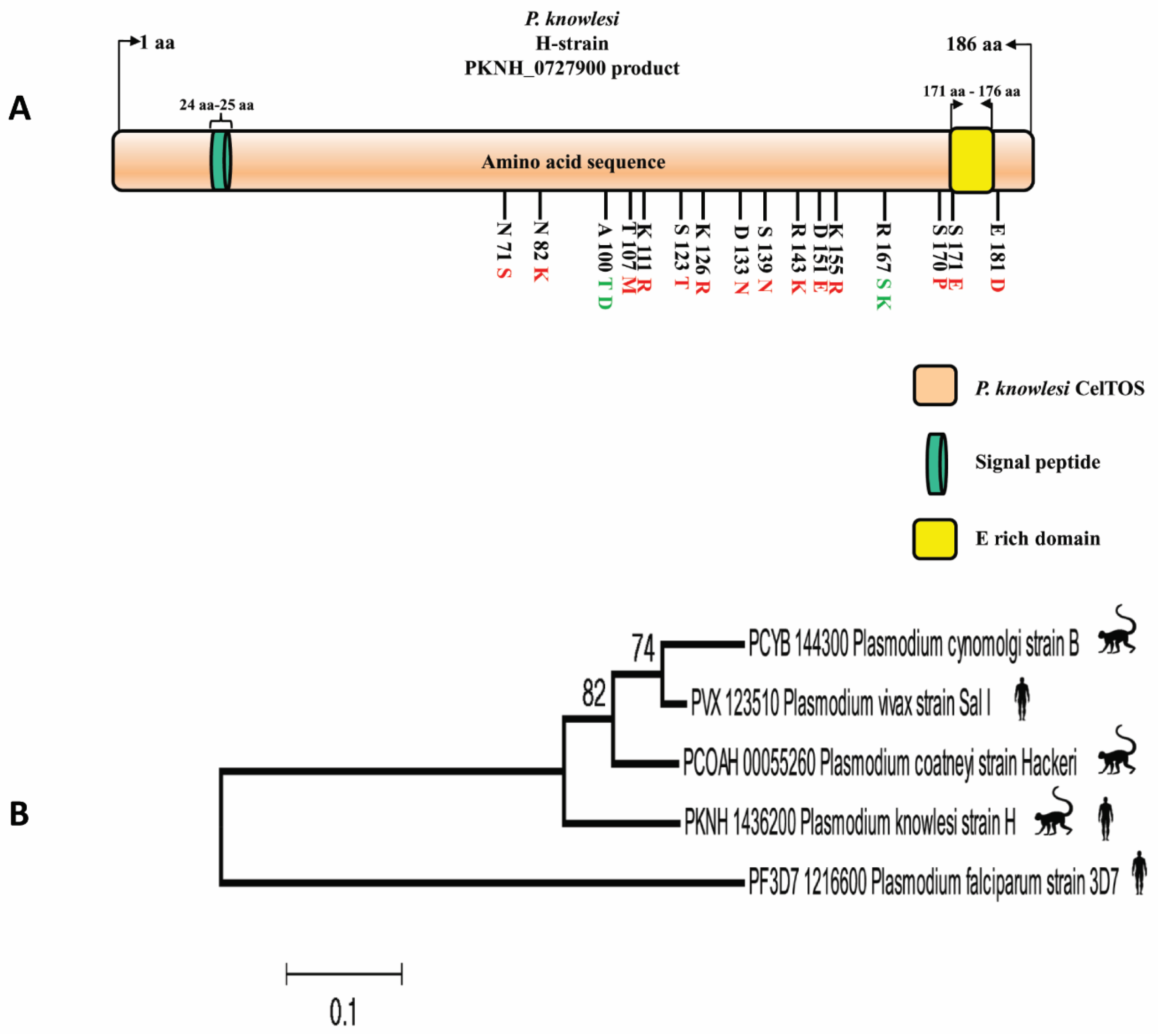
2.1. Sequence Information of PkCelTOS

A total of 34 PkCelTOS full-length gene (from 1 to 558 nt) sequences (4 laboratory strains including the reference H-strain PKNH_1436200 and 30 clinical samples) were obtained from the public database European Bioinformatics Institute (https://www.ebi.ac.uk/ena/browser/home, accessed on 20 March 2023) [18]. These sequences were derived from clinical samples obtained from Malaysian Borneo, as indicated in Supplementary Table S1, which shows the geographical locations of the sample collections [18]. Only high-quality, full-length PkCelTOS gene sequences were included for analysis. The sequences were aligned using the CLUSTAL-W program within the MegAlignLasergene v 7.0 software (DNASTAR, Madison, WI, USA). SignalP-5.0 prediction software (https://services.healthtech.dtu.dk/services/SignalP-5.0/) was used to predict the signal peptide for the full-length PkCelTOS protein [42].

2.2. Phylogenetic Analysis

Phylogenetic analysis was carried out to establish the intra-species relationship between amino acid sequences of PkCelTOS derived from clinical samples in Malaysian Borneo and long-term lab strains from Peninsular Malaysia and Philippines (H-strain, MR4, Philippine strain, and Malayan Strain). Phylogenetic analysis was conducted in MEGA 5.0 software utilizing Maximum Likelihood (ML) method on the basis of the Poisson correction model with 1000 bootstrap replicates [43]. Ortholog sequences of CelTOS in P. coatneyi (PCOAH_00055260), P. cynomolgi (PCYB_144300), P. vivax (PVX_123510), and P. falciparum (PF3D7_1216600) were also added to investigate the evolutionary relationship between the CelTOS protein of P. konwlesi along with its closest members in the genus Plasmodium.

2.3. Sequence Diversity and Polymorphism

Nucleotide diversity (π) in the PkCelTOS full-length gene was determined using DnaSP v6.12.03 software [44]. Other parameters, i.e., synonymous (S), non-synonymous substitutions (NS), polymorphic sites, the number of parsimony informative sites, singletons, the number of haplotypes (H), and haplotype diversity (HD) were also determined by the use of the same software. DnaSP software was utilized to visually represent nucleotide diversity by considering a window length of 50 and a step size of 10 bp.

2.4. Natural Selection Test

To determine the natural selection within PkCelTOS at the intra-species level, the neutrality tests of Tajima’s D, Fu, and Li’s F*and D* were computed using DnaSP software. Tajima’s D statistic yields a value of zero under conditions of neutrality. A negative Tajima’s D result determines population expansion, while a positive and significant Tajima’s D result signifies positive selection or balancing selection. Using the DnaSp software, the results of Tajima’s D were also shown graphically (with a window length of 50 and step size of 10 bp). Positive and significant values of Fu and Li’s F* and D* suggest that the population may have undergone a contraction. Conversely, an excess of singletons or negative values can indicate population expansion. In addition, the method of Nei and Gojobori was utilized for calculating the rate of non-synonymous substitution per non-synonymous site (dN) and the rate of synonymous substitution per synonymous site (dS) [43].
The robust McDonald and Kreitman (MK) test was carried out to examine natural selection at the inter-species level, utilizing DnaSP v6.12.03 software using 34 PkCelTOS gene sequences with their closely related ortholog sequences in P. coatneyi (PCOAH_00055260), P. cynomolgi (PCYB_144300) and P. vivax (PVX_123510).

2.5. Codon-Based Test Using Datamonkey Web Server for Natural Selection

Codon-based analysis of selection was performed on the PkCelTOS gene using the Datamonkey Web Server (https://www.datamonkey.org/ accessed on 1 July 2023) [45]. The analysis employed multiple methods, including single-likelihood ancestor counting (SLAC), mixed effects model of evolution (MEME), and fast unconstrained Bayesian approximation (FUBAR). Default settings for the significance level provided by the Datamonkey server were utilized for this analysis [45].

2.6. Epitope Prediction

B-cell epitopes are specific sites on a protein that can be recognized by the antigen-binding sites present on the immunoglobulin molecules. These epitopes play a crucial role in the formulation of peptide-based vaccines, disease detection, and allergy studies [46]. In this study, in silico prediction of B cell epitopes was carried out in the PkCelTOS sequence of P. knowlesi. The epitope prediction was conducted using two servers; the IEDB Analysis resource (http://tools.immuneepitope.org/bcell), with the Emini Surface Accessibility Prediction model to identify B-cell epitopes on the surface [47], and the Bcepred server (https://webs.iiitd.edu.in/raghava/bcepred/index.html) [46], using the Exposed surface method [48]. The method compares the possible conformations taken by protein side chains to energy calculation results using van der Waals interactions and steric hindrance. It assumes that the folded protein structure puts minimum strain on side-chain conformations, favoring low-energy positions during folding [48]. Since P. knowlesi and P. vivax are phylogenetically close and several studies have shown the importance of conserved cross-species vaccine candidates [11,49,50]. Therefore, in addition to identifying epitopes in P. knowlesi, this study examined the likelihood of epitope conservation with P. vivax.

3. Results

3.1. PkCelTOS Sequence Identity among Ortholog Members

A signal peptide spanning from amino acid position 24 to 25 was predicted by the Signal IP server within the PkCelTOS protein (Supplementary Figure S1). Alignment of full-length amino acid sequences of CelTOS of P. knowlesi strain H with its orthologs showed that PkCelTOS has 84.9%, 83.8%, 78.3%, and 44.5% amino acid sequence identities with P. viax, P. coatneyi, P. cynomolgi, and P. falciparum, respectively (Supplementary Figure S2). Figure 1A depicts a schematic structure of the PkCelTOS protein, highlighting the observed amino acid polymorphism, which includes both two and three variants.

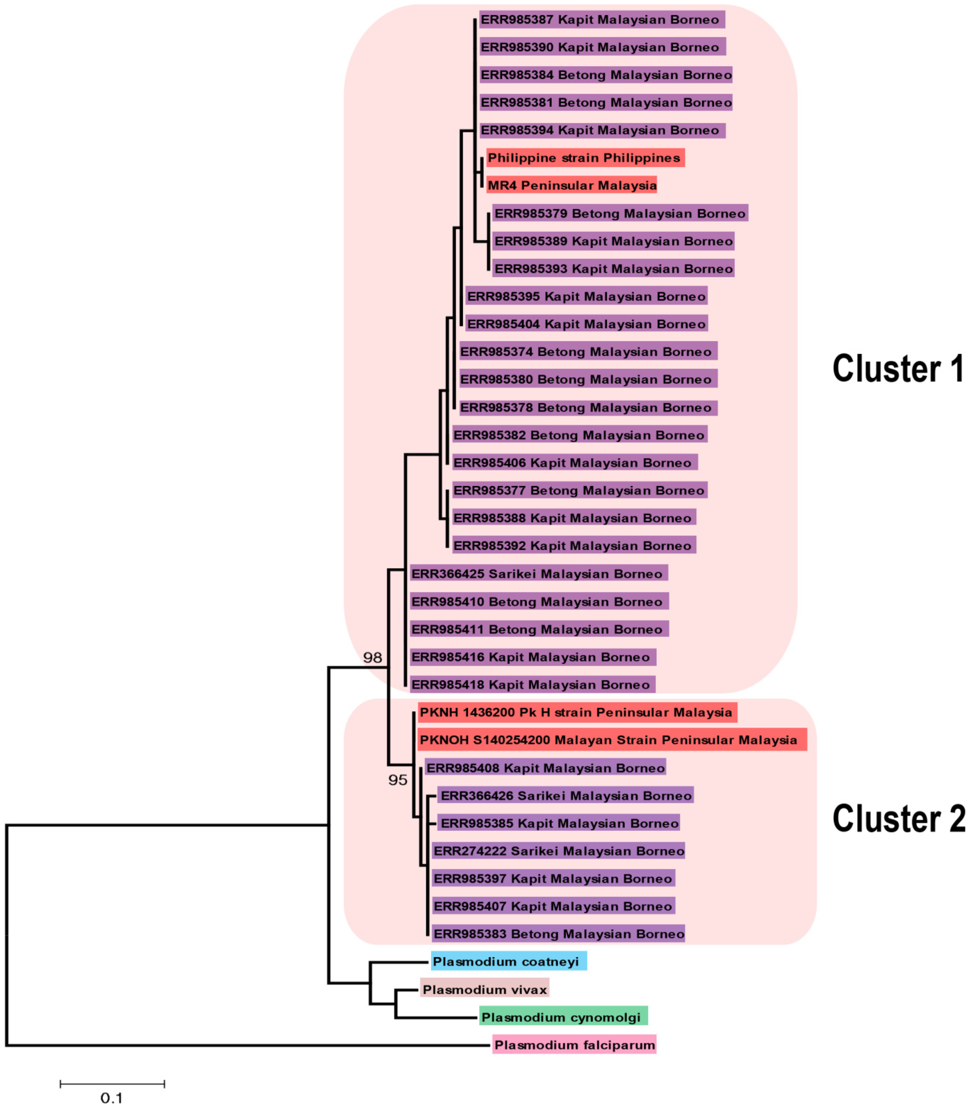
3.2. Phylogenetic Analysis of P. knowlesi CelTOS and Its Orthologs in Other Plasmodium Species

The inter-species phylogenetic relationship between PkCelTOS and its orthologs shows that PkCelTOS is phylogenetically closer to P. vivax than P. coatneyi and P. cynomolgi. While P. falciparum CelTOS is most distantly to PkCelTOS (Figure 1B).
A phylogenetic analysis of 34 full-length deduced amino acid sequences of PkCelTOS with its orthologs utilizing the Maximum Likelihood method showed no geographical clustering; however, samples were bifurcated into two major groups supported with strong bootstrap values (Figure 2).

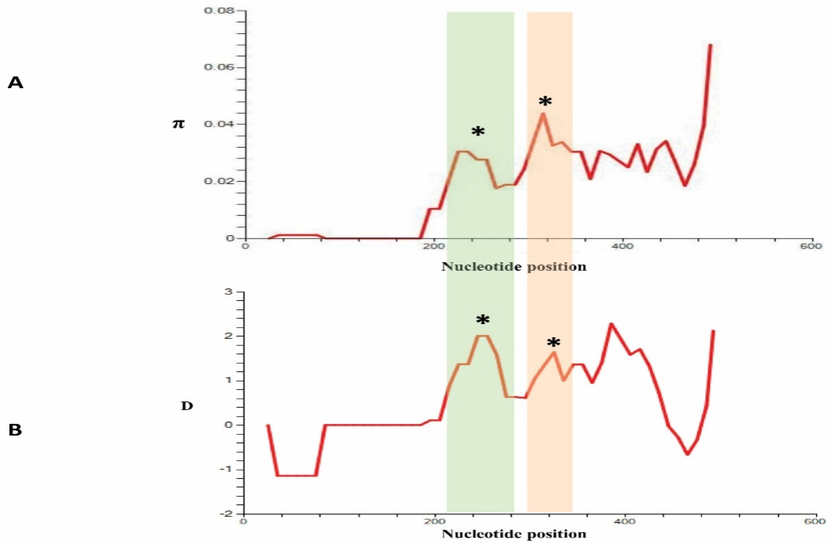
3.3. Nucleotide Diversity and Polymorphism of PkCelTOS in Clinical Samples

Analysis of 34 PkCelTOS nucleotide sequences showed 28 polymorphic sites, among which 12 were synonymous substitutions, and 16 were non-synonymous. The overall nucleotide diversity of PkCelTOS was determined to be π = 0.02111 + 0.00105 (Table 1), which is graphically represented in Figure 3A. Out of the 24 parsimony informative sites observed in PkCelTOS, 5 were singleton variable sites, 22 were with two variants, and 2 were with three variants. The analysis also revealed the presence of 17 haplotypes, which exhibited a high haplotype diversity (Hd) of 0.954 ± 0.016. Figure 1A illustrates a schematic representation of the 16 non-synonymous substitutions that were observed within the 34 samples, with respect to the P. knowlesi reference H-strain. The nucleotide and amino acid polymorphisms of the clinical strains with respect to the reference H-strain, along with nucleotide positions, were illustrated in Supplementary Figures S3 and S4.

3.4. Natural Selection in PkCelTOS

Analysis of the natural selection of PkCelTOS gene sequences obtained from 34 sequences showed overall Tajima’s D value to be D = 1.52705, p > 0.10, which was not significant (Table 1). Tajima’s D values indicated again that the PkCelTOS gene had undergone positive selection. The Tajima’s D result is shown graphically in Figure 3B. The values of Fu and Li’s F* and D* for PkCelTOS were found to be positive at 0.63460 and 1.10034, respectively, but they did not attain statistical significance (Table 1). In addition, the results from the codon-based Z-test for positive selection using the Nei and Gojobori method showed that the value was found to be negative (dN-dS = −1.058, p > 0.05) but not significant.
The robust MK test, which takes into consideration inter-species natural selection over time, suggested that the PkCelTOS gene may be undergone positive selection when tested with CelTOS orthologs in P. vivax, P. cynomolgi and P. coatneyi (Table 2). Though MK test results were not statistically significant, the NI values were indicative of a positive natural selection. This may be due to the low number of sequences analyzed in this study.

3.5. Codon-Wise Analysis

The departure of neutrality of the PkCelTOS gene was assessed using codon-based tests such as FUBAR, MEME, and SLAC, which detects sites with different selection pressure. The analysis conducted using FUBAR identified five sites (82, 100, 111, 170, and 171), while MEME detected one (171) site undergoing positive selection. Analysis conducted using the SLAC method 1 (111) site detected to be under positive selection. Remarkably, both FUBAR and MEME concurred on amino acid position 171, undergoing positive selection. Likewise, both FUBAR and SLAC concurred on a common site 111 to be under positive selection. In contrast, FUBAR identified four negatively selected sites (73, 80, 87, and 145), while SLAC identified two sites (73 and 87) undergoing strong negative selection. Notably, both methods agreed on amino acid positions 73 and 87 experiencing negative selection. A plot depicting positions for positive and negative selection of the PkCelTOS protein sequence generated using SLAC is shown in Supplementary Figure S5.

3.6. B Cell Epitopes in PkCelTOS

The prediction of potential B-cell epitopes using the IEDB server in P. knowlesi CelTOS identified two surface-exposed epitopes (96AQLKATA102 and 124TIKPPRIKED133) towards the C’ terminal region of the protein (Table 3, Supplementary Figures S6 and S7). The Bcpred server predicted a single potential epitope (125IKPPRIKED133). Both the servers predicted a common region of nine amino acids (IKPPRIKED), which can serve as a potential region for vaccine development research, while upon analysis of epitopes with its closest ortholog, P. vivax CelTOS. Three epitope regions were identified by the IEDB server and four by the Bcpred Server (Supplementary Table S2). A comparative analysis of the epitopes between the two species identified a conserved region of four amino acids (KPPR) in length.

4. Discussion

The life-cycle of Plasmodium spp. involves alternating between a definitive invertebrate host and intermediate human hosts. When a mosquito takes a blood meal, the parasite is released in sporozoite form into the host skin. It then initiates gliding motility and cell traversal for penetrating different cell types, e.g., endothelial cells of blood vessels, dermal fibroblasts, phagocytes in sinusoidal and dermal layers (Kupffer cells), and hepatocytes [6,31,51,52]. This enables the parasites to surpass cellular barriers and access the hepatocytes, where they occupy and mature into liver-stage parasites. [6,31,51,52]. Cell traversal plays a crucial role in protecting sporozoites from destruction by phagocytes and preventing their arrest by nonphagocytic cells in the host dermis [53]. Thus, a vaccine against these sporozoite proteins can inhibit parasite transmission in the pre-erythrocytic stage. To ensure effectiveness across different geographical regions and to prevent an allele-specific immune response, it is preferable for a candidate antigen to exhibit minimal polymorphism [31]. CelTOS is one such sporozoite protein, well studied as a potential candidate for vaccine development against P. vivax and P. falciparum, but it has not yet been explored in Plasmodium knowlesi. The present study is a pioneer in investigating the genetic diversity and polymorphisms within the PkCelTOS gene in clinical samples from Malaysia, as well as predicting the B-cell epitopes that can be further studied to develop sporozoite vaccines.
By aligning 34 full-length amino acid sequences of PkCelTOS, it was observed that it exhibits the highest sequence identity with PvCelTOS, which may indicate the ongoing adaptation for invading the physical barriers of the human host’s immune system. The signal peptide region was conserved among all clinical samples of P.knowlesi, indicating that it may play a critical role in directing the newly synthesized protein to its intended destination, i.e., sporozoites and ookinetes. The results of a phylogenetic analysis by ML method did not show any geographical clustering as both the clusters had laboratory lines originating from Peninsular Malaysia and the Philippines, together with clinical samples from Sarawak, Malaysian Borneo. Similar results were also found by Assefa et al., 2015 using the same set of sequences [18], except that the laboratory strains formed a separate cluster from the clinical samples. However, many previous studies on vaccine candidates, e.g., Pk41 [29], PkRhopH2 [11], MSP4 [25], MSP1P [54], nbpxa [55] along with P. knowlesi genome sequences from clinical samples [56] has shown geographical clustering. The reason could be that PkCelTOS is expressed in sporozoite and ookinete surfaces and may not be exposed to human immune pressure. PkCelTOS was found to be phylogenetically most related to PvCelTOS, its ortholog infecting humans. Among the simian malaria parasites, P. coatneyi CelTOS is closer to PkCelTOS. The overall nucleotide diversity of PkCelTOS (π = 0.02111 ± 0.00105) was higher than the nucleotide diversity of PfCelTOS (π = 0.01001 ± 0.00036) worldwide samples [32] and 20 times higher than PvCelTOS (π = 0.00141 ± 0.00014) [33]. The nucleotide diversity of PkCelTOS was also found to be higher than other sporozoite stage vaccine candidates, e.g., PkTRAP (π = 0.00908 ± 0.0006) [17] and merozoite stage vaccine candidates like PkTRAMP (π = 0.00652 ± 0.00028) [10], PkRhopH2 (π = 0.00936 ± 0.0013) [11], Pk41 (0.00959 ± 0.0001) [29] but lower than PkMSP7D (π = 0.052+ 0.002) [12]. The low number of non-synonymous mutations than synonymous mutations may again attribute to the parasite adaptation in the new host.
Natural selection tests (using Tajima’s D, Fu and Li’s F* and D*) yielded positive results, implying that the PkCelTOS gene may be under positive selection; however, the results were statistically not significant, which may be due to a smaller sample size. However, dN-dS results are found to be negative but not significant. A higher number of non-synonymous mutations than synonymous mutations was found in the McDonald and Kreitman (MK) neutrality test between PkCelTOS orthologs. Codon-based site-by-site selection analyses in Datamonkey identified five potential sites which could be under positive natural selection and four sites under purifying selection. This is probably due to protein folding, and the sites under positive selection might be exposed to host immune pressure, while the negatively selected sites are under functional constraints and not exposed to the host. These findings may indicate that the gene in question is undergoing long-term differential selective pressure from the human immune system, similar to the conclusions of PfCelTOS [32]. Both IEDB and Bcpred servers predicted a four amino acid epitope region, which may be a potential candidate for cross-species vaccine development against P. knowlesi and P. vivax. Nevertheless, further sequence analysis involving a more extensive sample size would be necessary to substantiate the presence of the epitope.

5. Conclusions

To conclude, this present study provides a valuable understanding of the genetic diversity and polymorphism across the full-length gene of CelTOS gene in Plasmodium knowlesi, a malaria parasite prevalent in Southeast Asia. The high degree of nucleotide diversity observed in the PkCelTOS gene suggests this protein may be under selective host immune pressure. However, the identification of two potential epitopes on the PkCelTOS protein provides a promising avenue for the development of a vaccine that can stimulate an effective immune response. The phylogenetic analysis of PkCelTOS revealed two distinct groups, indicating that there may be genetic differences between Plasmodium populations in different regions of Malaysia. The absence of geographic clustering suggests that the genetic variation observed in the PkCelTOS gene is likely the result of selective local pressures rather than geographic isolation. In conclusion, this study highlights the need for continued surveillance of PkCelTOS genetic diversity in different regions. The findings of this study offer valuable knowledge for the rational design of peptide-based vaccines against P. knowlesi malaria. However, further functional studies, as well as genetic studies with a higher number of samples, are needed to validate the effectiveness of a PkCelTOS-based vaccine.

Supplementary Materials

The following supporting information can be downloaded at: https://www.mdpi.com/article/10.3390/tropicalmed8080380/s1, Figure S1: Signal Peptide, Figure S2: Sequence identity between CelTOS othologs, Figures S3–S4: Nucleotide and Amino acid Polymorphisms, Figure S5: SLAC site graph, Figures S6–S7: Possible Epitopes; Table S1: Study samples with geographical origin.

Author Contributions

Conceptualization, M.A.A. and F.-S.Q.; methodology, M.A.A., P.B. and A.S.; validation, M.A.A., P.B., A.S., J.-H.H., M.A.-Z., S.W.W. and S.J.P.; formal analysis, M.A.A., P.B., A.S., J.-H.H., M.A.-Z., S.W.W., S.A., S.J.P. and M.S.A.-E.; investigation, M.A.A., P.B., A.S., J.-H.H., M.A.-Z., S.W.W., S.A., S.J.P. and M.S.A.-E.; resources, M.A.A. and F.-S.Q.; data curation, M.A.A., P.B. and A.S., J.-H.H., M.A.-Z., S.W.W., S.A., S.J.P. and M.S.A.-E.; writing—original draft preparation, M.A.A., P.B. and M.S.A.-E.; writing—review and editing, M.A.A., P.B., S.W.W., S.A., S.J.P. and M.S.A.-E.; visualization, M.A.A., P.B., A.S., J.-H.H., M.A.-Z., S.W.W., S.A., S.J.P. and M.S.A.-E.; supervision, F.-S.Q.; project administration, M.A.A. and F.-S.Q.; funding acquisition, M.S.A.-E. and F.-S.Q. All authors have read and agreed to the published version of the manuscript.

Funding

The authors extend their appreciation to the Deanship of Scientific Research at Imam Mohammad Ibn Saud Islamic University (IMSIU), Saudi Arabia, through Research Partnership Program no.RP-21-09-90 and Core Research Institute (CRI) Program, the Basic Science Research Program through the National Research Foundation of Korea (NRF), Ministry of Education (NRF-2018-R1A6A1A03025124) for funding and supporting this work.

Institutional Review Board Statement

Not applicable.

Informed Consent Statement

Not applicable.

Data Availability Statement

PkCelTOS gene sequences were downloaded from the public database European Bioinformatics Institute (EBI) (https://www.ebi.ac.uk/ena/browser/home accessed on 2 January 2023).

Conflicts of Interest

The authors declare no conflict of interest, and the funders had no role in the design of this study; in the collection, analysis, or interpretation of data; in the writing of the manuscript, or in the decision to publish the results.

References

  1. World Health Organization. Malaria. Available online: https://www.who.int/news-room/fact-sheets/detail/malaria (accessed on 30 March 2023).
  2. World Health Organization. World Malaria Report; World Health Organisation: Geneva, Switzerland, 2022. [Google Scholar]
  3. Oguike, M.C.; Betson, M.; Burke, M.; Nolder, D.; Stothard, J.R.; Kleinschmidt, I.; Proietti, C.; Bousema, T.; Ndounga, M.; Tanabe, K.; et al. Plasmodium ovale curtisi and Plasmodium ovale wallikeri circulate simultaneously in African communities. Int. J. Parasitol. 2011, 41, 677–683. [Google Scholar] [CrossRef] [Green Version]
  4. Ahmed, M.A.; Cox-Singh, J. Plasmodium knowlesi—An emerging pathogen. ISBT Sci. Ser. 2015, 10, 134–140. [Google Scholar] [CrossRef] [PubMed] [Green Version]
  5. Sutherland, C.J.; Tanomsing, N.; Nolder, D.; Oguike, M.; Jennison, C.; Pukrittayakamee, S.; Dolecek, C.; Hien, T.T.; do Rosario, V.E.; Arez, A.P.; et al. Two nonrecombining sympatric forms of the human malaria parasite Plasmodium ovale occur globally. J. Infect. Dis. 2010, 201, 1544–1550. [Google Scholar] [CrossRef] [Green Version]
  6. Tazi, L.; Ayala, F.J. Unresolved direction of host transfer of Plasmodium vivax v. P. simium and P. malariae v. P. brasilianum. Infect. Genet. Evol. 2011, 11, 209–221. [Google Scholar]
  7. Ta, T.H.; Hisam, S.; Lanza, M.; Jiram, A.I.; Ismail, N.; Rubio, J.M. First case of a naturally acquired human infection with Plasmodium cynomolgi. Malar. J. 2014, 13, 68. [Google Scholar] [CrossRef] [Green Version]
  8. Bogitsh, B.; Carter, C.; Oeltmann, T. Chapter 7—Blood and Tissue Protistans II: Human Malaria. In Human Parasitology; Elsevier: Amsterdam, The Netherlands, 2019; Volume 5, pp. 111–133. [Google Scholar]
  9. White, N.J. Plasmodium knowlesi: The fifth human malaria parasite. Clin. Infect. Dis. Off. Publ. Infect. Dis. Soc. Am. 2008, 46, 172–173. [Google Scholar] [CrossRef] [PubMed]
  10. Ahmed, M.A.; Zaidi, R.H.; Deshmukh, G.Y.; Saif, A.; Alshahrani, M.A.; Salam, S.S.; Elfaki, M.M.A.; Han, J.H.; Patgiri, S.J.; Quan, F.S. Genetic Diversity and Population Genetic Structure Analysis of Plasmodium knowlesi Thrombospondin-Related Apical Merozoite Protein (TRAMP) in Clinical Samples. Genes 2022, 13, 1944. [Google Scholar] [CrossRef] [PubMed]
  11. Ahmed, M.A.; Deshmukh, G.Y.; Zaidi, R.H.; Saif, A.; Alshahrani, M.A.; Wazid, S.W.; Patgiri, S.J.; Quan, F.S. Identification, Mapping, and Genetic Diversity of Novel Conserved Cross-Species Epitopes of RhopH2 in Plasmodium knowlesi with Plasmodium vivax. Front. Cell. Infect. Microbiol. 2021, 11, 810398. [Google Scholar] [CrossRef]
  12. Ahmed, M.A.; Quan, F.S. Plasmodium knowlesi clinical isolates from Malaysia show extensive diversity and strong differential selection pressure at the merozoite surface protein 7D (MSP7D). Malar. J. 2019, 18, 150. [Google Scholar] [CrossRef] [Green Version]
  13. Singh, B.; Kim Sung, L.; Matusop, A.; Radhakrishnan, A.; Shamsul, S.S.; Cox-Singh, J.; Thomas, A.; Conway, D.J. A large focus of naturally acquired Plasmodium knowlesi infections in human beings. Lancet 2004, 363, 1017–1024. [Google Scholar] [CrossRef]
  14. Cox-Singh, J.; Davis, T.M.; Lee, K.S.; Shamsul, S.S.; Matusop, A.; Ratnam, S.; Rahman, H.A.; Conway, D.J.; Singh, B. Plasmodium knowlesi malaria in humans is widely distributed and potentially life threatening. Clin. Infect. Dis. Off. Publ. Infect. Dis. Soc. Am. 2008, 46, 165–171. [Google Scholar] [CrossRef] [Green Version]
  15. Jeyaprakasam, N.K.; Liew, J.W.K.; Low, V.L.; Wan-Sulaiman, W.-Y.; Vythilingam, I. Plasmodium knowlesi infecting humans in Southeast Asia: What’s next? PLoS Neglected Trop. Dis. 2020, 14, e0008900. [Google Scholar] [CrossRef] [PubMed]
  16. Wharton, R.H.; Eyles, D.E. Anopheles hackeri, a vector of Plasmodium knowlesi in Malaya. Science 1961, 134, 279–280. [Google Scholar] [CrossRef] [PubMed]
  17. Ahmed, M.A.; Lau, Y.L.; Quan, F.S. Diversity and natural selection on the thrombospondin-related adhesive protein (TRAP) gene of Plasmodium knowlesi in Malaysia. Malar. J. 2018, 17, 274. [Google Scholar] [CrossRef] [Green Version]
  18. Assefa, S.; Lim, C.; Preston, M.D.; Duffy, C.W.; Nair, M.B.; Adroub, S.A.; Kadir, K.A.; Goldberg, J.M.; Neafsey, D.E.; Divis, P.; et al. Population genomic structure and adaptation in the zoonotic malaria parasite Plasmodium knowlesi. Proc. Natl. Acad. Sci. USA 2015, 112, 13027–13032. [Google Scholar] [CrossRef] [PubMed]
  19. Eyles, D.; Laing, A.; Warren, M.; Sandosham, A.; Wharton, R. Malaria parasites of the Malayan leaf monkeys of the genus Presbytis. Med J Malaya 1962, 17, 85–86. [Google Scholar]
  20. William, T.; Menon, J.; Rajahram, G.; Chan, L.; Ma, G.; Donaldson, S.; Khoo, S.; Frederick, C.; Jelip, J.; Anstey, N.M.; et al. Severe Plasmodium knowlesi malaria in a tertiary care hospital, Sabah, Malaysia. Emerg. Infect. Dis. 2011, 17, 1248–1255. [Google Scholar] [CrossRef] [PubMed]
  21. Knowles, R.; Gupta, B.M.D. A Study of Monkey-Malaria, and Its Experimental Transmission to Man. Indian Med. Gaz. 1932, 67, 301–320. [Google Scholar]
  22. Singh, B.; Daneshvar, C. Human infections and detection of Plasmodium knowlesi. Clin. Microbiol. Rev. 2013, 26, 165–184. [Google Scholar] [CrossRef] [Green Version]
  23. Barber, B.E.; William, T.; Grigg, M.J.; Yeo, T.W.; Anstey, N.M. Limitations of microscopy to differentiate Plasmodium species in a region co-endemic for Plasmodium falciparum, Plasmodium vivax and Plasmodium knowlesi. Malar. J. 2013, 12, 8. [Google Scholar] [CrossRef] [Green Version]
  24. Vythilingam, I.; Wong, M.; Wan-Yussof, W. Current status of Plasmodium knowlesi vectors: A public health concern? Parasitology 2018, 145, 32–40. [Google Scholar] [CrossRef] [Green Version]
  25. Ahmed, M.A.; Saif, A.; Quan, F.S. Diversity pattern of Plasmodium knowlesi merozoite surface protein 4 (MSP4) in natural population of Malaysia. PLoS ONE 2019, 14, e0224743. [Google Scholar] [CrossRef] [PubMed]
  26. Garzón-Ospina, D.; Buitrago, S.P.; Ramos, A.E.; Patarroyo, M.A. Identifying potential Plasmodium vivax sporozoite stage vaccine candidates: An analysis of genetic diversity and natural selection. Front. Genet. 2018, 9, 10. [Google Scholar] [CrossRef] [PubMed] [Green Version]
  27. Sinnis, P.; Fidock, D.A. The RTS,S vaccine-a chance to regain the upper hand against malaria? Cell 2022, 185, 750–754. [Google Scholar] [CrossRef] [PubMed]
  28. Neafsey, D.E.; Juraska, M.; Bedford, T.; Benkeser, D.; Valim, C.; Griggs, A.; Lievens, M.; Abdulla, S.; Adjei, S.; Agbenyega, T. Genetic diversity and protective efficacy of the RTS, S/AS01 malaria vaccine. N. Engl. J. Med. 2015, 373, 2025–2037. [Google Scholar] [CrossRef] [PubMed]
  29. Ahmed, M.A.; Chu, K.B.; Quan, F.S. The Plasmodium knowlesi Pk41 surface protein diversity, natural selection, sub population and geographical clustering: A 6-cysteine protein family member. PeerJ 2018, 6, e6141. [Google Scholar] [CrossRef] [Green Version]
  30. Fong, M.Y.; Ahmed, M.A.; Wong, S.S.; Lau, Y.L.; Sitam, F. Genetic diversity and natural selection of the Plasmodium knowlesi circumsporozoite protein nonrepeat regions. PLoS ONE 2015, 10, e0137734. [Google Scholar] [CrossRef] [Green Version]
  31. Arevalo-Pinzon, G.; Garzon-Ospina, D.; Pulido, F.A.; Bermudez, M.; Forero-Rodriguez, J.; Rodriguez-Mesa, X.M.; Reyes-Guarin, L.P.; Suarez, C.F.; Patarroyo, M.A. Plasmodium vivax Cell Traversal Protein for Ookinetes and Sporozoites (CelTOS) Functionally Restricted Regions Are Involved in Specific Host-Pathogen Interactions. Front. Cell. Infect. Microbiol. 2020, 10, 119. [Google Scholar] [CrossRef] [Green Version]
  32. Pirahmadi, S.; Zakeri, S.; Mehrizi, A.A.; Djadid, N.D. Analysis of genetic diversity and population structure of gene encoding cell-traversal protein for ookinetes and sporozoites (CelTOS) vaccine candidate antigen in global Plasmodium falciparum populations. Infect. Genet. Evol. J. Mol. Epidemiol. Evol. Genet. Infect. Dis. 2018, 59, 113–125. [Google Scholar] [CrossRef] [PubMed]
  33. Bitencourt Chaves, L.; Perce-da-Silva, D.S.; Rodrigues-da-Silva, R.N.; Martins da Silva, J.H.; Cassiano, G.C.; Machado, R.L.; Pratt-Riccio, L.R.; Banic, D.M.; Lima-Junior, J.D. Plasmodium vivax Cell Traversal Protein for Ookinetes and Sporozoites (PvCelTOS) gene sequence and potential epitopes are highly conserved among isolates from different regions of Brazilian Amazon. PLoS Negl. Trop. Dis. 2017, 11, e0005344. [Google Scholar] [CrossRef] [PubMed] [Green Version]
  34. Alves, E.; Salman, A.M.; Leoratti, F.; Lopez-Camacho, C.; Viveros-Sandoval, M.E.; Lall, A.; El-Turabi, A.; Bachmann, M.F.; Hill, A.V.; Janse, C.J.; et al. Evaluation of Plasmodium vivax Cell-Traversal Protein for Ookinetes and Sporozoites as a Preerythrocytic P. vivax Vaccine. Clin. Vaccine Immunol. CVI 2017, 24, e00501-16. [Google Scholar] [CrossRef] [Green Version]
  35. Kariu, T.; Ishino, T.; Yano, K.; Chinzei, Y.; Yuda, M. CelTOS, a novel malarial protein that mediates transmission to mosquito and vertebrate hosts. Mol. Microbiol. 2006, 59, 1369–1379. [Google Scholar] [CrossRef] [PubMed]
  36. Jimah, J.R.; Salinas, N.D.; Sala-Rabanal, M.; Jones, N.G.; Sibley, L.D.; Nichols, C.G.; Schlesinger, P.H.; Tolia, N.H. Malaria parasite CelTOS targets the inner leaflet of cell membranes for pore-dependent disruption. eLife 2016, 5, e20621. [Google Scholar] [CrossRef]
  37. Bergmann-Leitner, E.S.; Mease, R.M.; De La Vega, P.; Savranskaya, T.; Polhemus, M.; Ockenhouse, C.; Angov, E. Immunization with pre-erythrocytic antigen CelTOS from Plasmodium falciparum elicits cross-species protection against heterologous challenge with Plasmodium berghei. PLoS ONE 2010, 5, e12294. [Google Scholar] [CrossRef] [Green Version]
  38. Bergmann-Leitner, E.S.; Legler, P.M.; Savranskaya, T.; Ockenhouse, C.F.; Angov, E. Cellular and humoral immune effector mechanisms required for sterile protection against sporozoite challenge induced with the novel malaria vaccine candidate CelTOS. Vaccine 2011, 29, 5940–5949. [Google Scholar] [CrossRef] [PubMed]
  39. Anum, D.; Kusi, K.A.; Ganeshan, H.; Hollingdale, M.R.; Ofori, M.F.; Koram, K.A.; Gyan, B.A.; Adu-Amankwah, S.; Badji, E.; Huang, J. Measuring naturally acquired ex vivo IFN-γ responses to Plasmodium falciparum cell-traversal protein for ookinetes and sporozoites (CelTOS) in Ghanaian adults. Malar. J. 2015, 14, 20. [Google Scholar] [CrossRef] [PubMed] [Green Version]
  40. Espinosa, D.A.; Vega-Rodriguez, J.; Flores-Garcia, Y.; Noe, A.R.; Munoz, C.; Coleman, R.; Bruck, T.; Haney, K.; Stevens, A.; Retallack, D.; et al. The Plasmodium falciparum Cell-Traversal Protein for Ookinetes and Sporozoites as a Candidate for Preerythrocytic and Transmission-Blocking Vaccines. Infect. Immun. 2017, 85, 10–1128. [Google Scholar] [CrossRef] [Green Version]
  41. Trial of a Falciparum Malaria Protein (FMP012), E. Coli-expressed PfCelTOS, in Healthy Malaria-Naive Adults. ClinicalTrials.Gov. 2021. Available online: https://www.clinicaltrials.gov/study/NCT01540474?cond=Trial%20of%20a%20Falciparum%20Malaria%20Protein%20(FMP012),%20E.%20Coli-expressed%20PfCelTOS,%20in%20Healthy%20Malaria-Naive%20Adults&rank=1 (accessed on 20 March 2023).
  42. Petersen, T.N.; Brunak, S.; von Heijne, G.; Nielsen, H. SignalP 4.0: Discriminating signal peptides from transmembrane regions. Nat. Methods 2011, 8, 785–786. [Google Scholar] [CrossRef]
  43. Tamura, K.; Peterson, D.; Peterson, N.; Stecher, G.; Nei, M.; Kumar, S. MEGA5: Molecular evolutionary genetics analysis using maximum likelihood, evolutionary distance, and maximum parsimony methods. Mol. Biol. Evol. 2011, 28, 2731–2739. [Google Scholar] [CrossRef] [Green Version]
  44. Rozas, J.; Ferrer-Mata, A.; Sánchez-DelBarrio, J.C.; Guirao-Rico, S.; Librado, P.; Ramos-Onsins, S.E.; Sánchez-Gracia, A. DnaSP 6: DNA Sequence Polymorphism Analysis of Large Data Sets. Mol. Biol. Evol. 2017, 34, 3299–3302. [Google Scholar] [CrossRef] [PubMed]
  45. Pond, S.L.; Frost, S.D. Datamonkey: Rapid detection of selective pressure on individual sites of codon alignments. Bioinformatics 2005, 21, 2531–2533. [Google Scholar] [CrossRef] [Green Version]
  46. Saha, S.; Raghava, G.P. Prediction of continuous B-cell epitopes in an antigen using recurrent neural network. Proteins 2006, 65, 40–48. [Google Scholar] [CrossRef]
  47. Emini, E.A.; Hughes, J.V.; Perlow, D.S.; Boger, J. Induction of Hepatitis A Virus-Neutralizing Antibody by a Virus-Specific Synthetic Peptide. J. Virol. 1985, 55, 836–839. [Google Scholar] [CrossRef] [Green Version]
  48. Janin, J.; Wodak, S. Conformation of amino acid side-chains in proteins. J. Mol. Biol. 1978, 125, 357–386. [Google Scholar] [CrossRef]
  49. Muh, F.; Kim, N.; Nyunt, M.H.; Firdaus, E.R.; Han, J.-H.; Hoque, M.R.; Lee, S.-K.; Park, J.-H.; Moon, R.W.; Lau, Y.L. Cross-species reactivity of antibodies against Plasmodium vivax blood-stage antigens to Plasmodium knowlesi. PLoS Neglected Trop. Dis. 2020, 14, e0008323. [Google Scholar] [CrossRef]
  50. Longley, R.J.; Grigg, M.J.; Schoffer, K.; Obadia, T.; Hyslop, S.; Piera, K.A.; Nekkab, N.; Mazhari, R.; Takashima, E.; Tsuboi, T. Plasmodium vivax malaria serological exposure markers: Assessing the degree and implications of cross-reactivity with P. knowlesi. Cell Rep. Med. 2022, 3, 100662. [Google Scholar] [CrossRef]
  51. Mota, M.M.; Pradel, G.; Vanderberg, J.P.; Hafalla, J.C.; Frevert, U.; Nussenzweig, R.S.; Nussenzweig, V.; Rodriguez, A. Migration of Plasmodium sporozoites through cells before infection. Science 2001, 291, 141–144. [Google Scholar] [CrossRef]
  52. Vanderberg, J.P.; Chew, S.; Stewart, M.J. Plasmodium sporozoite interactions with macrophages in vitro: A videomicroscopic analysis. J. Protozool. 1990, 37, 528–536. [Google Scholar] [CrossRef]
  53. Amino, R.; Giovannini, D.; Thiberge, S.; Gueirard, P.; Boisson, B.; Dubremetz, J.F.; Prevost, M.C.; Ishino, T.; Yuda, M.; Menard, R. Host cell traversal is important for progression of the malaria parasite through the dermis to the liver. Cell Host Microbe 2008, 3, 88–96. [Google Scholar] [CrossRef] [Green Version]
  54. Ahmed, M.A.; Fauzi, M.; Han, E.T. Genetic diversity and natural selection of Plasmodium knowlesi merozoite surface protein 1 paralog gene in Malaysia. Malar. J. 2018, 17, 115. [Google Scholar] [CrossRef] [Green Version]
  55. Ahmed, M.A.; Fong, M.Y.; Lau, Y.L.; Yusof, R. Clustering and genetic differentiation of the normocyte binding protein (nbpxa) of Plasmodium knowlesi clinical isolates from Peninsular Malaysia and Malaysia Borneo. Malar. J. 2016, 15, 241. [Google Scholar] [CrossRef] [Green Version]
  56. Pinheiro, M.M.; Ahmed, M.A.; Millar, S.B.; Sanderson, T.; Otto, T.D.; Lu, W.C.; Krishna, S.; Rayner, J.C.; Cox-Singh, J. Plasmodium knowlesi genome sequences from clinical isolates reveal extensive genomic dimorphism. PLoS ONE 2015, 10, e0121303. [Google Scholar] [CrossRef] [Green Version]
Figure 1. (A) Distribution of the amino acid polymorphism found in 34 clinical samples of the PkCelTOS protein. The black lines with corresponding letters and numbers indicate the amino acid positions with polymorphism. The letter preceding the number represents the amino acid found in the reference H-strain. The polymorphic variants are shown in red (two variants) and green (three variants) colors. The signal peptide region lies between amino acid positions 24 to 25 (highlighted in dark green). An E-(Glutamic acid) rich region was identified spanning the amino acid positions 171 to 176 within the alignment. “aa” refers to amino acid positions. (B) Phylogenetic tree showing the inter-species relationship between sequences of amino acid of CelTOS of P. knowlesi reference strain H with its orthologs in P. vivax strain Sal I, P. coatneyi strain Hackeri, P. cynomolgi strain B and P. falciparum strain 3D7 constructed using MEGA 5.0 software using ML method and Poisson correction model.
Tropicalmed 08 00380 g001
Figure 2. Phylogenetic tree comprising 34 PkCelTOS amino acid sequences (including the reference H-strain) from different regions of Malaysia and its orthologs in P. vivax strain Sal I, P. coatneyi strain Hackeri, P. cynomolgi strain B and P. falciparum strain 3D7 gene constructed using by MEGA 5.0 software using Maximum-Likelihood Model and Poisson correction model with 1000 bootstrap replicates. Bootstrap values higher than 95 were only shown in the figure. Sequences originating from Peninsular Malaysia and the Philippines are shaded in red and Malaysian Borneo in violet.
Tropicalmed 08 00380 g002
Figure 3. (A) Graphical depiction of nucleotide diversity (π) and (B) Tajima’s D value (D) within 34 clinical samples of the P. knowlesi CelTOS gene. The window length and step size for both graphs were set at 50 and 10, respectively, as implemented in DnaSP software v6.12.03. Peaks marked with an asterisk (*) on the Tajima’s D graph may indicate possible epitope regions among the clinical samples analyzed. The shaded region highlights (green and red) areas of shared high nucleotide diversity and positive Tajima’s D values, which may have possible epitopes.
Tropicalmed 08 00380 g003
Table 1. Calculation of nucleotide diversity, haplotype diversity, and neutrality indices for the PkCelTOS gene.
GeneNo. SamplesSNPsSynNon-SynNo. HaplotypeDiversity ± SDTaj DFu and Li’s F*Fu and Li’s D*
HaplotypeNucleotide
PkCelTOS34281216170.954 ± 0.0160.02111 ± 0.001051.52705
p > 0.10
1.10034
p > 0.10
0.63460
p > 0.10
SNPs: Single nucleotide polymorphisms, Syn: Synonymous substitutions, Non-Syn: Non-synonymous substitutions, SD: Standard deviation.
Table 2. Results of MK tests performed using the full-length CelTOS sequence of P. knowlesi with its orthologs in P. caotneyi, P. vivax, and P. cynomolgi.
CelTOSPolymorphic Changes Observed in
P. knowlesi
Fixed Differences between SpeciesNeutrality Index
Pk vs. PcoPk vs. PcyPk vs. PvPk vs. PcoPk vs. PcyPk vs. Pv
SynNon-SynSynNonSynSynNon-SynSynNon-Syn
Full length12161522203221200.9090.9091.400
Syn: Synonymous sites, Non-Syn: Non-synonymous sites, Pk: Plasmodium knowlesi, Pco: Plasmodium coatneyi, Pcy: Plasmodium cynomolgi, Pv: Plasmodium vivax.
Table 3. Potential epitopes predicted in P. knowlesi CelTOS (ERR985408) by the IEDB and Bcpred server.
IEDB ServerBcpred Server
No.Start AAEnd AAPeptideLengthStart AAEnd AAPeptideLength
196102AQLKATA7125133IKPPRIKED9
2124133TIKPPRIKED10---NA
Bold font indicates the common amino acids within the predicted epitope as identified by both servers. The conserved epitope between the two species is highlighted in red amino acids. AA: amino acid.
Disclaimer/Publisher’s Note: The statements, opinions and data contained in all publications are solely those of the individual author(s) and contributor(s) and not of MDPI and/or the editor(s). MDPI and/or the editor(s) disclaim responsibility for any injury to people or property resulting from any ideas, methods, instructions or products referred to in the content.

