Article

Application of Multilocus Sequence Typing for the Characterization of Leptospira Strains in Malaysia

by
Fairuz Amran
1,
Nurul Atiqah Noor Halim
1,
Ayu Haslin Muhammad
1,
Mohd Khairul Nizam Mohd Khalid
2,
Nur Mukmina Dasiman
1,
Nadia Aqilla Shamsusah
1,3,
Abdul Khalif Adha Abd Talib
1,
Mohamed Asyraf Noh
1,4,
Mohammad Ridhuan Mohd Ali
1,* and
Rohaidah Hashim
1
1
Bacteriology Unit, Infectious Disease Research Center (IDRC), Institute for Medical Research (IMR), NIH Complex Setia Alam, Shah Alam 40170, Malaysia
2
Genetic Disorders and Inborn Error of Metabolism (IEM) Unit, Nutrition, Metabolic & Cardiovascular Research Centre (NMCRC), Institute for Medical Research (IMR), NIH Setia Alam, Shah Alam 40170, Malaysia
3
Department of Earth Sciences and Environment, Universiti Kebangsaan Malaysia, Bangi 43600, Malaysia
4
Department of Medical Microbiology, Faculty of Medicine and Health Sciences, Universiti Putra Malaysia (UPM), Serdang 43400, Malaysia
*
Author to whom correspondence should be addressed.
Trop. Med. Infect. Dis. 2023, 8(2), 69; https://doi.org/10.3390/tropicalmed8020069
Submission received: 26 September 2022 / Revised: 25 November 2022 / Accepted: 7 December 2022 / Published: 17 January 2023

Abstract

:
Leptospirosis is a common zoonotic disease in tropical and subtropical countries. It is considered an emerging disease in Malaysia and is a notifiable disease. This study was conducted to characterize Malaysian isolates from human, animal and environmental samples via MLST and rrs2 sequencing in an attempt to develop a Malaysian genotypic database. An existing polymerase chain reaction (PCR)-based MLST scheme was performed to facilitate subsequent sequencing. Out of 46 extracted DNA, 36 had complete MLST profiles whereby all six genes were amplified and sequenced. Most of the pathogenic Leptospira genotypes with full MLST profiles were L. interrogans serogroup Bataviae (n = 17), followed by L. borgpetersenii serogroup Javanica (n = 9), L. interrogans serogroup Sejroe (n = 2), L. interrogans serogroup Australis (n = 2), L. kirschneri (n = 2), L. interrogans serogroup Grippotyphosa (n = 1) and L. interrogans serogroup Pyrogenes (n = 3). Two samples (R3_SER/17 and R4_SER/17) were not closely related with any of the reference strains. For the samples with incomplete MLST profiles, leptospiral speciation was conducted through rrs2 analysis, in which four samples were identified as L. borgpetersenii, five samples were closely related to L. kmetyi and one sample was known as L. yasudae. This study shows that molecular approaches that combine both MLST and rrs2 sequencing have great potential in the comprehensive characterization of pathogenic Leptospira because they can be performed directly from cultured and clinical samples.

1. Introduction

Leptospirosis is a zoonotic infection caused by pathogenic Leptospira species and is identified as a global public health concern because of its increased mortality around the world, especially in tropical and humid subtropical regions. This disease can be transmitted to humans through indirect contact with environment contaminated with urine of infected animals, or direct contact with the latter [1]. Leptospira can infect a wide range of mammalian hosts, thus leading to a complex and dynamic epidemiology of the disease. Generally, the bacteria are sustained in the renal tubules of long-term carrier hosts, primarily rodents, and are eliminated through their urine. Leptospirosis resembles many other febrile illnesses in its early stages and may include severe diseases with jaundice, pulmonary hemorrhage or central nervous system involvement in its severe form [2,3,4,5].
Previously, the genus Leptospira has been classified into three main lineages (pathogenic, intermediate and saprophytic) based on the phylogenetic analyses of 16S rRNA genes. However, the lineages have been reclassified as a result of the isolation of more than 30 novel strains recently. This diverse genus is split into four groups: P1 (primary pathogenic strains such as L. interrogans, L. borgpetersenii, L. weilii, L. kirschneri, L. santarosai, L. mayottensis, L. noguchii, L. alexanderi, L. kmetyi, L. astonii, L. yasudae, L. stimsonii, L. astonii, L. barantonii, L. tipperaryensis, L. adleri, L. ellisii and L. gomenensis), P2 (pathogenic but low-virulence strains such as L. fainei, L. wolffii, L. broomii, L. licerasiae, L. inadai, L. hartskeerlii, L. dzoumogneensis, L. venezuelensis, L. selangorensis, L. andrefontaineae, L. haakeii, L. johnsonii, L. sarikeiensis and L. fletcheri), S1 (primary saprophytes such as L. biflexa, L. vanthielli, L. meyeri, L. wolbachii, L. idonii, L. terpstrae, and L. yanagawae) and S2 (new saprophytes such as L. jelokensis, L. kemamanensis, L. perdikensis, L. congkakensis, L. kobayashii and L. kanakyensis) [6,7].
The serovar is a basic identifier characterized based on serological criteria. To date, more than 300 serovars have been described under the Leptospira genus [8,9]. Antigenically similar serovars are clustered into >30 different serogroups [10]. Each serovar can infect and adapt to one or more animal hosts [8], making it challenging to track strains whose dynamically infect host and contaminate the environmental niches. Therefore, a complementary leptospiral typing is essential for identifying potential clusters, transmission pathways and host reservoir.
However, serological data are frequently presented without genetic data, or vice versa, which reduces the effectiveness of public health surveillance and investigations [11]. Molecular techniques are more advantageous compared to serological testing in clinical diagnostics due to their speed and higher sensitivity in the early stages of infection [12]. The two commonly applied molecular techniques are pulsed-field gel electrophoresis (PFGE) and multilocus sequence typing (MLST). MLST is robust and reproducible and can even be applied directly in clinical samples to type the infecting Leptospira [13,14,15]. MLST is widely used to characterize the subspeciation of Leptospira [16]. This method is a simple polymerase chain reaction (PCR)-based technique that identifies strains from a sequence of six or seven housekeeping with extremely slow evolutionary change [16,17]. For each strain of a specific species, the genes are amplified and sequenced. Each sequenced gene is then compared to every previously uploaded sequence (alleles) at that locus. Then, each of the six or seven loci is given an allele number. The allelic profile is interpreted using a combination of the six or seven allele numbers (assigned as sequence type, ST), which is used to describe the strain [18,19]. In this study, we aimed to characterize Malaysian isolates from human, animal and environmental samples via MLST and rrs2 sequencing in an attempt to develop a Malaysian genotypic database.

2. Materials and Methods

2.1. Sample Collection

Blood samples from patients suspected of leptospirosis infection were collected from local hospitals (n = 13), animal blood and environmental samples were obtained from rodents caught in wet markets (n = 30), wild dogs (n = 2) and soil (n = 1), respectively.

2.2. Isolation of Leptospira sp. and Dark-Field Microscopy Observation

To culture leptospires from rodent samples, kidneys from captured rodents were macerated in 2 mL of semi-solid Ellinghausen and McCullough modified by Johnson and Harris (EMJH) media and were allowed to settle for 20 min. The mixtures were then filtered into 5 mL of new media using 0.45 μm and 0.22 µm sterile syringe filters. For blood and urine samples collected from patients and animals, 2–3 drops of each sample were directly inoculated into 5 mL of EMJH media and incubated at 30 °C. Cultures were examined under a dark-field microscope at intervals of seven days. Leptospira were identified by their morphology and motility. Leptospira are very thin, long, and have hooked ends. Leptospires rotate on their longitudinal axis rapidly, moving forward and backward. The cultures were considered Leptospira-negative if there was no growth of the bacteria after three months and could thus be discarded.

2.3. DNA Extraction

For the MLST performed directly from clinical samples, DNA was extracted from 400 μL whole blood samples using the Maxwell 16 Automated Research Instrument. DNA from Leptospira isolates was extracted using the DNeasy Blood and Tissue Kit (Qiagen, Valencia, CA, Spain) following the manufacturer’s recommendation. Positive Leptospira cultures were extracted using the boiling method in which 2 mL of each sample was pelleted at 13,000× g for 30 min at 4 °C, washed two times with 1000 μL of phosphate-buffered saline (PBS), and re-suspended in 150 mL of TE buffer. Eluates were boiled at 100 °C for 7 min. DNA quantification was performed using a Thermo Scientific NanoDrop™ spectrophotometer to measure the purity and concentration of DNA. All DNA samples were stored in −20 °C to prevent degradation [20].

2.4. Detection and Characterization of Leptospira sp. in Cultured and Non-Cultured Samples Using rrs2 Genes

The following forward and reverse primers of the rrs2 gene were used: 5′-CATGCAAGTCAAGCGGAGTA-3′ and 5′-GCATCGAGAGGAATTAACATCA-3′ [21]. The PCR reactions were prepared containing 1 × PCR buffer, 0.2 mM of dNTPs, 0.5 μm of each primer, 1.5 mM of MgCl2, 0.5 U of Taq DNA polymerase (Qiagen, Hilden, Germany), DNA template and water (adjusted to 25 μL final reaction volume). The cycling conditions consisted of initial denaturation at 94 °C (3 min), followed by 35 cycles of 94 °C (1 min), 54 °C (1 min), 72 °C (2 min) and a final extension at 72 °C (10 min). The PCR amplicons were analyzed by electrophoresis on 1% agarose gel, purified and sequenced in forward direction via the Sanger sequencing method. The sequences were trimmed and compared to the GenBank database using the online BLAST webtool available at http://www.ncbi.nih.gov (accessed on 7 August 2017).

2.5. Detection and Characterization of Leptospira sp. in Cultured and Non-Cultured Samples through the Mlst

A previously published MLST scheme was adapted, based on the amplification of six genes, namely adk, icdA, LipL32, LipL41, rrs2 and secY (Table 1) [22]. The protocol was prepared using HotStar Taq Master Mix in a 25 μL reaction, consisting of a 100 nmol MgCl2 for tpiA only, 5 pmol of primers, and DNA template (40–60 ng of DNA from isolates or 5 μL of DNA from clinical samples). Cycling conditions were similar to the published guidelines, except for an additional 15-min initial 95 °C incubation to activate the enzyme. Restriction endonuclease-PCR was conducted if no bands were produced during the first trial to facilitate subsequent sequencing.
The PCR amplicons were analyzed using 1% agarose gel electrophoresis and sequenced. Individual sequences were assembled using Genius Pro 5.4.6 software. The assembled sequences were trimmed and aligned with the reference sequences downloaded from the sequences available in the database. The phylogenetic analysis of the neighbor-joining phylogenetic trees was constructed from the concatenated (2980 bp) sequences in the order of adk, icdA, LipL32, LipL41, rrs2 and secY using Molecular Evolutionary Genetic Analysis software version 7.

3. Results

Out of 46 extracted DNA, 36 had complete MLST profiles, of which all six genes were amplified and sequenced. Most of the pathogenic Leptospira genotypes with full MLST profiles were L. interrogans serogroup Bataviae (n = 17), followed by L. borgpetersenii serogroup Javanica (n = 9), L. interrogans serogroup Sejroe (n = 2), L. interrogans serogroup Australis (n = 2), L. kirschneri (n = 2), L. interrogans serogroup Grippotyphosa (n = 1) and L. interrogans serogroup Pyrogenes (n = 3). Two samples (R3_SER/17 and R4_SER/17) were not closely related with any of the reference strains (Figure 1). Table 2 shows the estimates of the average evolutionary divergence over sequence pairs within the groups.
An rrs2 phylogenetic tree was constructed for all 10 sample strains with incomplete MLST profiles (Tropicalmed 08 00069 i003
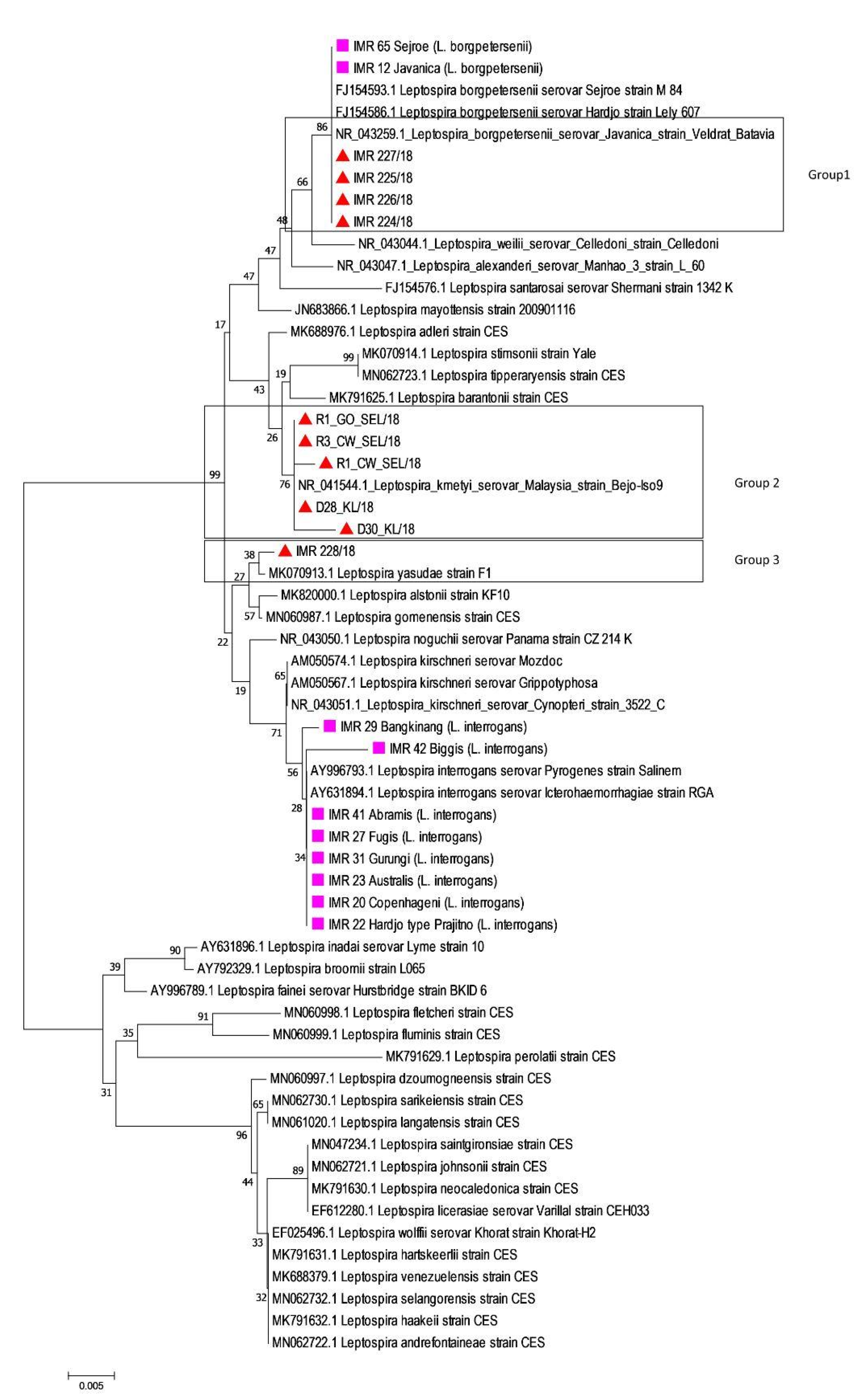
) (Figure 2). The rrs2 analysis showed a clustering of Leptospira into distinct species for both culture-positive and culture-negative samples. Four samples were identified as L. borgpetersenii, five samples were closely related to L. kmetyi and one sample was known as L. yasudae. Table 3 shows the estimates of the average evolutionary divergence over sequence pairs within the groups.

4. Discussion

The identification and typing of the infecting leptospiral can hint the animal source of the infections, as certain serovars are associated with either a single or an exclusive group of mammalian species [4,23]. Such information provides a vital contribution to the planning and implementation of effective prevention strategies [4,23,24]. As 16S rRNA gene sequencing has poor discriminatory strength and whole genome sequencing is currently impractical for routine strain typing, additional gene targets, such as secY and glmU genes, have been utilized to type Leptospira species [6,11,25,26]. LipL32 gene PCR sequencing has also been used extensively because it is a robust target for leptospirosis diagnosis and only detects pathogenic leptospires [27]. However, this technique lacks selective strength due to the conserved nature of the lipL32 gene sequence [11]. Furthermore, due to sequence polymorphism, the lipL32 gene PCR cannot detect P2 leptospires [27]. Thus, three different 6- or 7-loci MLST schemes have been most frequently utilized in typing Leptospira because of its ability to differentiate between seven pathogenic species of Leptospira up to the serovar level, in addition to the potential phylogenetic evidence that provide information on the relatedness between leptospiral species [16,21,27,28]. Ideally, Leptospira typing is carried out using isolates. Albeit culture is rarely performed in routine microbiology laboratories due to its low sensitivity and long incubation time, it plays a key role in elucidating both local and global epidemiology of infection [28,29,30,31].
As shown in Figure 1, most of the rodent samples from Kuala Lumpur wet markets and cities were closely related to L. interrogans serogroup Bataviae (Group 1) and L. borgpetersenii serogroup Javanica (Group 8), with a mean genetic distance within the groups of 0.0028 and 0.0049, respectively (Table 2). Comparable results were obtained by Benacer et al. [32] and Blasdell et al. [33]: L. borgpetersenii and L. interrogans were highly prevalent among urban rodents in Kuala Lumpur and Sarawak, respectively, indicating the potential role of the rat population as the maintenance hosts in the local urban transmission of Leptospira. Further, wet markets are a prime location for the spread of many infectious diseases and numerous studies have recently associated leptospirosis with them [34,35,36]. Leptospirosis may spread to humans due to a number of circumstances, including poor hygiene and management. Moreover, like other parts of the world, rapid urbanization, and high density of population in urban areas have attracted primary hosts of this disease, allowing them to harbor Leptospira, which then act as a persistent reservoir of infection to both livestock and humans cohabiting with rats [33]. Thus, regular monitoring of infections in wild rodents is essential to address important health problems and provide information that can lead to an effective public health policy [37].
Based on our results, L. interrogans was shown to be the main infecting Leptospira species in humans, with a broad range of infecting serogroups (Figure 1). Among these commonly identified serogroups in Malaysia, the most predominant serovars in humans were Bataviae and Sejroe (Figure 1). Interestingly, in the northeastern state of Malaysia, Shafei et al. [38] reported two predominant serogroups, Javanica and Bataviae, among town service laborers. Conversely, Rafizah et al. [39] reported the serogroup Sejroe as the common serogroup detected in febrile inpatient cases. Additionally, two samples of group 7 (R3_SER/17 and R4_SER/17) were not closely related with any of the reference strains based on the alignment of concatenated sequences of MLST Scheme 3 (Figure 1). This is because information on reference strains in the MLST database is limited to L. interrogans and L. kirschneri [16] and no allelic profiles have been assigned to new leptospiral strains. Furthermore, concatenated sequences of R3_SER/17 and R4_SER/17 might be unique with allelic variation at each locus, hence unable to find the exact/nearest match when compared with the database.
In this study, we were unable to isolate Leptospira from certain human and animal samples, although they were PCR-positive. We speculated that these subjects could become infected with isolates that favored natural conditions compared to the standard culture medium used in the laboratory, as Leptospira are fastidious microorganisms with high growth requirements. Not only that, clinical specimens often have very low bacterial burden [11]. Boonsilp et al. [40] also pointed out some possible explanations for patients who were PCR-positive but culture-negative. There is a possibility that the patients might have received an empirical antimicrobial drug. Furthermore, due to fluctuations in ambient temperature and delay, Leptospira might also become non-viable in the blood tube during the storage or transportation to the laboratory.
Therefore, in such instances, a direct identification of infecting Leptospira from clinical samples is favored due to the clinical and epidemiological importance [27,41,42]. However, because a very low amount of DNA is present in clinical samples, obtaining complete MLST results is laborious. For example, Chiani et al. [43] applied a seven-locus scheme to clinical samples, serum and whole blood from leptospirosis patients but failed to obtain complete MLST profiles. Thus, Boonsilp et al. [40] proposed a nested PCR for the rrs genes to detect and speciate pathogenic Leptospira in blood from patients with culture-negative leptospirosis. Thus, in our study, as rrs2 gene sequence analysis could be a reliable screening for species identification in culture-negative samples, isolates with incomplete MLST profiles were also included.
A phylogenetic tree based on rrs2 sequences (Figure 2) showed that Leptospira in all rodent samples were closely related to L. borgpetersenii, similar to the result in Group 8 (Figure 1). The isolates obtained from three cattle, two dogs, and one environmental sample were closely related to L. kmetyi serovar Malaysia (Group 2) and L. yasudae (Group 3) (Figure 2), with a mean genetic distance within group of 0.001 and 0.002, respectively (Table 3). These animals could become infected with Leptospira because of constant exposure to contaminated soil, as L. kmetyi was mostly isolated from environmental samples [44,45,46]. In addition, a novel species, L. yasudae was also isolated from the soil of an ex-situ wild animal conservation area in Malaysia recently [47]. Although both species are uncommonly isolated from clinical samples, L. kmetyi and L. yasudae have been classified under the P1 group based on the phylogenetic analysis of 16S rRNA gene as well as their genome compositions [7,44,47,48].
In this study, the inability of our local isolates to obtain complete information from all six MLST loci might be due to insufficient quantity of bacterial DNA in clinical specimens, which is necessary for MLST amplification. This is because Leptospira are only present in the blood within the first few days (4–6 days) post infection while in the urine, the bacteria can be detected after one week of illness [49,50]. Thus, the type of specimen and sampling window after symptom onset may prove crucial for MLST diagnosis directly from specimens. Furthermore, incomplete MLST profiles of our local isolates is attributable to the fact that they might be genetically unique, as the designing of primers was based on sequences from Leptospira recovered from other parts of the world. Conversely, the ability to recover the rrs2 sequence despite the incomplete coverage of six MLST loci could be due to the presence of multiple copies of the 16S rRNA gene in the leptospiral genome compared to the single copy genes used for MLST. Although a partial rrs2 sequence is adequate for molecular speciation, further sub-classification would require sequence information from complete MLST profiles.

5. Conclusions

This study indicates that molecular approaches by both MLST and rrs2 sequencing have immense potential in the comprehensive characterization of pathogenic Leptospira because they can be performed directly from clinical samples. MLST is also useful, especially in molecular epidemiological studies, for tracking the evolution and spread of this important human pathogen.

Author Contributions

Conceptualization, F.A.; methodology, N.A.N.H., A.H.M., N.M.D., A.K.A.A.T. and M.A.N.; software, M.K.N.M.K.; validation, F.A. and R.H.; formal analysis, M.K.N.M.K.; investigation, F.A.; resources, F.A.; data curation, F.A. and N.A.S.; writing—original draft preparation, N.A.S. and F.A.; writing—review and editing, M.R.M.A.; visualization, N.A.S., F.A. and M.K.N.M.K.; supervision, F.A.; project administration, F.A. All authors have read and agreed to the published version of the manuscript.

Funding

This research received no external funding.

Informed Consent Statement

Not applicable. This article utilized data from ongoing surveillance program and/or outbreak investigation related to leptospirosis.

Data Availability Statement

Available upon request.

Acknowledgments

We would like to thank the laboratory staff at the Institute for Medical Research (IMR) and veterinary personnel and officers who directly and indirectly contributed to this study. The authors would also like to thank the Director General of Health Malaysia for the permission to publish this paper. We would like to thank Nurul Najian Aminuddin Baki for her kind inputs and contribution to this paper.

Conflicts of Interest

The authors declare no conflict of interest.

References

  1. Philip, N.; Bahtiar Affendy, N.; Ramli, S.N.A.; Arif, M.; Raja, P.; Nagandran, E.; Renganathan, P.; Taib, N.M.; Masri, S.N.; Yuhana, M.Y.; et al. Leptospira interrogans and Leptospira kirschneri are the dominant Leptospira species causing human leptospirosis in Central Malaysia. PLoS Negl. Trop. Dis. 2020, 14, e0008197. [Google Scholar] [CrossRef] [PubMed]
  2. Barnacle, J.; Gurney, S.; Ledot, S.; Singh, S. Leptospirosis as an important differential of pulmonary haemorrhage on the intensive care unit: A case managed with VV-ECMO. J. Intensive Care 2020, 8, 31. [Google Scholar] [CrossRef] [PubMed]
  3. Dittrich, S.; Rattanavong, S.; Lee, S.J.; Panyanivong, P.; Craig, S.B.; Tulsiani, S.M.; Blacksell, S.D.; Dance, D.A.B.; FRCPath Dubot-Pérès, A.; Sengduangphachanh, A.; et al. Orientia, Rickettsia, and Leptospira pathogens as causes of CNS infections in Laos: A prospective study. Lancet Glob. Health 2015, 3, 104–112. [Google Scholar] [CrossRef] [PubMed]
  4. Levett, P.N. Leptospirosis. Clin. Microbiol. 2015, 14, 296–326. [Google Scholar] [CrossRef] [PubMed] [Green Version]
  5. Zuccarini, N.; Lutfi, S.; Piskorowski, T. Polyneuritis and rare sequelae of leptospirosis contracted while on an urban clean-up mission in Detroit: A case report of Weil’s disease and literature review. J. Med. Cases 2019, 10, 183–187. [Google Scholar] [CrossRef]
  6. Vincent, A.T.; Schiettekatte, O.; Goarant, C.; Neela, V.K.; Bernet, E.; Thibeaux, R.; Ismail, N.; Mohd Khalid, M.K.N.; Amran, F.; Masuzawa, T.; et al. Revisiting the taxonomy and evolution of pathogenicity of the genus Leptospira through the prism of genomics. PLoS Negl. Trop. Dis. 2019, 13, e0007270. [Google Scholar] [CrossRef] [Green Version]
  7. Casanovas-Massana, A.; Hamond, C.; Santos, L.A.; de Oliveira, D.; Hacker, K.P.; Balassiano, I.; Costa, F.; Medeiros, M.A.; Reis, M.G.; Ko, A.I.; et al. Leptospira yasudae sp. nov. and Leptospira stimsonii sp. nov., two new species of the pathogenic group isolated from environmental sources. Int. J. Syst. Evol. Microbiol. 2020, 70, 1450–1456. [Google Scholar]
  8. Picardeau, M. Virulence of the zoonotic agent of leptospirosis: Still terra incognita? Nat. Rev. Microbiol. 2017, 15, 297–307. [Google Scholar] [CrossRef]
  9. Thibeaux, R.; Girault, D.; Bierque, E.; Soupé-Gilbert, M.E.; Rettinger, A.; Douyère, A.; Meyer, M.; Iraola, G.; Picardeau, M.; Goarant, C. Biodiversity of environmental Leptospira: Improving identification and revisiting the diagnosis. Front. Microbiol. 2018, 9, 816. [Google Scholar] [CrossRef] [Green Version]
  10. Zhang, C.; Yang, H.; Li, X.; Cao, Z.; Zhou, H.; Zeng, L.; Xu, J.; Xu, Y.; Chang, Y.F.; Guo, X.; et al. Molecular typing of pathogenic Leptospira serogroup Icterohaemorrhagiae strains circulating in China during the past 50 years. PLoS Negl. Trop. Dis. 2015, 9, e0003762. [Google Scholar] [CrossRef] [Green Version]
  11. Guernier, V.; Allan, K.J.; Goarant, C. Advances and challenges in barcoding pathogenic and environmental Leptospira. Parasitology 2018, 145, 595–607. [Google Scholar] [CrossRef] [PubMed]
  12. Haake, D.A.; Levett, P.N. Leptospirosis in humans. Curr. Top. Microbiol. Immunol. 2015, 387, 65–97. [Google Scholar] [PubMed] [Green Version]
  13. Auch, A.F.; von Jan, M.; Klenk, H.P.; Göker, M. Digital DNA-DNA hybridization for microbial species delineation by means of genome-to-genome sequence comparison. Stand. Genom. Sci. 2010, 2, 117–134. [Google Scholar] [CrossRef] [PubMed] [Green Version]
  14. Goris, J.; Konstantinidis, K.T.; Klappenbach, J.A.; Coenye, T.; Vandamme, P.; Tiedje, J.M. DNA-DNA hybridization values and their relationship to whole-genome sequence similarities. Int. J. Syst. Evol. Microbiol. 2007, 57, 81–91. [Google Scholar] [CrossRef] [PubMed] [Green Version]
  15. Wolf, Y.I.; Rogozin, I.; Grishin, N.; Tatusov, R.; Koonin, E. Genome trees constructed using five different approaches suggest new major bacterial clades. BMC Evol. Biol. 2001, 1, 8. [Google Scholar] [CrossRef]
  16. Boonsilp, S.; Thaipadungpanit, J.; Amornchai, P.; Wuthiekanun, V.; Bailey, M.S.; Holden, M.T.G.; Zhang, C.; Jiang, X.; Koizumi, N.; Taylor, K.; et al. A single multilocus sequence typing (MLST) scheme for seven pathogenic Leptospira species. PLoS Negl. Trop. Dis. 2013, 7, e1954. [Google Scholar] [CrossRef] [Green Version]
  17. Leon, A.; Pronost, S.; Fortier, G.; Andre-Fontaine, G.; Leclercq, R. Multilocus sequence analysis for typing Leptospira interrogans and Leptospira kirschneri. J. Clin. Microbiol. 2010, 48, 581–585. [Google Scholar] [CrossRef] [Green Version]
  18. Aanensen, D.M.; Spratt, B.G. The multilocus sequence typing network: Mlst.net. Nucleic Acids Res. 2005, 33, W728–W733. [Google Scholar] [CrossRef] [Green Version]
  19. Inouye, M.; Dashnow, H.; Raven, L.A.; Schultz, M.B.; Pope, B.J.; Tomita, T.; Zobel, J.; Holt, K.E. SRST2: Rapid genomic surveillance for public health and hospital microbiology labs. Genome Med. 2014, 6, 90. [Google Scholar] [CrossRef] [Green Version]
  20. Baust, J.G. Strategies for the storage of DNA. Biopreserv. Biobank. 2008, 6, 251–252. [Google Scholar]
  21. Ahmed, N.; Devi, S.M.; Valverde, M.L.V.P.; Machang’u, R.S.; Ellis, W.A.; Hartskeerl, R.A. Multilocus sequence typing method for identification and genotypic classification of pathogenic Leptospira species. Ann. Clin. Microbiol. Antimicrob. 2006, 23, 28. [Google Scholar] [CrossRef] [PubMed] [Green Version]
  22. Ahmed, A.; Thaipadungpanit, J.; Boonsilp, S.; Wuthiekanun, V.; Nalam, K.; Spratt, B.G.; Aaensen, D.M.; Smythe, L.D.; Ahmed, N.; Feil, E.J.; et al. Comparison of two multilocus sequence based genotyping schemes for Leptospira species. PLoS Negl. Trop. Dis. 2011, 5, e1374. [Google Scholar] [CrossRef] [PubMed] [Green Version]
  23. Bharti, A.R.; Nally, J.E.; Ricaldi, J.N.; Matthias, M.A.; Diaz, M.M.; Lovett, M.A.; Levett, P.N.; Gilman, R.H.; Willig, M.R. Leptospirosis: A zoonotic disease of global importance. Lancet Infect. Dis. 2003, 3, 757–771. [Google Scholar] [CrossRef] [PubMed]
  24. Ko, A.I.; Goarant, C.; Picardeau, M. Leptospira: The dawn of the molecular genetics era for an emerging zoonotic pathogen. Nat. Rev. Microbiol. 2009, 7, 736–747. [Google Scholar] [CrossRef] [PubMed]
  25. Ahmed, A.; Engelberts, M.F.; Boer, K.R.; Ahmed, N.; Hartskeerl, R.A. Development and validation of a real-time PCR for detection of pathogenic Leptospira species in clinical materials. PLoS ONE 2009, 4, e7093. [Google Scholar] [CrossRef] [PubMed]
  26. Wilkinson, D.A.; Edwards, M.; Benschop, J.; Nisa, S. Identification of pathogenic Leptospira species and serovars in New Zealand using metabarcoding. PLoS ONE 2021, 16, e0257971. [Google Scholar] [CrossRef] [PubMed]
  27. Sykes, J.E.; Reagan, K.L.; Nally, J.E.; Galloway, R.L.; Haake, D.A. Role of diagnostics in epidemiology, management, surveillance, and control of leptospirosis. Pathogens 2022, 11, 395. [Google Scholar] [CrossRef]
  28. Thaipadungpanit, J.; Wuthiekanun, V.; Chierakul, W.; Smythe, L.D.; Petkanchanapong, W.; Limpaiboon, R.; Apiwatanaporn, A.; Slack, A.T.; Suputtamongkol, Y.; White, N.J.; et al. A dominant clone of Leptospira interrogans associated with human leptospirosis in Thailand. PLoS Negl. Trop. Dis. 2007, 1, e56. [Google Scholar] [CrossRef]
  29. Adler, B.; de la Peña Moctezuma, A. Leptospira and leptospirosis. Vet. Microbiol. 2010, 140, 287–296. [Google Scholar] [CrossRef]
  30. Slack, A.; Symonds, M.; Dohnt, M.; Harris, C.; Brookes, D.; Smythe, L. Evaluation of a modified Taqman assay detecting pathogenic Leptospira spp. against culture and Leptospira-specific IgM enzyme-linked immunosorbent assay in a clinical environment. Diagn. Microbiol. Infect. Dis. 2007, 57, 361–366. [Google Scholar] [CrossRef]
  31. Thaipadunpanit, J.; Chierakul, W.; Wuthiekanun, V.; Limmathurotsakul, D.; Amornchai, P.; Boonslip, S.; Smythe, L.D.; Limpaiboon, R.; Hoffmaster, A.R.; Day, N.P.J.; et al. Diagnostic accuracy of real-time PCR assays targeting 16S rRNA and lipl32 genes for human leptospirosis in Thailand: A case–control study. PLoS ONE 2011, 6, e16236. [Google Scholar] [CrossRef] [PubMed]
  32. Benacer, D.; Mohd Zain, S.N.; Amran, F.; Galloway, R.L.; Thong, K.L. Isolation and molecular characterization of Leptospira interrogans and Leptospira borgpetersenii isolates from the urban rat populations of Kuala Lumpur, Malaysia. Am. J. Trop. Med. Hyg. 2013, 88, 704–709. [Google Scholar] [CrossRef] [Green Version]
  33. Blasdell, K.R.; Morand, S.; Perera, D.; Firth, C. Association of rodent-borne Leptospira spp. with urban environments in Malaysian Borneo. PLoS Negl. Trop. Dis. 2019, 13, e0007141. [Google Scholar] [CrossRef] [PubMed] [Green Version]
  34. Bahtiar Affendy, N.; Mohd Desa, M.N.; Amran, F.; Sekawi, Z.; Masri, S.N. Isolation and molecular characterization of Leptospira interrogans and Leptospira borgpetersenii from small mammals in Selangor wet markets. Int. J. Infect. Dis. 2020, 101, 534. [Google Scholar] [CrossRef]
  35. Rahman, M.H.A.A.; Hairon, S.M.; Hamat, R.A.; Jamaluddin, T.Z.M.T.; Shafei, M.N.; Idris, N.; Osman, M.; Sukeri, S.; Wahab, Z.A.; Mohammad, W.M.Z.W.; et al. Seroprevalence and distribution of leptospirosis serovars among wet market workers in northeastern, Malaysia: A cross sectional study. BMC Infect. Dis. 2018, 18, 569. [Google Scholar] [CrossRef]
  36. Samsudin, S.; Saudi, S.N.S.; Masri, N.S.; Ithnin, N.R.; Jamaluddin, T.Z.M.T.; Hamat, R.A.; Wan Mohd, Z.W.M.; Nazri, M.S.; Surianti, S.; Daud, A.B.; et al. Awareness, knowledge, attitude and preventive practice of leptospirosis among healthy Malaysian and Non-Malaysian wet market workers in selected urban areas in Selangor, Malaysia. Int. J. Environ. Res. Public Health 2020, 17, 1346. [Google Scholar] [CrossRef] [PubMed] [Green Version]
  37. Benacer, D.; Mohd Zain, S.N.; Shin, Z.S.; Mohd Khalid, M.K.N.; Galloway, R.L.; Souris, M.; Thong, K.L. Determination of Leptospira borgpetersenii serovar Javanica and Leptospira interrogans serovar Bataviae as the persistent Leptospira serovars circulating in the urban rat populations in Peninsular Malaysia. Parasites Vectors 2016, 9, 117. [Google Scholar] [CrossRef] [Green Version]
  38. Shafei, M.N.; Sulong, M.R.; Yaacob, N.A.; Hassan, H.; Wan Mohd Zahiruddin, W.M.; Daud, A.; Ismail, Z.; Abdullah, M.R. Seroprevalence of leptospirosis among town service workers on northeastern state of Malaysia. Int. J. Collab. Res. Intern. Med. Public Health 2012, 4, 395–403. [Google Scholar]
  39. Rafizah, N.A.A.; Aziah, B.D.; Azwany, Y.N.; Imran, M.K.; Mohamed Rusli, A.; Mohd Nazri, S.; Mohd Nikman, A.; Nabilah, I.; Siti Asma’, H.; Zahiruddin, W.M.; et al. A hospital-based study on seroprevalence of leptospirosis among febrile cases in northeastern Malaysia. Int. J. Infect. Dis. 2013, 17, e394–e397. [Google Scholar] [CrossRef] [Green Version]
  40. Boonsilp, S.; Thaipadungpanit, J.; Amornchai, P.; Wuthiekanun, V.; Chierakul, W.; Limmathurotsakul, D.; Day, N.P.; Peacock, S.J. Molecular detection and speciation of pathogenic Leptospira spp. in blood from patients with culture-negative leptospirosis. BMC Infect. Dis. 2011, 11, 338. [Google Scholar] [CrossRef] [Green Version]
  41. Agampodi, S.B.; Moreno, A.C.; Vinetz, J.M.; Matthias, M.A. Utility and limitations of direct multi-locus sequence typing on qPCR-positive blood to determine infecting Leptospira strain. Am. J. Trop. Med. Hyg. 2013, 88, 184–185. [Google Scholar] [CrossRef] [Green Version]
  42. Podgoršek, D.; Ružić-Sabljić, E.; Logar, M.; Pavlović, A.; Remec, T.; Baklan, Z.; Pal, E.; Cerar, T. Evaluation of real-time PCR targeting the lipL32 gene for diagnosis of Leptospira infection. BMC Microbiol. 2020, 20, 59. [Google Scholar] [CrossRef] [PubMed]
  43. Chiani, Y.; Jacob, P.; Varni, V.; Lamdolt, N.; Schmeling, M.F.; Pujato, N.; Caimi, K.; Vanasco, B. Isolation and clinical sample typing of human leptospirosis cases in Argentina. Infect. Genet. Evol. 2016, 37, 245–251. [Google Scholar] [CrossRef]
  44. Slack, A.T.; Khairani-Bejo, S.; Symonds, M.L.; Dohnt, M.F.; Galloway, R.L.; Steigerwalt, A.G.; Bahaman, A.R.; Craig, S.; Harrower, B.J.; Smythe, L.D. Leptospira kmetyi sp nov., isolated from an environmental source in Malaysia. Int. J. Syst. Evol. Microbiol. 2009, 59, 705–708. [Google Scholar] [CrossRef] [PubMed]
  45. Tan, X.T.; Amran, F.; Chee Cheong, K.; Ahmad, N. In-house ELISA screening using a locally isolated Leptospira in Malaysia: Determination of its cut-off points BMC Infect. Dis. 2014, 14, 563. [Google Scholar]
  46. Zaki, A.M.; Hod, R.; Shamsusah, N.A.; Isa, Z.M.; Bejo, S.K.; Agustar, H.K. Detection of Leptospira kmetyi at recreational areas in Peninsular Malaysia. Environ. Monit. Assess. 2020, 192, 703. [Google Scholar] [CrossRef]
  47. Shamsusah, N.A.; Agustar, H.K.; Amran, F.; Hod, R. Draft genome sequence of Leptospira yasudae strain BJ3, isolated from the soil of an ex-situ wild animal conservation area. Microbiol. Resour. Announc. 2021, 10, e0072321. [Google Scholar] [CrossRef]
  48. Voronina, O.L.; Kunda, M.S.; Aksenova, E.I.; Ryzhova, N.N.; Semenov, A.N.; Petrov, E.M.; Didenko, L.V.; Lunin, V.G.; Ananyina, Y.V.; Gintsburg, A.L. The characteristics of ubiquitous and unique Leptospira strains from the collection of Russian centre for leptospirosis. BioMed Res. Int. 2014, 2014, 649034. [Google Scholar] [CrossRef] [Green Version]
  49. Sykes, J.E.; Gamage, C.D.; Haake, D.A.; Nally, J.E. Understanding leptospirosis: Application of state-of-the-art molecular typing tools with a One Health lens. Am. J. Vet. Res. 2022, 83, ajvr.22.06.0104. [Google Scholar] [CrossRef]
  50. Techawiwattanaboon, T.; Patarakul, K. Update on molecular diagnosis of human leptospirosis. Asian Biomed. 2019, 13, 207–216. [Google Scholar] [CrossRef]
Figure 1. Genetic relatedness comparing isolates from animal (Tropicalmed 08 00069 i001) and human (Tropicalmed 08 00069 i002) samples with reference strains from different countries based on the concatenated sequences of the six housekeeping genes using neighbor-joining phylogenetic trees with bootstraps of 100.
Tropicalmed 08 00069 g001
Figure 2. Genetic relatedness comparing strains from rodents, dogs and soil samples (Tropicalmed 08 00069 i004) to old reference Malaysian Leptospira isolates (Tropicalmed 08 00069 i005) and NCBI reference strains based on rrs2 genes using neighbor-joining phylogenetic trees with bootstraps of 100.
Tropicalmed 08 00069 g002
Table 1. Oligonucleotide sequence for both forward and reverse primers in MLST Scheme 3.
PrimersOligonucleotide Sequence (5′–3′)
adkF-GGG CTG GAA AAG GTA CAC AA
R-ACG CAA GCT CCT TTT GAA TC
icdAF-GGG ACG AGA TGA CCA GGA T
R-TTT TTT GAG ATC CGC AGC TTT
lipL32F-ATC TCC GTT GCA CTC TTT GC
R-ACC ATC ATC ATC ATC GTC CA
lipL41F-TAG GAA ATT GCG CAG CTA CA
R-GCA TCG AGA GGA ATT AAC ATC A
rrs2F-CAT GCA AGT CAA GCG GAG TA
R-AGT TGA GCC CGC AGT TTT C
secYF-ATG CCG ATC ATT TTT GCT TC
R-CCG TCC CTT AAT TTT AGA CTT CTT C
Table 2. Estimates of the average evolutionary divergence over sequence pairs within groups based on MLST analysis.
Group No.Mean Distance (within Groups)Standard Error (S.E)
10.00280.0004
20.00070.0005
30.00070.0005
40.00170.0008
50.00000.0000
60.00000.0000
70.01810.0024
80.00490.0011
Table 3. Estimates of the average evolutionary divergence over sequence pairs within groups on rrs genes.
Group No.Mean Distance (within Groups)Standard Error (S.E)
10.0000.000
20.0010.001
30.0020.002
Disclaimer/Publisher’s Note: The statements, opinions and data contained in all publications are solely those of the individual author(s) and contributor(s) and not of MDPI and/or the editor(s). MDPI and/or the editor(s) disclaim responsibility for any injury to people or property resulting from any ideas, methods, instructions or products referred to in the content.

