
A Systematic Review on the Viruses of Anopheles Mosquitoes: The Potential Importance for Public Health
 , , and
Abstract
:1. Introduction
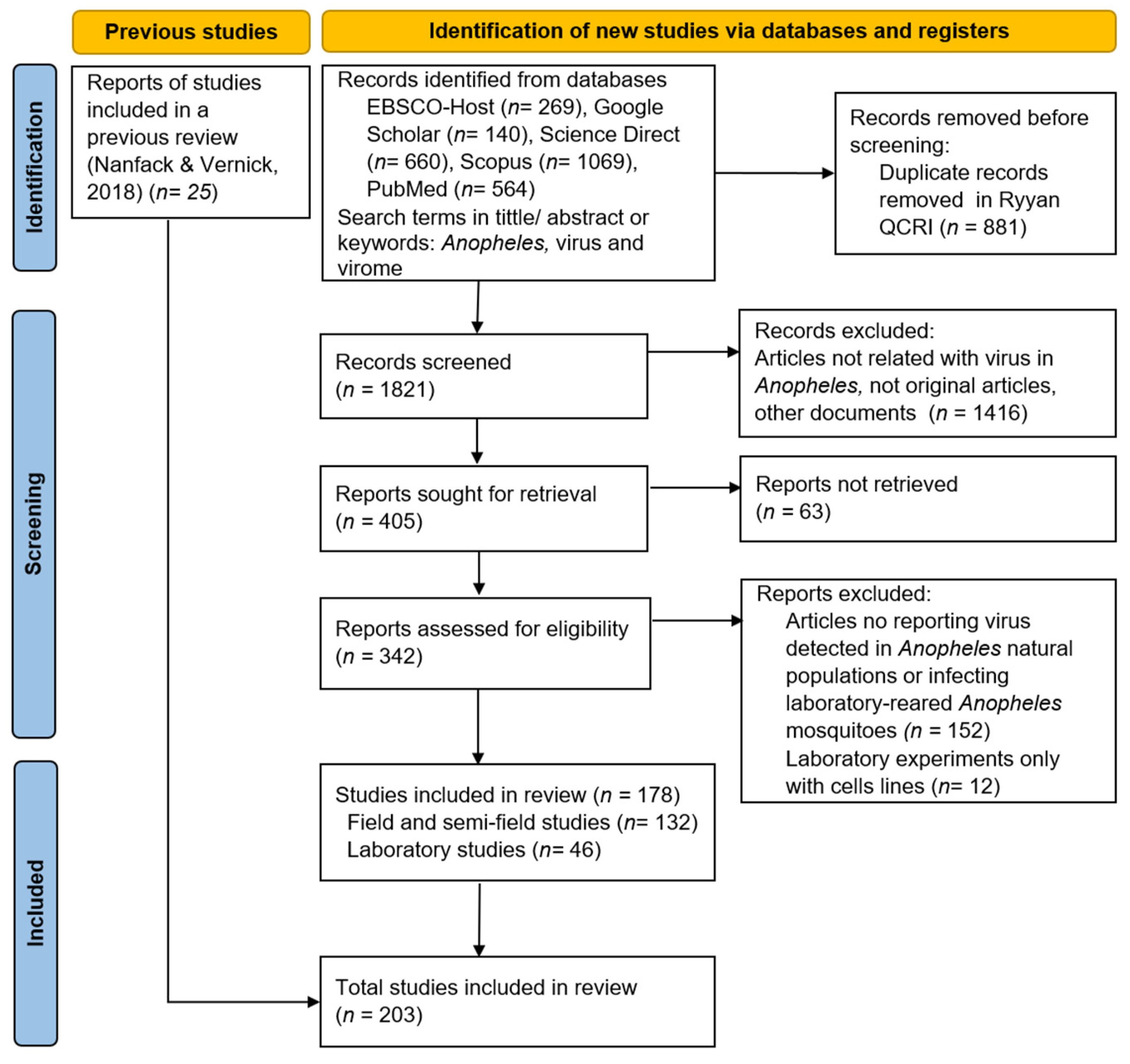
2. Materials and Methods
Scientific Literature Selection and Data Extraction
3. Results
3.1. Search Results
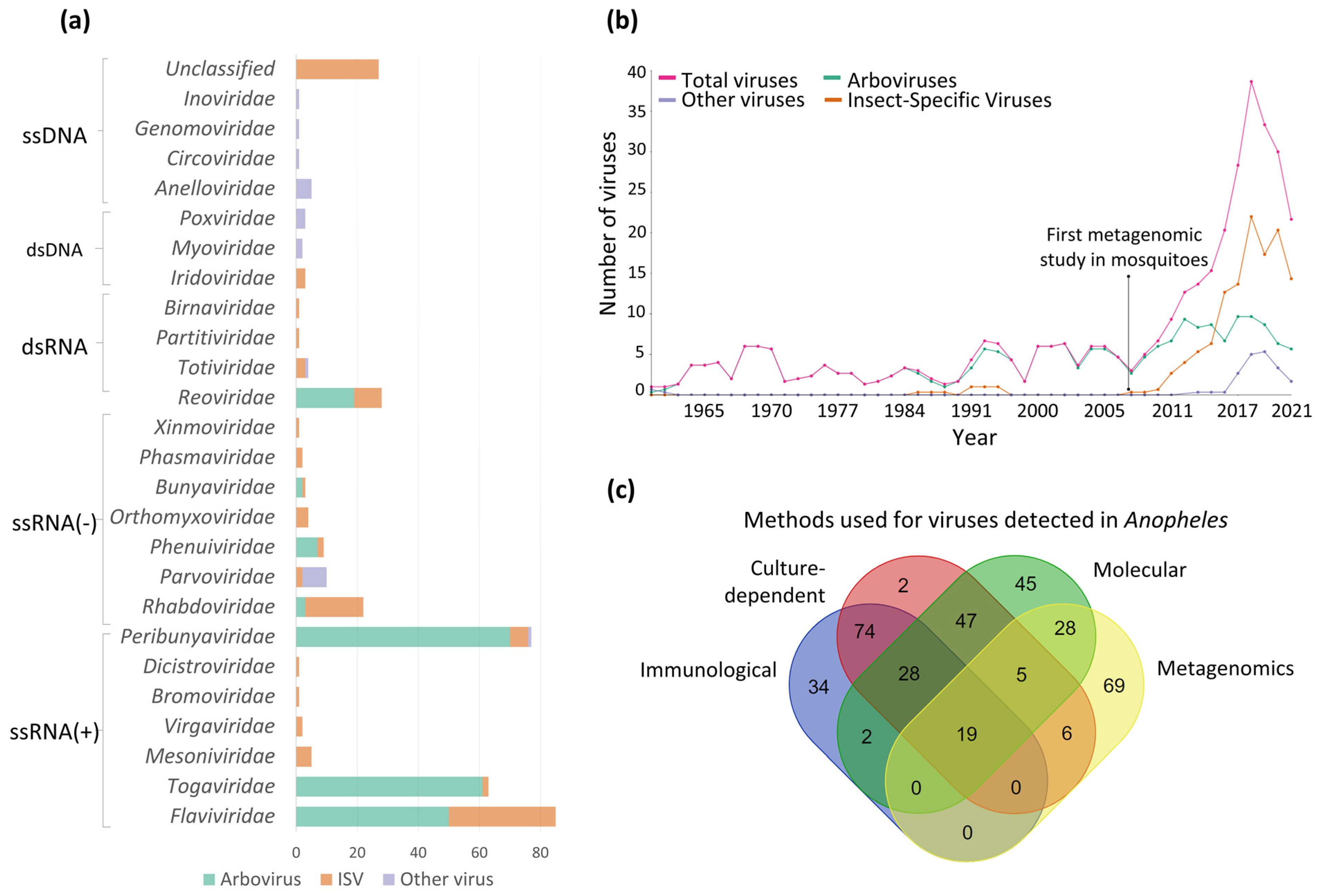
3.2. Viruses Detected in Anopheles Mosquitoes
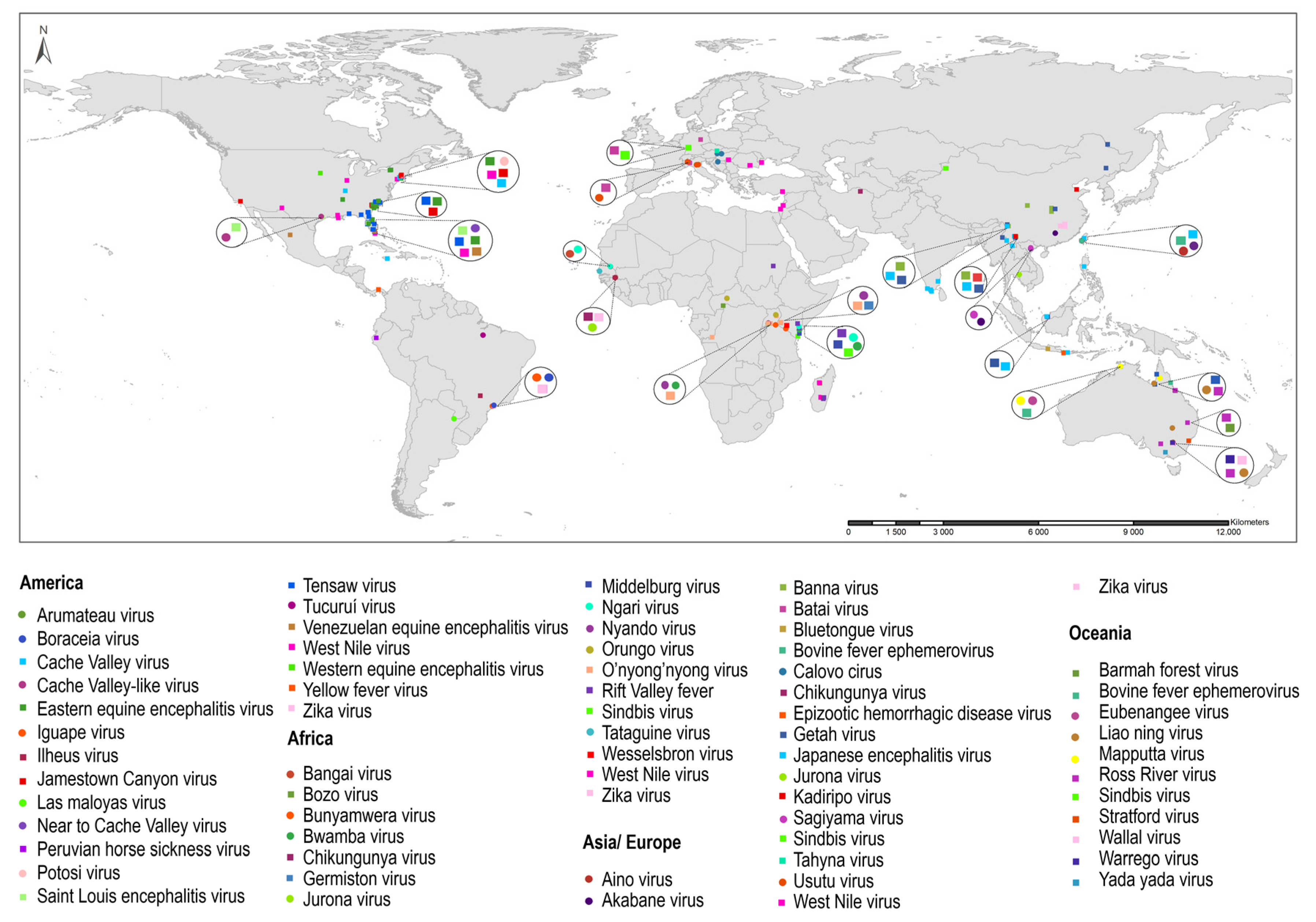
3.2.1. Arboviruses and Probable Arboviruses Detected in Anopheles
The Peribunyaviridae Family

| Virus Name (Abbreviation) | Anopheles Species | Detected in Natural Populations (Country/Number of Detections) | Detected during an Outbreak (Yes/No) | Results of Laboratory Studies (Viral Infection and Transmission) | References * |
|---|---|---|---|---|---|
| O’nyong-nyong virus ★ (ONNV) | An. gambie | Uganda/2, Kenya/1 | Yes [65] | IR 75% at 7 dpi with recombinant virus, TR not determined | [4] |
| Infection, IR not available, TR not determined | [146] | ||||
| Limited infection and spread, with no differences between transgenic and wild mosquitoes, TR 0% | [147] | ||||
| Studies with a recombinant virus, IR 78%, DR 15% at 6 dpi; IR 84%, DR 25% at 8 dpi, TR not determined | [148] | ||||
| IR 75%, TR 0% at 7 dpi; IR 95%, TR 57% at 14 dpi | [149] | ||||
| Rift Valley fever virus (RVFV) | An. coustani | Madagascar/1, Sudan/1 | Yes [12,74] | IR 50%, TR 100% at 8 dpi | [150] |
| Saint Louis encephalitis virus (SLEV) | An. quadrimaculatus | USA/1 | Yes [106] | Infection (IR not determined), transmission 0% | [151] |
| Tensaw virus (TENV) | An. quadrimaculatus | USA/4 | No | IR 100% at 10 and 20 dpi, transmission 20% at 14 dpi | [141] |
| Japanese encephalitis virus (JEV) | An. subpictus | India/4 × | Yes [54,55] | N/A | N/A |
| West Nile virus (WNV) | An. punctipennis | USA/3 | Yes [46,47] | N/A | N/A |
| An. maculipennis | Romania/1, Serbia/1 | Yes [37,40] | N/A | N/A | |
| Bunyamwera virus (BUNV) | An. gambiae | Kenya/1 | No | IR 38%, transmission 71% at 14 dpi | [144] |
| Cache Valley virus (CVV) | An. quadrimaculatus | USA/3 | No | IR 100%, transmission 20% at 7 dpi; IR 100%, transmission 33% at 14 dpi | [142] |
| No | IR 100%, TR 0% at 10–19 dpi | [143] | |||
| An. punctipennis | USA/2 | No | IR 85%, TR 30% at 14–18 dpi | [143] | |
| Eastern equine encephalitis virus (EEEV) | An. quadrimaculatus | USA/5 | No | Infection rate not determined; transmission 40% at 10 dpi, 50% at 11 dpi | [152] |
| Myxoma virus § (MYXV) | An. atroparvus | England/1 | Yes [153] | Infectious virion up to 220 dpi in mosquito mouthparts | [154] |
The Togaviridae Family
The Flaviviridae Family
Other Arboviruses
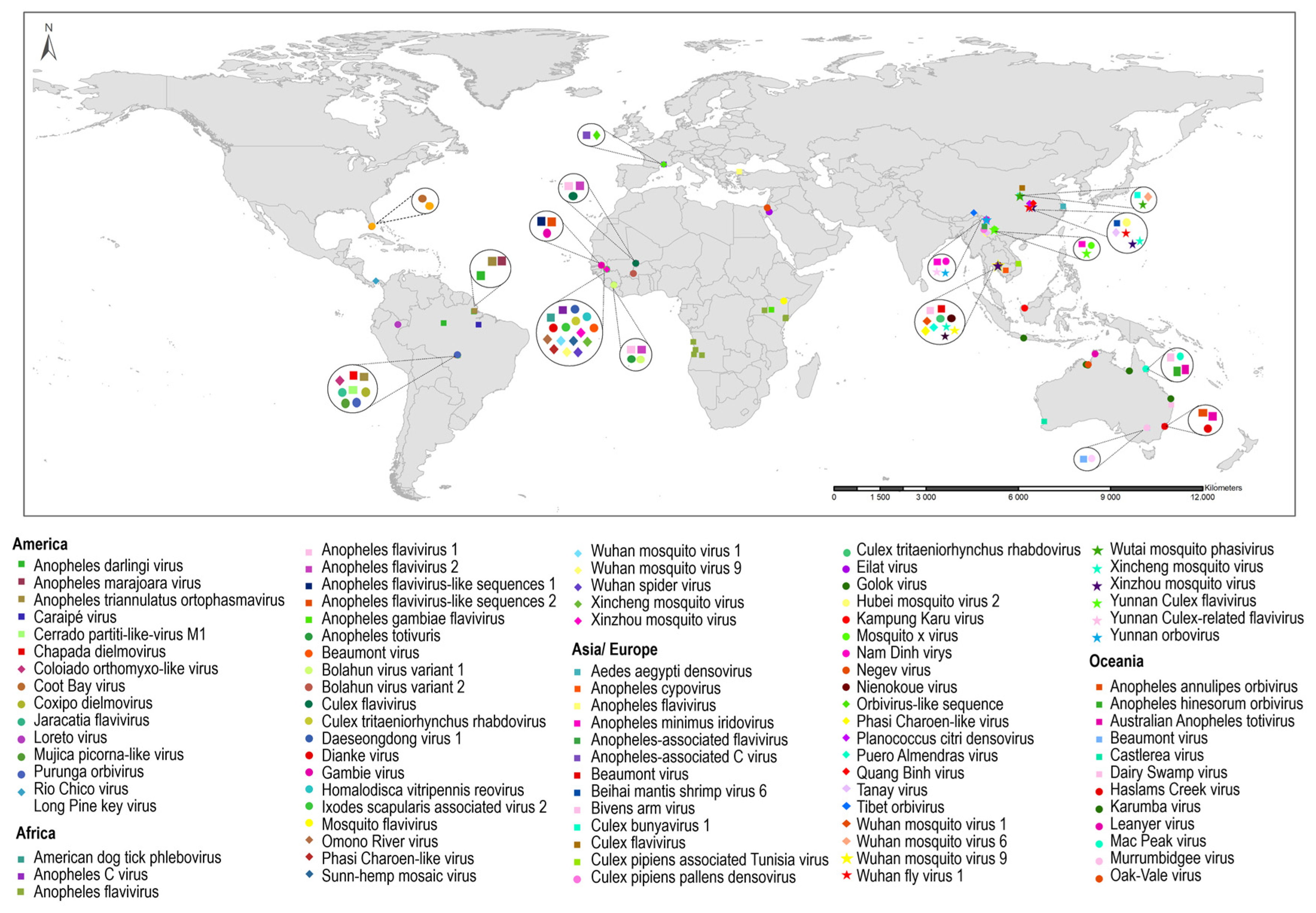
3.2.2. Insect-Specific Viruses (ISVs) Detected in Anopheles
| Virus Name/Abbreviation | Country | Anopheles Species | References * |
|---|---|---|---|
| Anopheles flavivirus (AnFV) | Angola | Anopheles spp. | [186] |
| Kenya | An. gambiae | [135] | |
| An. gambiae s.l. | [187] | ||
| An. squamosus | [135] | ||
| Turkey | An. maculipennis s.l. | [188] | |
| Karumba virus (KRBV) | Australia | An. meraukensis | [101,189] |
| Dianke virus (DKV) | Senegal | An. funestus | [190] |
| An. gambiae | [190] | ||
| An. pharoensis | [190] | ||
| An. rufipes | [190] | ||
| Xinzhou mosquito virus | Cambodia | Anopheles spp. | [116] |
| China | An. sinensis | [191] | |
| Senegal | Anopheles spp. | [148] | |
| Culex flavivirus (CxFV) | China | An. sinensis | [192] |
| Guinea/Mali | Anopheles spp. | [193] | |
| Beaumont virus | Australia | An. annulipes s.l. | [59] |
| Cambodia | Anopheles spp. | [116] | |
| Senegal | Anopheles spp. | [116] | |
| Xincheng mosquito virus | Cambodia | Anopheles spp. | [116] |
| China | An. sinensis | [191] | |
| Senegal | Anopheles spp. | [116] | |
| Tanay virus (TANAV) | China | An. sinensis | [89,194] |
| Hubei mosquito virus 2 (HMV2) | China | An. sinensis | [49,89] |
| Wuhan mosquito virus 1 | Cambodia | Anopheles spp. | [116] |
| Senegal | Anopheles spp. | [116] | |
| Wuhan mosquito virus 9 | Cambodia | Anopheles spp. | [116] |
| Senegal | Anopheles spp. | [116] | |
| Anopheles flavivirus 1 (AnFV1) | Guinea/Mali | Anopheles spp. | [193] |
| Liberia | An. gambiae | [195] | |
| Anopheles flavivirus 2 (AnFV2) | Guinea/Mali | Anopheles spp. | [193] |
| Liberia | An. gambiae | [195] | |
| Culex tritaeniorhynchus rhabdovirus | Cambodia | Anopheles spp. | [116] |
| Senegal | Anopheles spp. | [116] | |
| Anopheles minimus iridovirus (AMIV) | China | An. minimus | [50,196] |
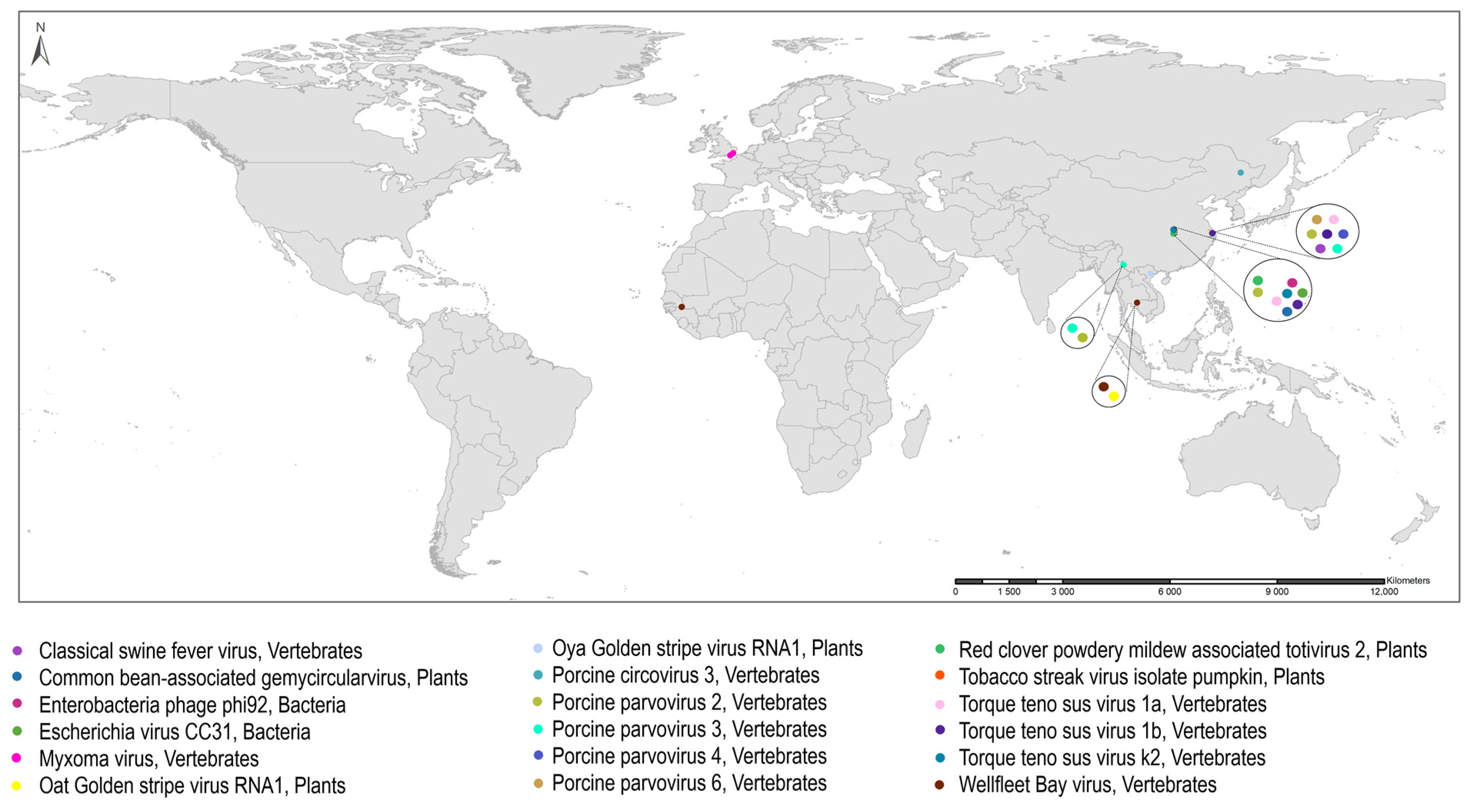
3.2.3. Other Viruses Detected in Anopheles
4. Discussion
Supplementary Materials
Author Contributions
Funding
Institutional Review Board Statement
Informed Consent Statement
Data Availability Statement
Conflicts of Interest
References
- Hay, S.I.; Guerra, C.A.; Tatem, A.J.; Noor, A.M.; Snow, R.W. The Global Distribution and Population at Risk of Malaria: Past, Present, and Future. Lancet Infect. Dis. 2004, 4, 327–336. [Google Scholar] [CrossRef] [PubMed]
- WHO. World Malaria Report 2021. 2021. Available online: https://www.who.int/teams/global-malaria-programme/reports/world-malaria-report-2022 (accessed on 25 August 2023).
- Manguin, S.; Bangs, M.J.; Pothikasikorn, J.; Chareonviriyaphap, T. Review on Global Co-Transmission of Human Plasmodium Species and Wuchereria bancrofti by Anopheles Mosquitoes. Infect. Genet. Evol. 2010, 10, 159–177. [Google Scholar] [CrossRef] [PubMed]
- Brault, A.C.; Foy, B.D.; Myles, K.M.; Kelly, C.L.H.; Higgs, S.; Weaver, S.C.; Olson, K.E.; Miller, B.R.; Powers, A.M. Infection Patterns of O’Nyong Nyong Virus in the Malaria-Transmitting Mosquito, Anopheles gambiae. Insect Mol. Biol. 2004, 13, 625–635. [Google Scholar] [CrossRef] [PubMed]
- Scott, T.W.; Takken, W. Feeding Strategies of Anthropophilic Mosquitoes Result in Increased Risk of Pathogen Transmission. Trends Parasitol. 2012, 28, 114–121. [Google Scholar] [CrossRef] [PubMed]
- Prasad, N.; Murdoch, D.R.; Reyburn, H.; Crump, J.A. Etiology of Severe Febrile Illness in Low- and Middle-Income Countries: A Systematic Review. PLoS ONE 2015, 10, e0127962. [Google Scholar] [CrossRef]
- Bolling, B.G.; Weaver, S.C.; Tesh, R.B.; Vasilakis, N. Insect-Specific Virus Discovery: Significance for the Arbovirus Community. Viruses 2015, 7, 4911–4928. [Google Scholar] [CrossRef]
- Tajudeen, Y.A.; Oladunjoye, I.O.; Mustapha, M.O.; Mustapha, S.T.; Ajide-Bamigboye, N.T. Tackling the Global Health Threat of Arboviruses: An Appraisal of the Three Holistic Approaches to Health. Health Promot. Perspect. 2021, 11, 371–381. [Google Scholar] [CrossRef]
- Chaves, L.S.M.; Fry, J.; Malik, A.; Geschke, A.; Sallum, M.A.M.; Lenzen, M. Global Consumption and International Trade in Deforestation-Associated Commodities Could Influence Malaria Risk. Nat. Commun. 2020, 11, 1258. [Google Scholar] [CrossRef]
- Hernández-Valencia, J.C.; Rincón, D.S.; Marín, A.; Naranjo-Díaz, N.; Correa, M.M. Effect of Land Cover and Landscape Fragmentation on Anopheline Mosquito Abundance and Diversity in an Important Colombian Malaria Endemic Region. PLoS ONE 2020, 15, e0240207. [Google Scholar] [CrossRef]
- Ellwanger, J.H.; Chies, J.A.B. Zoonotic Spillover: Understanding Basic Aspects for Better Prevention. Genet. Mol. Biol. 2021, 44, e20200355. [Google Scholar] [CrossRef]
- Seufi, A.E.M.; Galal, F.H. Role of Culex and Anopheles Mosquito Species as Potential Vectors of Rift Valley Fever Virus in Sudan Outbreak, 2007. BMC Infect. Dis. 2010, 10, 65. [Google Scholar] [CrossRef]
- Brustolin, M.; Pujhari, S.; Henderson, C.A.; Rasgon, J.L. Anopheles Mosquitoes May Drive Invasion and Transmission of Mayaro Virus across Geographically Diverse Regions. PLoS Negl. Trop. Dis. 2018, 12, e0006895. [Google Scholar] [CrossRef] [PubMed]
- Thenmozhi, V.; Balaji, T.; Venkatasubramani, K.; Dhananjeyan, K.; Selvam, A.; Rajamannar, V.; Tyagi, B. Role of Anopheles subpictus Grassi in Japanese Encephalitis Virus Transmission in Tirunelveli, South India. Indian J. Med. Res. 2016, 144, 477–481. [Google Scholar] [CrossRef] [PubMed]
- Greninger, A.L. A Decade of RNA Virus Metagenomics Is (Not) Enough. Virus Res. 2018, 244, 218–229. [Google Scholar] [CrossRef] [PubMed]
- Öhlund, P.; Lundén, H.; Blomström, A.-L. Insect-Specific Virus Evolution and Potential Effects on Vector Competence. Virus Genes 2019, 55, 127–137. [Google Scholar] [CrossRef]
- Roundy, C.M.; Azar, S.R.; Rossi, S.L.; Weaver, S.C.; Vasilakis, N. Chapter Four—Insect-Specific Viruses: A Historical Overview and Recent Developments. In Advances in Virus Research; Kielian, M., Mettenleiter, T.C., Roossinck, M.J., Eds.; Academic Press: Cambridge, MA, USA, 2017; Volume 98, pp. 119–146. [Google Scholar]
- PRISMA. Preferred Reporting Items for Systematic Reviews and Meta-Analyses (PRISMA). Available online: https://prisma-statement.org/ (accessed on 26 July 2022).
- Ouzzani, M.; Hammady, H.; Fedorowicz, Z.; Elmagarmid, A. Rayyan-a Web and Mobile App for Systematic Reviews. Syst. Rev. 2016, 5, 210. [Google Scholar] [CrossRef]
- Centers for Disease Control and Prevention. Arbovirus Catalog. Available online: https://wwwn.cdc.gov/arbocat/ (accessed on 17 July 2023).
- Nanfack-Minkeu, F.; Vernick, K.D. A Systematic Review of the Natural Virome of Anopheles Mosquitoes. Viruses 2018, 10, 222. [Google Scholar] [CrossRef]
- Chamberlain, R.W.; Sudia, W.D.; Coleman, P.H.; Johnston, J.G.; Work, T.H. Arbovirus Isolations from Mosquitoes Collected in Waycross, Georgia, 1963, during an Outbreak of Equine Encephalitis. Am. J. Epidemiol. 1969, 89, 82–88. [Google Scholar] [CrossRef]
- Cupp, E.W.; Tennessen, K.J.; Oldland, W.K.; Hassan, H.K.; Hill, G.E.; Katholi, C.R.; Unnasch, T.R. Mosquito and Arbovirus Activity during 1997–2002 in a Wetland in Northeastern Mississippi. J. Med. Entomol. 2004, 41, 495–501. [Google Scholar] [CrossRef]
- Day, J.; Stark, L. Eastern Equine Encephalitis Transmission to Emus (Dromaius novaehollandiae) in Volusia County, Florida: 1992 through 1994. J. Am. Mosq. Control Assoc. 1996, 12, 429–436. [Google Scholar]
- Wozniak, A.; Dowda, H.E.; Tolson, M.W.; Karabatsos, N.; Vaughan, D.R.; Turner, P.E.; Ortiz, D.I.; Wills, W. Arbovirus Surveillance in South Carolina, 1996–1998. J. Am. Mosq. Control Assoc. 2001, 17, 73–78. [Google Scholar] [PubMed]
- Ortiz, D.I.; Wozniak, A.; Tolson, M.W.; Turner, P.E. Arbovirus Circulation, Temporal Distribution, and Abundance of Mosquito Species in Two Carolina Bay Habitats. Vector-Borne Zoonotic Dis. 2005, 5, 20–32. [Google Scholar] [CrossRef]
- Oliver, J.; Lukacik, G.; Kokas, J.; Campbell, S.R.; Kramer, L.D.; Sherwood, J.A.; Howard, J.J. Twenty Years of Surveillance for Eastern Equine Encephalitis Virus in Mosquitoes in New York State from 1993 to 2012. Parasit. Vectors 2018, 11, 362. [Google Scholar] [CrossRef] [PubMed]
- Oliver, J.A.; Tan, Y.; Haight, J.D.; Tober, K.J.; Gall, W.K.; Zink, S.D.; Kramer, L.D.; Campbell, S.R.; Howard, J.J.; Das, S.R.; et al. Spatial and Temporal Expansions of Eastern Equine Encephalitis Virus and Phylogenetic Groups Isolated from Mosquitoes and Mammalian Cases in New York State from 2013 to 2019. Emerg. Microbes Infect. 2020, 9, 1638–1650. [Google Scholar] [CrossRef]
- Bingham, A.M.; Burkett-Cadena, N.D.; Hassan, H.K.; McClure, C.J.W.; Unnasch, T.R. Field Investigations of Winter Transmission of Eastern Equine Encephalitis Virus in Florida. Am. J. Trop. Med. Hyg. 2014, 91, 685–693. [Google Scholar] [CrossRef]
- Bond, J.O.; Quick, D.T.; Witte, J.J.; Oard, H.C. The 1962 Epidemic of St. Louis Encephalitis in Florida. Am. J. Epidemiol. 1965, 81, 392–404. [Google Scholar] [CrossRef] [PubMed]
- Chamberlain, R.W.; Sudia, W.D.; Coleman, P.H. Isolations of an Arbovirus of the Bunyamwera Group (Tensaw Virus) from Mosquitoes in the Southeastern United States, 1960–1963. Am. J. Trop. Med. Hyg. 1969, 18, 92–97. [Google Scholar] [CrossRef]
- Chamberlain, R.W.; Sudia, W.D.; Work, T.H.; Coleman, P.H.; Newhouse, V.F.; Johnston, J.G. Arbovirus Studies in South Florida, with Emphasis on Venezuelan Equine Encephalomyelitis Virus. Am. J. Epidemiol. 1969, 89, 197–210. [Google Scholar] [CrossRef]
- Mitchell, C.J.; Morris, C.D.; Smith, G.C.; Karabatsos, N.; Vanlandingham, D.; Cody, E. Arboviruses Associated with Mosquitoes from Nine Florida Counties during 1993. J. Am. Mosq. Control Assoc. 1996, 12, 255–262. [Google Scholar]
- Nayar, J.K.; Karabatsos, N.; Knight, J.W.; Godsey, M.; Chang, J.; Mitchell, C.J. Mosquito Hosts of Arboviruses from Indian River County, Florida, during 1998. Fla. Entomol. 2001, 84, 376–379. [Google Scholar] [CrossRef]
- Nir, Y.; Goldwasser, R.; Lasowski, Y.; Margalit, J. Isolation of West Nile Virus Strains from Mosquitoes in Israel. Am. J. Epidemiol. 1968, 87, 496–501. [Google Scholar] [CrossRef] [PubMed]
- Lustig, Y.; Hindiyeh, M.; Orshan, L.; Weiss, L.; Koren, R.; Katz-Likvornik, S.; Zadka, H.; Glatman-Freedman, A.; Mendelson, E.; Shulman, L.M. Mosquito Surveillance for 15 Years Reveals High Genetic Diversity among West Nile Viruses in Israel. J. Infect. Dis. 2016, 213, 1107–1114. [Google Scholar] [CrossRef] [PubMed]
- Maquart, M.; Boyer, S.; Rakotoharinome, V.M.; Ravaomanana, J.; Tantely, M.L.; Heraud, J.M.; Cardinale, E. High Prevalence of West Nile Virus in Domestic Birds and Detection in 2 New Mosquito Species in Madagascar. PLoS ONE 2016, 11, e0147589. [Google Scholar] [CrossRef] [PubMed]
- Tantely, L.M.; Cêtre-Sossah, C.; Rakotondranaivo, T.; Cardinale, E.; Boyer, S. Population Dynamics of Mosquito Species in a West Nile Virus Endemic Area in Madagascar. Parasite 2017, 24, 3. [Google Scholar] [CrossRef] [PubMed]
- Dinu, S.; Cotar, A.I.; Pănculescu-Gătej, I.R.; Fălcuţă, E.; Prioteasa, F.L.; Sîrbu, A.; Oprişan, G.; Bădescu, D.; Reiter, P.; Ceianu, C.S. West Nile Virus Circulation in South-Eastern Romania, 2011 to 2013. Eurosurveillance 2015, 20, 21130. [Google Scholar] [CrossRef]
- Kemenesi, G.; Krtinić, B.; Milankov, V.; Kutas, A.; Dallos, B.; Oldal, M.; Somogyi, N.; Németh, V.; Bányai, K.; Jakab, F. West Nile Virus Surveillance in Mosquitoes, April to October 2013, Vojvodina Province, Serbia: Implications for the 2014 Season. Eurosurveillance 2014, 19, 20779. [Google Scholar] [CrossRef]
- Reeves, W.K.; Miller, M.M.; Bayik, O.; Chapman, L. Operational Mosquito and Vector-Borne Diseases Surveillance at Incirlik Air Base, Turkey. US Army Med. Dep. J. 2017, 1, 86–89. [Google Scholar]
- Hribar, L.J.; Vlach, J.J.; Demay, D.J.; Stark, L.M.; Stoner, R.L.; Godsey, M.S.; Burkhalter, K.L.; Spoto, M.C.; James, S.S.; Smith, J.M.; et al. Mosquitoes Infected with West Nile Virus in the Florida Keys, Monroe County, Florida, USA. J. Med. Entomol. 2003, 40, 361–363. [Google Scholar] [CrossRef]
- Unlu, I.; Kramer, W.L.; Roy, A.F.; Foil, L.D. Detection of West Nile Virus RNA in Mosquitoes and Identification of Mosquito Blood Meals Collected at Alligator Farms in Louisiana. J. Med. Entomol. 2010, 47, 625–633. [Google Scholar] [CrossRef]
- Pitzer, J.B.; Byford, R.L.; Vuong, H.B.; Steiner, R.L.; Creamer, R.J.; Caccamise, D.F. Potential Vectors of West Nile Virus in a Semiarid Environment: Doa Ana County, New Mexico. J. Med. Entomol. 2009, 46, 1474–1482. [Google Scholar] [CrossRef]
- Andreadis, T.G.; Anderson, J.F.; Vossbrinck, C.R.; Main, A.J. Epidemiology of West Nile Virus in Connecticut: A Five-Year Analysis of Mosquito Data 1999–2003. Vector-Borne Zoonotic Dis. 2004, 4, 360–378. [Google Scholar] [CrossRef] [PubMed]
- Kulasekera, V.L.; Kramer, L.; Nasci, R.S.; Mostashari, F.; Cherry, B.; Trock, S.C.; Glaser, C.; Miller, J.R. West Nile Virus Infection in Mosquitoes, Birds, Horses, and Humans, Staten Island, New York, 2000. Emerg. Infect. Dis. 2001, 7, 722–725. [Google Scholar] [CrossRef] [PubMed]
- Yaremych, S.A.; Warner, R.E.; Mankin, P.C.; Brawn, J.D.; Raim, A.; Novak, R. West Nile Virus and High Death Rate in American Crows. Emerg. Infect. Dis. 2004, 10, 709–711. [Google Scholar] [CrossRef]
- Zhang, H.; Zi, D.; Shi, H.; Mi, Z.; Gong, Z.; Zhang, J.; Hou, Z. The Natural Infection Rate of Mosquitoes by Japanese Encephalitis B Virus in Yunnan Province, China. Chin. J. Prev. Med. 1990, 24, 265–267. [Google Scholar]
- Hameed, M.; Wahaab, A.; Shan, T.; Wang, X.; Khan, S.; Di, D.; Liu, X.; Zhang, J.J.; Anwar, M.N.; Nawaz, M.; et al. A Metagenomic Analysis of Mosquito Virome Collected from Different Animal Farms at Yunnan–Myanmar Border of China. Front. Microbiol. 2021, 11, 591478. [Google Scholar] [CrossRef]
- Li, L.; Guo, X.; Zhao, Q.; Tong, Y.; Fan, H.; Sun, Q.; Xing, S.; Zhou, H.; Zhang, J. Investigation on Mosquito-Borne Viruses at Lancang River and Nu River Watersheds in Southwestern China. Vector-Borne Zoonotic Dis. 2017, 17, 804–812. [Google Scholar] [CrossRef]
- Liu, H.; Lu, H.J.; Liu, Z.J.; Jing, J.; Ren, J.Q.; Liu, Y.Y.; Lu, F.; Jin, N.Y. Japanese Encephalitis Virus in Mosquitoes and Swine in Yunnan Province, China 2009–2010. Vector-Borne Zoonotic Dis. 2013, 13, 41–49. [Google Scholar] [CrossRef]
- Ksiazek, T.G.; Trosper, J.H.; Cross, J.H.; Basaca-Sevilla, V. Additional Isolations of Japanese Encephalitis Virus from the Philippines. Southeast Asian J. Trop. Med. Public Health 1980, 11, 507–509. [Google Scholar]
- Mourya, D.T.; Ilkal, M.A.; Mishra, A.C.; Jacob, P.G.; Pant, U.; Ramanujam, S.; Mavale, M.S.; Bhat, H.R.; Dhanda, V. Isolation of Japanese Encephalitis Virus from Mosquitoes Collected in Karnataka State, India from 1985 to 1987. Trans. R. Soc. Trop. Med. Hyg. 1989, 83, 550–552. [Google Scholar] [CrossRef]
- Thenmozhi, V.; Rajendran, R.; Ayanar, K.; Manavalan, R.; Tyagi, B.K. Long-Term Study of Japanese Encephalitis Virus Infection in Anopheles subpictus in Cuddalore District, Tamil Nadu, South India. Trop. Med. Int. Health 2006, 11, 288–293. [Google Scholar] [CrossRef]
- Dhanda, V.; Thenmozhi, V.; Kumar, N.P.; Hiriyan, J.; Arunachalam, N.; Balasubramanian, A.; Ilango, A.; Gajanana, A. Virus Isolation from Wild-Caught Mosquitoes during a Japanese Encephalitis Outbreak in Kerala in 1996. Indian J. Med. Res. 1997, 106, 4–6. [Google Scholar]
- Olson, J.G.; Ksiazek, T.G.; Lee, V.H.; Tan, R.; Shope, R.E. Isolation of Japanese Encephalitis Virus from Anopheles annularis and Anopheles vagus in Lombok, Indonesia. Trans. R. Soc. Trop. Med. Hyg. 1985, 79, 845–847. [Google Scholar] [CrossRef] [PubMed]
- Simpson, D.I.H.; Bowen, E.T.W.; Platt, G.S.; Way, H.; Smith, C.E.G.; Peto, S.; Kamath, S.; Lim, B.L.; Lim, T.W. Japanese Encephalitis in Sarawak: Virus Isolation and Serology in a Land Dyak Village. Trans. R. Soc. Trop. Med. Hyg. 1970, 64, 503–510. [Google Scholar] [CrossRef]
- Su, C.-L.; Yang, C.-F.; Teng, H.-J.; Lu, L.-C.; Lin, C.; Tsai, K.-H.; Chen, Y.-Y.; Chen, L.-Y.; Chang, S.-F.; Shu, P.-Y. Molecular Epidemiology of Japanese Encephalitis Virus in Mosquitoes in Taiwan during 2005–2012. PLoS Negl. Trop. Dis. 2014, 8, e3122. [Google Scholar] [CrossRef]
- Coffey, L.L.; Page, B.L.; Greninger, A.L.; Herring, B.L.; Russell, R.C.; Doggett, S.L.; Haniotis, J.; Wang, C.; Deng, X.; Delwart, E.L. Enhanced Arbovirus Surveillance with Deep Sequencing: Identification of Novel Rhabdoviruses and Bunyaviruses in Australian Mosquitoes. Virology 2014, 448, 146–158. [Google Scholar] [CrossRef] [PubMed]
- Kay, B.H.; Hearnden, M.N.; Oliveira, N.M.M.; Sellner, L.N.; Hall, R.A. Alphavirus Infection in Mosquitoes at the Ross River Reservoir, North Queensland, 1990–1993. J. Am. Mosq. Control Assoc. 1996, 12, 421–428. [Google Scholar] [PubMed]
- Van Den Hurk, A.F.; Nisbet, D.J.; Foley, P.N.; Ritchie, S.A.; Mackenzie, J.S.; Beebe, N.W. Isolation of Arboviruses from Mosquitoes (Diptera: Culicidae) Collected from the Gulf Plains Region of Northwest Queensland, Australia. J. Med. Entomol. 2002, 39, 786–792. [Google Scholar] [CrossRef]
- Azuolas, J.; Wishart, E.; Bibby, S.; Ainsworth, C. Isolation of Ross River Virus from Mosquitoes and from Horses with Signs of Musculo-Skeletal Disease. Med. J. Aust. 2003, 81, 344–347. [Google Scholar] [CrossRef]
- Mbanzulu, K.M.; Wumba, R.; Mukendi, J.P.K.; Zanga, J.K.; Shija, F.; Bobanga, T.L.; Aloni, M.N.; Misinzo, G. Mosquito-Borne Viruses Circulating in Kinshasa, Democratic Republic of the Congo. Int. J. Infect. Dis. 2017, 57, 32–37. [Google Scholar] [CrossRef]
- Johnson, B.K.; Gichogo, A.; Gitau, G.; Patel, N.; Ademba, G.; Highton, R.B.; Smith, D.H. Recovery of O’Nyong-Nyong Virus from Anopheles funestus in Western Kenya. Trans. R. Soc. Trop. Med. Hyg. 1981, 75, 239–241. [Google Scholar] [CrossRef]
- Williams, M.C.; Woodall, J.P.; Corbet, P.S.; Gillett, J.D. O’Nyong-Nyong Fever: An Epidemic Virus Disease in East Africa VIII. Virus Isolations from Anopheles Mosquitoes. Trans. R. Soc. Trop. Med. Hyg. 1965, 59, 300–306. [Google Scholar] [CrossRef] [PubMed]
- Lutwama, J.J.; Kayondo, J.; Savage, H.M.; Burkot, T.R.; Miller, B.R. Epidemic O’Nyong-Nyong Fever in Southcentral Uganda, 1996–1997: Entomologic Studies in Bbaale Village, Rakai District. Am. J. Trop. Med. Hyg. 1999, 61, 158–162. [Google Scholar] [CrossRef]
- Belle, E.A.; King, S.D.; Griffiths, B.B.; Grant, L.S. Epidemiological Investigation for Arboviruses in Jamaica, West Indies. Am. J. Trop. Med. Hyg. 1980, 29, 667–675. [Google Scholar] [CrossRef] [PubMed]
- Anderson, J.F.; Armstrong, P.M.; Misencik, M.J.; Bransfield, A.B.; Andreadis, T.G.; Molaei, G. Seasonal Distribution, Blood-Feeding Habits, and Viruses of Mosquitoes in an Open-Faced Quarry in Connecticut, 2010 and 2011. J. Am. Mosq. Control Assoc. 2018, 34, 1–10. [Google Scholar] [CrossRef] [PubMed]
- Andreadis, T.G.; Armstrong, P.M.; Anderson, J.F.; Main, A.J. Spatial-Temporal Analysis of Cache Valley Virus (Bunyaviridae: Orthobunyavirus) Infection in Anopheline and Culicine Mosquitoes (Diptera: Culicidae) in the Northeastern United States, 1997–2012. Vector-Borne Zoonotic Dis. 2014, 14, 763–773. [Google Scholar] [CrossRef]
- Kokernot, R.H.; Hayes, J.; Tempelis, C.H.; Chan, D.H.M.; Boyd, K.; Anderson, R.J. Arbovirus Studies in the Ohio-Mississippi Basin, 1964–1967. Am. J. Trop. Med. Hyg. 1969, 18, 768–773. [Google Scholar] [CrossRef]
- Kokernot, R.H.; Hayes, J.; Boyd, K.R.; Sullivan, P.S. Arbovirus Studies in Houston, Texas, 1968–1970. J. Med. Entomol. 1974, 11, 419–425. [Google Scholar] [CrossRef]
- Sang, R.; Kioko, E.; Lutomiah, J.; Warigia, M.; Ochieng, C.; O’Guinn, M.; Lee, J.S.; Koka, H.; Godsey, M.; Hoel, D.; et al. Rift Valley Fever Virus Epidemic in Kenya, 2006/2007: The Entomologic Investigations. Am. J. Trop. Med. Hyg. 2010, 83, 28–37. [Google Scholar] [CrossRef]
- LaBeaud, A.D.; Sutherland, L.J.; Muiruri, S.; Muchiri, E.M.; Gray, L.R.; Zimmerman, P.A.; Hise, A.G.; King, C.H. Arbovirus Prevalence in Mosquitoes, Kenya. Emerg. Infect. Dis. 2011, 17, 233–241. [Google Scholar] [CrossRef]
- Ratovonjato, J.; Olive, M.M.; Tantely, L.M.; Andrianaivolambo, L.; Tata, E.; Razainirina, J.; Jeanmaire, E.; Reynes, J.M.; Elissa, N. Detection, Isolation, and Genetic Characterization of Rift Valley Fever Virus from Anopheles (Anopheles) coustani, Anopheles (Anopheles) squamosus, and Culex (Culex) antennatus of the Haute Matsiatra Region, Madagascar. Vector-Borne Zoonotic Dis. 2011, 11, 753–759. [Google Scholar] [CrossRef]
- Fang, Y.; Zhang, W.; Xue, J.B.; Zhang, Y. Monitoring Mosquito-Borne Arbovirus in Various Insect Regions in China in 2018. Front. Cell. Infect. Microbiol. 2021, 11, 640993. [Google Scholar] [CrossRef]
- Liu, H.; Zhang, X.; Li, L.X.; Shi, N.; Sun, X.T.; Liu, Q.; Jin, N.Y.; Si, X.K. First Isolation and Characterization of Getah Virus from Cattle in Northeastern China. BMC Vet. Res. 2019, 15, 320. [Google Scholar] [CrossRef]
- Sun, X.; Fu, S.; Gong, Z.; Ge, J.; Meng, W.; Feng, Y.; Wang, J.; Zhai, Y.; Wang, H.; Nasci, R.; et al. Distribution of Arboviruses and Mosquitoes in Northwestern Yunnan Province, China. Vector-Borne Zoonotic Dis. 2009, 9, 623–630. [Google Scholar] [CrossRef] [PubMed]
- Zhang, H.L.; Zhang, Y.Z.; Yang, W.H.; Feng, Y.; Nasci, R.S.; Yang, J.; Liu, Y.H.; Dong, C.L.; Li, S.; Zhang, B.-S.; et al. Mosquitoes of Western Yunnan Province, China: Seasonal Abundance, Diversity, and Arbovirus Associations. PLoS ONE 2013, 8, e77017. [Google Scholar] [CrossRef]
- Simpson, D.I.H.; Way, H.J.; Platt, G.S.; Bowen, E.T.W.; Hill, M.N.; Kamath, S.; Bendell, P.J.E.; Heathcote, O.H.U. Arbovirus Infections in Sarawak, October 1968–February 1970: Getah Virus Isolations from Mosquitoes. Trans. R. Soc. Trop. Med. Hyg. 1975, 69, 35–38. [Google Scholar] [CrossRef] [PubMed]
- Chumakov, M.P.; Moshkin, A.V.; Andreeva, E.B. Isolation of Five Strains of Getah Virus from Mosquitoes in the Southern Part of the Amur Region, USSR. Tr. Instituta Polio. Virusn. Entsefalitov Akad. Meditsinskikh Nauk 1974, 22, 65–71. (In Russian) [Google Scholar]
- Scheuch, D.; Schäfer, M.; Eiden, M.; Heym, E.; Ziegler, U.; Walther, D.; Schmidt-Chanasit, J.; Keller, M.; Groschup, M.; Kampen, H. Detection of Usutu, Sindbis, and Batai Viruses in Mosquitoes (Diptera: Culicidae) Collected in Germany, 2011–2016. Viruses 2018, 10, 389. [Google Scholar] [CrossRef] [PubMed]
- Jöst, H.; Bialonski, A.; Schmetz, C.; Günther, S.; Becker, N.; Schmidt-Chanasit, J. Short Report: Isolation and Phylogenetic Analysis of Batai Virus, Germany. Am. J. Trop. Med. Hyg. 2011, 84, 241–243. [Google Scholar] [CrossRef]
- Calzolari, M.; Bonilauri, P.; Bellini, R.; Caimi, M.; Defilippo, F.; Maioli, G.; Albieri, A.; Medici, A.; Veronesi, R.; Pilani, R.; et al. Arboviral Survey of Mosquitoes in Two Northern Italian Regions in 2007 and 2008. Vector-Borne Zoonotic Dis. 2010, 10, 875–884. [Google Scholar] [CrossRef] [PubMed]
- Huhtamo, E.; Lambert, A.J.; Costantino, S.; Servino, L.; Krizmancic, L.; Boldorini, R.; Allegrini, S.; Grasso, I.; Korhonen, E.M.; Vapalahti, O.; et al. Isolation and Full Genomic Characterization of Batai Virus from Mosquitoes, Italy 2009. J. Gen. Virol. 2013, 94, 1242–1248. [Google Scholar] [CrossRef] [PubMed]
- Johansen, C.A.; Nisbet, D.J.; Zborowski, P.; Van Den Hurk, A.F.; Ritchie, S.A.; Mackenzie, J.S. Flavivirus Isolations from Mosquitoes Collected from Western Cape York Peninsula, Australia, 1999–2000. J. Am. Mosq. Control Assoc. 2003, 19, 392–396. [Google Scholar] [PubMed]
- Liang, G.D.; Li, L.; Zhou, G.L.; Fu, S.H.; Li, Q.P.; Li, F.S.; He, H.H.; Jin, Q.; He, Y.; Chen, B.Q.; et al. Isolation and Complete Nucleotide Sequence of a Chinese Sindbis-like Virus. J. Gen. Virol. 2000, 81, 1347–1351. [Google Scholar] [CrossRef]
- Jöst, H.; Bialonski, A.; Storch, V.; Günther, S.; Becker, N.; Schmidt-Chanasit, J. Isolation and Phylogenetic Analysis of Sindbis Viruses from Mosquitoes in Germany. J. Clin. Microbiol. 2010, 48, 1900–1903. [Google Scholar] [CrossRef] [PubMed]
- Johnson, B.K.; Shockley, P.; Chanas, A.C.; Squires, E.J.; Gardner, P.; Wallace, C.; Simpson, D.I.H.; Bowen, E.T.W.; Platt, G.S.; Way, H.; et al. Arbovirus Isolations from Mosquitoes: Kano Plain, Kenya. Trans. R. Soc. Trop. Med. Hyg. 1977, 71, 518–521. [Google Scholar] [CrossRef] [PubMed]
- Xia, H.; Wang, Y.; Shi, C.; Atoni, E.; Zhao, L.; Yuan, Z. Comparative Metagenomic Profiling of Viromes Associated with Four Common Mosquito Species in China. Virol. Sin. 2018, 33, 59–66. [Google Scholar] [CrossRef] [PubMed]
- Liu, H.; Li, M.H.; Zhai, Y.G.; Meng, W.S.; Sun, X.H.; Cao, Y.X.; Fu, S.H.; Wang, H.Y.; Xu, L.H.; Tang, Q.; et al. Banna Virus, China, 1987-2007. Emerg. Infect. Dis. 2010, 16, 514–517. [Google Scholar] [CrossRef]
- Xia, H.; Liu, H.; Zhao, L.; Atoni, E.; Wang, Y.; Yuan, Z. First Isolation and Characterization of a Group C Banna Virus (BAV) from Anopheles sinensis Mosquitoes in Hubei, China. Viruses 2018, 10, 555. [Google Scholar] [CrossRef]
- Barrio-Nuevo, K.M.; Cunha, M.S.; Luchs, A.; Fernandes, A.; Rocco, I.M.; Mucci, L.F.; DE Souza, R.P.; Medeiros-Sousa, A.R.; Ceretti-Junior, W.; Marrelli, M.T. Detection of Zika and Dengue Viruses in Wildcaught Mosquitoes Collected during Field Surveillance in an Environmental Protection Area in São Paulo, Brazil. PLoS ONE 2020, 15, e0227239. [Google Scholar] [CrossRef]
- Wang, J.; Xu, H.; Song, S.; Cheng, R.; Fan, N.; Fu, S.; Zhang, S.; Xu, Z.; He, Y.; Lei, W.; et al. Emergence of Zika Virus in Culex tritaeniorhynchus and Anopheles sinensis Mosquitoes in China. Virol. Sin. 2021, 36, 33–42. [Google Scholar] [CrossRef]
- Diallo, D.; Sall, A.A.; Diagne, C.T.; Faye, O.; Faye, O.; Ba, Y.; Hanley, K.A.; Buenemann, M.; Weaver, S.C.; Diallo, M. Zika Virus Emergence in Mosquitoes in Southeastern Senegal, 2011. PLoS ONE 2014, 9, e0109442. [Google Scholar] [CrossRef]
- Aspöck, V.H.; Kunz, C. Isolierung Des Calovo-(=batai-=Chitoor-) Virus Aus Stechmücken in Österreich. Wien. Med. Wochensschr. 1968, 22, 497–498. [Google Scholar]
- Aspöck, H.; Kunz, C. Überwinterung des Calovo-Virus in Experimentell Infizierten Weibchen von Anopheles maculipennis Messeae Fall. Bakteriol. Parasitenkd. Infekt. Hyg. 1970, 213, 429–433. [Google Scholar]
- Brudnjak, Z.; Danielova, V.; Ryba, J.; Vesenjak-Hirjan, J. Isolation of Calovo Virus from Anopheles maculipennis s.l. Mosquitoes in Yugoslavia. Folia Parasitol. 1970, 17, 323–324. [Google Scholar]
- Danielová, V.; Málková, D.; Minár, J.; Rehse-Küpper, B.; Hájková, Z.; Halgos, J.; Jedlicka, L. Arbovirus Isolations from Mosquitoes in South Slovakia. Folia Parasitol. 1978, 25, 187–191. [Google Scholar]
- Andreadis, T.G.; Anderson, J.F.; Armstrong, P.M.; Main, A.J. Isolations of Jamestown Canyon Virus (Bunyaviridae: Orthobunyavirus) from Field-Collected Mosquitoes (Diptera: Culicidae) in Connecticut, USA: A Ten-Year Analysis, 1997–2006. Vector-Borne Zoonotic Dis. 2008, 8, 175–188. [Google Scholar] [CrossRef]
- Heard, P.B.; Zhang, M.B.; Grimstad, P.R. Laboratory Transmission of Jamestown Canyon and Snowshoe Hare Viruses (Bunyaviridae: California Serogroup) by Several Species of Mosquitoes. J. Am. Mosq. Control Assoc. 1991, 7, 94–102. [Google Scholar] [PubMed]
- Prow, N.A.; Mah, M.G.; Deerain, J.M.; Warrilow, D.; Colmant, A.M.G.; O’Brien, C.A.; Harrison, J.J.; McLean, B.J.; Hewlett, E.K.; Piyasena, T.B.H.; et al. New Genotypes of Liao Ning Virus (LNV) in Australia Exhibit an Insect-Specific Phenotype. J. Gen. Virol. 2018, 99, 596–609. [Google Scholar] [CrossRef]
- Cybinski, D.H.; Muller, M.J. Isolation of Arboviruses from Cattle and Insects at Two Sentinel Sites in Queensland, Australia, 1979–1985. Aust. J. Zool. 1990, 38, 25–32. [Google Scholar] [CrossRef]
- Standfast, H.; Dyce, A.; St George, T.D.; JMuller, M.; Doherty, R.; Carley, J.; Filippich, C. Isolation of Arboviruses from Insects Collected at Beatrice Hill, Northern Territory of Australia, 1974–1976. Aust. J. Biol. Sci. 1984, 37, 351–366. [Google Scholar] [CrossRef]
- Tzeng, H.Y.; Wu, H.H.; Ting, L.J.; Chang, N.T.; Chou, Y.C.; Tu, W.C. Monitoring Taiwanese Bovine Arboviruses and Non-Arboviruses Using a Vector-Based Approach. Med. Vet. Entomol. 2019, 33, 195–202. [Google Scholar] [CrossRef]
- Chamberlain, R.W.; Sudia, W.D.; Coleman, P.H.; Beadle, L.D. Vector Studies in the St. Louis Encephalitis Tampa Bay Area, Florida, 1962. Am. J. Trop. Med. Hyg. 1964, 13, 456–461. [Google Scholar] [CrossRef] [PubMed]
- Sudia, W.D.; Coleman, P.H.; Chamberlain, R.W.; Wiseman, J.S.; Work, T.H. St. Louis Encephalitis Vector Studies in Houston, Texas, 1964. J. Med. Entomol. 1967, 4, 32–36. [Google Scholar] [CrossRef] [PubMed]
- Bryant, J.E.; Crabtree, M.B.; Nam, V.S.; Yen, N.T.; Duc, H.M.; Miller, B.R. Short Report: Isolation of Arboviruses from Mosquitoes Collected in Northern Vietnam. Am. J. Trop. Med. Hyg. 2005, 73, 470–473. [Google Scholar] [CrossRef] [PubMed]
- Cao, Y.; Fu, S.; Song, S.; Cai, L.; Zhang, H.; Gao, L.; Cao, L.; Li, M.; Gao, X.; He, Y.; et al. Isolation and Genome Phylogenetic Analysis of Arthropod-Borne Viruses, Including Akabane Virus, from Mosquitoes Collected in Hunan Province, China. Vector-Borne Zoonotic Dis. 2019, 19, 62–72. [Google Scholar] [CrossRef]
- Calzolari, M.; Bonilauri, P.; Bellini, R.; Albieri, A.; Defilippo, F.; Tamba, M.; Tassinari, M.; Gelati, A.; Cordioli, P.; Angelini, P.; et al. Usutu Virus Persistence and West Nile Virus Inactivity in the Emilia-Romagna Region (Italy) in 2011. PLoS ONE 2013, 8, e63978. [Google Scholar] [CrossRef] [PubMed]
- Mancini, G.; Montarsi, F.; Calzolari, M.; Capelli, G.; Dottori, M.; Ravagnan, S.; Lelli, D.; Chiari, M.; Santilli, A.; Quaglia, M.; et al. Specie di Zanzare Coinvolte Nella Circolazione dei Virus Della West Nile e Usutu in Italia. Vet. Ital. 2017, 53, 97–110. [Google Scholar] [CrossRef] [PubMed]
- Verna, F.; Modesto, P.; Radaelli, M.C.; Francese, D.R.; Monaci, E.; Desiato, R.; Grattarola, C.; Peletto, S.; Mosca, A.; Savini, G.; et al. Control of Mosquito-Borne Diseases in Northwestern Italy: Preparedness from One Season to the Next. Vector-Borne Zoonotic Dis. 2017, 17, 331–339. [Google Scholar] [CrossRef]
- Doherty, R.L.; Carley, J.G.; Mackerras, M.J.; Marks, E.N. Isolation and Characterization of Virus Strains from Wild-Caught Mosquitoes in North Queensland. Aust. J. Exp. Biol. Med. Sci. 1963, 41, 17–40. [Google Scholar] [CrossRef]
- Ochieng, C.; Lutomiah, J.; Makio, A.; Koka, H.; Chepkorir, E.; Yalwala, S.; Mutisya, J.; Musila, L.; Khamadi, S.; Richardson, J.; et al. Mosquito-Borne Arbovirus Surveillance at Selected Sites in Diverse Ecological Zones of Kenya; 2007–2012. Virol. J. 2013, 10, 140. [Google Scholar] [CrossRef]
- Gordon, S.W.; Tammariello, R.F.; Linthicum, K.J.; Dohm, D.J.; Digoutte, J.P.; Calvo-Wilson, M.A. Arbovirus Isolations from Mosquitoes Collected during 1988 in the Senegal River Basin. Am. J. Trop. Med. Hyg. 1992, 47, 742–748. [Google Scholar] [CrossRef]
- Metselaar, D.; Kirya, G.B.; Geus, A.D.E.; Fever, R.V.; Sickness, A.H. Isolation of Arboviruses in Kenya, 1966–1971. Trans. R. Soc. Trop. Med. Hyg. 1974, 68, 114–123. [Google Scholar] [CrossRef]
- Belda, E.; Nanfack-Minkeu, F.; Eiglmeier, K.; Carissimo, G.; Holm, I.; Diallo, M.; Diallo, D.; Vantaux, A.; Kim, S.; Sharakhov, I.V.; et al. De Novo Profiling of RNA Viruses in Anopheles Malaria Vector Mosquitoes from Forest Ecological Zones in Senegal and Cambodia. BMC Genom. 2019, 20, 664. [Google Scholar] [CrossRef]
- Sudia, W.D.; Newhouse, V.F.; Chlisher, C.H. Arbovirus Vector Ecology Studies in Mexico during the 1972 Venezuelan Equine Encephalitis Outbreak. Am. J. Epidemiol. 1975, 101, 51–58. [Google Scholar] [CrossRef] [PubMed]
- Williams, M.C.; Woodall, J.P.; Corbet, P.S. Nyando Virus: A Hitherto Undescribed Virus Isolated from Anopheles funestus Giles Collected in Kenya. Arch. Gesamte Virusforsch. 1965, 15, 422–427. [Google Scholar] [CrossRef]
- Zhang, W.; Li, F.; Liu, A.; Lin, X.; Fu, S.; Song, J.; Liu, G.; Shao, N.; Tao, Z.; Wang, Q.; et al. Identification and Genetic Analysis of Kadipiro Virus Isolated in Shandong Province, China. Virol. J. 2018, 15, 64. [Google Scholar] [CrossRef] [PubMed]
- De Souza Lopes, O.; Forattini, O.P.; Fonseca, I.E.M.; Lacerda, J.P.G.; Sacchetta, L.A.; Rabello, E.X. Boraceia Virus. A New Virus Related to Anopheles B Virus. EBM 1966, 123, 502–504. [Google Scholar] [CrossRef]
- De Souza Lopes, O.; De Abreu Sacchetta, L. Epidemiology of Boraceia Virus in a Forested Area in São Paulo, Brazil. Am. J. Epidemiol. 1974, 100, 410–413. [Google Scholar] [CrossRef]
- Bakhshi, H.; Mousson, L.; Moutailler, S.; Vazeille, M.; Piorkowski, G.; Zakeri, S.; Raz, A.; de Lamballerie, X.; Dinparast-Djadid, N.; Failloux, A.B. Detection of Arboviruses in Mosquitoes: Evidence of Circulation of Chikungunya Virus in Iran. PLoS Negl. Trop. Dis. 2020, 14, e0008135. [Google Scholar] [CrossRef]
- Diallo, D.; Fall, G.; Diagne, C.T.; Gaye, A.; Ba, Y.; Dia, I.; Faye, O.; Diallo, M. Concurrent Amplification of Zika, Chikungunya, and Yellow Fever Virus in a Sylvatic Focus of Arboviruses in Southeastern Senegal, 2015. BMC Microbiol. 2020, 20, 181. [Google Scholar] [CrossRef]
- Ajamma, Y.U.; Onchuru, T.O.; Ouso, D.O.; Omondi, D.; Masiga, D.K.; Villinger, J. Vertical Transmission of Naturally Occurring Bunyamwera and Insect-Specific Flavivirus Infections in Mosquitoes from Islands and Mainland Shores of Lakes Victoria and Baringo in Kenya. PLoS Negl. Trop. Dis. 2018, 12, e0006949. [Google Scholar] [CrossRef]
- Saluzzo, J.F. Étude Écologique du Virus Orungo en Afrique Centrale. Ann. Inst. Pasteur Virol. 1983, 134, 327–337. [Google Scholar] [CrossRef]
- Tomori, A.; Type, O.; Language, T.; Show, M. Orungo (UgMP 359) Virus: A Hitherto Undescribed Virus, Biochemical, Biophysical and Epidemiological Studies. Ph.D. Thesis, Faculty of Basic Medical Sciences, University of Ibadan, Oyo, Nigeria, 1976. [Google Scholar]
- Da Rosa, J.F.S.T.; de Andrade Travassos da Rosa, A.; Dégallier, N.; da Costa Vasconcelos, P.F. Caracterização e Relacionamento Antigênico de Três Novos Bunyavirus No Grupo Anopheles A (Bunyaviridae) Dos Arbovirus. Rev. Saúde Pública 1992, 26, 173–178. [Google Scholar] [CrossRef] [PubMed]
- Batovska, J.; Buchmann, J.P.; Holmes, E.C.; Lynch, S.E. Coding-Complete Genome Sequence of Yada Yada Virus, a Novel Alphavirus Detected in Australian Mosquitoes. Microbiol. Resour. Announc. 2020, 9, e01476-19. [Google Scholar] [CrossRef] [PubMed]
- Brown, S.E.; Gorman, B.M.; Tesh, R.B.; Knudson, D.L. Isolation of Bluetongue and Epizootic Hemorrhagic Disease Viruses from Mosquitoes Collected in Indonesia. Vet. Microbiol. 1992, 32, 241–252. [Google Scholar] [CrossRef] [PubMed]
- Simo Tchetgna, H.D.; Selekon, B.; Kazanji, M.; Berthet, N.; Nakoune, E. Complete Genome Sequence of the Tataguine Virus, Isolated in the Central African Republic in 1972 from a Human with an Acute Febrile Syndrome. Microbiol. Resour. Announc. 2019, 8, e01248-18. [Google Scholar] [CrossRef]
- Cunha, M.S.; Luchs, A.; Da Costa, A.C.; Ribeiro, G.; Dos Santos, F.C.P.; Nogueira, J.S.; Komninakis, S.V.; dos Santos Souza Marinho, R.; Witkin, S.S.; Villanova, F.; et al. Detection and Characterization of Ilheus and Iguape Virus Genomes in Historical Mosquito Samples from Southern Brazil. Acta Trop. 2020, 205. [Google Scholar] [CrossRef] [PubMed]
- Armstrong, P.M.; Andreadis, T.G.; Anderson, J.F.; Main, A.J. Isolations of Potosi Virus from Mosquitoes (Diptera: Culicidae) Collected in Connecticut. J. Med. Entomol. 2005, 42, 875–881. [Google Scholar] [CrossRef]
- Saluzzo, J.F.; Germain, M.; Huard, M.; Robin, Y.; Gonzalez, J.-P.; Herve, J.-P.; Georges, A.-J.; Heme, G.; Digoutte, J.-P. Le Virus Bozo (ArB 7343): Un Nouvel Arbovirus Du Groupe Bunyamwera Isolé En République Centrafricaine; Sa Transmission Expérimentale Par Aedes aegypti. Ann. Inst. Pasteur Virol. 1983, 134, 221–232. [Google Scholar] [CrossRef]
- Mitchell, C.J.; Monath, T.P.; Sabattini, M.S.; Cropp, C.B.; Daffner, J.F.; Calisher, C.H.; Jakob, W.L.; Christensen, H.A. Arbovirus Investigations in Argentina, 1977–1980. II. Arthropod Collections and Virus Isolations from Argentine Mosquitoes. Am. J. Trop. Med. Hyg. 1985, 34, 945–955. [Google Scholar] [CrossRef]
- Villinger, J.; Mbaya, M.K.; Ouso, D.; Kipanga, P.N.; Lutomiah, J.; Masiga, D.K. Arbovirus and Insect-Specific Virus Discovery in Kenya by Novel Six Genera Multiplex High-Resolution Melting Analysis. Mol. Ecol. Resour. 2017, 17, 466–480. [Google Scholar] [CrossRef]
- Rowley, W.A.; Wong, Y.W.; Dorsey, D.C.; Hausler, W.J. Field Studies on Mosquito-Arbovirus Relationships in Iowa, 1971. J. Med. Entomol. 1973, 10, 613–617. [Google Scholar] [CrossRef]
- De Rodaniche, E.; Galindo, P.; Johnson, C.M. Isolation of Yellow Fever Virus from Haemagogus lucifer, H. equinus, H. spegazzinii falco, Sabethes chloropterus and Anopheles neivai Captured in Panama in the Fall of 1956. Am. J. Trop. Med. Hyg. 1957, 6, 681–685. [Google Scholar] [CrossRef]
- Toi, C.S.; Webb, C.E.; Haniotis, J.; Clancy, J.; Doggett, S.L. Seasonal Activity, Vector Relationships and Genetic Analysis of Mosquito-Borne Stratford Virus. PLoS ONE 2017, 12, e0173105. [Google Scholar] [CrossRef] [PubMed]
- Hubalek, Z.; Sebesta, O.; Pesko, J.; Betasova, L.; Blazejova, H.; Venclikova, K.; Rudolf, I. Isolation of Tahyna Virus (California Encephalitis Group) from Anopheles hyrcanus (Diptera, Culicidae), a Mosquito Species New to, and Expanding in, Central Europe. J. Med. Entomol. 2014, 51, 1264–1267. [Google Scholar] [CrossRef] [PubMed]
- Méndez-López, M.R.; Attoui, H.; Florin, D.; Calisher, C.H.; Florian-Carrillo, J.C.; Montero, S. Association of Vectors and Environmental Conditions during the Emergence of Peruvian Horse Sickness Orbivirus and Yunnan Orbivirus in Northern Peru. J. Vector Ecol. 2015, 40, 355–363. [Google Scholar] [CrossRef]
- Collins, W.; Harrison, A. Studies of Tensaw Virus in Anopheles quadrimaculatus, A. albimanus, and A. maculatus. Mosq. News 1967, 27, 1–5. [Google Scholar]
- Blackmore, C.G.M.; Blackmore, M.S.; Grimstad, P.R. Role of Anopheles quadrimaculatus and Coquillettidia perturbans (Diptera: Culicidae) in the Transmission Cycle of Cache Valley Virus (Bunyaviridae: Bunyavirus) in the Midwest, U.S.A. J. Med. Entomol. 1998, 35, 660–664. [Google Scholar] [CrossRef] [PubMed]
- Saliba, E.K.; DeFoliart, G.R.; Yuill, T.M.; Hanson, R.P. Laboratory Transmission of Wisconsin Isolates of a Cache Valley like Virus by Mosquitoes. J. Med. Entomol. 1973, 10, 470–476. [Google Scholar] [CrossRef]
- Odhiambo, C.; Venter, M.; Chepkorir, E.; Mbaika, S.; Lutomiah, J.; Swanepoel, R.; Sang, R. Vector Competence of Selected Mosquito Species in Kenya for Ngari and Bunyamwera Viruses. J. Med. Entomol. 2014, 51, 1248–1253. [Google Scholar] [CrossRef]
- Johnson, B.K.; Chanas, A.C.; Squires, E.J.; Shockley, P.; Simpson, D.I.H.; Smith, D.H. The Isolation of a Bwamba Virus Variant from Man in Western Kenya. J. Med. Virol. 1978, 2, 15–20. [Google Scholar] [CrossRef] [PubMed]
- Chanas, A.C.; Hubalek, Z.; Johnson, B.K.; Simpson, D.I.H. A Comparative Study of O’Nyong Nyong Virus with Chikungunya Virus and Plaque Variants. Arch. Virol. 1979, 59, 231–238. [Google Scholar] [CrossRef]
- Mumford, J.D.; Long, C.A.; Weaver, S.C.; Miura, K.; Wang, E.; Rotenberry, R.; Dotson, E.M.; Benedict, M.Q. Plasmodium falciparum (Haemosporodia: Plasmodiidae) and O’Nyong-Nyong Virus Development in a Transgenic Anopheles gambiae (Diptera: Culicidae) Strain. J. Med. Entomol. 2019, 56, 936–941. [Google Scholar] [CrossRef]
- Myles, K.M.; Kelly, C.L.H.; Ledermann, J.P.; Powers, A.M. Effects of an Opal Termination Codon Preceding the NsP4 Gene Sequence in the O’Nyong-Nyong Virus Genome on Anopheles gambiae Infectivity. J. Virol. 2006, 80, 4992–4997. [Google Scholar] [CrossRef]
- Vanlandingham, D.L.; Hong, C.; Klingler, K.; Tsetsarkin, K.; McElroy, K.L.; Powers, A.M.; Lehane, M.J.; Higgs, S. Differential Infectivities of O’Nyong-Nyong and Chikungunya Virus Isolates in Anopheles gambiae and Aedes aegypti Mosquitoes. Am. J. Trop. Med. Hyg. 2005, 72, 616–621. [Google Scholar] [CrossRef] [PubMed]
- Nepomichene, T.N.J.J.; Raharimalala, F.N.; Andriamandimby, S.F.; Ravalohery, J.P.; Failloux, A.B.; Heraud, J.M.; Boyer, S. Vector Competence of Culex antennatus and Anopheles coustani Mosquitoes for Rift Valley Fever Virus in Madagascar. Med. Vet. Entomol. 2018, 32, 259–262. [Google Scholar] [CrossRef] [PubMed]
- Webster, L.T.; Clow, A.D.; Bauer, J.H. Experimental Studies on Encephalitis: III. Survival of Encephalitis Virus (St. Louis Type) in Anopheles quadrimaculatus. J. Exp. Med. 1935, 61, 479–487. [Google Scholar] [CrossRef] [PubMed]
- Collins, W.; Harrison, A.J.; Jumper, J.R. Infection and Transmission Studies with Eastern Encephalitis Virus and Anopheles albimanus and A. Quadrimaculatus. Mosq. News 1965, 25, 296–300. [Google Scholar]
- Muirhead-Thomson, R.C. Field Studies of the Role of Anopheles atroparvus in the Transmission of Myxomatosis in England. Epidemiol. Infect. 1956, 54, 472–477. [Google Scholar] [CrossRef]
- Andrewes, C.H.; Muirhead-Thomson, R.C.; Stevenson, J.P. Laboratory Studies of Anopheles atroparvus in Relation to Myxomatosis. J. Hyg. 1956, 54, 478–486. [Google Scholar] [CrossRef]
- Weaver, S.C. Incrimination of Mosquito Vectors. Nat. Microbiol. 2020, 5, 232–233. [Google Scholar] [CrossRef]
- Ward, T.W.; Jenkins, M.S.; Afanasiev, B.N.; Edwards, M.; Duda, B.A.; Suchman, E.; Jacobs-Lorena, M.; Beaty, B.J.; Carlson, J.O. Aedes aegypti Transducing Densovirus Pathogenesis and Expression in Aedes aegypti and Anopheles gambiae Larvae. Insect Mol. Biol. 2001, 10, 397–405. [Google Scholar] [CrossRef] [PubMed]
- Cook, S.; Moureau, G.; Kitchen, A.; Gould, E.A.; de Lamballerie, X.; Holmes, E.C.; Harbach, R.E. Molecular Evolution of the Insect-Specific Flaviviruses. J. Gen. Virol. 2012, 93, 223–234. [Google Scholar] [CrossRef] [PubMed]
- Nanfack-Minkeu, F.; Mitri, C.; Bischoff, E.; Belda, E.; Casademont, I.; Vernick, K.D. Interaction of RNA Viruses of the Natural Virome with the African Malaria Vector, Anopheles coluzzii. Sci. Rep. 2019, 9, 6319. [Google Scholar] [CrossRef] [PubMed]
- Barik, T.K.; Suzuki, Y.; Rasgon, J.L. Factors Influencing Infection and Transmission of Anopheles gambiae Densovirus (AgDNV) in Mosquitoes. PeerJ 2016, 4, e2691. [Google Scholar] [CrossRef] [PubMed]
- Ren, X.; Hoiczyk, E.; Rasgon, J.L. Viral Paratransgenesis in the Malaria Vector Anopheles gambiae. PLoS Pathog. 2008, 4, e1000135. [Google Scholar] [CrossRef] [PubMed]
- Hardy, J.L.; Lyness, R.N.; Rush, W.A. Experimental Vector and Wildlife Host Ranges of Buttonwillow Virus in Kern County, California. Am. J. Trop. Med. Hyg. 1972, 21, 100–109. [Google Scholar] [CrossRef]
- Gilotra, S.K.; Shah, K.V. Laboratory Studies on Transmission of Chikungunya Virus by Mosquitoes. Am. J. Epidemiol. 1967, 86, 379–385. [Google Scholar] [CrossRef]
- Yadav, P.; Gokhale, M.D.; Barde, P.V.; Singh, D.K.; Mishra, A.C.; Mourya, D.T. Experimental Transmission of Chikungunya Virus by Anopheles stephensi Mosquitoes. Acta Virol. 2003, 47, 45–47. [Google Scholar]
- Gaye, A.; Diagne, M.M.; Ndiaye, E.H.; Dior Ndione, M.H.; Faye, M.; Talla, C.; Fall, G.; Ba, Y.; Diallo, D.; Dia, I.; et al. Vector Competence of Anthropophilic Mosquitoes for a New Mesonivirus in Senegal. Emerg. Microbes Infect. 2020, 9, 496–504. [Google Scholar] [CrossRef]
- Vaidyanathan, R.; Edman, J.D.; Cooper, L.A.; Scott, T.W. Vector Competence of Mosquitoes (Diptera: Culicidae) from Massachusetts for a Sympatric Isolate of Eastern Equine Encephalomyelitis Virus. J. Med. Entomol. 1997, 34, 346–352. [Google Scholar] [CrossRef]
- Nasar, F.; Haddow, A.D.; Tesh, R.B.; Weaver, S.C. Eilat Virus Displays a Narrow Mosquito Vector Range. Parasit. Vectors 2014, 7, 595. [Google Scholar] [CrossRef] [PubMed]
- Dasgupta, R.; Cheng, L.L.; Bartholomay, L.C.; Christensen, B.M. Flock House Virus Replicates and Expresses Green Fluorescent Protein in Mosquitoes. J. Gen. Virol. 2003, 84, 1789–1797. [Google Scholar] [CrossRef] [PubMed]
- Dasgupta, R.; Free, H.M.; Zietlow, S.L.; Paskewitz, S.M.; Aksoy, S.; Lei, S.; Fuchs, J.; Changyun, H.; Christensen, B.M. Replication of Flock House Virus in Three Genera of Medically Important Insects. J. Med. Entomol. 2007, 44, 102–110. [Google Scholar] [CrossRef] [PubMed]
- Hirumi, H.; Burton, G.J.; Maramorosch, K. Electron Microscopy of Friend Murine Leukemia Virus in the Mid-Gut of Experimentally Infected Mosquitoes. J. Virol. 1971, 8, 801–804. [Google Scholar] [CrossRef]
- Blow, J.A.; Turell, M.J.; Walker, E.D.; Silverman, A.L. Post-Bloodmeal Diuretic Shedding of Hepatitis B Virus by Mosquitoes (Diptera: Culicidae). J. Med. Entomol. 2002, 39, 605–612. [Google Scholar] [CrossRef]
- Dieme, C.; Ciota, A.T.; Kramer, L.D. Transmission Potential of Mayaro Virus by Aedes albopictus, and Anopheles quadrimaculatus from the USA. Parasit. Vectors 2020, 13, 613. [Google Scholar] [CrossRef]
- Kramer, L.D.; Hardy, J.L.; Reeves, W.C.; Presser, S.B.; Bowen, M.D.; Eldridge, B.F. Vector Competence of Selected Mosquito Species (Diptera: Culicidae) for California Strains of Northway Virus (Bunyaviridae: Bunyavirus). J. Med. Entomol. 1993, 30, 607–613. [Google Scholar] [CrossRef]
- Gargan, T.P.; Clark, G.G.; Dohm, D.J.; Turell, M.J.; Bailey, C.L. Vector Potential of Selected North American Mosquito Species for Rift Valley Fever Virus. Am. J. Trop. Med. Hyg. 1988, 38, 440–446. [Google Scholar] [CrossRef]
- Turell, M.J.; Romoser, W.S. Effect of the Developmental Stage at Infection on the Ability of Adult Anopheles stephensi to Transmit Rift Valley Fever Virus. Am. J. Trop. Med. Hyg. 1994, 50, 448–451. [Google Scholar] [CrossRef]
- Vaughan, J.A.; Turell, M.J. Facilitation of Rift Valley Fever Virus Transmission by Plasmodium berghei Sporozoites in Anopheles stephensi Mosquitoes. Am. J. Trop. Med. Hyg. 1996, 55, 407–409. [Google Scholar] [CrossRef]
- Hammon, W.M.; Reeves, W.C. Laboratory Transmission of St. Louis Encephalitis Virus by Three Genera of Mosquitoes. J. Exp. Med. 1943, 78, 241–253. [Google Scholar] [CrossRef] [PubMed]
- Collins, W. Transmission of Semliki Forest Virus by Anopheles albimanus Using Membrane Feeding Techniques. Mosq. News 1963, 23, 96–99. [Google Scholar]
- Collins, W.; Harrison, A.J.; Skinner, J.C. The Use of a Membrane Feeding Technique to Determine Infection and Transmission Thresholds of Semliki Forest Virus in Anopheles quadrimaculatus and Anopheles albimanus. Mosq. News 1964, 24, 25–27. [Google Scholar]
- Collins, W.; Harrison, A.J.; Skinner, J.C. Studies on the Transmission of Semliki Forest Virus by Anopheles freeborni, A. stephensi, A. labranchiae atroparvus and A. sundaicus. Mosq. News 1965, 25, 54–57. [Google Scholar]
- Collins, W.; Harrison, A.J. Studies of Sindbis Virus in Anopheles albimanus and Aedes aegypti. Mosq. News 1966, 26, 91–93. [Google Scholar]
- Stollar, V.; Hardy, J.L. Host-Dependent Mutants of Sindbis Virus Whose Growth Is Restricted in Cultured Aedes albopictus Cells Produce Normal Yields of Virus in Intact Mosquitoes. Virology 1984, 134, 177–183. [Google Scholar] [CrossRef]
- Ledermann, J.P.; Zeidner, N.; Borland, E.M.; Mutebi, J.P.; Lanciotti, R.S.; Miller, B.R.; Lutwama, J.J.; Tendo, J.M.; Andama, V.; Powers, A.M. Sunguru Virus: A Novel Virus in the Family Rhabdoviridae Isolated from a Chicken in North-Western Uganda. J. Gen. Virol. 2014, 95, 1436–1443. [Google Scholar] [CrossRef]
- Rwegoshora, R.T.; Kittayapong, P. Pathogenicity and Infectivity of the Thai-Strain Densovirus (AThDNV) in Anopheles minimus s.l. Southeast Asian J. Trop. Med. Public Health 2004, 35, 630–634. [Google Scholar]
- Bautista Garfias, C.R.; Mercado Sanchez, S.; Morilla González, A. Experimental Infection of Anopheles albimanus and Culex thriambus Mosquitoes with Venezuelan Equine Encephalomyelitis Virus TC-83 Strain. Mosq. News 1977, 37, 15–18. [Google Scholar]
- Carissimo, G.; Eiglmeier, K.; Reveillaud, J.; Holm, I.; Diallo, M.; Diallo, D.; Vantaux, A.; Kim, S.; Ménard, D.; Siv, S.; et al. Identification and Characterization of Two Novel RNA Viruses from Anopheles gambiae Species Complex Mosquitoes. PLoS ONE 2016, 11, e0153881. [Google Scholar] [CrossRef]
- Morais, P.; Pinto, J.; Jorge, C.P.; Troco, A.D.; Fortes, F.; Sousa, C.A.; Parreira, R. Insect-Specific Flaviviruses and Densoviruses, Suggested to Have Been Transmitted Vertically, Found in Mosquitoes Collected in Angola: Genome Detection and Phylogenetic Characterization of Viral Sequences. Infect. Genet. Evol. 2020, 80, 104191. [Google Scholar] [CrossRef] [PubMed]
- Iwashita, H.; Higa, Y.; Futami, K.; Lutiali, P.A.; Njenga, S.M.; Nabeshima, T.; Minakawa, N. Mosquito Arbovirus Survey in Selected Areas of Kenya: Detection of Insect-Specific Virus. Trop. Med. Health 2018, 46, 19. [Google Scholar] [CrossRef] [PubMed]
- Öncü, C.; Brinkmann, A.; Günay, F.; Kar, S.; Öter, K.; Sarıkaya, Y.; Nitsche, A.; Linton, Y.-M.; Alten, B.; Ergünay, K. West Nile Virus, Anopheles flavivirus, a Novel Flavivirus as Well as Merida-like Rhabdovirus Turkey in Field-Collected Mosquitoes from Thrace and Anatolia. Infect. Genet. Evol. 2018, 57, 36–45. [Google Scholar] [CrossRef] [PubMed]
- Colmant, A.M.G.; Hobson-Peters, J.; Bielefeldt-Ohmann, H.; van den Hurk, A.F.; Hall-Mendelin, S.; Chow, W.K.; Johansen, C.A.; Fros, J.; Simmonds, P.; Watterson, D.; et al. A New Clade of Insect-Specific Flaviviruses from Australian Anopheles Mosquitoes Displays Species-Specific Host Restriction. mSphere 2017, 2, e00262-17. [Google Scholar] [CrossRef]
- Diagne, M.M.; Gaye, A.; Ndione, M.H.D.; Faye, M.; Fall, G.; Dieng, I.; Widen, S.G.; Wood, T.G.; Popov, V.; Guzman, H.; et al. Dianke Virus: A New Mesonivirus Species Isolated from Mosquitoes in Eastern Senegal. Virus Res. 2020, 275, 197802. [Google Scholar] [CrossRef]
- Li, C.X.; Shi, M.; Tian, J.H.; Lin, X.D.; Kang, Y.J.; Chen, L.J.; Qin, X.C.; Xu, J.; Holmes, E.C.; Zhang, Y.Z. Unprecedented Genomic Diversity of RNA Viruses in Arthropods Reveals the Ancestry of Negative-Sense RNA Viruses. eLife 2015, 4, e05378. [Google Scholar] [CrossRef]
- Liang, W.; He, X.; Liu, G.; Zhang, S.; Fu, S.; Wang, M.; Chen, W.; He, Y.; Tao, X.; Jiang, H.; et al. Distribution and Phylogenetic Analysis of Culex flavivirus in Mosquitoes in China. Arch. Virol. 2015, 160, 2259–2268. [Google Scholar] [CrossRef]
- Cannon, M.V.; Bogale, H.N.; Bhalerao, D.; Keita, K.; Camara, D.; Barry, Y.; Keita, M.; Coulibaly, D.; Kone, A.K.; Doumbo, O.K.; et al. High-Throughput Detection of Eukaryotic Parasites and Arboviruses in Mosquitoes. Biol. Open 2021, 10, bio058855. [Google Scholar] [CrossRef]
- Zhao, L.; Mwaliko, C.; Atoni, E.; Wang, Y.; Zhang, Y.; Zhan, J.; Hu, X.; Xia, H.; Yuan, Z. Characterization of a Novel Tanay Virus Isolated from Anopheles sinensis Mosquitoes in Yunnan, China. Front. Microbiol. 2019, 10, 1963. [Google Scholar] [CrossRef]
- Fauver, J.R.; Grubaugh, N.D.; Krajacich, B.J.; Weger-Lucarelli, J.; Lakin, S.M.; Fakoli, L.S.; Bolay, F.K.; Diclaro, J.W.; Dabiré, K.R.; Foy, B.D.; et al. West African Anopheles gambiae Mosquitoes Harbor a Taxonomically Diverse Virome Including New Insect-Specific Flaviviruses, Mononegaviruses, and Totiviruses. Virology 2016, 498, 288–299. [Google Scholar] [CrossRef]
- Huang, Y.; Li, S.; Zhao, Q.; Pei, G.; An, X.; Guo, X.; Zhou, H.; Zhang, Z.; Zhang, J.; Tong, Y. Isolation and Characterization of a Novel Invertebrate Iridovirus from Adult Anopheles minimus (AMIV) in China. J. Invertebr. Pathol. 2015, 127, 1–5. [Google Scholar] [CrossRef] [PubMed]
- Da Silva Neves, N.A.; Pinto, A.Z.L.; Melo, F.L.; Maia, L.M.S.; da Silva Ferreira, R.; de Carvalho, M.S.; de Campos Júnior, F.A.; Nunes, M.R.T.; Ribeiro, B.M.; Slhessarenko, R.D. Sialovirome of Brazilian Tropical Anophelines. Virus Res. 2021, 302, 198494. [Google Scholar] [CrossRef]
- Scarpassa, V.M.; Debat, H.J.; Alencar, R.B.; Saraiva, J.F.; Calvo, E.; Arcà, B.; Ribeiro, J.M.C. An Insight into the Sialotranscriptome and Virome of Amazonian Anophelines. BMC Genom. 2019, 20, 166. [Google Scholar] [CrossRef] [PubMed]
- Colmant, A.M.G.; Etebari, K.; Webb, C.E.; Ritchie, S.A.; Jansen, C.C.; van den Hurk, A.F.; Bielefeldt-Ohmann, H.; Hobson-Peters, J.; Asgari, S.; Hall, R.A. Discovery of New Orbiviruses and Totivirus from Anopheles Mosquitoes in Eastern Australia. Arch. Virol. 2017, 162, 3529–3534. [Google Scholar] [CrossRef] [PubMed]
- Hameed, M.; Liu, K.; Anwar, M.N.; Wahaab, A.; Li, C.; Di, D.; Wang, X.; Khan, S.; Xu, J.; Li, B.; et al. A Viral Metagenomic Analysis Reveals Rich Viral Abundance and Diversity in Mosquitoes from Pig Farms. Transbound. Emerg. Dis. 2019, 67, 328–343. [Google Scholar] [CrossRef]
- Fang, Y.; Li, X.S.; Zhang, W.; Xue, J.B.; Wang, J.Z.; Yin, S.Q.; Li, S.G.; Li, X.H.; Zhang, Y. Molecular Epidemiology of Mosquito-Borne Viruses at the China–Myanmar Border: Discovery of a Potential Epidemic Focus of Japanese Encephalitis. Infect. Dis. Poverty 2021, 10, 57. [Google Scholar] [CrossRef]
- Cook, S.; Chung, B.Y.W.; Bass, D.; Moureau, G.; Tang, S.; McAlister, E.; Culverwell, C.L.; Glücksman, E.; Wang, H.; Brown, T.D.K.; et al. Novel Virus Discovery and Genome Reconstruction from Field Rna Samples Reveals Highly Divergent Viruses in Dipteran Hosts. PLoS ONE 2013, 8, e80720. [Google Scholar] [CrossRef]
- O’Brien, C.A.; McLean, B.J.; Colmant, A.M.G.; Harrison, J.J.; Hall-Mendelin, S.; van den Hurk, A.F.; Johansen, C.A.; Watterson, D.; Bielefeldt-Ohmann, H.; Newton, N.D.; et al. Discovery and Characterisation of Castlerea Virus, a New Species of Negevirus Isolated in Australia. Evol. Bioinform. 2017, 13, 1176934317691269. [Google Scholar] [CrossRef]
- Contreras, M.A.; Eastwood, G.; Guzman, H.; Popov, V.; Savit, C.; Uribe, S.; Kramer, L.D.; Wood, T.G.; Widen, S.G.; Fish, D.; et al. Almendravirus: A Proposed New Genus of Rhabdoviruses Isolated from Mosquitoes in Tropical Regions of the Americas. Am. J. Trop. Med. Hyg. 2017, 96, 100–109. [Google Scholar] [CrossRef]
- He, X.; Yin, Q.; Zhou, L.; Meng, L.; Hu, W.; Li, F.; Li, Y.; Han, K.; Zhang, S.; Fu, S.; et al. Metagenomic Sequencing Reveals Viral Abundance and Diversity in Mosquitoes from the Shaanxi-Gansu-Ningxia Region, China. PLoS Negl. Trop. Dis. 2021, 15, e0009381. [Google Scholar] [CrossRef]
- Debat, H.J.; Ribeiro, J.M. A Divergent Strain of Culex pipiens-Associated Tunisia Virus in the Malaria Vector Anopheles epiroticus. Microbiol. Resour. Announc. 2018, 7, e01026-18. [Google Scholar] [CrossRef] [PubMed]
- Samina, I.; Margalit, J.; Peleg, J. Isolation of Viruses from Mosquitoes of the Negev, Israel. Trans. R. Soc. Trop. Med. Hyg. 1986, 80, 471–472. [Google Scholar] [CrossRef] [PubMed]
- Guzman, H.; Contreras-Gutierrez, M.A.; Travassos da Rosa, A.P.A.; Nunes, M.R.T.; Cardoso, J.F.; Popov, V.L.; Young, K.I.; Savit, C.; Wood, T.G.; Widen, S.G.; et al. Characterization of Three New Insect-Specific Flaviviruses: Their Relationship to the Mosquito-Borne Flavivirus Pathogens. Am. J. Trop. Med. Hyg. 2018, 98, 410–419. [Google Scholar] [CrossRef] [PubMed]
- Doherty, R.L.; Carley, J.G.; Filippich, C.; Kay, B.H.; Gorman, B.M.; Rajapaksa, N. Isolation of Sindbis (Alphavirus) and Leanyer Viruses from Mosquitoes Collected in the Northern Territory of Australia, 1974. Aust. J. Exp. Biol. Med. Sci. 1977, 55, 485–489. [Google Scholar] [CrossRef] [PubMed]
- Vasilakis, N.; Forrester, N.L.; Palacios, G.; Nasar, F.; Savji, N.; Rossi, S.L.; Guzman, H.; Wood, T.G.; Popov, V.; Gorchakov, R.; et al. Negevirus: A Proposed New Taxon of Insect-Specific Viruses with Wide Geographic Distribution. J. Virol. 2013, 87, 2475–2488. [Google Scholar] [CrossRef]
- Huang, Y.; Mi, Z.; Zhuang, L.; Ma, M.; An, X.; Liu, W.; Cao, W.; Tong, Y. Presence of Entomobirnaviruses in Chinese Mosquitoes in the Absence of Dengue Virus Coinfection. J. Gen. Virol. 2013, 94, 663–667. [Google Scholar] [CrossRef] [PubMed]
- Quan, P.-L.; Williams, D.T.; Johansen, C.A.; Jain, K.; Petrosov, A.; Diviney, S.M.; Tashmukhamedova, A.; Hutchison, S.K.; Tesh, R.B.; Mackenzie, J.S.; et al. Genetic Characterization of K13965, a Strain of Oak Vale Virus from Western Australia. Virus Res. 2011, 160, 206–213. [Google Scholar] [CrossRef]
- Li, M.; Zheng, Y.; Zhao, G.; Fu, S.; Wang, D.; Wang, Z.; Liang, G. Tibet Orbivirus, a Novel Orbivirus Species Isolated from Anopheles maculatus Mosquitoes in Tibet, China. PLoS ONE 2014, 9, e88738. [Google Scholar] [CrossRef]
- Zuo, S.; Zhao, Q.; Guo, X.; Zhou, H.; Cao, W.; Zhang, J. Detection of Quang Binh Virus from Mosquitoes in China. Virus Res. 2014, 180, 31–38. [Google Scholar] [CrossRef]
- Brugman, V.A.; Hernández-Triana, L.M.; Prosser, S.W.J.; Weland, C.; Westcott, D.G.; Fooks, A.R.; Johnson, N. Molecular Species Identification, Host Preference and Detection of Myxoma Virus in the Anopheles maculipennis Complex (Diptera: Culicidae) in Southern England, UK. Parasit. Vectors 2015, 8, 421. [Google Scholar] [CrossRef]
- Ha, Z.; Li, J.F.; Xie, C.Z.; Li, C.H.; Zhou, H.N.; Zhang, Y.; Hao, P.F.; Nan, F.L.; Zhang, J.Y.; Han, J.C.; et al. First Detection and Genomic Characterization of Porcine Circovirus 3 in Mosquitoes from Pig Farms in China. Vet. Microbiol. 2020, 240, 108522. [Google Scholar] [CrossRef]
- Organisation for Economic Co-operation and Development (OECD). Gross Domestic Spending on Total, % of GDP 2000 – 2022, Annual 2022. Available online: https://data.oecd.org/chart/7bTc (accessed on 23 August 2023).
- National Research Council (US) Committee on Metagenomics. The New Science of Metagenomics: Revealing the Secrets of Our Microbial Planet; National Academies Press: Washington, DC, USA, 2017. [Google Scholar]
- De Almeida, J.P.P.; Aguiar, E.R.G.R.; Armache, J.N.; Olmo, R.P.; Marques, J.T. The Virome of Vector Mosquitoes. Curr. Opin. Virol. 2021, 49, 7–12. [Google Scholar] [CrossRef]
- Moonen, J.P.; Schinkel, M.; van der Most, T.; Miesen, P.; van Rij, R.P. Composition and Global Distribution of the Mosquito Virome—A Comprehensive Database of Insect-Specific Viruses. One Health 2023, 16, 100490. [Google Scholar] [CrossRef] [PubMed]
- Ren, X.; Rasgon, J.L. Potential for the Anopheles gambiae Densonucleosis Virus to Act as an “Evolution-Proof” Biopesticide. J. Virol. 2010, 84, 7726–7729. [Google Scholar] [CrossRef] [PubMed]
- Carvalho, V.L.; Long, M.T. Insect-Specific Viruses: An Overview and Their Relationship to Arboviruses of Concern to Humans and Animals. Virology 2021, 557, 34–43. [Google Scholar] [CrossRef]
- Nasar, F.; Palacios, G.; Gorchakov, R.V.; Guzman, H.; Da Rosa, A.P.T.; Savji, N.; Popov, V.L.; Sherman, M.B.; Lipkin, W.I.; Tesh, R.B.; et al. Eilat Virus, a Unique Alphavirus with Host Range Restricted to Insects by RNA Replication. Proc. Natl. Acad. Sci. USA 2012, 109, 14622–14627. [Google Scholar] [CrossRef]
- Hermanns, K.; Zirkel, F.; Kopp, A.; Marklewitz, M.; Rwego, I.B.; Estrada, A.; Gillespie, T.R.; Drosten, C.; Junglen, S. Discovery of a Novel Alphavirus Related to Eilat Virus. J. Gen. Virol. 2017, 98, 43–49. [Google Scholar] [CrossRef]
- Nasar, F.; Erasmus, J.H.; Haddow, A.D.; Tesh, R.B.; Weaver, S.C. Eilat Virus Induces Both Homologous and Heterologous Interference. Virology 2015, 484, 51–58. [Google Scholar] [CrossRef]
- Ballinger, M.J.; Bruenn, J.A.; Hay, J.; Czechowski, D.; Taylor, D.J. Discovery and Evolution of Bunyavirids in Arctic Phantom Midges and Ancient Bunyavirid-like Sequences in Insect Genomes. J. Virol. 2014, 88, 8783–8794. [Google Scholar] [CrossRef] [PubMed]
- Hanley, K.A.; Weaver, S.C. CHAPTER 16—Arbovirus Evolution. In Origin and Evolution of Viruses, 2nd ed.; Domingo, E., Parrish, C.R., Holland, J.J., Eds.; Academic Press: London, UK, 2008; pp. 351–391. ISBN 978-0-12-374153-0. [Google Scholar]
- Erasmus, J.H.; Seymour, R.L.; Kaelber, J.T.; Kim, D.Y.; Leal, G.; Sherman, M.B.; Frolov, I.; Chiu, W.; Weaver, S.C.; Nasar, F. Novel Insect-Specific Eilat Virus-Based Chimeric Vaccine Candidates Provide Durable, Mono- and Multivalent, Single-Dose Protection against Lethal Alphavirus Challenge. J. Virol. 2018, 92, e01274-17. [Google Scholar] [CrossRef] [PubMed]
- Erasmus, J.H.; Needham, J.; Raychaudhuri, S.; Diamond, M.S.; Beasley, D.W.C.; Morkowski, S.; Salje, H.; Fernandez Salas, I.; Kim, D.Y.; Frolov, I.; et al. Utilization of an Eilat Virus-Based Chimera for Serological Detection of Chikungunya Infection. PLoS Negl. Trop. Dis. 2015, 9, e0004119. [Google Scholar] [CrossRef] [PubMed]
- Öhlund, P.; Hayer, J.; Lundén, H.; Hesson, J.C.; Blomström, A.-L. Viromics Reveal a Number of Novel RNA Viruses in Swedish Mosquitoes. Viruses 2019, 11, 1027. [Google Scholar] [CrossRef] [PubMed]
- Gray, S.M.; Banerjee, N. Mechanisms of Arthropod Transmission of Plant and Animal Viruses. Microbiol. Mol. Biol. Rev. 1999, 63, 128–148. [Google Scholar] [CrossRef] [PubMed]
- Dietzgen, R.G.; Mann, K.S.; Johnson, K.N. Plant Virus-Insect Vector Interactions: Current and Potential Future Research Directions. Viruses 2016, 8, 303. [Google Scholar] [CrossRef]
- Ng, T.F.F.; Willner, D.L.; Lim, Y.W.; Schmieder, R.; Chau, B.; Nilsson, C.; Anthony, S.; Ruan, Y.; Rohwer, F.; Breitbart, M. Broad Surveys of DNA Viral Diversity Obtained through Viral Metagenomics of Mosquitoes. PLoS ONE 2011, 6, e20579. [Google Scholar] [CrossRef] [PubMed]
- Chamberlain, R.W.; Sudia, W.D. Mechanism of Transmission of Viruses by Mosquitoes. Annu. Rev. Entomol. 1961, 6, 371–390. [Google Scholar] [CrossRef]
- Chihota, C.M.; Rennie, L.F.; Kitching, R.P.; Mellor, P.S. Mechanical Transmission of Lumpy Skin Disease Virus by Aedes aegypti (Diptera: Culicidae). Epidemiol. Infect. 2001, 126, 317–321. [Google Scholar] [CrossRef]
- Eterpi, M.; McDonnell, G.; Thomas, V. Disinfection Efficacy against Parvoviruses Compared with Reference Viruses. J. Hosp. Infect. 2009, 73, 64–70. [Google Scholar] [CrossRef]
- Bagshaw, C.; Isdell, A.E.; Thiruvaiyaru, D.S.; Brisbin, I.L., Jr.; Sanchez, S. Molecular Detection of Canine Parvovirus in Flies (Diptera) at Open and Closed Canine Facilities in the Eastern United States. Prev. Vet. Med. 2014, 114, 276–284. [Google Scholar] [CrossRef]
- Garnham, P.C.; Bird, R.G.; Baker, J.R. Electron Microscope Studies of Motile Stages of Malaria Parasites. III The Ookinetes of Haemamoeba and Plasmodium. Trans. R. Soc. Trop. Med. Hyg. 1962, 56, 116–120. [Google Scholar] [CrossRef]
- Kaya, A.; Ergul, N.; Kaya, S.Y.; Kilic, F.; Yilmaz, M.H.; Besirli, K.; Ozaras, R. The Management and the Diagnosis of Fever of Unknown Origin. Expert Rev. Anti-Infect. Ther. 2013, 11, 805–815. [Google Scholar] [CrossRef] [PubMed]
- Odaga, J.; Sinclair, D.; Lokong, J.A.; Donegan, S.; Hopkins, H.; Garner, P. Rapid Diagnostic Tests versus Clinical Diagnosis for Managing People with Fever in Malaria Endemic Settings. Cochrane Database Syst. Rev. 2014, 2014, CD008998. [Google Scholar] [CrossRef] [PubMed]
- Afrane, Y.A.; Githeko, A.K.; Yan, G. The Ecology of Anopheles Mosquitoes under Climate Change: Case Studies from the Effects of Deforestation in East African Highlands. Ann. N. Y. Acad. Sci. 2012, 1249, 204–210. [Google Scholar] [CrossRef] [PubMed]
- Hertig, E. Distribution of Anopheles Vectors and Potential Malaria Transmission Stability in Europe and the Mediterranean Area under Future Climate Change. Parasit. Vectors 2019, 12, 18. [Google Scholar] [CrossRef] [PubMed]


| Virus Name (Abbreviation) | Country | Anopheles Species | References * |
|---|---|---|---|
| Eastern equine encephalitis virus (EEE) | USA | An. crucians | [22,23,24,25] |
| An. crucians complex | [26] | ||
| An. punctipennis | [25,27,28] | ||
| An. quadrimaculatus | [23,25,27,28,29] | ||
| Tensaw virus (TENV) | USA | An. crucians | [22,25,30,31,32,33,34] |
| An. crucians complex | [26] | ||
| An. quadrimaculatus | [22,31,32] | ||
| West Nile virus (WNV) | Israel | An. coustani | [35] |
| An. tenebrosus | [36] | ||
| Madagascar | An. coustani | [37] | |
| An. pauliani | [37,38] | ||
| Romania | An. hyrcanus | [39] | |
| An. maculipennis | [39] | ||
| Serbia | An. maculipennis | [40] | |
| Turkey | An. claviger | [41] | |
| USA | An. atropos | [42] | |
| An. crucians | [43] | ||
| An. franciscanus | [44] | ||
| An. punctipennis | [45,46,47] | ||
| An. quadrimaculatus | [43] | ||
| An. walkeri | [45] | ||
| Japanese encephalitis virus (JEV) | China | An. sinensis | [48,49,50,51] |
| Philippines | An. annularis | [52] | |
| India | An. barbirostris | [14] | |
| An. pallidus | [14] | ||
| An. peditaeniatus | [53] | ||
| An. subpictus | [14,54,55] | ||
| Indonesia | An. annularis | [56] | |
| An. vagus | [56] | ||
| Malaysia | Anopheles spp. | [57] | |
| Taiwan | An. sinensis | [58] | |
| Ross River virus (RRV) | Australia | An. amictus | [59,60,61] |
| An. annulipes | [62] | ||
| An. annulipes s.l. | [59] | ||
| An. bancroftii | [61] | ||
| O’nyong-nyong virus (ONNV) | Democratic Republic of Congo | Anopheles spp. | [63] |
| Kenya | An. funestus | [64,65] | |
| An. gambiae | [65] | ||
| Uganda | An. funestus | [65,66] | |
| An. gambiae | [65] | ||
| Cache Valley virus (CVV) | Jamaica | An. grabhami | [67] |
| USA | An. punctipennis | [68,69] | |
| An. quadrimaculatus | [68,69,70,71] | ||
| An. walkeri | [69] | ||
| Rift Valley fever virus (RVFV) | Kenya | An. squamosus | [72] |
| Anopheles spp. | [73] | ||
| Madagascar | An. coustani | [74] | |
| An. squamosus | [74] | ||
| Sudan | An. arabiensis | [12] | |
| An. coustani | [12] | ||
| Getah virus (GETV) | China | An. sinensis | [50,75,76,77,78] |
| Malaysia | Anopheles spp. | [79] | |
| Russia | An. hyrcanus | [80] | |
| Batai virus (BATV) | Germany | An. daciae | [81] |
| An. maculipennus s.l. | [82] | ||
| An. messeae | [81] | ||
| Italy | An. maculipennis | [83,84] |
| Virus Name/ Abbreviation | Category | Country | Anopheles Species | References |
|---|---|---|---|---|
| Classical swine fever virus (CSFV) | Vertebrates | China | Anopheles spp. | [200] |
| Common bean-associated gemycircularvirus (CBaGmV) | Plants | China | An. sinensis | [89] |
| Enterobacteria phage phi92 | Bacteria | China | An. sinensis | [89] |
| Escherichia virus CC31 | Bacteria | China | An. sinensis | [89] |
| Myxoma virus (MYXV) | Vertebrates | England | An. atroparvus | [153] |
| An. maculipennis s.l. | [215] | |||
| Oat golden stripe virus RNA1 | Plants | Cambodia | Anopheles spp. | [116] |
| Oya virus (OYAV) | Vertebrates | Vietnam | An. sinensis | [107] |
| An. vagus | [107] | |||
| Porcine circovirus 3 (PCV3) | Vertebrates | China | An. sinensis | [216] |
| Porcine parvovirus 2 (PPV2) | Vertebrates | China | An. sinensis | [49,89] |
| Anopheles spp. | [200] | |||
| Porcine parvovirus 3 (PPV3) | Vertebrates | China | An. sinensis | [49] |
| Anopheles spp. | [200] | |||
| Porcine parvovirus 4 (PPV4) | Vertebrates | China | Anopheles spp. | [200] |
| Porcine parvovirus 6 (PPV6) | Vertebrates | China | Anopheles spp. | [200] |
| Red clover powdery Mildew-associated totivirus 2 | Plants | China | An. sinensis | [89] |
| Tobacco streak virus isolate pumpkin | Plants | Cambodia | Anopheles spp. | [116] |
| Torque teno sus virus 1a (TTSV) | Vertebrates | China | An. sinensis | [89] |
| Anopheles spp. | [200] | |||
| Torque teno sus virus 1b (TTSV) | Vertebrates | China | An. sinensis | [89] |
| Vertebrates | Anopheles spp. | [200] | ||
| Torque teno sus virus k2 (TTSV) | Vertebrates | China | An. sinensis | [89] |
| Wellfleet Bay virus (WBV) | Vertebrates | Cambodia | Anopheles spp. | [116] |
| Vertebrates | Senegal | Anopheles spp. | [116] |
Disclaimer/Publisher’s Note: The statements, opinions and data contained in all publications are solely those of the individual author(s) and contributor(s) and not of MDPI and/or the editor(s). MDPI and/or the editor(s) disclaim responsibility for any injury to people or property resulting from any ideas, methods, instructions or products referred to in the content. |
© 2023 by the authors. Licensee MDPI, Basel, Switzerland. This article is an open access article distributed under the terms and conditions of the Creative Commons Attribution (CC BY) license (https://creativecommons.org/licenses/by/4.0/).
Share and Cite
Hernandez-Valencia, J.C.; Muñoz-Laiton, P.; Gómez, G.F.; Correa, M.M. A Systematic Review on the Viruses of Anopheles Mosquitoes: The Potential Importance for Public Health. Trop. Med. Infect. Dis. 2023, 8, 459. https://doi.org/10.3390/tropicalmed8100459
Hernandez-Valencia JC, Muñoz-Laiton P, Gómez GF, Correa MM. A Systematic Review on the Viruses of Anopheles Mosquitoes: The Potential Importance for Public Health. Tropical Medicine and Infectious Disease. 2023; 8(10):459. https://doi.org/10.3390/tropicalmed8100459
Chicago/Turabian StyleHernandez-Valencia, Juan C., Paola Muñoz-Laiton, Giovan F. Gómez, and Margarita M. Correa. 2023. "A Systematic Review on the Viruses of Anopheles Mosquitoes: The Potential Importance for Public Health" Tropical Medicine and Infectious Disease 8, no. 10: 459. https://doi.org/10.3390/tropicalmed8100459