Article

A Histone Deacetylase Inhibitor Manifests Synergistic Interaction with Artesunate by Suppressing DNA Repair Activity

by
Asif Raza
1,2,*,
Raghuram Kandimalla
3,
Sanjeeb Kalita
3 and
Siddhartha Sankar Ghosh
2,4
1
Department of Pharmacology, Penn State College of Medicine, Hershey, PA 17033, USA
2
Department of Biosciences and Bioengineering, Indian Institute of Technology Guwahati, Guwahati 781039, AS, India
3
Drug Discovery Lab, Institute of Advanced Study in Science and Technology, Guwahati 781035, AS, India
4
Centre for Nanotechnology, Indian Institute of Technology Guwahati, Guwahati 781039, AS, India
*
Author to whom correspondence should be addressed.
Submission received: 8 September 2022 / Revised: 12 October 2022 / Accepted: 19 October 2022 / Published: 26 October 2022
(This article belongs to the Special Issue Research Progress in Bioorganic Medicinal Chemistry)

Abstract

:
Artesunate (ART), a plant based semi-synthetic antimalarial drug, is emerging as a new class of effective cancer chemotherapeutics. However, the dosage of ART required to have an anti-cancer effect on cancer cells is greater than that needed to exterminate malarial parasites. The goal of this study was to develop an effective combination therapy to reduce the dose-dependent side effects of ART both in vitro and in vivo. In our study, 4-phenylbutyrate (4-PB), a histone deacetylase inhibitor (HDAC), exhibited significant synergistic induction of apoptosis in MCF-7 cells in combination with ART. The IC50 of ART decreased significantly from 55.56 ± 5.21 µM to 24.71 ± 3.44 µM in MCF-7 cells. ART treatment increased cellular oxidative stress, and the resulting generation of intracellular reactive oxygen species (ROS) caused extensive DNA damage in the cell. The extent of ROS production and cell cycle arrest were further enhanced by 4-PB treatment. In further investigation, we found that 4-PB attenuated mRNA expression of crucial DNA damage response (DDR) elements of the nonhomologous end-joining (NHEJ) pathway, consequently enhancing the DNA damaging effect of ART. Furthermore, the combination therapy resulted in improvement in the life expectancy of the treated mice and a prominent reduction in tumour volume without interfering with the normal biochemical, haematological and histological parameters of the mice. Overall, our study revealed a novel combination therapy in which 4-PB potentiated the cytotoxicity of ART synergistically and provided a promising combination drug for effective cancer therapy.

1. Introduction

In cellular oncogenesis, aberrant activity of histone deacetylases (HDACs) and histone acetyltransferases (HATs) has been observed in various instances. Much evidence suggests that the inactivation of HATs and abnormal HDACs expression results in the suppression of several antiproliferative genes in cancer cells, which further mediate tumour onset or progression [1]. Apart from regulating acetylation of histones, HDACs also participate in the modifications of various non-histone proteins, including repression of several transcription factors, chaperones, and signalling molecules, which may contribute to cancer cell survival [2].
HDACi modulates chromatin structure, resulting in the relaxation of chromatin and making it easier for transcription factors to load onto DNA [3]. This further regulates the expression of various tumour related genes involved in cell cycle control and apoptosis of cancer cells [4]. Histone deacetylase inhibitors (HDACi) have been known to inhibit cancer cell progression and cause programmed cancer cell death, both in vivo and in several pre-clinical models. Various HDACi have shown their potential as anti-cancer drugs, particularly in combination with other chemotherapeutic drugs in clinical studies [5].
4-phenylbutyrate (4-PB), a non-cytotoxic stable short chain fatty acid HDACi, is known for its characteristics to act as an ammonia sink and a chemical chaperone [6]. To date, 4-PB has been effectively tried in patients having a myelodysplastic disorder or recurring malignant gliomas in clinical trials. Furthermore, 4-PB has been approved by the FDA and recommended as well-tolerated drug for patients with hyperammonemia and several urea-cycle disorders, and is an enticing possibility for cancer treatments with fewer or no side effects [7]. Moreover, HDACi has been known to suppress the capacity of the cell to repair ionising radiation- induced DNA damage, both at the level of DDR signalling and by affecting essential DNA repair pathways, in a wide range of cell types in vitro [8]. It complimented the effect of radiation-induced DNA damage and enhanced its efficacy. Because HDACi disrupts vital cell processes, it is also possible that a combination of this drug with other cancer treatments may provide a therapeutic advantage.
Keeping this in mind, we decided to explore the role of 4-PB on ROS-mediated DNA damage and the complementing effect of 4-PB with artesunate on the human breast cancer cell lines, MCF-7 and MDA-MB-231. Artesunate (ART) is a semi-synthetic derivative of the plant-based anti-malarial drug artemisinin, which has been used to treat malaria traditionally [9]. The recent exploration of its strong anti-cancer activity makes it a good choice for combination therapies [10,11]. Because artesunate gives rise to ROS leading to oxidative DNA damage [12], it is tempting to hypothesise that the ART-induced DNA damage may be enhanced by the DDR suppressing activity of 4-PB, which ultimately accounts for the enhancement of cytotoxic activity observed in cancer cells. The required dosage of ART to substantially affect tumour cells is considerably higher than that required to terminate malarial parasites. Consequently, an effective mode of combination therapy would be required to limit the dose-dependent adverse effects of ART [13].
In the current study, we designed a combination therapy involving 4-PB and ART and investigated its efficacy both in vitro and in vivo. We also investigated whether 4-PB and ART act synergistically in inducing apoptosis in MCF-7 cells. The outcome of this study could provide information about the potentiality of this two-drug combination as an effective and less toxic treatment for cancer.

2. Materials and Methods

2.1. Chemicals and Reagents

Chemicals, reagents, and kits used as a part of the accompanying investigations were obtained from Sigma-Aldrich, unless specified.

2.2. Cell Culture and Drug Treatment

MDA-MB-231 (human breast adenocarcinoma) and MCF-7 (human breast adenocarcinoma) cells were purchased from the National Centre for Cell Science (NCCS), Pune, India. The cell line was routinely maintained in 10% fetal bovine serum (FBS) supplemented with Dulbecco’s modified Eagle’s medium (DMEM high glucose) containing 1% penicillin/streptomycin (100 U/mL; 0.1 mg/mL). The cells were grown in humidified air containing 5% CO2 at 37 °C. 4-PB was freshly prepared as a 100 mM stock in PBS before each use. ART was resuspended in DMSO as a 100 mM stock solution.

2.3. Assessment of Cell Viability

The cell cytotoxicity activities of 4-PB and ART were assessed by the MTT assay (HiMedia, Mumbai, India) as previously described [14]. Briefly, 7 × 10³ cells were seeded in 96-well plate in triplicates (n = 4). After 24 h of seeding, the cells were treated with different doses of drugs and combinations (keeping 5 mM of 4-PB constant) for 48 h. Fractional cell survival was assessed by taking absorbance at 570 nm (Infinite® 200 PRO, Tecan, Switzerland) and normalising the background measurement at 650 nm. The viability of the untreated cells (or control group) was normalised at 100%.

2.4. HDAC Inhibition Assay

4-PB-induced HDAC inhibition was calculated using a fluorometric HDAC Activity Assay Kit. Briefly, cells were incubated with 5 mM of 4-PB and/or 50 µM ART for 4 h. The cells were then lysed in a solution containing protease inhibitor cocktail, 0.1% Triton X-100, 150 mM NaCl, and 50 mM HEPES. Following the manufacturer’s instructions, the resulting cell lysates were processed using a sonicator. A plate reader (Infinite® 200 PRO, Tecan, Switzerland) was used to measure the amount of fluorescence, with excitation at 350 nm and emission occurring at 440 nm.

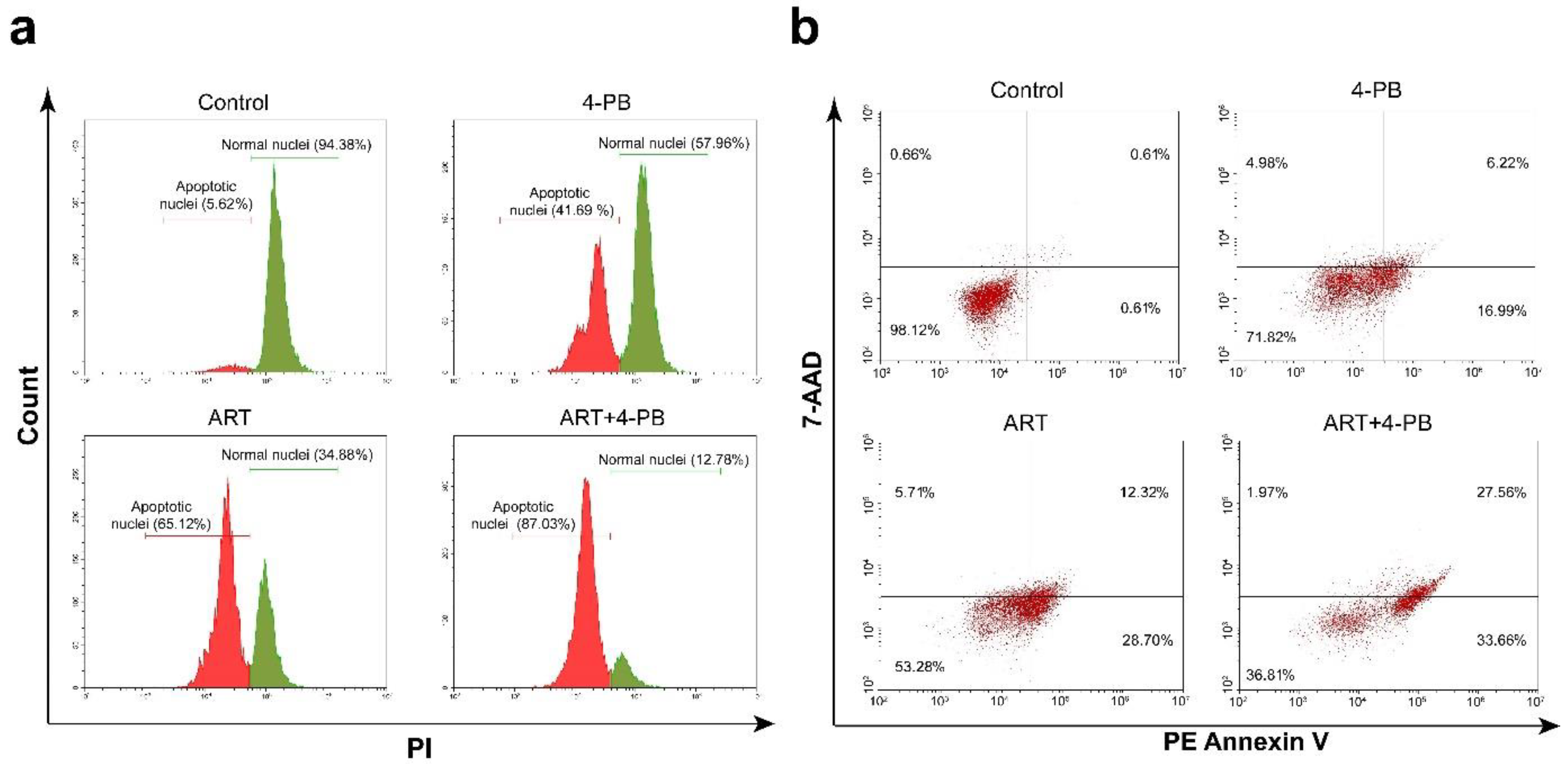
2.5. Propidium Iodide (PI) Apoptosis Assay

The apoptotic population of the cells were analysed using the protocol described earlier [15]. MCF-7 cells were treated with 4-PB and/or ART for 48 h. The cells were collected, fixed in 70% ethanol, and processed in accordance with the aforementioned technique when the treatment period was over. After that, the cells were examined using a Beckman Coulter CytoFLEX flow cytometer with a 488 nm laser line as the excitation source. The PE-A channel was used to measure the red fluorescence of PI.

2.6. Fluoremetric Analyses of Apoptotic Cells Using PE Annexin V and 7-AAD

Following treatment with 5 mM 4-PB and/or 50 µM ART, the various apoptotic cell populations were counted using PE (phycoerythrin), Annexin V, and 7-AAD (7-aminoactinomycin D). Briefly, MCF-7 cells were treated with ART and/or 4-PB for 48 h. Following the manufacturer’s instructions, the cells were harvested and stained with PE Annexin V and 7-AAD (BD Biosciences). The degree of apoptosis in the cells was examined using a flow cytometer (CytoFLEX, Beckman Coulter) as described earlier [16].

2.7. Combination Index (CI) Curve and Isobologram Analyses

The data generated in the cell viability experiment were analysed in COMPUSYN software to study the type of interaction between 4-PB and ART. The normalized isobologram generated by the software, based on Median-Effect Equation (Chou) and the Combination Index Theorem (Chou-Talalay), were then analysed. The CI was used to express antagonism (CI > 1), additivity (CI = 1) or synergism (CI < 1). The analyses were repeated up to four times. The average effect values (fa’s) were used to plot Fa-Log (CI) curve.

2.8. Intracellular ROS Measurement

The levels of ROS generated after treatment with drugs were assessed using the dichlorodihydrofluorescein diacetate (DCF-DA) method [17]. Briefly, cells were treated with the 4-PB and/or ART for 6 h in the presence or absence of 500 µM of N-acetylcysteine (NAC) in the medium. Further, the cells were washed multiple times with PBS and incubated in serum-free medium containing 1 μM of DCFH-DA for 30 min at 37 °C. The data were then acquired using a CytoFLEX flow cytometer (Beckman Coulter) and analysed using WinList 3D 9.0.1 software (Verity Software House, Topsham, ME, USA).

2.9. Cell Cycle Analysis

The cell cycle analysis was performed as described previously [18]. MCF-7 cells were synchronised by serum starvation for 24 h. Further, the cells were treated with 5 mM 4-PB and/or 50 µM ART for 36 h. After the completion of this treatment, the cells were fixed using 70 % ethanol and stained with 25 µM propidium iodide stain following RNAse (10 µg/mL) and 0.1% triton X-100 treatment. The data were acquired using a CytoFLEX flow cytometer (Beckman Coulter) and analysed using ModFit LT software (Verity Software House).

2.10. Relative mRNA Expression Analysis

Quantitative real-time PCR (qPCR) was performed to estimate the fold change in the expression of Ku70, Ku80, XRCC4, and DNA-PKcs using SYBR Green as a reporter dye (iTaq Universal SYBR Green Supermix, Bio-Rad, Hercules, CA, USA) in Rotor-Gene Q (Qiagen, Hilden, Germany). β-actin was taken as the endogenous control. The ΔΔCt method was used to calculate the relative mRNA expression of Ku70, Ku80, XRCC4, and DNA-PKcs in LinRegPCR software.

2.11. In Vivo Study

For the in vivo study, male Swiss albino mice four months old weighing 22–25 g were purchased from Chakraborty enterprise (1443/PO/b/11/CPCSEA), Kolkata, India. All the animals were acclimatised to the laboratory environment at the Central Animal Facility, Institute of Advanced Study in Science and Technology (IASST), Guwahati, Assam, before conducting experiments. Animals were kept in polypropylene cages and maintained at 22 ± 2 °C and 60–70% relative humidity with a 12 h light–dark cycle. Throughout the experimental period, the animals were fed with a standard rodent pellet diet (Provimi Animal Nutrition India Pvt. Ltd., Bengaluru, KA, India). All the protocols were approved by the Institutional Animal Ethics Committee (IAEC) of IASST (IASST/IAEC/2016-17/002) and conducted following the guidelines of Committee for the Purpose of Control and Supervision of Experiments on Animals (CPCSEA), Government of India.

2.12. Dalton’s Ascites Lymphoma Development in Mice

1 × 10⁶ viable Dalton’s ascites lymphoma (DLA) cells were injected into the mice through the intraperitoneal (I.P.) route. Tumour growth was confirmed by belly swelling and increased body weight, which was visible from 8–10 days post transplantation.

2.13. In Vivo Experimental Design

A total of 46 Swiss albino mice were selected for this study (n = 10), where for group-I n = 6. ART and 4-PB doses were scaled up according to the in vitro results. To develop DLA tumours, 0.2 mL of 1 × 10⁶ DLA cells per mouse were inoculated. Drug treatments were started from the 8th day of DLA induction and continued for 8 days with 24 h interval.
Group-I: animals with no DLA tumour + 0.2 mL PBS (I.P).
Group-II: animals with DLA tumour + 0.2 mL PBS (I.P).
Group-III: animals with DLA tumour + 20 mg/kg ART (I.P).
Group-IV: animals with DLA tumour + 50 mg/kg 4-PB (I.P).
Group-V: animals with DLA tumour + 50 mg/kg 4-PB (I.P) + 20 mg/kg ART (I.P).
Body weight changes were measured throughout the experimental period and cells from all treatment groups except Group-I were collected at the 16th day of the experiment to measure cell viability. At the 17th day, five animals from each group were sacrificed by decapitation and specimens of blood, liver and kidney were collected for biochemical and histopathological analysis. The other five animals were observed for a period of 50 days to estimate the mean survival time (MST) and percentage increase in lifespan (% ILS).

2.14. Histopathological Analysis

Pathological changes in liver and kidney were observed by histopathology analysis (Leica Biosystems, Nußloch, Germany). The livers and kidneys were collected in 10% buffered formaldehyde and kept for at least for 24 h. The tissues were dehydrated by gradient alcohol concentrations (50, 70, 90 & 100%) and xylene (1 h in each concentration). Further, the tissues were transferred to melted paraffin and kept for 1 h before embedding in paraffin blocks. Thin 5 µm sections were prepared using a microtome and the sections stained in hematoxylin and eosin. Stained sections were observed under a light microscope (10X) to observe pathological changes.

2.15. Cell Viability Assay

The trypan blue assay was conducted to determine cell viability and to observe cell cytotoxicity. Briefly, 0.1 mL of trypan blue (0.4 %) solution was mixed with 0.1 mL of DLA cells that were collected on day 17. The cells were quantified using an automated cell counter (Countess II FL, Life Technologies, Bothell, WA, USA). The percentage of viable cells was calculated using the following formula-
%   cell   cytotoxicity = 100 n u m b e r   o f   v i a b l e   c e l l s i n   t h e   t r e a t e d   g r o u p n u m b e r   o f   v i a b l e   c e l l s   i n   t h e u n t r e a t e d   c o n t r o l   g r o u p   ×   100
Measurement of percent survival fraction.
Five animals in each group (Except G–I) were observed for 50 days to measure mortality. A Kaplan-Meier curve was plotted to represent percent survival fraction using Prism, version 6.01 software (GraphPad Software Inc., San Diego, CA, USA).

2.16. Statistical Analysis

The degree of significance was determined using an unpaired, two-tailed t-test, and the IC50 dosage was determined using non-linear curve regression analysis. Experiments were carried out at least three times in duplicate and data points were represented as the mean ± SD (SD = standard deviation). One-way and two-way analysis of variance (ANOVA) and Tukey’s post hoc test were applied for pairwise comparisons to determine the statistical significance of differences. Prism, version 6.01 (GraphPad Software Inc., San Diego, CA, USA), was used to analyze the data. Statistically significant values (p-value) for unpaired two-tailed t-test are provided in the Result section. For one-way and two-way analysis of variance (ANOVA) the p-values are * p < 0.05, ** p < 0.01, *** p < 0.001 and **** p < 0.0001.

3. Results

3.1. 4-PB Cytotoxicity on MCF-7 and MDA-MB-231 Cells as a Consequence of HDAC Activity Inhibition

Initial MTT date assay suggested that the 4-PB significantly reduced the cell viability of MCF-7 and MDA-MB-231 cells at different concentrations when administered for 48 h (Figure 1a). However, the effect of 4-PB on MCF-7 cells was more pronounced than on MDA-MB-231 cells. Using a 1 mM concentration of 4-PB, we observed 84% cell viability of MCF-7 cells, which further decreased to 68% at 5 mM concentration. We chose an intermediate dose of 5 mM of 4-PB to further assess the synergism of 4-PB with ART in MCF-7 cells.
As 4-PB-mediated growth inhibition was known to act by inhibiting HDAC [19], we further performed an HDAC activity assay by treating MCF-7 cells with 4-PB with or without ART (Figure 1b). HDAC activity analysis using treated MCF-7 cell lysate showed a significant drop in the 4-PB (p = 0.0079) and 4-PB cells (p = 0.0082) treated with ART when compared with untreated control lysate fluorescence. No inhibition of HDAC was observed in ART-only treated cells.

3.2. 4-PB Enhanced the Dose-Dependent Cytotoxicity of ART

To probe into the effect of ART on MCF-7 cells, a cell viability assay was performed. MCF-7 cells were treated with different doses of ART in the presence or absence of a constant dose of 4-PB (5-mM). Initially, ART treatment for 48 h showed a dose-dependent decrease in the cell viability of MCF-7 cells as depicted in Figure 1c. However, a significant (p = 0.0020) enhancement in the cell cytotoxicity of MCF-7 cells was observed when a similar treatment was performed in the presence of 4-PB. In particular, we observed that 55.56 ± 5.21 µM concentration of ART was required to reach its IC50, while for ART + 4-PB the IC50 concentration was significantly reduced to only 24.71 ± 3.44 µM (Figure 1d).

3.3. Induction of Apoptosis by ART and/or 4-PB

Two independent sets of flow cytometry-based tests were carried out to establish that ART and/or 4-PB treated cells were undergoing apoptosis. First, PI dye was used to analyze the apoptotic nuclei, and then PE Annexin V and the 7-AAD test were used to distinguish between early, late, and necrotic apoptotic cells.
Separation of the apoptotic cell population from the living cell population may be accomplished quickly, accurately, and repeatedly using PI staining. After 48 h of treatment, 4-PB showed an apoptotic cell population of 41.69%, and ART showed an apoptotic cell population of 65.12%. When we treated the MCF-7 cells with the drug combination, the extent of the apoptotic cell population increased drastically to 87.03% (Figure 2a).
We used the PE Annexin V and 7-AAD flow cytometric test to further confirm the results generated by the PI apoptotic assay (Figure 2b). The cells were stained with PE Annexin V and 7-AAD once the treatment period was over. Data from flow cytometry supported the observation that 4-PB increased ART’s cytotoxic impact. We found 23.21% and 41.02% apoptotic cell populations (early and late apoptotic combined) after treatment with 4-PB and ART, respectively. When a combination of drugs was used, the effect was enhanced to 61.22% for the apoptotic cell population.

3.4. Isobologram Depicted the Interaction of 4-PB with ART Is Synergistic

When MCF-7 cells were treated with 4-PB and ART, each of the drugs alone was able to induce apoptosis. However, when treated with the two drugs combined, the effect was substantially greater compared to that of the individual drugs. We investigated the nature of the interaction between these two drugs by plotting isobologram based on the Median-Effect Equation (Chou) and the Combination Index Theorem (Chou-Talalay). The combination index (CI) was used to express antagonism (CI > 1), additivity (CI = 1) or synergism (CI < 1). The CI was calculated using CompuSyn software.
For computing the nature of the interaction between 4-PB and ART, we designed an experiment keeping the concentration of 4-PB constant (i.e., 5-mM) while the concentration of ART was varied. Based on the available information about the mode of action of 4-PB and ART, we assumed that the 4-PB does not preclude the effect of ART and vice versa, thus they can be regarded as mutually non-exclusive. At the lowest concentration (4-PB + 10 µM ART), 23% apoptosis was induced in MCF-7 cells, with a CI value of 1.3, indicating the interaction was close to additive. In contrast, when the concentration of ART was increased (20, 50 100, and 250 µM) the CI value was decreased to 0.66, 0.77, 0.48, and 0.9 (indicating synergism) with an apoptosis of 49, 60, 85, and 89% of cells, respectively. We observed that when the concentration of 4-PB was kept constant and the concentration of ART was increased, the value of CI also decreased with up to a 250 µM concentration of ART, suggesting an increase in synergism between the two drugs (Figure 3a,b).

3.5. 4-PB and ART Increased Oxidative Stress in MCF7 Cells

To investigate the mechanism of cell death induced by 4-PB and ART, we initially examined the role of these drugs in increasing oxidative stress in the cells. The level of intracellular ROS generated was estimated using intracellular peroxide-dependent oxidation of DCFH-DA to form fluorescent DCF. Fluorescence of DCF increases proportionally with the increase in the intracellular ROS generation. To measure ROS, the MCF-7 cells were treated with ART and/or 4-PB in the presence or absence of NAC for 8 h at 37 °C. An increase in DCF fluorescence was noticed in the treated samples with 4-PB or ART (Figure 4a); however, the fluorescence intensity was considerably more in case of ART-treated samples, suggesting an extensive ROS generation. Consequently, the fluorescence intensity increased sharply in samples treated with the combination of both the drugs. The addition of NAC to the medium of the treated samples attenuated DCF fluorescence to that of control fluorescence, implying that the increase in fluorescence was due to ROS generation.

3.6. Cell Cycle Perturbation after Treatment with ART and/or 4-PB

Cytofluorimetric analysis of cells after synchronisation and treatment with ART and/or 4-PB showed that MCF-7 cells were progressively blocked in the G0/G1 phase of the cell cycle. However, after treatment with the drug combination, the percentage of the cell population in the G0/G1 phase was considerably higher than in cells exposed to individual drugs alone. When treated with 4-PB and ART alone, the percentages of cells in the G0/G1 phase of the cell cycle were 69.38% and 71.82%, respectively. The G0/G1 phase population reached 83.84% after treatment with both 4-PB + ART combined. The data suggested that 4-PB enhanced the G0/G1 arrest induced by the ART treatment (Figure 4b).

3.7. Mechanism Underlying 4-PB Mediated Enhanced ART DNA Damaging Effect

We provided evidence that 4-PB enhances the effect of ART in MCF-7 cells, which led us to find the underlying mechanism behind the synergism showed by 4-PB and ART.
As an initial investigation into the underlying mechanism, we examined the effect of 4-PB on the expression of well-known DDR proteins, with or without the treatment involving ART. Figure 5 shows real-time PCR analysis of the mRNA expression of Ku70, Ku80, XRCC4, and DNA-PKcs performed on the MCF-7 cells after treatment with ART and/or 4-PB. The data show a significant decrease in the expression of all the four proteins at the mRNA level when treated with 4-PB alone. However, the expression of Ku70, Ku80, XRCC4, and DNA-PKcs increased significantly following treatment with ART.

3.8. Therapeutic Effectivity of Combinated Drusg in a Murine Model

For the assessment of the potentiality of the drug combination, the mice were split into five groups (n = 10). Except in group 1, DLA was induced in all other groups. It took 8 days for the tumour to attain an appropriate volume for drug treatment (swelling of the peritoneum was due to the accumulation of ascitic fluid). Administration of the drug was started after the tumour was formed (from the 8th day). On the 17th day, a cell viability assay was performed and the acquired data (Table 1) showed a significant decrease in the viable cell count after treatment with the combination drug compared with the other treatment groups (4-PB or ART). Along with cell viability, reduction in the volume of the tumour followed the same pattern as when 4-PB and ART were administered simultaneously in DLA mice, and the effectivity was greater compared with single drug treatments (Figure 6a). The above finding was also reflected in the lifespan of the mice (Figure 6b). In the control group, all the DLA mice died at the end of the 18th day after tumour inoculation, while the 4-PB or ART-treated mice were alive for up to 59 days but eventually died at the end of the observation period. However, the group receiving the combination treatment was alive till the end of the observation period. Although both drugs individually caused an effective reduction in cell viability as well as shrinking of tumour volume, both failed to increase the lifespan of the mice, while the combination drug did successfully increase the lifespan of the mice.
DLA-bearing mice were observed to have elevated levels of WBC and reduced levels of Hb and RBC count. Treatment with the combination drugs reversed these conditions and restored the levels of WBC, RBC, and Hb counts to normal (Table S1). A significant decline in the liver function enzymes SGOT, SGPT & ALP was observed compared to the control group (Table S2). In Figure 6c, histopathological examination of liver and kidney tissues show a remarkable recovery in conditions that changed due to the DLA induction. For instance, in kidney, glomerular atrophy, tubular congestion, infiltration of cells and deformed epithelial cells were seen; however, the conditions reverted to normal with a decreased occurrence of tubular, glomerular and blood vessel congestion in the treated groups. In the liver, an abnormal arrangement of hepatocytes with polymorphic neutrophil infiltration, hepatic fibrillation, haemorrhage and perinuclear clumping of cytoplasm was observed in control group. Interestingly, in the treated group, reduction of damage of normal cellular ultrastructures comparable to normal animals was observed. Overall, these changes were more profound in the combination drug-treated group compared to the individual drug treatment regime.

4. Discussion

In this study, we showed that 4-PB enhanced the cytotoxicity of the traditional antiparasitic drug ART against a breast cancer cell line, as well as DLA, in mice. 4-PB synergistically ameliorated the effect of ART in a dose-dependent manner by contributing to G1 arrest of the cell cycle, increasing ROS production. Additionally, it could subjugate the DDR pathways in the MCF-7 cells, thus amplifying the DNA damage effect of ART.
ART is recognised as a remarkable new generation of antimalarial drugs with no noticeable side effects or obvious adverse reactions in humans [20]. Recent experimental evidence suggested that ART may provide a therapeutic alternative in rapid disseminating cancers, without inducing drug-based resistance [21]. Although, a few reports of cell lines and laboratory animal toxicity have raised concerns due to high doses and prolonged use of ART [13], these findings have not been reported to date in humans taking numerous doses of artemisinin. Keeping this in mind, we devised a novel combination therapy involving ART and 4-PB, in which, 4-PB reduced the effective cytotoxic ART concentration synergistically when administered to an MCF-7 cell line and in a murine model.
4-PB has been effectively used in patients with hyperammonemia and urea cycle disorders (UCDs) involving deficiencies of carbamyl phosphate synthetase (CPS), ornithine transcarbamylase (OTC), or argininosuccinic acid synthetase (AS) [22]. Although, two HDACi, namely vorinostat (suberoylanilide hydroxamic acid, Zolinza) and depsipeptide (romidepsin, Istodax), received approval from the FDA for the treatment of cutaneous T-cell lymphoma in 2006 and 2009 [23], respectively, 4-PB’s potentiality as an anti-cancer drug is still under evaluation in various clinical trials [24].
Our observations indicated the enhancement of the cytotoxity of ART when combined with 4-PB. We further investigated the role of 4-PB in heightening the effect of ART by analysing the distribution of cells in several phases of the cell cycle. 4-PB has been known to cause a G1 arrest in cells by increasing the expression of p21 protein [25]. Thus, when synchronised MCF-7 cells were treated with a combination drug, they showed a substantial accumulation of cells in G1 phase. Basically, 4-PB amplified G1 arrest after treatment with ART. In a similar fashion, we also observed the enhancement in ROS generation inside the cells after the combination treatment.
Our data imply that 4-PB has strong synergism with ART in inducing a profound level of apoptosis in dose-dependent manner. Based on CI values, we deduced that the interaction between 4-PB and ART was synergistic rather than additive. To figure out the underlying pathway responsible for the enhanced effect, we checked for the expression levels of DDR elements after drug treatment. To date, there has been no report of downregulation of DDR elements after the treatment with 4-PB in MCF-7 cells. The only notable exception is a report that demonstrated that the role of 4-PB in the attenuation of the expression of Bcl-XL, DNA-PK, Caveolin-1, and VEGF was behind the increase in the sensitivity towards radiation-induced apoptosis in prostate cancer cells [26]. To elucidate the exact molecular mechanism by which 4-PB exerts a synergistic effect with ART, we analysed DNA repair processes, as it was reported earlier that ART induces DNA damage in cancer cells mediated by ROS [27,28]. Based on these reports, we investigated the expression levels of key proteins involved in the nonhomologous end joining pathway, namely Ku70, Ku86, XRCC4, and DNA-PKcs, which is crucial in DDR processes [29,30]. The qPCR analysis of the expression of these proteins showed downregulation at mRNA level after treatment with 4-PB. However, when treated with ART alone, the levels of these proteins increased significantly. Interestingly, the elevation nin the expression of these proteins diminished when 4-PB was introduced with ART in the medium, suggesting the involvement of attenuation of Ku70, Ku86, XRCC4, and DNA-PKcs by 4-PB in enhancing the effect of the combination drug.
To further examine the efficacy of our combination drug cocktail, we used a murine model. We used DLA mice for a preliminary investigation of the drug cytotoxicity for our in vivo studies. As DLA mimics most of the cancer types and is easy to maintain, it was used to provide fast and reliable information of the cytotoxic effects of the drug in vivo [31,32,33].
DLA-bearing Swiss albino mice were initially tested for tolerable doses of ART and/or 4-PB. Once done, we started the treatment of the DLA mice with ART and/or 4-PB as described in Materials and Methods section. We found a reduction in the volume of the tumour after treatment with ART and/or 4-PB, both visually and by weighing the mice. However, the reduction in tumour volume was more pronounced in mice treated with the combination drug. The survival probability data corroborated with the above findings. After an observation period of 40 days, mice treated with the combination drug survived over 40 days while others died within the observation period. The histological parameters of the treated mice were improved drastically compared with the control DLA mice.
In conclusion, we present a novel combination of drugs that are already in use for the treatment of other diseases, as a probable anti-cancer therapy. This drug combination holds immense potential for further testing in clinical trials. In this study, we showed that 4-PB synergistically enhanced the efficacy of ART and potentiated its cytotoxicity against MCF-7 cells. The underlying mechanism behind this revolves around the overall attenuation of the DDR elements inside the cell. The combination therapy showed its effectivity in DLA mice model. Overall, a comprehensive understanding of these findings requires further studies. However, the observations reported here emphasise the need for persistent improvement of strategies for making human tumour cells susceptible to cancer therapies that kill cancer cells by inducing DNA damage.

Supplementary Materials

The following supporting information can be downloaded at: https://www.mdpi.com/article/10.3390/sci4040041/s1, Table S1: Effect of drug treatment on hematological parameters. Table S2: Effect of drug treatment on serum biochemical enzymes. Figure S1: Reduction in the tumour volume was easily visualised in above image, taken after the respective drug treatment.

Author Contributions

Conceptualization, A.R. and S.S.G.; methodology, A.R.; software, A.R.; validation, A.R., S.S.G., R.K. and S.K.; investigation, A.R., S.S.G. and R.K.; resources, S.S.G.; data curation, A.R. and S.S.G.; writing—original draft preparation, A.R.; writing—review and editing, S.S.G., R.K. and S.K.; visualization, A.R.; supervision, S.S.G.; project administration, S.S.G.; funding acquisition, S.S.G. All authors have read and agreed to the published version of the manuscript.

Funding

The authors primarily acknowledge the financial support of the Department of Biotechnology, Government of India, for DBT Program Support (BT/PR13560/COE/34/44/2015) grant. Partial support by the Department of Biotechnology (DBT-NER/Health/47/2015) and the Department of Electronics and Information Technology (No. 5(9)/2012-NANO (Vol. II)), Government of India are also acknowledged.

Institutional Review Board Statement

All the protocols were approved by the Institutional Animal Ethics Committee (IAEC) of IASST (IASST/IAEC/2016-17/002) and conducted by following the guidelines of Committee for the Purpose of Control and Supervision of Experiments on Animals (CPCSEA), Government of India.

Informed Consent Statement

Not applicable.

Data Availability Statement

The datasets during and/or analysed during the current study are available from the corresponding author on reasonable request. All data generated or analysed during this study are included in this published article (and its Supplementary Information Files).

Acknowledgments

Authors acknowledge the aid of the Central Instruments Facility (CIF) and the Centre for Nanotechnology, IIT Guwahati.

Conflicts of Interest

The authors declare no conflict of interest. The funders had no role in the design of the study; in the collection, analyses or interpretation of the data; in the writing of the manuscript; or in the decision to publish the results.

Ethics Approval

All the protocols were approved by the Institutional Animal Ethics Committee (IAEC) of IASST (IASST/IAEC/2016-17/002) and were conducted by following the guidelines of Committee for the Purpose of Control and Supervision of Experiments on Animals (CPCSEA), Government of India.

References

  1. Sakuma, T.; Uzawa, K.; Onda, T.; Shiiba, M.; Yokoe, H.; Shibahara, T.; Tanzawa, H. Aberrant expression of histone deacetylase 6 in oral squamous cell carcinoma. Int. J. Oncol. 2006, 29, 117–124. [Google Scholar] [CrossRef] [PubMed]
  2. Kumar, S.; Attrish, D.; Srivastava, A.; Banerjee, J.; Tripathi, M.; Chandra, P.S.; Dixit, A.B. Non-histone substrates of histone deacetylases as potential therapeutic targets in epilepsy. Expert Opin. Ther. Targets 2021, 25, 75–85. [Google Scholar] [CrossRef] [PubMed]
  3. Stypula-Cyrus, Y.; Damania, D.; Kunte, D.P.; Cruz, M.D.; Subramanian, H.; Roy, H.K.; Backman, V. HDAC up-regulation in early colon field carcinogenesis is involved in cell tumorigenicity through regulation of chromatin structure. PLoS ONE 2013, 8, e64600. [Google Scholar] [CrossRef] [PubMed] [Green Version]
  4. Peart, M.J.; Smyth, G.K.; Van Laar, R.K.; Bowtell, D.D.; Richon, V.M.; Marks, P.A.; Holloway, A.J.; Johnstone, R.W. Identification and functional significance of genes regulated by structurally different histone deacetylase inhibitors. Proc. Natl. Acad. Sci. USA 2005, 102, 3697–3702. [Google Scholar] [CrossRef] [PubMed] [Green Version]
  5. Kelly, W.K.; O’Connor, O.A.; Marks, P.A. Histone deacetylase inhibitors: From target to clinical trials. Expert Opin. Investig. Drugs 2002, 11, 1695–1713. [Google Scholar] [CrossRef]
  6. Brusilow, S.W.; Finkelstien, J. Restoration of nitrogen homeostasis in a man with ornithine transcarbamylase deficiency. Metabolism 1993, 42, 1336–1339. [Google Scholar] [CrossRef]
  7. Rubenstein, R.C.; Zeitlin, P.L. A Pilot Clinical Trial of Oral Sodium 4-Phenylbutyrate (Buphenyl) in Δ F508-Homozygous Cystic Fibrosis Patients: Partial Restoration of Nasal Epithelial CFTR function. Am. J. Respir. Crit. Care Med. 1998, 157, 484–490. [Google Scholar] [CrossRef] [PubMed]
  8. Munshi, A.; Kurland, J.F.; Nishikawa, T.; Tanaka, T.; Hobbs, M.L.; Tucker, S.L.; Ismail, S.; Stevens, C.; Meyn, R.E. Histone deacetylase inhibitors radiosensitize human melanoma cells by suppressing DNA repair activity. Clin. Cancer Res. 2005, 11, 4912–4922. [Google Scholar] [CrossRef] [Green Version]
  9. Rosenthal, P.J. Artesunate for the treatment of severe falciparum malaria. N. Engl. J. Med. 2008, 358, 1829–1836. [Google Scholar] [CrossRef] [Green Version]
  10. Crespo-Ortiz, M.P.; Wei, M.Q. Antitumor activity of artemisinin and its derivatives: From a well-known antimalarial agent to a potential anticancer drug. J. Biomed. Biotechnol. 2012, 2012, 247597. [Google Scholar] [CrossRef] [Green Version]
  11. Raza, A.; Ghoshal, A.; Chockalingam, S.; Ghosh, S.S. Connexin-43 enhances tumor suppressing activity of artesunate via gap junction-dependent as well as independent pathways in human breast cancer cells. Sci. Rep. 2017, 7, 7580. [Google Scholar] [CrossRef] [PubMed]
  12. Hosoya, N.; Miyagawa, K. Targeting DNA damage response in cancer therapy. Cancer Sci. 2014, 105, 370–388. [Google Scholar] [CrossRef] [PubMed] [Green Version]
  13. Gordi, T.; Lepist, E.-I. Artemisinin derivatives: Toxic for laboratory animals, safe for humans? Toxicol. Lett. 2004, 147, 99–107. [Google Scholar] [CrossRef] [PubMed]
  14. Raza, A.; Kohila, V.; Ghosh, S.S. Redesigned Escherichia coli cytosine deaminase: A new facet of suicide gene therapy. J. Gene Med. 2015, 17, 132–139. [Google Scholar] [CrossRef]
  15. Raza, A.; Ghosh, S.S. Connexin-43 Enhances the Redesigned Cytosine Deaminase Activity for Suicide Gene Therapy in Human Breast Cancer Cells. Biochem. Insights 2019, 12, 1178626418818182. [Google Scholar] [CrossRef]
  16. Goswami, U.; Dutta, A.; Raza, A.; Kandimalla, R.; Kalita, S.; Ghosh, S.S.; Chattopadhyay, A. Transferrin–copper nanocluster–doxorubicin nanoparticles as targeted theranostic cancer Nanodrug. ACS Appl. Mater. Interfaces 2018, 10, 3282–3294. [Google Scholar] [CrossRef]
  17. Raza, A.; Singh, A.; Amin, S.; Spallholz, J.E.; Sharma, A.K. Identification and biotin receptor-mediated activity of a novel seleno-biotin compound that inhibits viability of and induces apoptosis in ovarian cancer cells. Chem.-Biol. Interact. 2022, 365, 110071. [Google Scholar] [CrossRef]
  18. Ghoshal, A.; Goswami, U.; Raza, A.; Chattopadhyay, A.; Ghosh, S.S. Recombinant sFRP4 bound chitosan–alginate composite nanoparticles embedded with silver nanoclusters for Wnt/β-catenin targeting in cancer theranostics. RSC Adv. 2016, 6, 85763–85772. [Google Scholar] [CrossRef]
  19. Gore, S.D.; Carducci, M.A. Modifying histones to tame cancer: Clinical development of sodium phenylbutyrate and other histone deacetylase inhibitors. Expert Opin. Investig. Drugs 2000, 9, 2923–2934. [Google Scholar] [CrossRef]
  20. Benakis, A.; Paris, M.; Loutan, L.; Plessas, C.T.; Plessas, S.T. Pharmacokinetics of artemisinin and artesunate after oral administration in healthy volunteers. Am. J. Trop. Med. Hyg. 1997, 56, 17–23. [Google Scholar] [CrossRef]
  21. Fishwick, J.; McLean, W.G.; Edwards, G.; Ward, S.A. The toxicity of artemisinin and related compounds on neuronal and glial cells in culture. Chem.-Biol. Interact. 1995, 96, 263–271. [Google Scholar] [CrossRef]
  22. Batshaw, M.L.; MacArthur, R.B.; Tuchman, M. Alternative pathway therapy for urea cycle disorders: Twenty years later. J. Pediatrics 2001, 138, S46–S55. [Google Scholar] [CrossRef] [PubMed] [Green Version]
  23. Ververis, K.; Hiong, A.; Karagiannis, T.C.; Licciardi, P.V. Histone deacetylase inhibitors (HDACIs): Multitargeted anticancer agents. Biol. Targets Ther. 2013, 7, 47. [Google Scholar]
  24. Smith, K.T.; Workman, J.L. Histone deacetylase inhibitors: Anticancer compounds. Int. J. Biochem. Cell Biol. 2009, 41, 21–25. [Google Scholar] [CrossRef] [PubMed]
  25. Blagosklonny, M.V.; Robey, R.; Sackett, D.L.; Du, L.; Traganos, F.; Darzynkiewicz, Z.; Fojo, T.; Bates, S.E. Histone deacetylase inhibitors all induce p21 but differentially cause tubulin acetylation, mitotic arrest, and cytotoxicity. Mol. Cancer Ther. 2002, 1, 937–941. [Google Scholar] [PubMed]
  26. Goh, M.; Chen, F.; Paulsen, M.T.; Yeager, A.M.; Dyer, E.S.; Ljungman, M. Phenylbutyrate attenuates the expression of Bcl-X(L), DNA-PK, caveolin-1, and VEGF in prostate cancer cells. Neoplasia 2001, 3, 331–338. [Google Scholar] [CrossRef] [Green Version]
  27. Berdelle, N.; Nikolova, T.; Quiros, S.; Efferth, T.; Kaina, B. Artesunate induces oxidative DNA damage, sustained DNA double-strand breaks, and the ATM/ATR damage response in cancer cells. Mol. Cancer Ther. 2011, 10, 2224–2233. [Google Scholar] [CrossRef] [Green Version]
  28. Li, P.C.; Lam, E.; Roos, W.P.; Zdzienicka, M.Z.; Kaina, B.; Efferth, T. Artesunate derived from traditional Chinese medicine induces DNA damage and repair. Cancer Res. 2008, 68, 4347–4351. [Google Scholar] [CrossRef] [Green Version]
  29. Haber, J.E. Partners and pathways: Repairing a double-strand break. Trends Genet. 2000, 16, 259–264. [Google Scholar] [CrossRef]
  30. Khanna, K.K.; Jackson, S.P. DNA double-strand breaks: Signaling, repair and the cancer connection. Nat. Genet. 2001, 27, 247–254. [Google Scholar] [CrossRef]
  31. Koiri, R.K.; Mehrotra, A.; Trigun, S.K. Dalton’s lymphoma as a murine model for understanding the progression and development of t-cell lymphoma and its role in drug discovery. Int. J. Immunother. Cancer Res. 2017, 3, 001–006. [Google Scholar]
  32. Choudhury, B.; Kandimalla, R.; Bharali, R.; Monisha, J.; Kunnumakara, A.B.; Kalita, K.; Kotoky, J. Anticancer activity of Garcinia morella on T-cell murine lymphoma via apoptotic induction. Front. Pharmacol. 2016, 7, 3. [Google Scholar] [CrossRef] [PubMed]
  33. Shanker, A.; Singh, S.M.; Sodhi, A. Ascitic growth of a spontaneous transplantable T cell lymphoma induces thymic involution. Tumor Biol. 2000, 21, 315–327. [Google Scholar] [CrossRef] [PubMed]
Figure 1. Dose dependent cell viability and HDAC inhibition assay. (a) Reduction in cell viability was assessed after treating MCF-7 and MDA-MB-231 cells with different concentrations of 4-PB. (b) 4-PB acted by inhibiting HDAC activity in MCF-7 cells. (c) Dose dependent cell viability of ART and in combination with 5 mM 4-PB was examined by MTT based cell viability assay. (d) IC50 value of ART and ART in combination with 5 mM 4-PB was calculated using non-linear curve regression analysis and the level of significance were calculated using unpaired two tailed t-test (n = 3, p = 0.0020). Data are represented as mean ± SD (n = 4) in all the graphs. * p < 0.05, ** p < 0.01, *** p < 0.001 and **** p < 0.0001.
Sci 04 00041 g001
Figure 2. Enhanced cell cytotoxicity of ART in combination with 4-PB was assessed by flow cytometry-based apoptotic assays. (a) PI based apoptotic cell analysis using flow cytometer. (b) Assessment of early apoptotic (Annexin V), late apoptotic (Annexin V + 7-AAD) and necrotic cells (7-AAD) was performed using PE Annexin V and 7-AAD-based assay. Four distinct populations of cells were produced by the method: healthy cells (Annexin-V negative and 7-ADD negative; lower right quadrant), early apoptotic cells (positive for Annexin-V and negative for 7-ADD; lower right corner), late apoptotic/dead cells (both Annexin V and 7-ADD positive; upper right quadrant), and necrotic cells (only 7-ADD positive).
Sci 04 00041 g002
Figure 3. The nature of interaction between 4-PB and ART was analysed using the Median-Effect Principle (Chou) and the Combination Index-Isobologram Theorem (Chou-Talalay). (a) CI plot showing substantial synergism at higher Fa values. (b) Isobologram showing visual, not quantitative, determination of synergism or antagonism. Combination data points on the isobologram show the nature of interaction with the combination treatment at different Fa.
Sci 04 00041 g003
Figure 4. ROS generation and cell cycle analysis. (a) ROS generation in MCF-7 cells was examined by using DCFH-DA dye. The fluorescence level of DCF corresponds to the amount of ROS generated inside the cell. (b) The synchronised MCF-7 cells were treated with ART and/or 4-PB. After the end of the treatment duration, the phases of cell cycle were evaluated using PI dye. The data were acquired in the flow cytometer and analysed using ModFit LT software.
Sci 04 00041 g004
Figure 5. Expression analyses of DDR pathways. mRNA expression levels of DDR elements of the NHEJ pathway. Ku70, Ku80, XRCC4 and DNA-PKcs were assessed using qPCR (n = 3). Statistically significant values were taken as * p < 0.05, ** p < 0.01 and *** p < 0.001.
Sci 04 00041 g005
Figure 6. In vivo assessment of the combination drug treatment. (a) Reduction in the weight of the mice indicates reduction in volume of the tumour. The effect of drugs on the tumour volume after 15 days of DLA induction and seven days of drug treatment were investigated by weighing mice at different time intervals. (b) Changes in the lifespan of the mice were analysed after calculating the percent survival fraction of the mice after treatment. The percent survival curve is plotted using the Kaplan-Meier curve. (c) Histopathological analysis of liver and kidney tissues of mice (magnification 20×). * p < 0.05.
Sci 04 00041 g006
Table 1. Effect of drug treatment on DLA cell viability in mice.
S. NoTreatmentViable Cell Count (105 Cells/mL)
1DLA + Saline418.4 ± 19.7
2DLA + 4-PB 10.35 ± 1.5 *
3DLA + Artesunate5.82 ± 1.4 *
4DLA + 4-PB + Artesunate2.47 ± 0.8 *
All the results ae expressed as mean ± S.D. * p < 0.05 comparing drug-treated groups with the untreated group.
Publisher’s Note: MDPI stays neutral with regard to jurisdictional claims in published maps and institutional affiliations.

