Article

Dietary Nanocurcumin Impacts Blood Biochemical Parameters and Works Synergistically with Florfenicol in African Catfish Challenged with Aeromonas veronii

1
Zoology Department, Faculty of Science, South Valley University, Qena 83523, Egypt
2
Department of Fish Diseases, Faculty of Veterinary Medicine, South Valley University, Qena 83523, Egypt
3
Botany and Microbiology Department, Faculty of Science, South Valley University, Qena 83523, Egypt
4
Department of Physiology, Faculty of Veterinary Medicine, South Valley University, Qena 83523, Egypt
5
General Authority for Veterinary Services, Qena Veterinary Directorate, Qena 83511, Egypt
6
Department of Rangeland, Wildlife and Fisheries Management, Texas A&M University, College Station, TX 77843, USA
*
Authors to whom correspondence should be addressed.
Fishes 2023, 8(6), 298; https://doi.org/10.3390/fishes8060298
Submission received: 6 May 2023 / Revised: 29 May 2023 / Accepted: 29 May 2023 / Published: 2 June 2023

Abstract

:
The present study investigated the effects of dietary supplementation of nanocurcumin (NCur = 150 ppm) in African catfish feed for 15 days along with two doses of florfenicol—a therapeutic dose (FFC1 = 10 mg/kg.BW) and a high dose (FFC2 = 30 mg/kg.BW). Serum biochemical parameters and histopathological changes in liver and kidney tissues were determined post-feeding. Additionally, fish were experimentally challenged with a virulent strain of Aeromonas veronii to explore the fish’s capacity to resist infections. Results indicated that the serum level of total protein was significantly elevated in the FFC2 + Ncur group compared with control, Ncur, and FFC2 groups. Total protein level was also higher in FFC1 + Ncur compared with that of the FFC1 group. Moreover, feeding NCur, FFC1 + Ncur, and FFC2 + Ncur resulted in a significant increase in globulin levels compared to control, FFC1, and FFC2-Fed groups. However, the albumin level was unaffected. Serum levels of AST, ALT, ALP, and urea significantly increased in FFC2-fed fish compared to controls indicating liver damage. The higher dosage of FFC2 induced histological alterations in the liver and kidney architecture, represented by a rupture of the central vein, cytoplasmic vacuolation, hepatocytes necrosis, and severe inflammation. Surprisingly, NCur inclusion improved most of the altered biochemical parameters and mitigated FFC-induced histological damage in the hepatic and renal tissues. Similarly, fish that were fed a combination of both FFC and Ncur showed a higher resistance when exposed to A. veronii infection, as the mortality rates were significantly lower than those of the control group. Our findings indicated that incorporating NCur into the diet can have multiple benefits for the sustainable aquaculture of African catfish. These advantages encompass mitigating drug-induced damage to the liver and kidney tissues, augmenting the therapeutic efficacy of FFC in a synergistic manner and providing protection against A. veronii infections.
Key Contribution: Nanocurcumin (NCur) inclusion in African catfish feed with florfenicol (FFC) improved blood biochemical parameters, mitigated FFC-induced histological damage to hepatic and renal tissues, augmented FFC therapeutic efficacy, and increased resistance to an A. veronii infection. The study highlights the potential benefits of incorporating NCur into the feed for more environmentally friendly and sustainable African catfish aquaculture.

Graphical Abstract

1. Introduction

The global population growth and the limitations in expanding capture fisheries have made it necessary to rely on aquaculture to meet the increasing demand for seafood. As of 2018, global fish production totaled 179 million tonnes, with a total sale value estimated at 401 billion USD (FAO, 2020), of which 82 million tonnes, valued at 250 billion USD, were produced by aquaculture [1]. However, infectious diseases cause substantial production losses in aquaculture every year [2]. Among the most common and troublesome diseases of fish raised in aquaculture ponds are bacterial infections caused by motile members of the genus Aeromonas [3]. Traditionally, antibiotics have been used in aquaculture to prevent and treat bacterial diseases. There are currently only three antibiotics approved by the U.S. Food and Drug Administration for use in aquaculture: oxytetracycline (Terramycin 200), ormetoprim-sulfadimethoxine (Romet-30), and florfenicol (Aquaflor). Florfenicol (FFC) is one of the most frequently used antibiotics in aquacultural settings globally [4]. FFC is a veterinary antibiotic with a broad-spectrum activity that inhibits the synthesis of protein in many Gram-negative and -positive bacteria [5,6]. It is a fluorinated derivative analog of thiamphenicol, and its mechanism of action resembles that of chloramphenicol. FFC inhibits protein synthesis by binding to the 50S subunit of the ribosome of the bacteria [7,8]. As a result of its widespread use and application, FFC has been detected at low concentrations of about 0.01 μg L−1 in aquatic environments [9,10].
The recommended therapeutic dose of FFC is 10–15 mg/kg fish for 10 consecutive days [11], which has been approved for treatment of a range of fish diseases in various warm- and cold-water cultured species as a medicated feed [12,13]. Despite the fact that FFC and other antibiotics have many benefits, their abuse or misuse in aquaculture can have a number of adverse effects. The rampant utilization of antibiotics in industrial aquaculture has led to the accelerated development of antibiotic resistance in bacteria that can cause diseases in humans. Furthermore, commercial fish products have been found to contain traces of residual antibiotics, which presents a significant risk to public health. It also suppresses the immune system of the host, slows growth, and causes other ecological problems not only for fish, but also for humans, animals, and the environment as well [14,15,16]. One alternative strategy to control bacterial infections among farmed fish involves the use of natural plant-derived bioactives or phytocompounds.
Fish farmers are constantly looking for alternative sources to improve fish growth, health, and overall performance. Functional feed additives, including phytogenics, are gaining more attention in recent times as a sustainable approach to promoting fish health, stimulating their immune system, and providing physiological benefits that go beyond traditional diets [17,18]. Curcumin or diferuloylmethane (1,7-bis (4-hydroxy-3 methoxyphenyl)-1,6-heptadiene-3,5-dione) is a polyphenol energetic product extracted from turmeric Rhizome roots (Curcuma longa) and has countless therapeutic benefits [19]. Numerous studies conducted in the past have shown the beneficial effects of turmeric and curcumin on human health, as well as on animals, in terms of their anti-cancer, anti-bacterial, and anti-inflammatory properties. In addition, they have been found to act as growth promoters and provide antioxidant protection for various organs, including the liver, stomach, and nervous system [20,21]. Despite the numerous benefits associated with curcumin, its impact on animal health may be limited since it is rapidly metabolized and excreted from the body [22]. Recently, various studies have investigated the incorporation of curcumin into the diets of different animals—including fish—either alone or in combination with other feed additives [22,23,24,25]. In addition to using the raw form of curcumin, there have been several studies exploring the use of curcumin nanoparticles [26,27].
The African sharptooth catfish (Clarias gariepinus) is a commercially important, native fish species to Africa but has been widely introduced in various countries around the world. Due to its broad distribution, accessibility throughout the year, and adaptability to laboratory-controlled conditions, C. gariepinus is a highly suitable model species for toxicological studies [28]. The purpose of this study was to investigate the potential effects of feeding NCur (150 ppm) and florfenicol (10 and 30 mg/kg.BW) separately and combined for 15 days on the serum biochemical indices and histological architecture of liver and kidney tissues of C. gariepinus. In addition, a challenge experiment was conducted to assess resistance of C. gariepinus to Aeromonas veronii infection.

2. Materials and Methods

2.1. Florfenicol Medicated Feed

Florfenicol, as an antimicrobial powder (Panflor®), was obtained from Arabcomed, Marcyrl Animal Health (Marcyrl Pharmaceutical Industries, Cairo, Egypt). The recommended dose of FFC is 10 mg/kg body weight/day for 10 days. FFC was incorporated into commercial feed pellets (Skretting, Egypt) at a 200 mg/kg and 600 mg/kg diet and administrated to fish (feeding rate = 5% body weight) to deliver final doses of 10 mg florfenicol per kg and 30 mg FFC per kg of body weight, respectively. The desired amount of FFC was mixed well with vegetable oil, then this emulsion was strongly mixed with feed to achieve the required concentrations. The prepared diets were air-dried and kept in plastic containers in a dark and cool place until use [29]. Medicated feed was the sole ration for 15 consecutive days prior to sampling and experimental infection. The high dose was chosen according to a previous study [30], in which the pharmacokinetics and biosafety of FFC were evaluated at different doses.

2.2. Preparation of Nanocurcumin

Curcumin nanoparticles (NPs) were biosynthesized using the marine brown alga/seaweed, Dictyota dicotoma, following the methods described by Kumar et al. [31]. Briefly, tufts of D. dichotoma species growing on the coral reefs were harvested from coastal areas in Hurghada, Egypt. Epiphytes were removed from healthy algae, and the necrotic and extra material were discarded. Samples were thoroughly cleaned with sea water and then sterile distilled water. The samples were air-dried and then chopped into little pieces before being ground into a fine powder using a tissue grinder IKA A10 (IKA®-Werke GmbH, Staufen, Germany). One gram of the fine powder was dissolved in 100 mL of distilled water at 100 °C for 1 h in an Erlenmeyer flask. The extract was filtered using Rotilabo® Tyb 601P filter paper, heat sterilized by autoclaving at 121 °C for 15 min, and stored in a refrigerator at 4 °C for future use. To exclude the antibacterial properties of D. dichotoma extract against A. veronii, we used a disk-diffusion susceptibility test. There was 15 µL of the extract applied onto a blank disk and placed on a tryptic soy agar plate inoculated with A. veronii. After 48 h, no growth inhibition was observed, indicating no antibacterial activity.
Curcumin (Cur) powder was provided by SD-Fine Chemicals Limited (SDFCL, Mumbai, India). For synthesis of Curcumin/D. dichotoma nanoparticles, a nanoprecipitation method was utilized [32,33], where the extract of D. dichotoma was used as a reducing and capping agent. Briefly, a 100 mL aliquot of freshly prepared D. dichotoma extract was transferred to a 250 mL conical flask. Curcumin powder (0.5 g) was dissolved in 150 mL of distilled water under continuous stirring at 45 °C and the aqueous solution of curcumin was added in a dropwise manner to the D. dichotoma extract. Within 1 h, the color of the solution changed from bright yellow to orange with a precipitate, signifying the creation of Cur/D. dichotoma NPs. The resulting solution was kept under stirring for a further 4 h to ensure a full reaction. To separate the Cur/D NPs, the solution was centrifuged at 6000 rpm/min for 10 min, followed by extracting the pellets.
For characterization of the prepared Cur/D. dichotoma NPs, the absorption spectra were recorded by a JASCO UV-2300 spectrophotometer (JASCO INTERNATIONAL CO., LTD. Tokyo, Japan) operated at a resolution of 1 nm. To determine the diameter, size, morphology, and structure, Cur/D. dichotoma NPs were imaged using a transmission electron microscope (TEM, JEOL JEM-1010, JEOL LTD., Tokyo, Japan), fitted with a digital “Kodak Megaplus® 1.6i camera and image-processing software (AMT advantage camera system V2. 25.4, Ford City, PA, USA). Fourier transform infrared (FTIR) spectroscopy was used to determine the functional groups of fresh curcumin and Cur/D. dichotoma NPs using a FTIR Model 560 (Nicolet, Madison, WI, USA) with a resolution of 4.00 cm−1, and covers the wave number range of 4000–400 cm−1. To ensure that there were no remnants of algal extract, byproducts, or any unbound materials in the curcumin nanoparticles, they were washed with sterile distilled water. After that, the purified nanoparticles were separated from the rinsing solution through centrifugation. To complete the process, the purified curcumin nanoparticles were dried by heating them to 90 °C. The purified curcumin nanoparticles were used to supplement the fish diet.

2.3. Fish Acclimation and Husbandry Conditions

The Research Ethics Committee of the Faculty of Veterinary Medicine at South Valley University has reviewed and approved the research conducted in this study under the approval number (No. 44/06.09.2022).
One hundred and fifty-six apparently healthy C. gariepinus juveniles (initial body weight of 150.7 ± 4.6 g) were purchased from a commercial fish farm in Assiut governorate, Egypt, and delivered in oxygenated hauling tanks to the Department of Fish Diseases, Faculty of Veterinary Medicine at South Valley University, Qena. Fish were held under quarantine in a separate room in a fiberglass tank—270 gallons with the dimensions of 72 × 36 × 24 in—for acclimation to the laboratory conditions for two weeks prior to allocation to study groups in 160 L tanks. During this acclimation period, the fish were monitored for the development of any clinical signs of disease. Ten catfish were randomly sampled and confirmed negative for A. veronii via gyrB PCR, as indicated below. The fish were fed once a day (5% of the fish’s body weight) with commercial feed pellets purchased from Skretting Animal Nutrition. The basal feed contained 30% crude protein, 5.22% crude fiber, 6% crude lipid, 9.5% ash, and a gross energy of 3900 kCal. Tanks were supplied with fresh water (0–5 ppt) and the water flow rate to tanks was ~3 L/min (ca. one tank exchange per hour). Dissolved oxygen (DO) was maintained at 7.5 ± 0.4 mg L−1 and the photoperiod was set to 12-h light:12-h dark. Residual feces and feed were removed daily from the bottom of tanks by siphoning and one-third of each tank’s water was exchanged daily and replaced with fresh dechlorinated water.

2.4. Experiment I (Sampling Tanks)

2.4.1. Experimental Conditions

After acclimation, fish (n = 72) were distributed into 12 fish-rearing flow-through tanks which were supplied with constant aeration through air stones and air pumps. The study contained six experimental groups in duplicate tanks, and each tank contained 6 fish. Florfenicol (FFC) and nanocurcumin (Ncur) were added to the fish basal diet (as medicated feed during the experimental period) as follows: (i) control group fed a basal diet free from FFC and Ncur; (ii) fish fed a diet supplemented with nanocurcumin 150 ppm (Ncur); (iii) fish fed a diet with florfenicol 10 mg/kg.BW (FFC1); (iv) fish fed a diet with florfenicol 30 mg/kg.BW (FFC2); (v) fish fed a diet with florfenicol 10 mg/kg.BW + nanocurcumin 150 ppm (FFC1 + Ncur); (vi) fish fed a diet with florfenicol 30 mg/kg.BW + nanocurcumin 150 ppm (FFC2 + Ncur). Fish from all treatment groups were housed in the same study room and all tanks shared the same water source. The experiment was carried out and continued for 15 days. During this period, fish were fed the experimental diets twice daily at the same feeding rate as before (5% of the fish’s body weight). The physiochemical parameters of tank water were measured daily, recorded, and maintained at water temperature 26 ± 0.5 °C, DO 7.5 ± 0.4 mg L−1, and pH 7.5 ± 0.3.

2.4.2. Analysis of Serum Biochemical Indices

After the experimental period was completed, four randomly selected fish from each group were netted out of their holding tanks and anesthetized in external containers containing an anesthetic dose of eugenol (clove oil) for blood sampling. Blood was collected from the caudal veins of sacrificed C. gariepinus using a needle attached to plain vacutainer blood collection tubes. Blood samples were allowed to coagulate for 2–4 h at room temperature, then centrifuged at 5000 rpm for 20 min at 4 °C. The serum was pipetted and transferred into labeled sterile Eppendorf tubes without disturbing the blood clot. Serum samples were stored at −20 °C until future analysis. The sera were used for biochemical analysis using commercially available kits following the manufacturer’s recommended procedures: total protein, albumin (Biomed Diagnostics, Badr city, Egypt—Cat. NO. TP 2020 and AB 1010, respectively); alanine aminotransferase (ALT, Cat. NO. 292005), aspartate aminotransferase (AST, Cat. NO. 291005), and alkaline phosphatase (ALP, Cat. NO. 215001); urea (Cat. NO. 318002), and creatinine (Cat. NO. 234001) (Spectrum Diagnostics, Cairo Governorate, Egypt). Assay results were measured through colorimetric analysis using the Spectrophotometer: T80 Spectrophotometer (PG Instrument, Leicestershire, UK). Globulin level (g/dL) was calculated mathematically by subtracting the albumin value from the total protein value [34].

2.4.3. Histopathological Examination

Following a full necropsy of the experimental fish, small fresh biopsies from the liver and kidney tissues were dissected and thoroughly fixed in a 10% neutral buffered formalin solution for histopathological examinations. After fixation, tissue biopsies were immediately dehydrated in ascending grades of ethanol, followed by clearance in xylene. Fixed tissues were then embedded in blocks of paraffin wax according to previously published protocols [35]. Photomicrographs of the transverse sections stained with hematoxylin and eosin (H&E) were examined under an advanced trinocular biological microscope (Leitz, Ortholux). Histological sections were selected from each experimental group for the histopathological scoring system based on the severity of lesions as follows: not detected (−); mild (+); moderate (++); and severe (+++).

2.5. Experiment II (Bacterial Challenge)

The challenge experiment was conducted on catfish groups similar to that in the first experiment (duplicate tanks per group), but with the addition of a negative control group (non-challenged, duplicate tanks of 6 fish each). This negative control group received the basic diet throughout the study period at the same feeding rate and was injected with a sterile saline solution at the time of challenge. The total number of catfish used for this challenge trial in all 7 groups (n = 84).

2.5.1. Bacterial Strain, Suspension, and Counts

The pathogenic wild-type strain of Aeromonas veronii used in the challenge experiment was previously isolated from a clinical case of infected African catfish showing typical signs of hemorrhagic septicemia during a motile Aeromonas septicemia outbreak in a commercial pond in Qena, Egypt. The strain was identified biochemically and molecularly according to Austin and Austin [36]. A. veronii colonies were revived from a cryostock by plating on tryptic soy agar plates and incubated at 28 °C for 24 h. Next, a single colony was picked and inoculated into 100 mL of tryptic soy broth and incubated overnight (~16 h) at 28 °C. The optical density and colony-forming unit (cfu) counts of the bacterial suspension used for catfish infection were determined by both measuring spectrophotometric absorbance at a wavelength of 600 nm and a standard plate-count method [37]. Before the actual challenge experiment, the strain was passed 3 times in healthy catfish via intraperitoneal injection (500 µL broth culture) to regain virulence. A. veronii was isolated from the internal organs (posterior kidney) of the injected catfish and presumptively identified based on the morphological characteristics of colonies and biochemical profile. The identification was definitively confirmed by the PCR method using Aeromonas species-specific gyrB primers [38].

2.5.2. Bacterial Infection

After 15 days of feeding the experimental diets, 12 fish from each of the treated groups (i–vi, n = 72 catfish) were netted out of their holding tanks, anesthetized, and challenged with a 500 µL intraperitoneal injection of A. veronii 1 × 10⁶ cfu/mL. The negative control group (n = 12 catfish) was treated identically to the infection groups except that the fish were intraperitoneally injected with a 500 µL sterile physiological saline solution. Catfish daily mortality rate was monitored for 7 days post-challenge and recorded. Additionally, moribund, or freshly dead fish (approximately 30%) were sampled from the kidney to confirm the presence of A. veronii as the cause of the observed infection signs and mortality.

2.6. Statistical Analysis

The data were expressed as mean ± SEM and differences between the groups were evaluated statistically through a One-way analysis of variance test (ANOVA), followed by Tukey’s Post-Hoc test for multiple comparisons. The Shapiro–Wilk and Kolmogorov–Smirnov normality tests validated the data for normality and homogeneity of variance. When the data was uneven, it was transformed into a logarithmic form or reciprocals. Significant differences were accepted when p < 0.05. The computer program GraphPad Prism, version 7.0 (ISI Software, Philadelphia, PA, USA) was used for data analysis and plotting.

3. Results

3.1. Characterization of Cur/D. dichotoma NPs

The absorption spectra of fresh curcumin and Cur/D. dichotoma NPs were measured by UV-Visible spectroscopy and presented in (Figure 1). While Cur/D. dichotoma NPs showed a characteristic peak at 413.95 nm, a low flat curve with a small intensity peak was observed for fresh curcumin. The presence of the sharp absorption peak at 413.95 nm confirms the successful formation of Cur/D. dichotoma NPs, as suggested by previously reported results [39,40].
The TEM image of Cur/D. dichotoma NPs, (Figure 2A), showed that most of the particles were spherical in shape, while certain aggregates and some of the nanoparticles’ edges were found to be smoother than their cores. The average particle size, as calculated from the TEM image (n = 100), was 4.257 ± 0.897 nm, (Figure 2B), which reflects the nano size of the prepared Cur/D. dichotoma NPs.
Different functional groups can be assigned to fresh curcumin and Cur/D. dichotoma NPs due to the presence of biomolecules (Figure 3). The broad band observed at ~3400 to 3500 cm−1 was assigned to stretching vibration of hydrogen-bonded O−H. The vibrational bands at 2928 and 2840 cm−1 are attributed to Csp2−H and Csp3−H bonds, respectively. The C-H stretching vibrational band was observed at 1633 cm−1, while stretching vibration of the conjugated carbonyl (C=O) was observed at 1512 cm−1. Bands at 1278 and 1120 cm−1 were due to the stretching vibrations of Csp2−O and Csp3−O bonds, respectively. The presence of all the characteristic functional groups of fresh curcumin in the biosynthesized Cur/D. dichotoma NPs confirms the size reduction and the successful formation of Cur/D. dichotoma NPs [33,41,42].

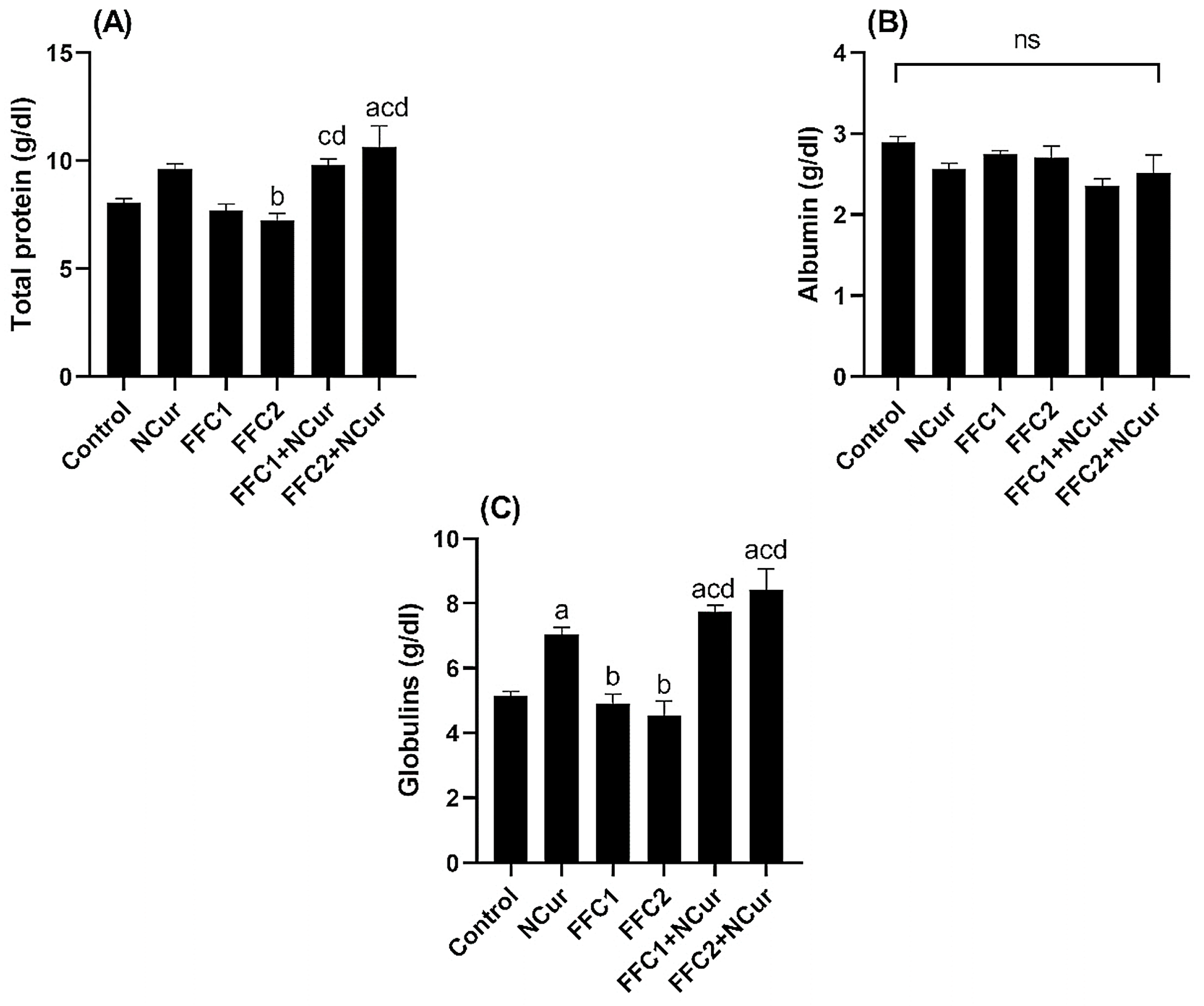
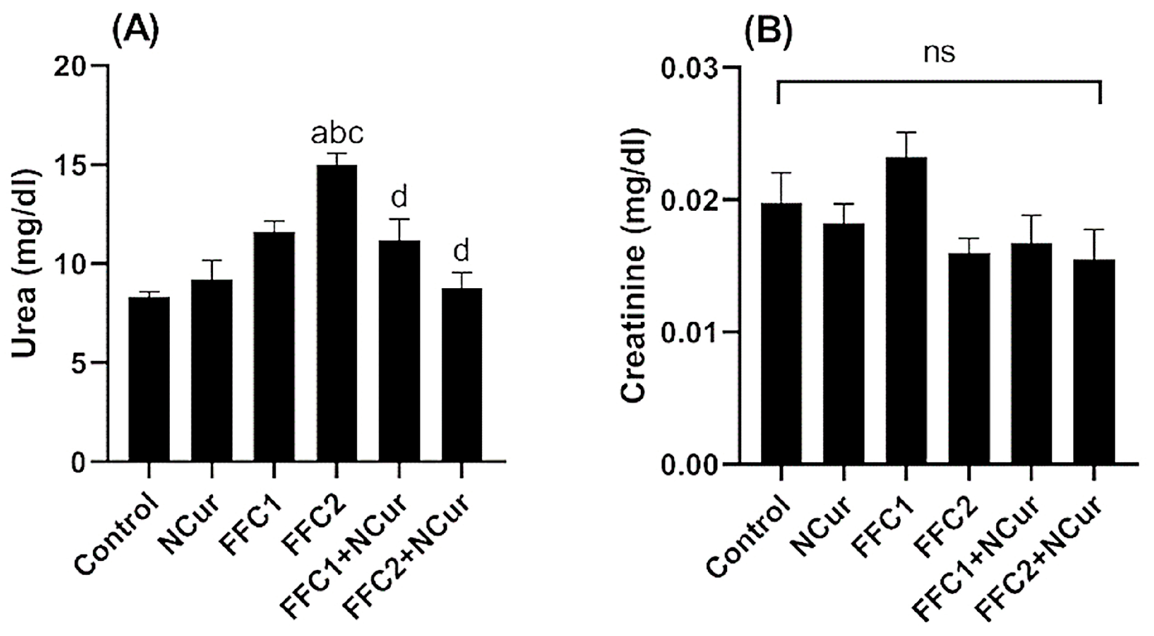
3.2. Biochemical Parameters

The level of serum total protein was elevated in the FFC2 + Ncur group compared with control, Ncur, and FFC2. Similarly, the serum total protein level increased in FFC1 + Ncur compared with the FFC1 group (Figure 4A). However, the level of serum albumin showed no change among the treated groups, as shown in (Figure 4B). Feeding NCur alone, FFC1 + NCur, and FFC2 + NCur for 15 days resulted in a significant increase in globulins levels compared to control, FFC1- and FFC2-treated groups (Figure 4C). Furthermore, serum levels of AST, ALT, and ALP significantly increased in FFC2-fed catfish compared to the control group. These elevated levels were restored to normal levels in the FFC2 + NCur-fed group (Figure 5A, B and C, respectively). In addition, the serum urea level was elevated significantly in FFC2-treated catfish compared with other groups, including the control group (Figure 6A). Surprisingly, the elevated serum urea level was unaffected when the catfish were fed a combination of FFC2 and Ncur and remained comparable to the level of the controls. On the contrary, no change was observed in serum creatinine levels across all groups (Figure 6B).

3.3. Histological Studies

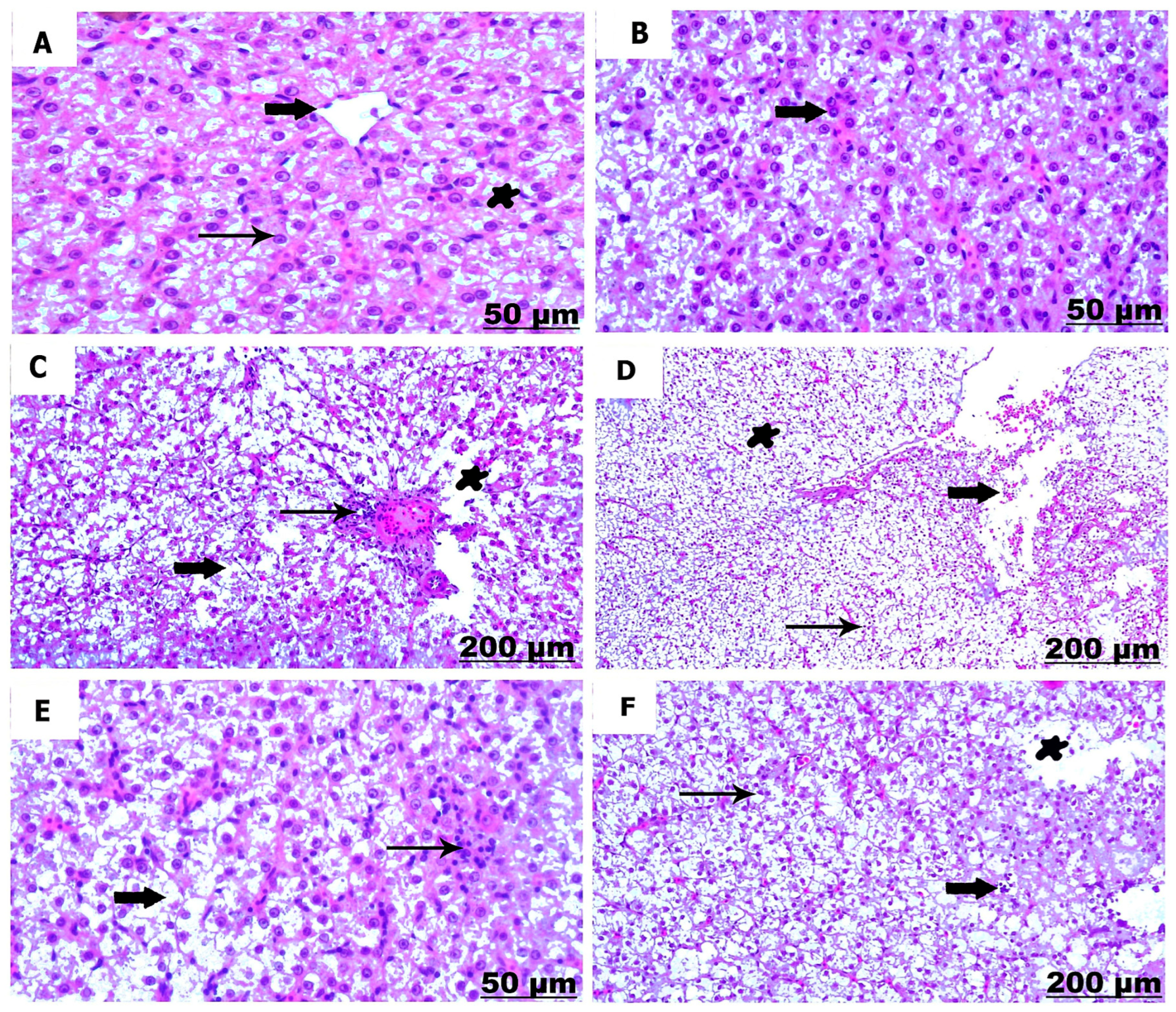
3.3.1. Histological Examination of the Liver

The liver samples of the control C. gariepinus fed the basal diet for 15 days showed normal morphology of the hepatic structure with coordinated hepatic cords (Table 1, Figure 7A). Histological examination of liver tissue sections from catfish fed diets supplemented with NCur (150 ppm) for 15 days exhibited an improvement in the hepatocytes histology when compared with catfish groups fed diets supplemented with FFC. It was observed that the borders between cells were more clearly defined and the nuclei of liver cells were larger and more hyperchromatic (Table 1, Figure 7B). The liver of catfish fed diets supplemented with 10 mg/Kg fish of FFC (FFC1) displayed mild deformation of the hepatic morphology in the form of a marked hepatocytes degeneration distinguished with vacuolated cytoplasm, necrotic hepatocytes, and rupture in the central vein. Dilated and congested blood vessels, hemorrhage, and a few inflammatory cells within the hepatic central vein were also noticed (Table 1, Figure 7C). The liver of catfish fed diets supplemented with 30 mg/Kg fish of FFC (FFC2) for 15 days showed severe damage in the structure of hepatocytes. Hepatic degeneration was observed, necrotic hepatocytes, dilation, rupture, and hemorrhage in the central vein. Additionally, focal aggregation of inflammatory cells was also noticed (Table 1, Figure 7D). Administration of NCur in combination with FFC 10 mg/kg (FFC1 + NCur) to catfish for 15 days improved the histological structure of liver hepatocytes and maintained their normal polygonal shape. However, some cells suffered from mild hydropic degeneration and minimal infiltration of inflammatory cells was observed (Table 1, Figure 7E). C. gariepinus fed both 30 mg/kg FFC and 150 ppm NCur (FFC2 + NCur) had normal hepatocytes, but with some infiltrations of mononuclear and intracytoplasmic granulated pigment. However, some hepatocytes suffered from degeneration and necrosis, besides focal aggregation of melanomacrophages. An aggregation of hepatocytes in clumps was noticed and the boundaries between cells were not clear. The nuclei of the aggregated cells were seen to be enlarged and hyperchromatic (Table 1, Figure 7F).

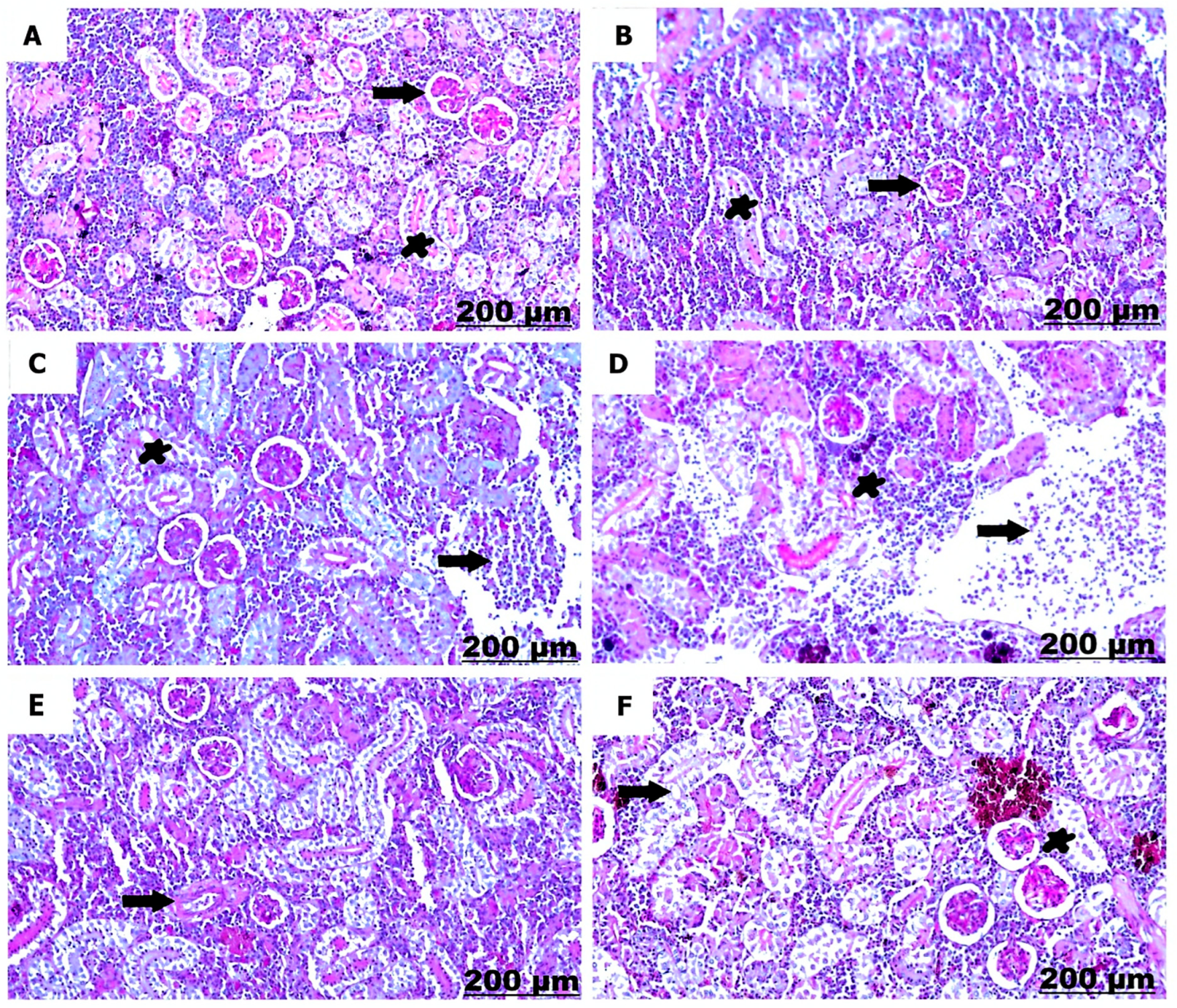
3.3.2. Histological Examination of the Kidney

The light microscopy of the posterior kidney of the control catfish group demonstrated a normal architecture with a normal arrangement of the glomeruli and renal tubules (Table 1, Figure 8A). The group of catfish fed diets supplemented with 150 ppm NCur manifested normal histological arrangement of the nephrons mainly of intact renal tubules and glomeruli (Table 1, Figure 8B). Histopathological assessment of tissue sections from catfish fed diets supplemented with FFC 10 mg/kg (FFC1) revealed a mild degree of hemorrhagic inflammation characterized by erythrocytic infiltration (Table 1, Figure 8C). The catfish group treated with FFC 30 mg/kg (iv, FFC2) displayed morphological lesions ranging from congestion of the blood vessels to sharp infiltration of erythrocytes (Table 1, Figure 8D), besides necrobiotic changes of the renal tubules were detected and infiltration of melanomacrophages. The blood vessels were fairly thickened in the catfish group v fed the diet with a combination of NCur plus FFC 10 mg/kg (FFC1 + Ncur—Table 1, Figure 8E). Vacuolar degeneration and necrosis of the tubules were discovered upon histopathological investigation of kidney tissues sections prepared from the catfish group vi fed the diet with a combination of NCur plus FFC 30 mg/kg (FFC2 + Ncur—Table 1, Figure 8F).

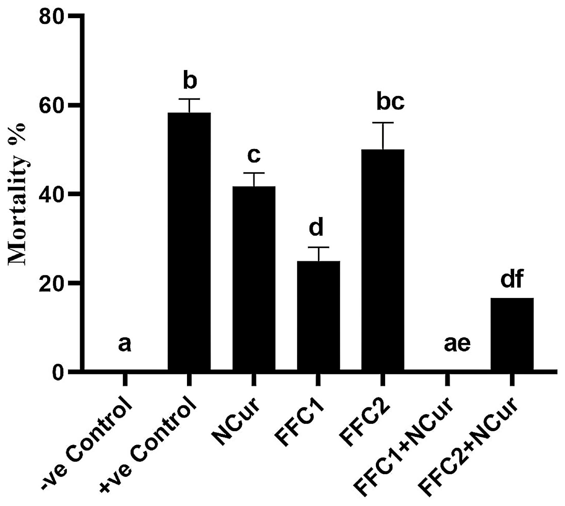
3.4. Bacterial Challenge Experiment

Neither signs nor lesions were noticed on the catfish in the negative control group throughout the study. However, in the positive control, NCur, FFC1, and FFC2 challenged catfish groups with A. veronii. Typical clinical signs associated with motile Aeromonas septicemia started two days post-challenge, including reddening of the skin, fins, and eyes; skin ulceration; diffuse hemorrhages; and internal signs of hemorrhagic septicemia. motile Aeromonas septicemia usually results in an acute mortality within a few days post-infection. Mortality rates were 58.33%, 41.66%, 25%, and 50% for positive control, NCur, FFC1, and FFC2, respectively. The catfish group challenged with A. veronii post-feeding FFC2 + NCur displayed mild clinical signs, such as focal hemorrhages and shallow skin ulcers. The cumulative mortality rate of catfish in this group (FFC2 + NCur) was 16.66%, which was a significant reduction in mortality compared with the positive control group, whereas the catfish group challenged post-feeding FFC1 + NCur showed no mortalities (Figure 9).

4. Discussion

In aquaculture, several studies have demonstrated that dietary curcumin can improve the growth rate, antioxidant status, immunity, and disease resistance of various finfish species [43]. Curcumin has been shown to have powerful immunomodulatory properties in both animals and humans. Nevertheless, curcumin’s potential role has not been evaluated in African catfish yet. Although curcumin and its nanoparticles exhibit beneficial biological functions, their bioavailability is often limited due to rapid metabolism and clearance. The current study was performed to investigate the potential beneficial and ameliorative effect of NCur when fed in combination with therapeutic and high doses of FFC (15 and 30 mg/kg.BW, respectively). FFC is a broad-spectrum bactericidal antibiotic extensively used in the fields of veterinary medicine and aquaculture. FAO has approved the use of FFC, among three antibiotics, for the treatment of septicemia and columnaris diseases in freshwater fish. While there are existing reports on the effectiveness of FFC in managing bacterial infections in finfish aquaculture in other regions, the impact and safety of administering FFC orally to African catfish have not been extensively investigated. This was the main reason for including the higher dose of FFC in the study. Previous safety studies conducted in tropical conditions have shown various alterations in the kidney and liver tissues of Nile tilapia (Oreochromis niloticus) in response to oral FFC [44].
In the present study, C. gariepinus fed diets supplemented with NCur combined with FFC showed a significant elevation in serum total protein levels compared to catfish fed FFC alone. Previous studies have demonstrated the immunomodulatory impacts of dietary NCur in fish [45]. There was a significant elevation in the levels of serum globulins of catfish that received dietary supplementation with NCur alone or combined with FFC, which is in accordance with the findings of Eissa et al. [45] who reported an improvement in globulins of O. niloticus after supplementation of different diets with NCur. Additionally, levels of globulins were elevated in C. gariepinus and O. niloticus administrated diets supplemented with curcumin compared to fish in the control groups [46,47,48]. Globulins are principal components of the fish’s immune defense mechanisms, play critical functions in the innate and adaptive immunity against pathogenic organisms [49], and compose an indivisible part of all the immunoglobulins of fish blood [50]. The observed elevation of globulin levels in catfish fed diets supplemented with NCur in the current study supports the immunomodulatory properties of curcumin in fish [51,52,53]. Sustainable aquaculture producers are increasingly using immunomodulatory products to reduce the use of therapeutic treatments and enhance the effectiveness of vaccines.
The increased levels of ALT and AST are believed to be caused by the damage or dysfunction of liver tissue that occurs because of exposure to stress-inducing conditions or hepatotoxic substances [54,55]. It has been reported that many antibiotics [56], as well as amphenicols [57], are hepatotoxic. Administration of high doses (200–300 mg/kg body weight) of amphenicol antibiotics (chloramphenicol, thiamphenicol, and FFC) induced toxic changes in the hematopoietic tissues of laboratory rats and dogs [58]. Generally, determination of hepatic tissue status can be assessed through measuring the biomarkers AST and ALT, which are crucial aminotransferases [59,60]. Results of the current study revealed a significant rise in enzyme activities (AST, ALT, and ALP) of C. gariepinus fed diets supplemented with the high dose of FFC (FFC2) for 15 days. These results were consistent with the previous studies [61,62,63] that suggested an increase of AST, ALT, and ALP in O. niloticus after FFC supplementation. Additionally, Er and Dik [64] recorded an elevation in AST levels upon FFC treatment in Oncorhynchus mykiss. Similarly, Zhang et al. [65] observed an increase in AST, ALT, and ALP in European seabass (Dicentrarchus labrax) post-feeding dietary FFC-contained feeds. Abnormal liver enzyme levels typically signal liver damage. Elevation of these enzyme levels may be due to liver inflammation resulting from FFC use and the enzyme levels are usually corresponding to the degree of liver damage or injury [66]. Liver dysfunction can cause an increase in certain serum enzymes due to their impaired removal from the bloodstream [67].
Results of the current investigation revealed a significant increase in urea levels in C. gariepinus fed diets supplemented with the high dose of FFC (FFC2) for 15 days. Elevation of urea level is attributed to the pathological changes in the kidney of FFC-treated fish, as shown previously [63]. Amer et al. [68] recorded the same results in fish after FFC dosing. When the kidney is stimulated by antibiotics and other nephrotoxic medications, many reactive oxygen radicals will be produced. These free radicals can attack the cell membrane of the kidney, resulting in kidney dysfunction [69]. Previous studies revealed that the liver and kidney functions of healthy brown trout can be affected by FFC administration even at the therapeutic dose [64]. In the present study, dietary supplementation of NCur, in combination with the high dose of FFC (FFC2 + Ncur), maintained the urea at a normal level—comparable to that of the control catfish group. Similar results were also obtained in African catfish fed turmeric powder diets and exposed to cadmium toxicity where turmeric showed a potential for reversing the cadmium-induced toxic effects [70]. Curcumin extracts are known to contain several compounds with significant pharmacological and biological properties, such as flavonoids and antioxidants. These compounds can help to maintain or restore normal levels of biochemical markers and play a crucial role in maintaining overall cellular health. They can enhance antioxidant capacity and stabilize cell membranes during oxidative stress, thereby exerting a protective effect on biochemical markers [71,72]. Furthermore, curcumin is known to effectively prevent cellular damage caused by oxidative stress by reducing the production of reactive oxygen species [73].
The liver of the catfish fed FFC exhibited several hepatic alterations, including vacuolation, necrosis, congestion of blood vessels, and infiltration of melanomacrophages, according to the histopathological results. The observed lesions may have been caused by the direct or indirect deleterious effects of FFC on liver tissue. The observed pathological lesions observed in the African catfish fed FFC in our study have been similarly reported in C. gariepinus [74], Oreochromis sp. [11,71], channel catfish [72], hybrid striped bass [73], and silver catfish (Rhamdia quelen) [74], following FFC or chloramphenicol treatments. Moreover, the increased vacuolization of hepatocytes observed after exposure to FFC is indicative of a degenerative process that could potentially cause metabolic damage, likely because of the medication [29,75]. Changes in cell membrane integrity led to ion pump damage, which is the initial process of vacuolar degeneration [76]. In addition, the degeneration and necrosis observed may be a result of the accumulation of the antibiotic compound in hepatic tissue [77].
Administration of NCur as a dietary supplement in African catfish ration in the present study exerted a protective antioxidant effect and restored the general structure of liver tissue. These results agree with Khorshidi et al. [78], who observed no lesions in the liver tissue of common carp (Cyprinus carpio) fed with 0.75 g/kg curcumin. Manju et al. [79] reported no histological changes in the liver of curcumin-treated Anabas sp. after feeding 2 doses of curcumin (5 and 10 g/kg). Moreover, Zhang et al. [65] observed that diets supplemented with 120 mg/kg curcumin had the strongest protective effect on carbon tetrachloride (CCl4) induced liver damage in common carp. Similarly, Mohamed et al. [48] concluded that O. niloticus fed diets supplemented with curcumin (200 mg/kg diet) showed a normal liver structure. Curcumin ameliorates hexavalent chromium (Cr6+) induced hepatoxicity and oxidative stress in Channa punctatus, as Awasthi et al. [80] observed histological recovery of the liver tissue after curcumin supplementation (1, 2, and 3 mg/L).
Dietary inclusion of NCur was found to mitigate the potential toxic impacts of FFC and reduce the histopathological alterations in liver and kidney structures induced by feeding diets supplemented with FFC alone. This is in accordance with [81], who reported that curcumin exhibited an ameliorative effect against the impacts of aflatoxicosis in O. niloticus by reducing the oxidative stress and hence, resulting in enhanced growth performance. FFC-induced nephrotoxicity in the present study resulted in necrosis and degeneration of the renal tubules distinguished by focal infiltration with mononuclear cells, which is like the findings by [75]. The incidence and severity of epithelial degeneration also demonstrated a dose-dependent effect on kidney tissue, which may lead to severe renal failure [82]. Observations of the degeneration of the renal tubular epithelium indicated that the FFC-induced injury may be toxic, ischemic, inflammatory, and obstructive. The persistence of such changes implied the nephrotoxic potential of FFC, even at the therapeutic dose [83]. The administration of NCur as a dietary supplement in our study showed remarkable protective effects and improved the overall kidney structure, possibly due to its antioxidant activity. The results obtained by Mohamed et al. [48] demonstrated that dietary curcumin protected the renal tissue of O. niloticus against sub-lethal concentrations of hexavalent chromium.
Our study demonstrated that the concomitant use of NCur with FFC had a positive impact on the overall health and resistance of African catfish against A. veronii infection. While administering NCur alone did not provide sufficient protection against the infection, the mortality rate of catfish in FFC1 + NCur and FFC2 + NCur groups was significantly lower than that of the positive control catfish fed the basal diet. NCur has a well-established antimicrobial activity and has been shown to exhibit synergy with antibiotics. Moreover, the observed improvement in blood biochemical parameters in the experimental groups fed a combination of FFC and NCur indicates that NCur has the potential to offer a protective effect to African catfish. Previous in vivo studies in silver catfish (Rhamdia quelen) confirmed the synergistic activity of other phytochemical compounds, such as linalool, when administered in combination with FFC against A. hydrophila infection [84]. A number of earlier studies have also demonstrated curcumin’s antibacterial synergy with antibiotics against sixty biofilm-producing bacterial isolates and have recommended curcumin as a beneficial supplement to antibiotic therapy [85]. Although previous research has shown that FFC-medicated feeds are effective in managing bacterial diseases in a range of fish species, such as Atlantic salmon (Salmo salar), Atlantic cod (Gadus morhua), and channel catfish (Ictalurus punctatus) [3,86,87], supplementing FFC with NCur in African catfish diets may offer a promising approach for treating bacterial infections.

5. Conclusions

In summary, the present study revealed that FFC at a therapeutic dose (10 mg/kg body weight fish) is a safe treatment option for African catfish, despite causing mild damage to hepatic and renal tissues, as evidenced through histopathological findings. However, a high dose (30 mg/kg body weight fish) of FFC may induce hepato- and nephrotoxicity in African catfish. The addition of NCur to African catfish feed has the potential to improve the fish’s overall health status by restoring biochemical indices to normal levels and protecting against possible liver and kidney damage associated with FFC treatment. Furthermore, the experimental challenge with A. veronii demonstrated that NCur has a synergistic effect when supplemented in combination with FFC, making it a superior medicated feed formulation for protecting African sharptooth catfish against motile Aeromonas septicemia. Overall, these findings contribute to our understanding of how to achieve sustainability in African catfish aquaculture.

Author Contributions

Conceptualization, S.M. and W.F.A.E.; Data curation, H.A. and W.F.A.E.; Formal analysis, S.M., H.A. and W.F.A.E.; Methodology, S.M., K.A.B., E.A.A., H.A., Z.A.-A. and W.F.A.E.; Software, S.M., E.A.A., H.A. and W.F.A.E.; Supervision, W.F.A.E.; Writing—original draft, S.M. and W.F.A.E.; Writing—review & editing, S.M., K.A.B., H.A., Z.A.-A., H.H.M. and W.F.A.E. All authors have read and agreed to the published version of the manuscript.

Funding

This research received no external funding.

Institutional Review Board Statement

The Research Ethics Committee of the Faculty of Veterinary Medicine at South Valley University has reviewed and approved the research conducted in this study under the approval number (No. 44/06.09.2022).

Informed Consent Statement

Not applicable.

Data Availability Statement

Data is available upon request.

Acknowledgments

The authors gratefully acknowledge Cell and Molecular Physiology Research Center, Department of Physiology, Faculty of Veterinary Medicine, South Valley University, Qena, Egypt, for providing all facilities and equipment used in the current study.

Conflicts of Interest

The authors declare no conflict of interest.

References

  1. FAO. The State of World Fisheries and Aquaculture 2020; Sustainability in Action: Rome, Italy, 2020. [Google Scholar]
  2. Pulkkinen, K.; Suomalainen, L.R.; Read, A.F.; Ebert, D.; Rintamäki, P.; Valtonen, E.T. Intensive fish farming and the evolution of pathogen virulence: The case of columnaris disease in Finland. Proc. Biol. Sci. 2010, 277, 593–600. [Google Scholar] [CrossRef] [Green Version]
  3. Roberts, R.J. Efficacy of orally administered florfenicol and oxolinic acid for the treatment of vibriosis in cod (Gadus morhua). Aquaculture 2001, 235, 27–35. [Google Scholar]
  4. Assane, I.M.; Gozi, K.S.; Valladao, G.M.R.; Pilarski, F. Combination of antimicrobials as an approach to reduce their application in aquaculture: Emphasis on the use of thiamphenicol/florfenicol against Aeromonas hydrophila. Aquaculture 2019, 507, 238–245. [Google Scholar] [CrossRef]
  5. Carraschi, S.P.; Cruz, C.; Machado Neto, J.G.; Castro, M.P.; Bortoluzzi, N.L.; Gírio, A.C.F. Eficácia do florfenicol e da oxitetraciclina no controle de Aeromonas hydrophila em pacu (Piaractus mesopotamicus). Arq. Bras. Med. Vet. Zootec. 2011, 63, 579–583. [Google Scholar] [CrossRef] [Green Version]
  6. Dowling, P.M. Chloramphenicol, Thiamphenicol, and Florfenicol. In Antimicrobial Therapy in Veterinary Medicine, 5th ed.; John Wiley & Sons Inc.: Hoboken, NJ, USA, 2013; pp. 269–278. [Google Scholar]
  7. Orlando, E.A.; Costa Roque, A.G.; Losekann, M.E.; Colnaghi Simionato, A.V. UPLC-MS/MS determination of florfenicol and florfenicol amine antimicrobial residues in tilapia muscle. J. Chromatogr. B Analyt. Technol. Biomed. Life Sci. 2016, 1035, 8–15. [Google Scholar] [CrossRef] [PubMed]
  8. Fukui, H.; Fujihara, Y.; Kano, T. In vitro and in vivo antibacterial activities of florfenicol, a new fluorinated analog of thiamphenicol, against fish pathogens. Fish Pathol. 1987, 22, 201–207. [Google Scholar] [CrossRef]
  9. Sørensen, L.K.; Elbæk, T.H. Simultaneous determination of trimethoprim sulfadiazine, florfenicol and oxolinic acid in surface water by liquid chromatography tandem mass spectrometry. Chromatographia 2004, 60, 287–291. [Google Scholar] [CrossRef]
  10. Gordon, L.; Giraud, E.; Ganière, J.-P.; Armand, F.; Bouju-Albert, A.; De La Cotte, N.; Mangion, C.; Le Bris, H. Antimicrobial resistance survey in a river receiving effluents from freshwater fish farms. J. Appl. Microbiol. 2007, 102, 1167–1176. [Google Scholar] [CrossRef]
  11. USFWS. Approved Drugs for Use in Aquaculture, 2nd ed.; U.S. Fish and Wildlife Service’s Aquatic Animal Drug Approval Partnership Program, American Fisheries Society’s Fish Culture and Fish Health Sections; Association of Fish and Wildlife Agencies and Fisheries and Water Resources Policy Committee’s Drug Approval Working Group: Hoboken, NJ, USA, 2015. [Google Scholar]
  12. Klein, S.; Bueno, G.W.; Lorenz, E.K.; Diemer, O.; Feiden, A.; Boscolo, W.R. Additives in the water during transportation of Piaractus mesopotamicus: Effects of survival rates after stocking in cages. Zootec. Trop. 2013, 31, 255–260. [Google Scholar]
  13. Darwish, A.M. Laboratory efficacy of florfenicol against Streptococcus iniae infection in Sunshine Bass. J. Aquat. Anim. Health 2007, 19, 1–7. [Google Scholar] [CrossRef]
  14. Saglam, N.; Yonar, M.E. Effects of sulfamerazine on selected haematological and immunological parameters in rainbow trout (Onchorhynchus mykiss, Walbaum, 1792). Aquacult. Res. 2009, 40, 395–404. [Google Scholar] [CrossRef]
  15. Binh, V.N.; Dang, N.; Anh, N.T.K.; Ky, L.X.; Thai, P.K. Antibiotics in the aquatic environment of Vietnam: Sources, concentrations, risk and control strategy. Chemosphere 2018, 197, 438–450. [Google Scholar] [CrossRef] [Green Version]
  16. Henriksson, A.P.J.G.; Rico, M.; Troell, D.H.; Klinger, A.H.; Buschmann, S.; Saksida, M.V.; Chadag, W.Z. Unpacking factors influencing antimicrobial use in global aquaculture and their implication for management: A review from a systems perspective. Sustain. Sci. 2018, 13, 1105–1120. [Google Scholar] [CrossRef] [Green Version]
  17. Dossou, S.; Dawood, M.A.O.; Zaineldin, A.I.; Abouelsaad, I.A.; Mzengereza, K.; Shadrack, R.S.; Zhang, Y.; El-Sharnouby, M.; Ahmed, H.A.; El Basuini, M.F. Dynamical hybrid system for optimizing and controlling efficacy of plantbased protein in aquafeeds. Complexity 2021, 2021, 9957723. [Google Scholar] [CrossRef]
  18. Cheng, G.; Hao, H.; Xie, S.; Wang, X.; Dai, M.; Huang, L.; Yuan, Z. Antibiotic alternatives: The substitution of antibiotics in animal husbandry. Front. Microbiol. 2014, 5, 217. [Google Scholar] [CrossRef] [Green Version]
  19. Priyadarsini, K.I. The chemistry of curcumin: From extraction to therapeutic agent. Molecules 2014, 19, 20091–20112. [Google Scholar] [CrossRef] [Green Version]
  20. Alagawany, M.; Farag, M.R.; Abdelnour, S.A.; Dawood, M.A.O.; Elnesr, S.S.; Dhama, K. Curcumin and its different forms: A review on fish nutrition. Aquaculture 2021, 532, 736030. [Google Scholar] [CrossRef]
  21. Hewlings, S.; Kalman, D. Curcumin: A review of its effects on human health. Foods 2017, 6, 92. [Google Scholar] [CrossRef] [Green Version]
  22. Ahmadifar, E.; Yousefi, M.; Karimi, M.; Fadaei Raieni, R.; Dadar, M.; Yilmaz, S.; Dawood, M.A.O.; Abdel-Latif, H.M.R. Benefits of dietary polyphenols and polyphenol-rich additives to aquatic animal health: An overview. Rev. Fish. Sci. Aquac. 2021, 29, 478–511. [Google Scholar] [CrossRef]
  23. Ashry, A.M.; Hassan, A.M.; Habiba, M.M.; El-Zayat, A.; El-Sharnouby, M.E.; Sewilam, H.; Dawood, M.A.O. The impact of dietary curcumin on the growth performance, intestinal antibacterial capacity, and haemato-biochemical parameters of gilthead seabream (sparus aurata). Animals 2021, 11, 1779. [Google Scholar] [CrossRef] [PubMed]
  24. Galli, G.M.; Da Silva, A.S.; Biazus, A.H.; Reis, J.H.; Boiago, M.M.; Topazio, J.P.; Migliorini, M.J.; Guarda, N.S.; Moresco, R.N.; Ourique, A.F.; et al. Feed addition of curcumin to laying hens showed anticoccidial effect, and improved egg quality and animal health. Res. Vet. Sci. 2018, 118, 101–106. [Google Scholar] [CrossRef]
  25. Galli, G.M.; Gerbet, R.R.; Griss, L.G.; Fortuoso, B.F.; Petrolli, T.G.; Boiago, M.M.; Souza, C.F.; Baldissera, M.D.; Mesadri, J.; Wagner, R.; et al. Combination of herbal components (curcumin carvacrol, thymol, cinnamaldehyde) in broiler chicken feed: Impacts on response parameters, performance, fatty acid profiles, meat quality and control of coccidian and bacteria. Microb. Pathog. 2020, 139, 103916. [Google Scholar] [CrossRef]
  26. de Almeida, M.; da Rocha, B.A.; Francisco, C.R.L.; Miranda, C.G.; Santos, P.D.F.; de Araújo, P.H.H.; Sayer, C.; Leimann, F.V.; Gonçalves, O.H.; Bersani-Amado, C.A. Evaluation of the in vivo acute anti-inflammatory response of curcumin loaded nanoparticles. Food Funct. 2018, 9, 440–449. [Google Scholar] [CrossRef] [PubMed]
  27. Reda, F.M.; El-Saadony, M.T.; Elnesr, S.S.; Alagawany, M.; Tufarelli, V. Effect of dietary supplementation of biological curcumin nanoparticles on growth and carcass traits, antioxidant status, immunity and caecal microbiota of Japanese quails. Animals 2020, 10, 754. [Google Scholar] [CrossRef] [PubMed]
  28. Nwani, C.D.; Mkpadobi, B.N.; Onyishi, G.; Echi, P.C.; Chukwuka, C.O.; Oluah, S.N.; Ivoke, N. Changes in behavior and hematological parameters of freshwater African catfish Clarias gariepinus (Burchell 1822) following sublethal exposure to chloramphenicol. Drug Chem. Toxicol. 2013, 37, 107–113. [Google Scholar] [CrossRef]
  29. Bardhan, A.; Abraham, T.J.; Singha, J.; Sar, T.K.; Rajisha, R.; Krishna, E.K.N.; Kumar, K.A.; Patil, P.K. Histopathological aberrations and oxidative stress responses in Nile tilapia Oreochromis niloticus as influenced by dietary florfenicol and its metabolites. Aquaculture 2022, 559, 738447. [Google Scholar] [CrossRef]
  30. Mallik, S.K.; Shahi, N.; Pathak, R.; Kala, K.; Patil, P.K.; Singh, B.; Ravindran, R.; Krishna, N.; Pandey, P.K. Pharmacokinetics and biosafety evaluation of a veterinary drug florfenicol in rainbow trout, Oncorhynchus mykiss (Walbaum 1792) as a model cultivable fish species in temperate water. Front. Pharmacol. 2023, 14, 1033170. [Google Scholar] [CrossRef]
  31. Kumar, R.V.; Vinoth, S.; Baskar, V.; Arun, M.; Gurusaravanan, P. Synthesis of zinc oxide nanoparticles mediated by Dictyota dichotoma endophytic fungi and its photocatalytic degradation of fast green dye and antibacterial applications. S. Afr. J. Bot. 2022, 151, 337–344. [Google Scholar] [CrossRef]
  32. Carvalho, D.M.; Takeuchi, K.P.; Geraldine, R.M.; Moura, C.J.d.; Torres, M.C.L. Production, Solubility and Antioxidant Activity of Curcumin Nanosuspension. Food Sci. Technol. 2015, 35, 115–119. [Google Scholar] [CrossRef] [Green Version]
  33. Pandit, R.S.; Gaikwad, S.C.; Agarkar, G.A.; Gade, A.K.; Rai, M. Curcumin Nanoparticles: Physico-Chemical Fabrication and Its in Vitro Efficacy against Human Pathogens. 3 Biotech. 2015, 5, 991–997. [Google Scholar] [CrossRef] [Green Version]
  34. Busher, J. Serum albumins and globulin. In Clinical Methods: The History, Physical, and Laboratory Examinations, 3rd ed.; Walker, H.K., Hall, W.D., Hurst, J.W., Eds.; Butterworths: Boston, UK, 1990; Chapter 101. [Google Scholar]
  35. Bacha, W.J.J.; Bacha, L.M. Color Atlas of Veterinary Histology, 2nd ed.; Lippincott Williams & Wilkins: Philadelphia, PA, USA, 2000. [Google Scholar]
  36. Austin, B.; Austin, D.A. Aeromonadaceae representatives (Motile Aeromonads). In Bacterial Fish Pathogens. Disease of Farmed and Wild Fish, 6th ed.; Springer Nature Publishing: Berlin/Heidelberg, Germany, 2016; pp. 161–214. [Google Scholar]
  37. Goldman, E.; Green, L.H. (Eds.) Practical Handbook of Microbiology, 3rd ed.; CRC Press: Boca Raton, FL, USA, 2015. [Google Scholar]
  38. Yanez, M.; Catalán, V.; Apráiz, D.; Figueras, M.; Martínez-Murcia, A. Phylogenetic analysis of members of the genus Aeromonas based on gyrB gene sequences. Int. J. Syst. Evol. Microbiol. 2003, 53, 875–883. [Google Scholar] [CrossRef] [PubMed] [Green Version]
  39. Pawar, H.A.; Gavasane, A.J.; Choudhary, P.D. A Novel and Simple Approach for Extraction and Isolation of Curcuminoids from Turmeric Rhizomes. Adv. Recycl. Waste Manag. 2018, 6, 2. [Google Scholar] [CrossRef]
  40. Yadav, D.; Kumar, N. Nanonization of Curcumin by Antisolvent Precipitation: Process Development, Characterization, Freeze Drying and Stability Performance. Int. J. Pharm. 2014, 477, 564–577. [Google Scholar] [CrossRef] [PubMed]
  41. Hanna, D.H.; Saad, G.R. Nanocurcumin: Preparation, Characterization and Cytotoxic Effects towards Human Laryngeal Cancer Cells. RSC Adv. 2020, 10, 20724–20737. [Google Scholar] [CrossRef] [PubMed]
  42. Siregar, C.E.; Martono, S.; Rohman, A. Application of Fourier Transform Infrared (FTIR) Spectroscopy Coupled with Multivariate Calibration for Quantitative Analysis of Curcuminoid in Tablet Dosage Form. J. Appl. Pharm. Sci. 2018, 8, 151–156. [Google Scholar]
  43. Zou, W.; Ma, Y.; Ai, C.; Yu, W.; Gao, X.; Liu, S.; Luo, X.; You, W.; Ke, C. Dietary curcumin influence on growth, antioxidant status, immunity, gut flora and resistance to Vibrio harveyi AP37 in Haliotis discus hannai. Aquac. Rep. 2022, 26, 101336. [Google Scholar] [CrossRef]
  44. Bardhan, A.; Abraham, T.J.; Singha, J.; Rajisha, R.; Krishna, E.K.N.; Panda, S.K.; Patil, P.K. Impacts of Oral Florfenicol Medication and Residues on the Kidney and Liver of Nile Tilapia Oreochromis niloticus (L.). Vet. Sci. 2023, 10, 36. [Google Scholar] [CrossRef]
  45. Eissa, E.H.; Ezzo, O.H.; Khalil, H.S.; Tawfik, W.A.; El-Badaw, A.A.; Abd Elghany, N.A.; Mossa, M.I.; Hassan, M.M.; Hassan, M.M.; Eissa, M.E.H.; et al. The effect of dietary nanocurcumin on the growth performance, body composition, haemato-biochemical parameters and histopathological scores of the Nile tilapia (Oreochromis niloticus) challenged with Aspergillus flavus. Aquac. Res. 2022, 53, 6098–6111. [Google Scholar] [CrossRef]
  46. Abbas, W.T.; El Ibrahim, T.B.; Elgendy, M.Y.; Abdel Zaher, M.F. Effect of Curcumin on Iron Toxicity and Bacterial Infection in Catfish (Clarias gariepinus). Pak. J. Biol. Sci. 2019, 22, 510–517. [Google Scholar] [CrossRef]
  47. Elgendy, M.Y.; Hakim, A.S.; Ibrahim, T.B.; Soliman, W.S.; Ali, S.E. Immunomodulatory effects of curcumin on Nile tilapia, Oreochromis niloticus and its antimicrobial properties against Vibrio alginolyticus. J. Fish Aquatic. Sci. 2016, 11, 206–215. [Google Scholar] [CrossRef] [Green Version]
  48. Mohamed, A.A.R.; El-Houseiny, W.; EL-Murr, A.E.; Ebraheim, L.L.M.; Ahmed, A.I.; ElHakim, Y.M.A. Effect of hexavalent chromium exposure on the liver and kidney tissues related to the expression of CYP450 and GST genes of Oreochromis niloticus fish: Role of curcumin supplemented diet. Ecotoxicol. Environ. Saf. 2020, 188, 109890. [Google Scholar] [CrossRef] [PubMed]
  49. Sahoo, P.K.; Mukherjee, S.C. Effect of dietary $-1,3 glucan on immune responses and disease resistance of healthy and aflatoxin B1-induced immunocompromised rohu (Labeo rohita Hamilton). Fish Shellfish Immunol. 2001, 11, 683–695. [Google Scholar] [CrossRef] [PubMed]
  50. Misra, C.K.; Das, B.K.; Mukherjee, S.C.; Pattnaik, P. Effect of long-term administration of dietary $-glucan on immunity growth and survival of Labeo rohita fingerlings. Aquaculture 2006, 255, 82–94. [Google Scholar] [CrossRef]
  51. Abdelrazek, H.M.A.; Tag, H.M.; Kilany, O.E.; Reddy, P.G.; Hassan, A.M. Immuomodulatory effect of dietary turmeric supplementation on Nile tilapia (Oreochromis niloticus). Aquac. Nutr. 2017, 23, 1048–1054. [Google Scholar] [CrossRef]
  52. Eissa, I.A.M.; Ismail, T.; Yousseff, F.; Mansour, S. Trials for control of pseudomonas septicemia in tilapia Zillii using turmeric powder as a medicated feed. Suez Canal Vet. Med. J. 2019, 24, 273–285. [Google Scholar] [CrossRef] [Green Version]
  53. Kord, M.I.; Maulu, S.; Srour, T.M.; Omar, E.A.; Farag, A.A.; Nour, A.A.M.; Hasimuna, O.J.; Abdel-Tawwab, M.; Khalil, H.S. Impacts of water additives on water quality, production efficiency, intestinal morphology, gut microbiota, and immunological responses of Nile tilapia fingerlings under a zero-water-exchange system. Aquaculture 2021, 547, 737503. [Google Scholar] [CrossRef]
  54. Julinta, R.B.; Abraham, T.J.; Roy, A.; Singha, J.; Boda, S.; Patil, P.K. Dietary influences of oxytetracycline on the growth and serum biomarkers of Oreochromis niloticus (L.). Ecotoxicol. Environ. Saf. 2019, 186, 109752. [Google Scholar] [CrossRef]
  55. Bojarski, B.; Kot, B.; Witeska, M. Antibacterials in aquatic environment and their toxicity to fish. Pharmaceuticals 2020, 13, 189. [Google Scholar] [CrossRef]
  56. Chang, C.Y.; Schiano, T.D. Review article: Drug hepatotoxicity. Aliment. Pharmacol. Ther. 2007, 25, 1135–1151. [Google Scholar] [CrossRef]
  57. Memik, F. Opinions on drugs and liver. Eurasian J. Med. 1975, 4, 399–402. [Google Scholar]
  58. Gaikowski, M.P.; Wolf, J.C.; Endris, R.G.; Gingerich, W.H. Safety of Aquaflor1 (Florfenicol, 50% type A medicated article), administered in feed to channel catfish, Ictalurus punctatus. Toxicol. Pathol. 2003, 31, 689–697. [Google Scholar] [CrossRef] [PubMed]
  59. Liu, F.; Qu, Y.K.; Wang, A.M.; Yu, Y.B.; Yang, W.P.; Lv, F.; Nie, Q. Effects of carotenoids on the growth performance, biochemical parameters, immune responses, and disease resistance of yellow catfish (Pelteobagrus fulvidraco) under hightemperature stress. Aquaculture 2019, 503, 293–303. [Google Scholar] [CrossRef]
  60. Kong, Y.D.; Li, M.; Chu, G.S.; Liu, H.J.; Shan, X.F.; Wang, G.Q.; Han, G.H. The positive effects of single or conjoint administration of lactic acid bacteria on Channa argus: Digestive enzyme activity, antioxidant capacity, intestinal microbiota, and morphology. Aquaculture 2021, 531, 735852. [Google Scholar] [CrossRef]
  61. Bardhan, A.; Abraham, T.J.; Singha, J.; Saha, S.; Sarker, S.; Patil, P.K. The effects of extended feeding of florfenicol coated medicated diets on the safety, serum biomarkers and blood cells morphology of Nile tilapia Oreochromis niloticus (L.). Environ. Sci. Pollut. Res. 2022, 29, 39914–39927. [Google Scholar] [CrossRef] [PubMed]
  62. Soltan, M.A.; Agouz, H.M.; Mohamed, M.G. Effect of oxytetracycline and florfenicol drugs on the physiological activities and its residues of Oreochromis niloticus. Egypt J. Aquat. Biol. Fish 2013, 17, 25–36. [Google Scholar]
  63. Reda, R.M.; Ibrahim, R.; Ahmed, E.N.G.; El-Bouhy, Z. Effect of oxytetracycline and florfenicol as growth promoters on the health status of cultured Oreochromis niloticus. Egypt J. Aquat. Res. 2013, 39, 241–248. [Google Scholar] [CrossRef] [Green Version]
  64. Er, A.; Dik, B. The effects of florfenicol on the values of serum tumor necrosis factor- and other biochemical markers in lipopolysaccharide-induced endotoxemia in brown trout. Mediat. Inflamm. 2014, 1, 1–5. [Google Scholar] [CrossRef] [Green Version]
  65. Zhang, Q.; Zhang, Y.; Zhang, X.; Rabbi, M.H.; Guo, R.; Shi, S.; Ma, Z.; Liu, Y. Effects of dietary florfenicol contained feeds on growth and immunity of European seabass (Dicentrarchus labrax) in flow-through and recirculating aquaculture system. Aquac. Rep. 2021, 19, 100602. [Google Scholar] [CrossRef]
  66. James, R.B. A division of J. B. Lippincott Company Philadelphia. In Drug, Facts and Comparisons, 1985th ed.; J. B. Lippincott Company: Toronto, ON, Canada, 1985; pp. 1296–1333. [Google Scholar]
  67. Barraze, M.I.; Coppock, C.E.; Brooks, K.N.; Wilks, D.L.; Saunders, R.G.; Latimer, J.R. Iron sulfate and feed pelleting to detoxify free gossypol in cotton seed diets for dairy cattle. J. Dairy Sci. 1991, 74, 3457–3467. [Google Scholar] [CrossRef]
  68. Amer, M.S.; El-Sayed, M.G.; Abd El-Fatah, R.A. Pharmacological studies on some antibacterial drugs in fish. J. Vet. Med. Mansoura Univ. 2009, 9, 165–184. [Google Scholar]
  69. Ghorbel, I.; Elwej, A.; Fendri, N.; Mnif, H.; Jamoussi, K.; Boudawara, T.; Grati Kamoun, N.; Zeghal, N. Olive oil abrogates acrylamide induced nephrotoxicity by modulating biochemical and histological changes in rats. Ren. Fail. 2017, 39, 236–245. [Google Scholar] [CrossRef] [Green Version]
  70. El-Houseiny, W.; Khalil, A.A.; Abd-Elhakim, Y.M.; Badr, H.A. The potential role of turmeric and black pepper powder diet supplements in reversing cadmium-induced growth retardation, ATP depletion, hepatorenal damage, and testicular toxicity in Clarias gariepinus. Aquaculture 2019, 510, 109–121. [Google Scholar] [CrossRef]
  71. Handy, R.D.; Von Der Kammer, F.; Lead, J.R.; Hassellöv, M.; Owen, R.; Crane, M. The ecotoxicology and chemistry of manufactured nanoparticles. Ecotoxicology 2008, 17, 287–314. [Google Scholar] [CrossRef]
  72. Banaee, M.; Sureda, A.; Shahaf, S.; Fazilat, N. Protective effects of silymarin extract on malthion-induced zebra cichlid (Cichlasoma nigrofasciatum) hepatotoxicity. Iran. J. Toxicol. 2015, 9, 1239–1246. [Google Scholar]
  73. Zhu, Y.G.; Chen, X.C.; Chen, Z.Z.; Zeng, Y.Q.; Shi, G.B.; Su, Y.H.; Peng, X. Curcumin protects mitochondria from oxidative damage and attenuates apoptosis in cortical neurons. Acta. Pharmacol. Sin. 2004, 25, 1606–1612. [Google Scholar] [PubMed]
  74. Nkwuda, P.J.; Awoke, J.S.; Nwakpa, J.N. Histological Changes in Liver and Kidney of Clarias gariepinus (Burchell, 1822) Juvenile Exposed to Sub-lethal Doses of Chloramphenicol (CAP). Aquac. Stud. 2019, 20, 165–174. [Google Scholar] [CrossRef] [PubMed]
  75. Gaikowski, M.P.; Wolf, J.C.; Schleis, S.M.; Tuomari, D.; Endris, R.G. Safety of florfenicol administered in feed to tilapia (Oreochromis sp.). Toxicol. Pathol. 2013, 41, 639–652. [Google Scholar] [CrossRef]
  76. Saravpreet, K.; Kuldeep, S.K.; Jasjit, K.K. Heavy metal induced histopathological alterations in liver, muscle and kidney of freshwater cyprinid, Labeo rohita (Hamilton). J. Entomol. Zool. Stud. 2018, 6, 2137–2144. [Google Scholar]
  77. Ali, F.A.Z.; Abdel-Maksoud, F.M.; Abd Elaziz, H.O.; Al-Brakati, A.; Elmahallawy, E.K. Descriptive histopathological and ultrastructural study of hepatocellular alterations induced by aflatoxin B1 in rats. Animals 2021, 11, 509. [Google Scholar] [CrossRef] [PubMed]
  78. Khorshidi, Z.; Moghanlou, K.S.; Imani, A.; Behrouzi, S.; Policar, T.; Rahimnejad, S. Interactive Effects of Curcumin and Silver Nanoparticles on Growth, Hemato-Biochemical Parameters, Digestive Enzymes Activity and Histology of Common Carp (Cyprinus Carpio). Res. Sq. 2021. [Google Scholar] [CrossRef]
  79. Manju, M.; Akbarsha, M.A.; Oommen, O.V. In vivo protective effect of dietary curcumin in fish Anabas testudineus (Bloch). Fish Physiol. Biochem. 2012, 38, 309–318. [Google Scholar] [CrossRef] [PubMed]
  80. Awasthi, Y.; Ratn, A.; Prasad, R.; Kumar, M.; Abha Trivedi, A.; Shukla, J.P.; Trivedi, S.P. A protective study of curcumin associated with Cr6+ induced oxidative stress, genetic damage, transcription of genes related to apoptosis and histopathology of fish, Channa punctatus (Bloch, 1793). Environ. Toxicol. Pharmacol. 2019, 71, 103209. [Google Scholar] [CrossRef]
  81. Mahfouz, M.E. Ameliorative Effect of Curcumin on Aflatoxin B1 Induced Changes in Liver Gene Expression of Oreochromis niloticus. J. Mol. Biol. 2015, 49, 275–286. [Google Scholar] [CrossRef]
  82. Seely, J.C.; Francke, S.; Mog, S.R.; Frazier, K.S.; Hard, G.C. Renal papillary rarefaction: An artifact mimicking papillary necrosis. Toxicol. Pathol. 2019, 47, 645–648. [Google Scholar] [CrossRef]
  83. Wang, W.; Luo, L.; Jiang, L.; Chang, O.; Ju, J.; Xiao, H.; Zhao, F. Blood concentration and histological toxicology of different dosages of florfenicol in GIFT Nile tilapia. J. Ocean Univ. 2014, 23, 90–94. [Google Scholar]
  84. Junior, G.B.; de Souza, C.F.; da Silva, H.N.; Bianchini, A.E.; Rodrigues, P.; da Costa, S.T.; Heinzmann, B.M.; Cargnelutti, J.F.; Baldisserotto, B. Combined effect of florfenicol with linalool via bath in combating Aeromonas hydrophila infection in silver catfish (Rhamdia quelen). Aquaculture 2021, 545, 737247. [Google Scholar] [CrossRef]
  85. Kali, A.; Bhuvaneshwar, D.; Charles, P.M.; Seetha, K.S. Antibacterial synergy of curcumin with antibiotics against biofilm producing clinical bacterial isolates. J. Basic Clin. Pharm. 2016, 7, 93–96. [Google Scholar] [CrossRef] [Green Version]
  86. Inglis, V.; Richards, R.H.; Varma, K.J.; Sutherland, I.H.; Brokken, E.S. Florfenicol in Atlantic salmon, Salmo salar L., parr: Tolerance and assessment of efficacy against furunculosis. J. Fish Dis. 1991, 14, 343–352. [Google Scholar] [CrossRef]
  87. Gaunt, P.S.; Endris, R.G.; Khoo, L.; Howard, R.; McGinnis, A.L.; Santucci, T.D.; Katz, T. Determination of dose rate of florfenicol in feed for control of mortality in channel catfish Ictalurus punctatus (Rafinesque) infected with Edwardsiella ictaluri, etiological agent of enteric septicemia. J. World Aquacult. Soc. 2004, 35, 257–267. [Google Scholar] [CrossRef]
Figure 1. UV-Vis spectra of fresh curcumin and Cur/D. dichotoma NPs showing a characteristic peak at 413.95 nm.
Fishes 08 00298 g001
Figure 2. TEM images of Cur/D. dichotoma NPs (A), particle size distribution as calculated from TEM images (B).
Fishes 08 00298 g002
Figure 3. FTIR spectra of fresh curcumin and Cur/D. dichotoma NPs.
Fishes 08 00298 g003
Figure 4. Effect of feeding nanocurcumin along with 2 doses of florfenicol and their combination to C. gariepinus for 15 days on serum total protein (A), albumin (B), and globulins (C) levels. Data are presented as the mean ± standard error (n = 4). a p < 0.05 vs. Control, b p < 0.05 vs. NCur, c p < 0.05 vs. FFC1, and d p < 0.05 vs. FFC2, ns: non-significant. NCur = nanocurcumin (150 ppm), FFC 1 = Florfenicol (10 mg/kg.BW), and FFC 2 = Florfenicol (30 mg/kg.BW).
Fishes 08 00298 g004
Figure 5. Effect of feeding nanocurcumin along with 2 doses of florfenicol and their combination to C. gariepinus for 15 days on serum AST (A), ALT (B), and ALP (C) levels. Data are presented as the mean ± standard error (n = 4). a p < 0.05 vs. Control, b p < 0.05 vs. NCur, c p < 0.05 vs. FFC1, and d p < 0.05 vs. FFC2. NCur = nanocurcumin (150 ppm), FFC 1 = Florfenicol (10 mg/kg.BW), and FFC 2 = Florfenicol (30 mg/kg.BW).
Fishes 08 00298 g005
Figure 6. Effects of feeding nanocurcumin along with 2 doses of florfenicol and their combination to C. gariepinus for 15 days on serum urea (A) and creatinine (B) levels. Data are presented as the mean ± standard error (n = 4). a p < 0.05 vs. Control, b p < 0.05 vs. NCur, c p < 0.05 vs. FFC1, and d p < 0.05 vs. FFC2, ns: non-significant. NCur = nanocurcumin (150 ppm), FFC1 = Florfenicol (10 mg/kg.BW), and FFC2 = Florfenicol (30 mg/kg.BW).
Fishes 08 00298 g006
Figure 7. Transverse sections of the liver of C. gariepinus—control and supplemented groups (H&E, scale bar = 50 & 200 μm). (A) Liver of control catfish showing normal histology of central vein (thick arrow), hepatocytes (thin arrow) and Küpffer cell (star). (B) Liver of catfish fed the diet supplemented with nanocurcumin (150 ppm) for 15 days showing intact hepatic cells with enlarged and hyperchromatic nuclei (arrow). (C) Liver of catfish fed the diet supplemented with 10 mg/Kg florfenicol for 15 days showing cytoplasmic vacuolation (thick arrow), congested blood sinusoids (thin arrow), and hepatic necrosis (star). (D) Liver of catfish fed the diet supplemented with 30 mg/Kg florfenicol for 15 days showing rupture in the blood vessels (thick arrow), inflammatory cells (thin arrow), and pyknotic hepatocytes (star). (E) Liver of catfish fed the diet supplemented with nanocurcumin (150 ppm) plus florfenicol (10 mg/kg) for 15 days showing vacuolar degeneration (thick arrow) and lymphocytes infiltration (thin arrow). (F) Liver of catfish fed the diet supplemented with nanocurcumin (150 ppm) plus florfenicol (30 mg/kg) for 15 days showing inflammatory cells infiltration (thick arrow), vacuolated hepatocytes (thin arrow), and necrotic hepatocytes (star).
Fishes 08 00298 g007
Figure 8. Transverse sections of the posterior kidney of C. gariepinus—control and supplemented groups (H&E, scale bar = 200 μm). (A) Posterior kidney of the control catfish showing normal parenchymatous tissues composed of intact glomeruli (arrow) and normally coordinated renal tubules (star). (B) The posterior kidney of catfish fed the diet supplemented with nanocurcumin (150 ppm) for 15 days showing apparent normally-arranged glomeruli (arrow) and nephritic tubules (star). (C) The posterior kidney of catfish fed the diet supplemented with 10 mg/Kg florfenicol for 15 days showing hemorrhagic inflammation with red blood cells infiltration (arrow). (D) The posterior kidney of catfish fed the diet supplemented with 30 mg/Kg florfenicol for 15 days showing prominent erythrocytic infiltration (arrow) and heavily accumulated inflammatory cells replaced with necrotic tubules (star). (E) The posterior kidney of catfish fed the diet supplemented with nanocurcumin plus florfenicol (10 mg/kg) for 15 days showing slight thickening in the blood vessels (arrow). (F) The posterior kidney of catfish fed the diet supplemented with nanocurcumin plus florfenicol (30 mg/kg) for 15 days showing degenerated (arrow) and necrotic tubules (star).
Fishes 08 00298 g008
Figure 9. The effect of feeding a diet supplemented with nanocurcumin along with 2 doses of florfenicol and their combination to C. gariepinus for 15 days on the mortality rate following intraperitoneal injection of a pathogenic strain of A. veronii. Data are presented as the means of two replicates ± standard error. Bars labeled with different lowercase letters are significantly different (p < 0.05).
Fishes 08 00298 g009
Table 1. Semi-quantitative histopathological scoring of the liver and kidney of C. gariepinus fed the diet supplemented with nanocurcumin along with 2 doses of florfenicol and their combinations for 15 days.
ControlNCurFFC1FFC2FFC1+ NCurFFC2 + NCur
Liver
Dilation and congestion of blood vessels++++++++
Infiltrations of inflammatory cell+++++++++
Melanomacrophage aggregation+++++++++
Hepatic necrosis and degeneration+++++
Dilation of blood vessel+++++
Hemorrhage+++++
Kidney
Necrosis of renal tubules++++++++
Degenerative changes of renal tubules++++++++
Hemorrhage with erythrocytic infiltration+++++++++
Thickening of the renal capsule+++++
Focal aggregation of mononuclear cells+++++++
Dilation and congestion of blood vessel+++++
Thickening of blood vessel++++++++
Score: (−) absent, (+) Mild, (++) moderate, (+++) severe. NCur = nanocurcumin (150 ppm), FFC 1 = Florfenicol (10 mg/kg.BW), and FFC 2 = Florfenicol (30 mg/kg.BW).
Disclaimer/Publisher’s Note: The statements, opinions and data contained in all publications are solely those of the individual author(s) and contributor(s) and not of MDPI and/or the editor(s). MDPI and/or the editor(s) disclaim responsibility for any injury to people or property resulting from any ideas, methods, instructions or products referred to in the content.

