Article

Growth Retardation, Oxidative Stress, Immunosuppression, and Inflammatory Disturbances Induced by Herbicide Exposure of Catfish, Clarias gariepinus, and the Alleviation Effect of Dietary Wormwood, Artemisia cina

by
Walaa El-Houseiny
1,*,
Reham G. A. Anter
2,
Ahmed H. Arisha
3,4,
Abdallah Tageldein Mansour
5,6,*,
Fatmah Ahmed Safhi
7,
Khairiah Mubarak Alwutayd
7,
Gehad E. Elshopakey
8,
Yasmina M. Abd El-Hakim
9 and
Engy M. M. Mohamed
10
1
Department of Aquatic Animal Medicine, Faculty of Veterinary Medicine, Zagazig University, Zagazig 44511, Egypt
2
Department of Parasitology, Faculty of Veterinary Medicine, Zagazig University, Zagazig 44511, Egypt
3
Department of Animal Physiology and Biochemistry, Faculty of Veterinary Medicine, Badr University in Cairo (BUC), Badr City 11829, Egypt
4
Department of Physiology, Faculty of Veterinary Medicine, Zagazig University, Zagazig 44511, Egypt
5
Animal and Fish Production Department, College of Agricultural and Food Sciences, King Faisal University, P.O. Box 420, Hofuf 31982, Al-Ahsa, Saudi Arabia
6
Fish and Animal Production Department, Faculty of Agriculture (Saba Basha), Alexandria University, Alexandria 21531, Egypt
7
Department of Biology, College of Science, Princess Nourah bint Abdulrahman University, P.O. Box 84428, Riyadh 11671, Saudi Arabia
8
Department of Clinical Pathology, Faculty of Veterinary Medicine, Mansoura University, Mansoura 35516, Egypt
9
Department of Forensic Medicine and Toxicology, Faculty of Veterinary Medicine, Zagazig University, Zagazig 44511, Egypt
10
Fish Diseases and Management, Veterinary Clinic, Faculty of Veterinary Medicine, Zagazig University, Zagazig 44511, Egypt
*
Authors to whom correspondence should be addressed.
Fishes 2023, 8(6), 297; https://doi.org/10.3390/fishes8060297
Submission received: 26 April 2023 / Revised: 27 May 2023 / Accepted: 29 May 2023 / Published: 1 June 2023
(This article belongs to the Special Issue Immune Response in Fish)

Abstract

:
The present study evaluated the impact of chronic herbicide (oxyfluorfen; OXY) exposure on catfish, Clarias gariepinus, in terms of growth, hematobiochemical parameters, immune response, antioxidant- and immune-related gene expression, and resistance to monogenean parasites, Quadriacanthus aegypticus. In addition, the protective role of Wormwood, Artemisia cina (AC) against OXY exposure through diet inclusion was also analyzed. The catfish fingerlings were exposed to OXY (1.16 mg/L) for 60 days and fed diets without AC supplementation (control) and with 5% AC supplementation. The results demonstrated that exposure to OXY stunted growth; decreased survival, erythrograms and leukograms, serum protein, and acetylcholinesterase; and negatively altered the antioxidant status. On the contrary, AC supplementation significantly reduced OXY’s negative impacts on growth and hematological, biochemical, and antioxidant balance. In addition, exposure to OXY markedly increased levels of biomarkers of hepatorenal damage, stress indicators, and DNA damage, which were alleviated with AC supplementation. OXY exposure induced immunosuppression manifested by a decrease in lysozyme activities, complement c3, nitric oxide levels, and phagocytic activity. Furthermore, exposure to OXY negatively regulated the expression of immune-antioxidant genes (CAT, GPX1, SOD1, GST, and TGF-Β1). However, it upregulated the expression of CYP1a, IL-1β, and TNF-α in the liver, anterior kidney, and intestine of C. gariepinus. Meanwhile, the addition of AC to the OXY-exposed fish diets notably restored immune components and remedied the altered immune-related gene expressions. Likewise, the AC supplementation significantly alleviated the OXY-induced reduction in the fish survival rate after Q. aegypticus challenge. Accordingly, AC dietary supplementation in catfish diets could alleviate the negative impact of exposure to OXY on growth performance, physiological status, and some immune-antioxidant-related gene expression.
Key Contribution: Chronic exposure to oxyfluorfen (OXY) induced growth retardation, immunosuppression, and hematobiochemical and antioxidant disturbances in C. gariepinus. Exposure to OXY significantly modulated inflammatory and antioxidant-related gene expression. Dietary Artemisia cina restored the physiological status, antioxidant balance, and disease resistance of OXY-exposed fish.

1. Introduction

The African catfish (Clarias gariepinus, Burchell, 1822) is one of the most popular fish species farmed today, both in and out of its natural habitats [1]. Due to its excellent nutritional value and fatty acid profile, the demand for C. gariepinus is increasing. So, many trials have been carried out recently to enhance its growth and health aspects [2,3,4]. Despite C. gariepinus being a fast-growing species resistant to harsh farming circumstances, infectious diseases may occur when exposed to various disease agents, parasites [5], and aquatic contaminations [2], thus inducing production losses [6].
Globally, herbicides are increasingly used in agriculture with a consequent contamination of the water systems through the water drainage systems, which exert toxicological impacts on the aquatic environment [4,7]. Due to its negative impact on living organisms, the contamination of herbicides in aquatic and terrestrial environments has received increased attention to identify the problem and provide solutions. Oxyfluorfen (OXY) is a common pre- and post-emergence herbicide used in vegetable, rice, soybean, and peanut fields to control broad-leaved and annual grass weeds [8]. The principal consequences of OXY exposure studies in living hosts were protoporphyrinogen oxidase inhibition, heme production suppression, anemia, and liver damage [9]. Due to several physicochemical characteristics, such as its poor biodegradability, low vapor pressure, and low solubility in water, OXY could pose a major threat to aquatic creatures, including fish [10]. The half-life of OXY in the soil is between 72 and 150 days [11], and its presence in fresh water bodies and sediment was recorded [12,13]. The bioconcentration of OXY in the organs of fish and whole fishes has been documented [14].
In freshwater aquaculture systems, the rate of fish development must be continuously monitored during the growing season. Any deviation from the expected growth rate may be a sign of disease, poor water quality, or feeding problems [15]. A strong indicator of the external changes in the aquatic medium in which fish live is the antioxidant system in fish organs such as the kidney, intestinal tract, and liver, which can be tested biochemically and molecularly [16]. Moreover, biochemical variables in blood are critical for general health, toxicity, and fish biomonitoring [17,18].
The primary line of defense against invasive diseases for fish is their innate immune system, which is more important for fish than mammals [19]. Lysozyme activity, nitric oxide (NO), and complement 3 are key indicators of fish innate immunological defense [20,21]. Cytokines play a crucial role in the immune system via their unique receptors that attach to the cellular membrane, enhancing the cascade-enhancing induction, stimulation, or repression of genes regulated by nuclear cytokines [22]. Several essential cytokines, including tumor necrosis factor-α (TNF-α), interleukin-1β (IL-1β), transforming growth factor-Β (TGF-Β), interferon (IFN), and various chemokines, have been identified in teleost fish [23,24]. Additionally, a positive correlation between antioxidant enzyme activity (CAT, SOD, GSH, and GPx) and innate immune response in fish has been proven [25].
Multiple illnesses in fish and other aquatic organisms have been linked to OXY. For example, exposure to OXY in Paramisgurnus dabryanus resulted in DNA damage [26]. Similarly, stunted skeletal growth and negative genome-level consequences were observed in several fish species [27]. Furthermore, Gambusia affinis and Oreochromis niloticus had less acetylcholinesterase (AChE) in their brains after exposure to OXY [28]. Moreover, heat shock protein 70 was upregulated in the kidney and liver of O. niloticus exposed to OXY [29]. Exposure to sublethal OXY levels resulted in leukopenia, anemia, lymphopenia, monocytopenia, eosinopenia, and hepato-renal damage in C. gariepinus [8,30] and O. niloticus [31]. Accordingly, the need for antistress, antitoxic, and growth enhancers to alleviate the negative impact of stressful conditions has been demonstrated [32]. Natural plants can serve as better alternatives to antimicrobials in aquaculture, since they have a diverse range of functions, including growth promotion, antioxidant and antimicrobial activities, and their ability to reduce stress [33,34].
Artemisia cina (AC), also known as Santonica, wormwood, and worm seed, is a perennial herbaceous plant in the daisy family [35]. Since ancient times, it has been and continues to be applied in traditional remedies to cure various diseases, including bacterial, fungal and viral infections, cancer, and malaria [36]. Artemisinin, quercetin, guaianolide, barrelin, capillarisin, artemalin, fisetin, and barrelierin are among the examples of the physiologically active compounds isolated from plants of the Artemisia genus [37]. The terpenoid santonin is the most prevalent and well-researched component of this plant. This substance has potent anthelmintic properties [38]. Additionally, medications containing artemisinin have an antiparasitic capability, and earlier studies have demonstrated artemisinin to be an effective anticancer agent [39]. Adding AC at 3% and 5% to C. gariepinus diets considerably enhanced growth and immune response [40]. AC diet supplementation in concentrations showed efficacy in treating Gyrodactylus rysavyi of Cyprinus carpio [41]. Moreover, the addition of A. infra at 5% in C. gariepinus diets was shown to improve innate immune response and hematological parameters while increasing resistance to Aeromonas hydrophila [6]. In light of the previous assumptions, this study was designed to define the effects of the co-dietary administration of AC and OXY exposure on C. gariepinus’s productive performance, physiological and health indicators, and ability to fight against monogeneous parasites of Quadriacanthus aegypticus.

2. Material and Methods

2.1. Tested Compounds

OXY (2-chloro-1-(3-ethoxy-4-nitrophenoxy)-4-(trifluoromethyl) benzene goal, C15H11CIF3NO4), was purchased from Chem (West Chester, PA, USA). Artemisia cina L. leaves or wormseed plants (Shieh Baladi) were purchased from a perfumery shop in Zagazig city, Egypt. A. cina leaves were properly cleaned under running water to remove sand and other impurities. Then, they were dried for three days in a sun shed room before being placed in a hot-air oven for eight hours at 60 °C. A mortar and pestle were used to grind the dried leaves to fine powder. The other chemicals used in the biochemical analysis were of analytical grade and obtained from Sigma Co. (St. Louis, MO, USA).

2.2. Fish Rearing Conditions

Two hundred apparently healthy African catfish (C. gariepinus; 70.0 ± 1.15 g average body weight) were obtained from the Abbassa fish hatchery in the Egyptian province of Sharkia. Before the trial, the fish had a 15-day adaptation period and were fed a basal diet without feed supplements. The health of the fish was routinely examined prior to the experiment [42]. The fish were randomly stocked in 100 L tanks at an initial stocking density of 20 fish per tank (three tanks per group) with continuous aeration via an air stone from a central air compressor. The water in the tanks was renewed daily at a 50% rate, including the disposal of water through the siphoning of the deposited waste and feces. A controlled photoperiod of 10 h light: 14 h dark was maintained in the laboratory throughout the experiment. The water parameters were monitored and were within the varieties necessary for fish growth throughout the experiment [43] (pH = 6.5 ± 0.3; ammonia = 0.03 ± 0.001 mg/L; dissolved oxygen= 6.2 mg/L; nitrite = 0.015 ± 0.001 mg/L; and water temperature= 26 ± 0.3 °C). The Zagazig University EAURC (Ethics of Animal Use in Research Committee) approved this protocol. All experimental techniques met the NIH general criteria for the care and use of laboratory animals in research.

2.3. Experimental Design and Diets

The fish were allocated into four groups, with five replicates per group and ten fish in each replicate. The control group was fed a basal diet without supplementation. The AC group was fed a basal diet containing 5% AC [40]. The OXY group was exposed to a 1/10 LC50 of OXY (1.16 mg/L) according to Abd El-Rahman, Ahmed [8] and fed a basal diet. The AC+OXY group was fed 5% AC supplemented diet and exposed to 1.16 mg OXY/L. The concentration of OXY was kept constant within the experimental period in OXY-exposed groups during the water exchange rate by using a new water volume with the same OXY level (1.16 mg/L).
A pellet machine mechanically mixed the diet ingredients into 1.5 mm pellets. Once the diets were prepared, they were air dried for 24 h at room temperature and then placed in a refrigerator at 4 °C for later use. The ingredients and chemical composition of the tested diets are detailed in Table 1. Using a forced-air oven, every experimental diet was analyzed for moisture, crude protein using the macro-Kjeldahl method, crude fat using the ether extraction technique, total ash using the muffle furnace, and crude fiber using the AOAC [44] procedures. The fish were fed with an experimental diet twice daily (at 8:00 a.m. and 3:00 p.m.), for 60 days, at a rate of 3% of the total biomass. Based on the weight of the fish, the diet was adjusted every two weeks.

2.4. Growth Efficiency

Every two weeks, fish were sampled for growth performance evaluation via sensitive weight balance. The following parameters were calculated:
Specific growth rate (SGR) = 100 (Lin W2 − Lin W1)/days where W1 = Initial fish weight and W2 = Final fish weight.
Weight gain = Final weight of fish–Initial weight of fish
Feed intake (FI) = amount of feed consumed/number of survival fish number.
Feed conversion rate (FCR) = Consumed feed by fish (g)/Fish weight gain (g).

2.5. Sampling

After 60 days of the experiment, the fish were anesthetized with tricaine methanesulfonate (MS-222, Argent Chemical Labs, Redmond, WA, USA). Blood samples were collected from 10 fish randomly selected from each tank. Blood was extracted from the caudal veins of five and placed in heparinized vials to be analyzed for hematological parameters. Another five blood samples were collected using sterile syringes without anticoagulants and centrifuged for 20 min at 1075× g; 4 °C to extract serum samples, which were then kept at −20 °C until they were used. The biochemical and immunological assays were performed and oxidant/antioxidant statuses were determined on serum. Six fish from each group were aseptically necropsied, and liver, anterior kidney, and intestinal samples were obtained. These tissue samples were placed on an RNAlater and stored at −20 °C for real-time PCR application.

2.6. Estimation of Health-Related Indicators

2.6.1. Hematological Examination

According to the protocol outlined by Feldman, Zinkl [45], hematological parameters involving red blood cell count (RBC), hemoglobin concentration (Hb), mean corpuscular volume (MCV), packed cell volume (PCV), mean corpuscular hemoglobin (MCH), and mean corpuscular hemoglobin concentration (MCHC) were measured using an automated hematology analyzer, Hema Screen 18 (Hospitex Diagnostics, Sesto Fiorentino, Italy). Following the Dacie and Lewis manual method, total leukocyte counts (WBCs) and differential leukocyte counts (heterophils, lymphocytes, eosinophil, monocytes, and basophils) were carried out [46].

2.6.2. Hepatorenal Function Indices

Following the methods designed by Henry [47] and Reinhold [48], the total serum proteins and albumin were evaluated spectrophotometrically. Serum globulins were obtained following the procedures of Coles [49] by deducting albumins from the total proteins. According to Adeyemi, Osilesi [50], the hepatic enzymes including alanine transaminase (ALT), aspartate transaminase (AST), and serum alkaline phosphatase (ALP) were measured colorimetrically at a wavelength of 540 nm. Urea and creatinine were determined using the Ajeniyi and Solomon [51] technique. Bilirubin was determined according to Kaplan [52].

2.6.3. Stress Indicators

The serum glucose was calculated using the Trinder [53] method and colorimetric diagnostic tools from Spectrum-Bioscience (Egyptian Company for Biotechnology, Cairo, Egypt). The method described by Tunn, Möllmann [54] was used to determine the serum cortisol level. Serum cholesterol was measured using Spinreact kits (Esteve De Bas, Girona, Spain) following the method of Naito [55].

2.6.4. Evaluation of Oxidant/Antioxidant Status

The serum’s antioxidant activity was evaluated using commercial colorimetric kits from Biodiagnostic Co, Cairo, Egypt. The technique of Aebi [56] was used to measure catalase activity (CAT). The methodology developed by Nishikimi, Rao [57] was used to measure the activity of superoxide dismutase (SOD). Using Bio-Assay Systems’ EnzyChromTM Glutathione Peroxidase Assay Kit (EGPX-100), the quantitative colorimetric measurement of glutathione peroxidase (GPx) was carried out (BioAssay Systems, CA 94545, USA) [58]. Quantitative colorimetric glutathione dehydrogenase (GSH) followed the Beutler [59] protocol. The Uchiyama and Mihara [60] method was used to detect malondialdehyde (MDA).

2.6.5. Evaluation of Nonspecific Immunological Indicators

Following the manufacturer’s recommendations, ELISA kits from MyBioSource, San Diego, USA, were used to measure the levels of some immunological parameters, including nitric oxide (NO) and complement 3 (C3). Using a Micrococcus lysodeikticus suspension, the turbidimetric method [61] was used to measure the serum lysozyme activity (Sigma-Aldrich, St. Louis, MO, USA). The test depends on the lysis of a lysozyme-sensitive Gram-positive bacteria (M. lysodeikticus). The approach of Sakai, Kobayashi [62] was used to measure phagocytic activity. Blood that had been anticoagulant treated (heparinized blood) was mixed with 1.0 × 10⁵ Staphylococcus albus cells/mL in PBS with 7.2 pH, and the mixture was then incubated at 37 °C for 30 min. A drop of the mixture was transferred and flattened on a microscope slide. Cells were fixed for 30 min with methanol. After 1–2 min of Levowitz–Weber staining, they received three rounds of distilled water washing. Under an oil immersion light microscope, the data were read. The number of phagocytic cells that engulfed bacteria were counted. The following formula was used to calculate the activity of phagocytosis:
PA = 100 × the number of phagocytosing cells/number of total cells.

2.6.6. Neuro-Stress and DNA Damage Assays

Using acetylcholine iodide, the degree of acetylcholine esterase (AChE) activity in serum was determined spectrophotometrically [63]. 8-hydroxy-2-deoxyguanosine (8-OHdG activity), a biomarker of oxidative DNA damage, was measured using a commercial MyBioSource ELISA kit (San Diego, CA, USA) according to the manufacturer’s guidelines, as stated by Setyaningsih, Husodo [64].

2.7. Transcriptional Analysis of Stress and Immune-Related Genes

From intestine, liver, and kidney tissues, total RNA was extracted using Trizol (Invitrogen; Thermo Fisher Scientific, Inc., MA, USA). A total of 1 μg of RNA was used for cDNA synthesis using a HiSenScriptTM RH (-) cDNA kit (iNtRON Biotechnology Co., Sangdaewon-Dong, Jungwon-Gu, Seongnam-Si, Gyeonggi-do, Republic of Korea), which synthesized cDNA. In-keeping with the company’s instructions, the RT-PCR was carried out in the Mx3005P real-time PCR system (Agilent Stratagene, Santa Clara, CA, USA) using TOPrealTM qPCR 2X PreMIX (SYBR Green with low ROX) (Enzynomics, Yuseong-gu, Daejeon, Republic of Korea). Following the guidelines provided by the manufacturer, the PCR cycling conditions comprised initial denaturation for 15 min at 95 °C, followed by 40 cycles of denaturation for 10 s at 95 °C, annealing for 15 s at 60 °C, and extension for 30 s at 72 °C. Sangon Biotech (Beijing, China) created the oligonucleotide-specific primers depicted in Table 2 [65,66]. The level of GAPDH gene expression was used to standardize the level of the examined genes, and the relative fold changes in gene expression were calculated using the 2−ΔΔCT comparative method [67].

2.8. Parasitic Challenge Test

At the end of the testing period (60 days), five fishes per replicate were challenged with gill monogeneans (Quadriacanthus aegypticus) earlier isolated from naturally infected C. gariepinus at the Aquatic Animal Medicine Department. Gills from freshly killed fish were detached and observed under a dissecting microscope in bottled water. With tiny needles, live monogeneans were removed from the gills and immediately processed. Some samples were prepared for morphological investigation. The collected Q. aegypticus monogenean parasites were identified according to the studies of Paperna [68] and El-Naggar and Serag [69]. Following the feeding period, the fish were exposed using the bath exposure method to a solution of Q. aegypticus (40 individuals/L) [70]. After 24 h of parasite exposure, the fish were fed with the corresponding diets administered in the feeding trial. For 14 days, all groups were closely monitored to note any clinical signs and mortality. The mean intensity and infestation prevalence (%) were calculated following the Bush, Lafferty [71] protocol. The prevalence (%) was measured as the proportion of infected fish relative to the total number of fish inspected. The mean intensity was determined by dividing the total number of infected fish by the average number of parasites.

2.9. Statistical Analysis

The One-Way Analysis of Variance (ANOVA) was used to statistically evaluate the data using SPSS V. 16 (released 2007. SPSS for Windows, Version 16.0., SPSS Inc., Chicago, IL, USA). The means of groups were compared using Tukey’s multiple comparisons post hoc test at a p value of <0.05. The results were displayed as means ± SE (standard error).

3. Results

3.1. Survival Percent and Growth Performance

Catfish’s, C. Gariepinus’s, exposure to OXY for 60 days caused a substantial decrease in fish survival to 75%. Meanwhile, supplementation with AC at 5% alleviated OXY’s negative impacts on survival to be not significantly different from that observed in the control group (Table 3). Compared to the control fish, there was a substantial increase in weight gain and final body weight in the AC-supplemented groups. Meanwhile, exposure to OXY for 60 days resulted in a significant (p < 0.05) decrease in final body weight, specific growth rate, and FI with a significant increase in FCR. Meanwhile, feeding AC ameliorated the above negative alterations of OXY (Table 3).

3.2. Hematological Indices

The erythrogram of fish exposed to OXY revealed the lowest PCV, Hb, and RBCs and the highest MCH and MCV, as presented in Table 4. The leukogram revealed a significant (p < 0.05) decline in most leukocytes in the OXY-subjected group. At the same time, AC alone improved Hb content, leukocytosis, lymphocytosis, eosinophilia, heterocytosis, and monocytosis. In the AC+OXY treatment, an enhancement in the recorded deviations in all hematological indices to levels of the control.

3.3. Protein Profile and Hepatorenal Function

As shown in Table 5, the protein profile did not reveal significant differences between the AC and the control. However, the fish exposed to OXY had significantly (p < 0.05) lower levels of total protein, globulin, and albumin levels than the control. Adding AC alongside OXY exposure reduced the total protein and albumin in the AC+OXY groups. However, except for globulin, the protein profile differed significantly from the controls. When comparing the OXY group to the control, hepatorenal function indicators (ALT, AST, ALP, bilirubin, creatinine, and urea) displayed a significant (p < 0.05) rise in the OXY group while revealing no significant difference between all other experimental groups, except for bilirubin, which attained the lowest value in AC-supplemented groups.

3.4. Immunological Response Parameters

Table 5 displays the immune response in fish in various groups. It was shown that including AC in the diet improved the immunological response, as evidenced by an increase in lysozyme activity, C3, NO levels, and phagocytic activity, all below normal in the OXY group. The NO and lysozyme activity level were significantly modulated in the AC-fortified groups with OXY exposure, where they reached the control level. However, in the AC+OXY groups, the enhancement in C3 and PA% was still noticeably lower than in the control group.

3.5. Activity of Antioxidant Enzymes

As shown in Table 6, there was remarkable lowering in the SOD, GSH, GPX, and CAT activities with an increase in MDA in the OXY group relative to the control group. The above-mentioned data were reversed in the AC and the control. In the AC+OXY group, there was a significant improvement in these indicators, but they did not attain the control levels.

3.6. Stress, DNA Damage Markers, and AChE Activity

The serum cortisol, glucose, and cholesterol assessment showed a more substantial increase in the OXY group than the control group (Table 6). Fish that received AC-enriched diets exhibited a significant reduction (p < 0.05) in serum cortisol and glucose levels, which was insignificant compared to the control. These levels in the AC+OXY group were high above the control. The serum 8-OHdG level significantly increased when OXY was administered alone relative to the control. Compared to the OXY-exposed group, supplementing with an AC-based diet concurrently with exposure to OXY reduced the rise in 8-OHdG, although it did not reach the control value. Compared to the non-exposed groups (control and AC), OXY and co-treated groups had lower AChE enzyme activity levels. The fish that received an AC-enriched diet simultaneously with OXY exposure significantly (p< 0.05) increased the AChE activity compared to the OXY group (Table 6).

3.7. Relative Expression of Immune and Antioxidant-Related Genes

The control and AC groups revealed the lowest expression of the liver, intestinal, and anterior kidney TNF-α, IL-1β, and Cyp1a genes among all groups (Figure 1, Figure 2 and Figure 3). Moreover, the AC group showed the highest SOD1, GPX1, GST, CAT, and TGF-β1 gene expression. However, in fish exposed to OXY, mRNA expression rates were significantly diminished relative to the control. Furthermore, OXY caused the significantly higher expression of IL-1β, TNF-α, and Cyp1a. The addition of AC to the fish diet in conjunction with OXY improved these metrics mentioned above toward the control.

3.8. Quadriacanthus Aegypticus Challenge

The survival rate after parasitic exposure was significantly increased (p < 0.05) in C. gariepinus fed with AC-based diets compared to the control of fish without AC supplementation. On the contrary, the lowest survival rate was reported in the OXY-exposed group. Interestingly, AC supplementation alone or mixed with OXY groups showed the lowest prevalence % of parasites compared to the control. The mean intensity achieved its lowest rate in the AC+OXY group, followed by AC-supplemented and OXY groups. Similarly, the group showed severe signs of asphyxia and postmortem gill lesions. Instead, the AC groups showed mild respiratory symptoms without gill lesions (Table 7).

4. Discussion

This study analyzed the effect of OXY sub-lethal exposure on the growth performance and biological response of catfish, C. gariepinus, as well as the potential beneficial role of AC dietary inclusion against OXY exposure. Fish exposed to OXY for 60 days displayed substantial growth retardation revealed by reduced FBW, WG, SGR, and feed intake relative to the control group. Similar growth reduction was observed in Nile tilapia after exposure to OXY [31]. Furthermore, OXY resulted in the stunted skeletal growth of Japanese medaka [27]. The growth inhibition effect of OXY could be triggered by the negative impact on the health status of exposed fish, a great loss of the digestive cells and decreased digestive enzyme activity [12], and severe kidney injury combined with the poor health condition of organisms [30,72].
Meanwhile, AC dietary supplementation successfully improved the growth parameters and survival of catfish. It also alleviated the negative impact of OXY exposure on zootechnical performance. Comparably, Saleh, Sakr [40] has shown that AC dietary supplementation can boost the growth performance of C. gariepinus. AC was recently shown to possess a valuable array of active compounds, such as phenolic compounds, alkaloids, terpenoids, sterols, acetylenes, coumarins, and caffeoylquinic acids [35]. The AC-growth-stimulating effects may be owed to the phytochemicals which act as growth promoters, such as santonin, betaine, choline, and tannins [73]. In fish, phytochemicals and their metabolic products may act as prebiotics by selectively inhibiting harmful intestine bacteria and providing growth factors and fermentation substrates for healthy gastrointestinal bacteria [74]. As a result, farmed fish have improved growth characteristics, feed effectiveness, and resistance to disease. Additionally, the AC-growth-enhancing effect could be correlated to its strong antibacterial effect [75].
Here, fish exposed to OXY displayed obvious anemic conditions and a substantial decrease in total leukocyte count with a parallel decline in lymphocytes, eosinophils, heterophil, and monocytes. Comparable shifts in hematological parameters have been revealed for OXY-intoxicated C. gariepinus [8] and Nile tilapia [31]. Furthermore, comparable anemic symptoms with various kinds of herbicides such as atrazine [76], MCPA herbicide [77], and pendimethalin [78] have been reported in C. gariepinus and C. carpio, respectively. The increased rate of erythrocyte destruction caused by the stress situation imposed by prolonged OXY exposure, which has a detrimental impact on the erythrogram, hastened the unsaturated fatty acids’ peroxidation in the erythrocyte membranes [31]. It has been noted that OXY prevents human protoporphyrinogen oxidase from producing hemoglobin [79]. Additionally, the kidney’s interstitium is the primarily hematopoietic system in fish. Therefore, renal injury due to OXY, as presented here, may have caused a decline in the hematological markers. AC dietary supplementation, on the other hand, effectively corrected OXY-induced hematological changes. A similar positive effect of dietary supplementation with A. infra [75] and A. cina [40] on hematoimmunological indicators has been previously demonstrated in C. gariepinus. Additionally, Soares, Cardoso [80] reported that the 30-day dietary supplementation of A. annua extract increased fish leukocytes and red blood cell function. A rise in RBC, HB, and PCV may indicate a plant’s ability to promote erythropoiesis [6]. An increased RBC count may be caused by the RBC release from the spleen’s storage pool, which assists the erythrocytes in producing HB [81]. Furthermore, these favorable outcomes could be linked to augmented oxygen-carrying efficacy, decreased physiological stress, and enhanced health status [6].
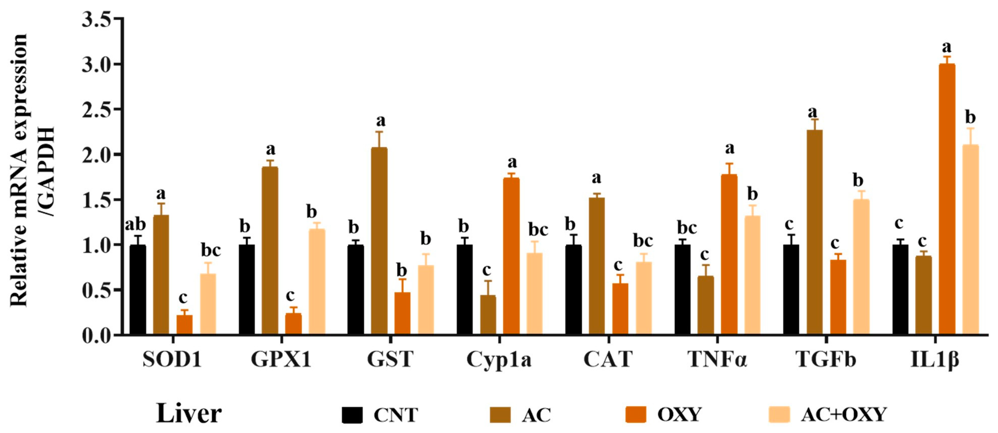
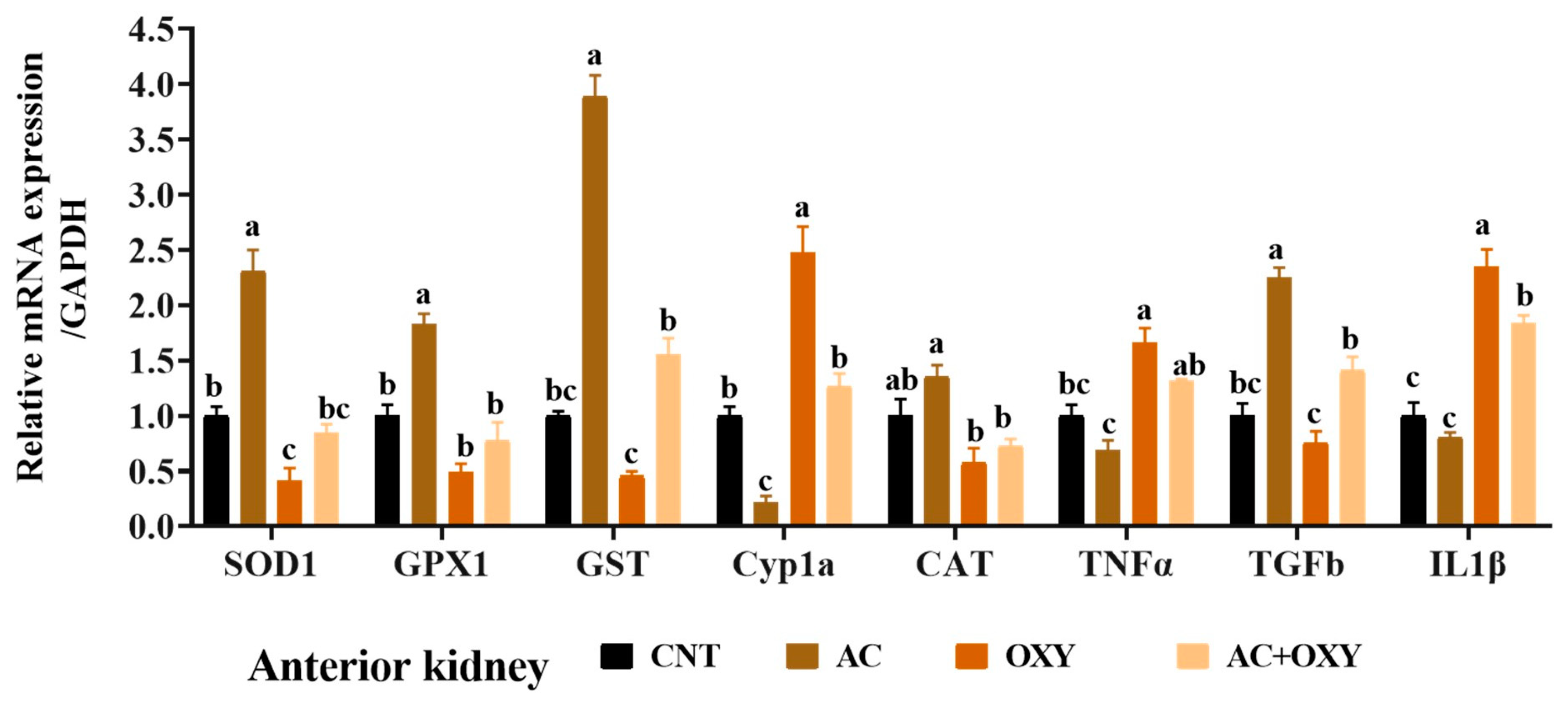
Due to their roles in xenobiotic detoxification, biotransformation, and excretion, the liver and kidneys are the most crucial target organs for most environmental pollutants. Exposure to OXY increases AST, ALT, ALP, creatinine, urea, and bilirubin. When aquatic organisms are exposed to OXY, their livers can bioaccumulate it at high bioconcentration factors of up to 5000, as observed in channel catfish [8]. Consequently, OXY may disrupt the functional integrity and increase their permeability due to its potent pro-oxidative effects, which could release liver enzymes into the bloodstream and affect liver function [9]. According to Hassanein, Banhawy [29], protoporphyrinogen oxidase may be a possible mechanism by which OXY causes hepatorenal damage, as previously demonstrated in mouse kidneys and livers [82]. Similarly, significant hepatorenal damage due to OXY has been described in C. gariepinus by Abd El-Rahman, Ahmed [8] and in tilapia fish by Ibrahim, Ghamry [31]. Huang, Jia [72] verified that OXY increased serum levels of kidney injury markers, including creatinine and urea, in zebrafish.
In the present study, C. gariepinus displayed hypoproteinemia, hypoalbuminemia, hypoglobulinemia, and hypercholesterolemia. Overall, these results point to a fish liver that is less functional, especially in producing blood proteins and controlling lipid metabolism. Furthermore, the reduction in total proteins, albumin, and globulin may be caused by the greater emigration caused by renal failure spurred by exposure to OXY. The results demonstrated that an AC-enriched diet prevents the toxicity of OXY, as evidenced by the above maintenance of the markers at levels similar to or even close to those of the control, confirming the preventive properties of AC on the liver and kidneys. In this regard, Saleh, Sakr [40] revealed a development pattern of the liver and spleen of C. gariepinus fed AC.
The results of the current investigation confirmed that fish exposed to OXY exhibited immune-toxic damage and increased fish stress. This was demonstrated by a significant decrease in lysozyme activity, nitric oxide, C3, and phagocytic activity but an increase in cortisol and glucose levels. Likewise, Ibrahim, Ghamry [31] described a considerable decrease in lysozyme activity and C3 and phagocytic activity following the exposure of O. niloticus to OXY. Increased cortisol and glucose levels may result from severe liver damage via inhibiting lipid metabolism and glycolysis pathways in the liver [83]. As opposed to this, fish fed a diet supplemented with AC exhibit improved immune response, enhanced immunological biomarkers, increased survivability, and decreased cortisol levels. Similarly, adding A. afra leaf powder to the diet of O. mossambicus increased lysozyme activity [84]. Moreover, there was an increase in both lysozyme and phagocytic activity in C. gariepinus fed with 5% A. afra [6]. An earlier report has also shown that AC supplementation at a rate of 5% can boost immune response in C. gariepinus. An increase in white blood cells indicates that AC inclusion in the diet enhances immunity [85]. Artemisinin and its derivatives can also actively modify the host’s immune system [86].
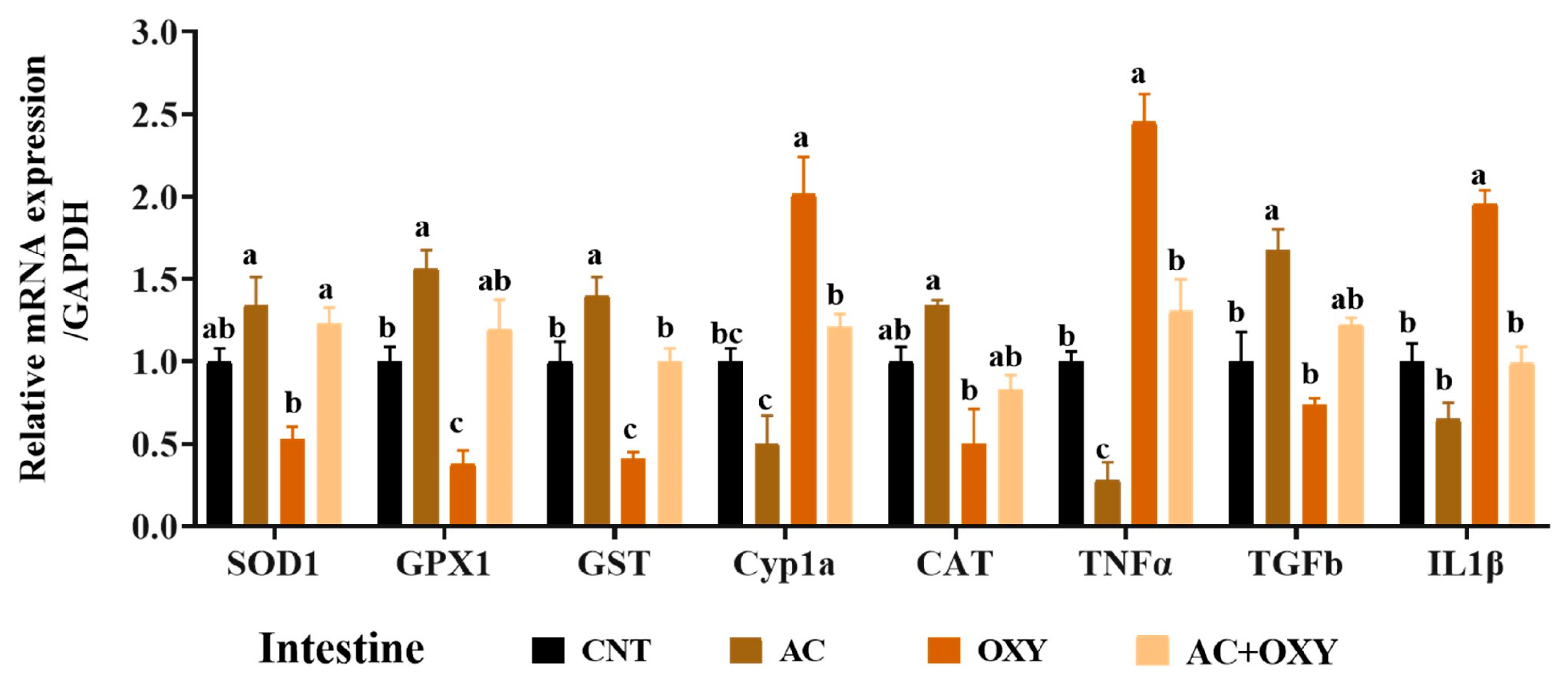
To study the immunotoxic effect of chronic OXY exposure on fish, the mRNA expression of some immune-related genes was assessed in the head kidney, liver, and intestine of C. gariepinus. The genes analyzed, including TNF-α, IL-1β, and TGF-β1, have been reported to play an important role in the immune defense of C. gariepinus [87]. IL-1β and TNF-α are examples of the inflammatory cytokines generated during inflammation [88]. The anti-inflammatory cytokine TGF-β1 reduces inflammation in fish [89]. The levels of inflammatory and anti-inflammatory cytokines were upregulated in the tissues studied after chronic exposure to OXY. Similarly, O. niloticus’s toxicity to OXY causes a down-regulation of immune-related gene expression [31]. In zebrafish, OXY also triggered inflammation by increasing the inflammatory factors TNF-α, IL-8, and IL-6 [83]. On the other hand, AC dietary supplementation alone or with OXY significantly decreased the expression of TNF-α and IL-1β genes while upregulating TGF-β1 in contrast to the only impact of OXY exposure. Consequently, the immunostimulant action of AC may have an anti-inflammatory impact as its underlying mechanism. The anti-inflammatory and immunomodulatory effects of sesquiterpene lactones from the Artemisia genus could improve the management of chronic diseases and, consequently, the efficacy of therapy [90]. Sakipova, Giorno [36] concluded that AC has probable anti-inflammatory and antinociceptive actions.
According to the results of the numerous assays used to assess the oxidative status, the most likely reason for the reduced organ function in C. gariepinus intoxicated with OXY was the incidence of oxidative stress damage. The high release of reactive oxygen species (ROS) and free radicals, as well as the incapacity of endogenous antioxidant mechanisms such as CAT, SOD, GST, and GPx to mitigate the harmful effects of these free radicals, are the usual causes of oxidative stress [91]. Our investigation demonstrated that serum levels of SOD, CAT, GPx, and reduced GSH all significantly decreased after exposure to OXY. Additionally, among all experimental groups, the OXY group had the highest MDA levels, which is a lipid peroxidation marker. Our observations of decreased antioxidant enzyme activity in the serum of C. gariepinus exposed to OXY matched the outcomes of gene expression analyses. Indeed, we revealed that the liver, kidney, and intestinal tissues of the fish subjected to sublethal OXY dosages considerably decreased the levels of mRNA expression of the genes coding for these antioxidant enzymes (SOD1, CAT, GST, and GPX1). These results indicated the inadequacy of the antioxidant systems to defend against oxidative stress damage triggered by the excessive generation of free radicals in response to exposure to OXY. Similar oxidative stress was described by Abd El-Rahman, Ahmed [8] in C. gariepinus and Ibrahim, Ghamry [31] in O. niloticus exposed to OXY. The results of the tests used in our investigation to define the level of Cyp1a gene mRNA expression in the tissues of OXY-intoxicated C. gariepinus offered additional proof in favor of the development of an oxidative stressful environment in these organs. Additionally, this expression can be related to DNA damage [92]. The proposed mechanism of immunobiotics that intensify the activities of antioxidant enzymes may increase resistance to toxin exposure [3]. Interestingly, adding AC, either separately or combined with OXY, enhanced the resistance against the oxidative damage imposed by OXY. The bioactive components of AC, such as thymol, carvacrol, phenolic compounds, betaine, choline, tannins, pigments, essential oil, and santonin, may contribute to its protective impact against oxidative damage induced by OXY [93]. 8-OHdG is commonly used to diagnose ROS-induced oxidative DNA damage [94]. In the current investigation, fish exposed to OXY for 60 days had considerably lower levels of AChE and increased 8-OHdG levels. Similarly, significant drops in AChE levels have been observed in the brain of Gambusia affinis, O. niloticus, and C. gariepinus after exposure to sublethal doses of OXY [8,28,31]. The increase in 8-OHdG occurring in serum might have resulted from oxidative stress induced by OXY [95]. Dietary AC administration restored AChE levels in fish that had been exposed to OXY, perhaps as a result of the supplement’s capacity to mitigate the oxidative stress caused by OXY [96].
In the current trial, the C. gariepinus was challenged with Q. aegypticus gill monogenea after OXY exposure for 60 days. The results showed that the percentage of OXY-exposed fish that survived the challenge reduced significantly. However, the number of infested fish with monogenea in the AC and OXY groups was lower than that in the control. AC’s efficacy as an antiparasitic agent in combating the monogenetic parasite, Heterobranchus longifilis, has been reported in C. carpio [41]. The World Health Organization recommends the use of artemisinin to treat malaria, and this plant is the only known source of this compound [97]. Artemisinin attacks the parasite’s mitochondria, which are essential for the parasite’s effective functioning [96]. Additionally, the main component of wormseed is the crystalline primary, santonin, from which the drug’s anthelmintic effects are derived. The immunomodulatory, anti-inflammatory, antimicrobial, and antiulcer activities help in promoting the health of fish, and consequently, the relative survival rate. Exposure to OXY could disrupt parasite transmission via direct or indirect effects on hosts and parasites; this can explain the decreased number of infested fish and prevalence percentage. Sublethal exposure to OXY significantly altered the health state of intoxicated fish in our study, and these changes had a deleterious effect on the general well-being, and consequently, survival rate.

5. Conclusions

The results of this study revealed that catfish, Clarias gariepinus, exposed to sublethal levels of OXY experienced growth suppression and hematologic, immunological, and antioxidant disturbances. It also induced detrimental effects on the expression of antioxidant and immune-related genes expression and resistance to monogenean parasites, Quadriacanthus aegypticus infection. Meanwhile, wormwood dietary supplementation, Artemisia cina, at a level of 5% alleviated the negative impacts of OXY on the zootechnical performance, physiological status, and monogenean parasite resistance of fish. It could be recommended as an aquafeed supplementation to mitigate the adverse effects of agriculture chemicals and infection.

Author Contributions

W.E.-H.: Conceptualization, Methodology, Resources, Writing—Original Draft, Writing—Review and Editing. R.G.A.A.: Conceptualization, Methodology, Formal Analysis, Investigation, Resources. A.H.A.: Methodology, Formal Analysis, Investigation, Resources. A.T.M.: Validation, Writing—Review and Editing. E.M.M.M.; F.A.S. and K.M.A.: Project Administration, Resources, Funding Acquisition, Review and Editing. G.E.E.: Conceptualization, Methodology, Formal Analysis, Investigation, Resources. Y.M.A.E.-H.: Conceptualization, Formal Analysis, Investigation, Resources, Writing—Original Draft, Writing—Review and Editing. All authors have read and agreed to the published version of the manuscript.

Funding

Princess Nourah bint Abdulrahman University Researchers Supporting Project number (PNURSP2023R318), Princess Nourah bint Abdulrahman University, Riyadh, Saudi Arabia.

Institutional Review Board Statement

The Zagazig University EAURC (Ethics of Animal Use in Research Committee) approved this protocol (ZU-IACUC/2/F/188/2023). All experimental techniques met the NIH general criteria for the care and use of laboratory animals in research.

Informed Consent Statement

Not applicable.

Data Availability Statement

Data will be made available on request.

Acknowledgments

The authors acknowledge Princess Nourah bint Abdulrahman University Researchers Supporting Project number (PNURSP2023R318), Princess Nourah bint Abdulrahman University, Riyadh, Saudi Arabia.

Conflicts of Interest

The authors declare no conflict of interest.

References

  1. Adewolu, M.A.; Adeniji, C.A.; Adejobi, A.B. Feed utilization, growth and survival of Clarias gariepinus (Burchell 1822) fingerlings cultured under different photoperiods. Aquaculture 2008, 283, 64–67. [Google Scholar] [CrossRef]
  2. El-Houseiny, W.; Khalil, A.A.; Abd-Elhakim, Y.M.; Badr, H.A. The potential role of turmeric and black pepper powder diet supplements in reversing cadmium-induced growth retardation, ATP depletion, hepatorenal damage, and testicular toxicity in Clarias gariepinus. Aquaculture 2019, 510, 109–121. [Google Scholar] [CrossRef]
  3. El-Houseiny, W.; Abd El-Hakim, Y.M.; Metwally, M.M.; Ghfar, S.S.A.; Khalil, A.A. The single or combined Silybum marianum and co-enzyme Q10 role in alleviating fluoride-induced impaired growth, immune suppression, oxidative stress, histological alterations, and reduced resistance to Aeromonas sobria in African catfish (Clarias gariepinus). Aquaculture 2022, 548, 737693. [Google Scholar]
  4. Mansour, A.T.; Hamed, H.S.; El-Beltagi, H.S.; Mohamed, W.F. Modulatory Effect of Papaya Extract against Chlorpyrifos-Induced Oxidative Stress, Immune Suppression, Endocrine Disruption, and DNA Damage in Female Clarias gariepinus. Int. J. Environ. Res. Public 2022, 19, 4640. [Google Scholar] [CrossRef] [PubMed]
  5. Ikpi, G.; Offem, B. Bacterial infection of mudfish Clarias gariepinus (Siluriformes: Clariidae) fingerlings in tropical nursery ponds. Rev. Biol. Trop. 2011, 59, 751–759. [Google Scholar] [CrossRef]
  6. Mbokane, E.M.; Moyo, N.A.G. Effect of dietary Artemisia afra on growth, some innate immunological parameters in Clarias gariepinus challenged with Aeromonas hydrophila. Aquac. Int. 2020, 28, 539–553. [Google Scholar] [CrossRef]
  7. Moustafa, G.G.; Shaaban, F.; Hadeed, A.A.; Elhady, W.M. Immunotoxicological, biochemical, and histopathological studies on Roundup and Stomp herbicides in Nile catfish (Clarias gariepinus). Vet. World 2016, 9, 638. [Google Scholar] [CrossRef]
  8. Abd El-Rahman, G.I.; Ahmed, S.A.; Khalil, A.A.; Abd-Elhakim, Y.M. Assessment of hematological, hepato-renal, antioxidant, and hormonal responses of Clarias gariepinus exposed to sub-lethal concentrations of oxyfluorfen. Aquat. Toxicol. 2019, 217, 105329. [Google Scholar] [CrossRef]
  9. Stagg, N.J.; LeBaron, M.J.; Eisenbrandt, D.L.; Gollapudi, B.B.; Klaunig, J.E. Assessment of possible carcinogenicity of oxyfluorfen to humans using mode of action analysis of rodent liver effects. Toxicol. Sci. 2012, 128, 334–345. [Google Scholar] [CrossRef]
  10. Mantzos, N.; Karakitsou, A.; Hela, D.; Patakioutas, G.; Leneti, E.; Konstantinou, I. Persistence of oxyfluorfen in soil, runoff water, sediment and plants of a sunflower cultivation. Sci. Total Environ. 2014, 472, 767–777. [Google Scholar] [CrossRef]
  11. Hall, K.E.; Ray, C.; Ki, S.J.; Spokas, K.A.; Koskinen, W.C. Pesticide sorption and leaching potential on three Hawaiian soils. J. Environ. Manag. 2015, 159, 227–234. [Google Scholar] [CrossRef] [PubMed]
  12. Ibrahim, A.M.; Sayed, D.A. Toxicological impact of oxyfluorfen 24% herbicide on the reproductive system, antioxidant enzymes, and endocrine disruption of Biomphalaria alexandrina (Ehrenberg, 1831) snails. Environ. Sci. Pollut. Res. 2019, 26, 7960–7968. [Google Scholar] [CrossRef] [PubMed]
  13. Riley, M.B.; Keese, R.J.; Camper, N.D.; Whitwell, T.; Wilson, P.C. Pendimethalin and oxyfluorfen residues in pond water and sediment from container plant nurseries. Weed Technol. 1994, 8, 299–303. [Google Scholar] [CrossRef]
  14. Nilsen, E.; Zaugg, S.; Alvarez, D.; Morace, J.; Waite, I.; Counihan, T.; Hardiman, J.; Torres, L.; Patiño, R.; Mesa, M. Contaminants of legacy and emerging concern in largescale suckers (Catostomus macrocheilus) and the foodweb in the lower Columbia River, Oregon and Washington, USA. Sci. Total Environ. 2014, 484, 344–352. [Google Scholar] [CrossRef] [PubMed]
  15. El-Houseiny, W.; AbdelMageed, M.; Abd-Elhakim, Y.M.; Abdel-Warith, A.-W.A.; Younis, E.M.; Abd-Allah, N.A.; Davies, S.J.; El-Kholy, M.S.; Ahmed, S.A. The effect of dietary Crataegus Sinaica on the growth performance, immune responses, hemato-biochemical and oxidative stress indices, tissues architecture, and resistance to Aeromonas sobria infection of acrylamide-exposed Clarias gariepinus. Aquac. Rep. 2023, 30, 101576. [Google Scholar] [CrossRef]
  16. Cheung, C.; Zheng, G.J.; Li, A.M.Y.; Richardson, B.J.; Lam, P.K.S. Relationships between tissue concentrations of polycyclic aromatic hydrocarbons and antioxidative responses of marine mussels, Perna viridis. Aquat. Toxicol. 2001, 52, 189–203. [Google Scholar] [CrossRef]
  17. El-Bouhy, Z.M.; Mohamed, F.A.; Elashhab, M.W.; El-Houseiny, W. Toxicity bioassay and sub-lethal effects of profenofos-based insecticide on behavior, biochemical, hematological, and histopathological responses in Grass carp (Ctenopharyngodon idella). Ecotoxicology 2023, 32, 196–210. [Google Scholar] [CrossRef]
  18. Khalil, A.A.; Abd-Elhakim, Y.M.; Said, E.N.; Moselhy, A.A.; Abu-Elsaoud, A.M.; El-Houseiny, W. Milk thistle and co-enzyme Q10 fortified diets lessen the nickel chloride-induced neurotoxic and neurobehavioral impairments in Oreochromis niloticus via regulating the oxidative stress response, acetylcholinesterase activity, and brain nickel content. Aquaculture 2022, 553, 738102. [Google Scholar] [CrossRef]
  19. Saurabh, S.; Sahoo, P. Lysozyme: An important defence molecule of fish innate immune system. Aquac. Res. 2008, 39, 223–239. [Google Scholar] [CrossRef]
  20. Mohamed, W.A.; El-Houseiny, W.; Ibrahim, R.E.; Abd-Elhakim, Y.M. Palliative effects of zinc sulfate against the immunosuppressive, hepato- and nephrotoxic impacts of nonylphenol in Nile tilapia (Oreochromis niloticus). Aquaculture 2019, 504, 227–238. [Google Scholar] [CrossRef]
  21. Abd El-Hakim, Y.M.; El-Houseiny, W.; Abd Elhakeem, E.-M.; Ebraheim, L.L.; Moustafa, A.A.; Mohamed, A.A.R. Melamine and curcumin enriched diets modulate the haemato-immune response, growth performance, oxidative stress, disease resistance, and cytokine production in Oreochromis niloticus. Aquat. Toxicol. 2020, 220, 105406. [Google Scholar] [CrossRef] [PubMed]
  22. Mokhtar, D.M.; Zaccone, G.; Alesci, A.; Kuciel, M.; Hussein, M.T.; Sayed, R.K. Main components of fish immunity: An overview of the fish immune system. Fishes 2023, 8, 93. [Google Scholar] [CrossRef]
  23. Laing, K.J.; Wang, T.; Zou, J.; Holland, J.; Hong, S.; Bols, N.; Hirono, I.; Aoki, T.; Secombes, C.J. Cloning and expression analysis of rainbow trout Oncorhynchus mykiss tumour necrosis factor-α. Eur. J. Biochem. 2001, 268, 1315–1322. [Google Scholar] [CrossRef]
  24. Laing, K.J.; Secombes, C.J. Chemokines. Dev. Comp. Immunol. 2004, 28, 443–460. [Google Scholar] [CrossRef] [PubMed]
  25. El-Houseiny, W.; Khalil, A.A.; Abd-Elhakim, Y.M.; Arisha, A.H.; Moselhy, A.A.; Dahshan, H.; Saber, T.; Saber, T.M.; Ahmed, M.M. Alleviative effects of dietary Silybum marianum and co-enzyme Q10 on waterborne nickel-induced impaired growth, immunosuppression, tissue damage, immune-related genes dysregulation, and reduced resistance to Pseudomonas aeruginosa in Oreochromis niloticus. Aquac. Rep. 2022, 26, 101308. [Google Scholar] [CrossRef]
  26. Xia, X.; Lu, C.; Li, M.; Wang, P.; Dong, H.; Gao, Y.; Chang, Z. Toxic effects of oxyfluorfen on Paramisgurnus dabryanus. J. Henan Agric. Sci. 2016, 45, 122–126. [Google Scholar]
  27. Powe, D.K.; Dasmahapatra, A.K.; Russell, J.L.; Tchounwou, P.B. Toxicity implications for early life stage Japanese medaka (Oryzias latipes) exposed to oxyfluorfen. Environ. Toxicol. 2018, 33, 555–568. [Google Scholar] [CrossRef]
  28. Hassanein, H.M. Toxicological effects of the herbicide oxyfluorfen on acetylcholinesterase in two fish species: Oreochromis niloticus and Gambusia affinis. J. Environ. Sci. Health Part A 2002, 37, 521–527. [Google Scholar] [CrossRef]
  29. Hassanein, H.; Banhawy, M.; Soliman, F.; Abdel-Rehim, S.; Müller, W.; Schröder, H. Induction of hsp70 by the herbicide oxyfluorfen (Goal) in the Egyptian Nile fish, Oreochromis niloticus. Arch. Environ. Contam. Toxicol. 1999, 37, 78–84. [Google Scholar]
  30. Mansour, A.T.; Amen, R.M.; Mahboub, H.H.; Shawky, S.M.; Orabi, S.H.; Ramah, A.; Hamed, H.S. Exposure to oxyfluorfen-induced hematobiochemical alterations, oxidative stress, genotoxicity, and disruption of sex hormones in male African catfish and the potential to confront by Chlorella vulgaris. Comp. Biochem. Physiol. Part C Toxicol. Pharmacol. 2023, 267, 109583. [Google Scholar] [CrossRef]
  31. Ibrahim, R.E.; Ghamry, H.I.; Althobaiti, S.A.; Almalki, D.A.; Shakweer, M.S.; Hassan, M.A.; Khamis, T.; Abdel-Ghany, H.M.; Ahmed, S.A. Moringa oleifera and Azadirachta indica Leaves enriched diets mitigate chronic oxyfluorfen toxicity induced immunosuppression through disruption of pro/anti-inflammatory gene pathways, alteration of antioxidant gene expression, and histopathological Alteration in Oreochromis niloticus. Fishes 2022, 8, 15. [Google Scholar]
  32. Almarri, S.H.; Khalil, A.A.; Mansour, A.T.; El-Houseiny, W. Antioxidant, Immunostimulant, and Growth-Promoting Effects of Dietary Annona squamosa Leaf Extract on Nile Tilapia, Oreochromis niloticus, and Its Tolerance to Thermal Stress and Aeromonas sobria Infection. Animals 2023, 13, 746. [Google Scholar] [CrossRef] [PubMed]
  33. El-Houseiny, W.; Algharib, S.A.; Mohamed, E.A.; Metwally, M.M.; Mahmoud, Y.K.; Alghamdi, Y.S.; Soliman, M.M.; Abd-Elhakim, Y.M.; El-Murr, A.E. Dietary parsley seed mitigates methomyl-induced impaired growth performance, hemato-immune suppression, oxidative stress, hepato-renal damage, and Pseudomonas aeruginosa susceptibility in Oreochromis niloticus. Antioxidants 2022, 11, 1185. [Google Scholar] [CrossRef] [PubMed]
  34. Mansour, A.T.; Mahboub, H.H.; Elshopakey, G.E.; Aziz, E.K.; Alhajji, A.H.; Rayan, G.; Ghazzawy, H.S.; El-Houseiny, W. Physiological Performance, Antioxidant and Immune Status, Columnaris Resistance, and Growth of Nile Tilapia That Received Alchemilla vulgaris-Supplemented Diets. Antioxidants 2022, 11, 1494. [Google Scholar] [CrossRef] [PubMed]
  35. Anibogwu, R.; Jesus, K.D.; Pradhan, S.; Pashikanti, S.; Mateen, S.; Sharma, K. Extraction, Isolation and Characterization of Bioactive Compounds from Artemisia and Their Biological Significance: A Review. Molecules 2021, 26, 6995. [Google Scholar] [CrossRef]
  36. Sakipova, Z.; Giorno, T.B.S.; Bekezhanova, T.; Siu Hai Wong, N.; Shukirbekova, A.; Fernandes, P.D.; Boylan, F. Pharmacological evaluation of Artemisia cina crude CO2 subcritical extract after the removal of santonin by means of high speed countercurrent chromatography. Molecules 2020, 25, 2728. [Google Scholar] [CrossRef]
  37. Tan, R.X.; Zheng, W.; Tang, H. Biologically active substances from the genus Artemisia. Planta Med. 1998, 64, 295–302. [Google Scholar] [CrossRef]
  38. Miraldi, E.; Ferri, S.; Franchi, G.G. Santonin: A new method of extraction from, and quantitative determination in Artemisia caerulescens ssp. cretacea (fiori) br.-catt. & gubell. by high-performance liquid chromatography. Phytochem. Anal. Int. J. Plant Chem. Biochem. Tech. 1998, 9, 296–298. [Google Scholar]
  39. Barton, B.A.; Iwama, G.K. Physiological changes in fish from stress in aquaculture with emphasis on the response and effects of corticosteroids. Annu. Rev. Fish Dis. 1991, 1, 3–26. [Google Scholar] [CrossRef]
  40. Saleh, O.A.; Sakr, S.F.; Abdelhadi, Y.M. Effect of wormseed plants; Artemisia cina L. and chamomile; Matricaria chamomilla L. on non specific immune response of Clarias gariepinus (African catfish). Abbassa Int. J. Aqua. 2010, 3, 246–259. [Google Scholar]
  41. Ramadan, R.; Doaa, A. Use of Artemisia cina against Gyrodactylus rysavyi Infecting Cyprinus carpio in Comparison with Praziquntel. J. Arab. Aquac. Soc. 2012, 7, 239–258. [Google Scholar]
  42. Canadian Council on Animal Care. Canadian Council on Animal Care Guidelines on: The Care and Use of Fish in Research, Teaching and Testing; CCAC: Ottawa, ON, Canada, 2005; p. 84. [Google Scholar]
  43. Boyd, C.E.; Tucker, C.S. Pond Aquaculture Water Quality Management; Springer Science & Business Media: Berlin, Germany, 2012. [Google Scholar]
  44. AOAC. Official Methods of Analysis, 18th ed.; Association of Official Agricultural Chemists: Washington, DC, USA, 2006. [Google Scholar]
  45. Feldman, B.V.; Zinkl, J.G.; Jain, N.C.; Schalm, O.W. Schalm’s Veterinary Hematology, 5th ed.; Lippincott Williams & Wilkins: Philadelphia, PA, USA, 2000. [Google Scholar]
  46. Bain, B.J.; Bates, I.; Laffan, M.A. Dacie and Lewis Practical Haematology E-Book; Expert Consult: Online and Print; Elsevier Health Sciences: Amsterdam, The Netherlands, 2016. [Google Scholar]
  47. Henry, R.J. Clinical Chemistry, Principles and Technics; Hoeber Medical Division, Harper & Row: New York, NY, USA, 1964. [Google Scholar]
  48. Reinhold, R. Determination of serum albumin. Clin. Chem. 1953, 21, 1370–1372. [Google Scholar]
  49. Coles, E. Veterinary Clinical Pathology; WB Saunders Company: Philadelphia, PA, USA; London, UK, 1986. [Google Scholar]
  50. Adeyemi, O.T.; Osilesi, O.; Adebawo, O.O.; Onajobi, F.D.; Oyedemi, S.O.; Afolayan, A. Alkaline phosphatase (ALP), aspartate aminotransferase (AST) and alanine aminotransferase (ALT) activities in selected tissues of rats fed on processed atlantic horse mackerel (Trachurus trachurus). Adv. Biosci. Biotechnol. 2015, 6, 139. [Google Scholar] [CrossRef]
  51. Ajeniyi, S.; Solomon, R. Urea and creatinine of Clarias gariepinus in three different commercial ponds. Nat. Sci. 2014, 12, 124–138. [Google Scholar]
  52. Kaplan, A. Glucose. In Clinical Chemistry; The CV Mosby Co.: St. Louis, MO, USA; Toronto, ON, Canada; Princeton, NJ, USA, 1984; 418p. [Google Scholar]
  53. Trinder, P. Determination of blood glucose using 4-amino phenazone as oxygen acceptor. J. Clin. Pathol. 1969, 22, 246. [Google Scholar] [CrossRef]
  54. Tunn, S.; Möllmann, H.; Barth, J.; Derendorf, H.; Krieg, M. Simultaneous measurement of cortisol in serum and saliva after different forms of cortisol administration. Clin. Chem. 1992, 38, 1491–1494. [Google Scholar] [CrossRef]
  55. Naito, H. Cholesterol. In Clinical Chemistry; The CV Mosby Co.: St. Louis, MO, USA; Toronto, ON, Canada; Princeton, NJ, USA, 1984. [Google Scholar]
  56. Aebi, H. Catalase in vitro. In Methods in Enzymology; Elsevier: Amsterdam, The Netherlands, 1984; Volume 105, pp. 121–126. [Google Scholar]
  57. Nishikimi, M.; Rao, N.A.; Yagi, K. The occurrence of superoxide anion in the reaction of reduced phenazine methosulfate and molecular oxygen. Biochem. Biophys. Res. Commun. 1972, 46, 849–854. [Google Scholar] [CrossRef]
  58. Paglia, D.E.; Valentine, W.N. Studies on the quantitative and qualitative characterization of erythrocyte glutathione peroxidase. J. Lab. Clin. Med. 1967, 70, 158–169. [Google Scholar]
  59. Beutler, E. Improved method for the determination of blood glutathione. J. Lab. Clin. Med. 1963, 61, 882–888. [Google Scholar]
  60. Uchiyama, M.; Mihara, M. Determination of malonaldehyde precursor in tissues by thiobarbituric acid test. Anal. Biochem. 1978, 86, 271–278. [Google Scholar] [CrossRef]
  61. Ellis, A.E. Lysozyme assays. Tech. Fish Immunol. 1990, 1, 101–103. [Google Scholar]
  62. Sakai, M.; Kobayashi, M.; Yoshida, T. Activation of rainbow trout, Oncorhynchus mykiss, phagocytic cells by administration of bovine lactoferrin. Comp. Biochem. Physiol. Part B Biochem. Mol. Biol. 1995, 110, 755–759. [Google Scholar] [CrossRef]
  63. Ellman, G.L.; Courtney, K.D.; Andres, V., Jr.; Featherstone, R.M. A new and rapid colorimetric determination of acetylcholinesterase activity. Biochem. Pharmacol. 1961, 7, 88–95. [Google Scholar] [CrossRef] [PubMed]
  64. Setyaningsih, Y.; Husodo, A.H.; Astuti, I. Detection of urinary 8-hydroxydeoxyguanosine (8-OHdG) levels as a biomarker of oxidative DNA damage among home industry workers exposed to chromium. Procedia Environ. Sci. 2015, 23, 290–296. [Google Scholar] [CrossRef]
  65. Shi, Y.; Hu, Y.; Wang, Z.; Zhou, J.; Zhang, J.; Zhong, H.; Fu, G.; Zhong, L. The Protective Effect of Taurine on Oxidized Fish-Oil-Induced Liver Oxidative Stress and Intestinal Barrier-Function Impairment in Juvenile Ictalurus punctatus. Antioxidants 2021, 10, 1690. [Google Scholar] [CrossRef]
  66. Ibor, O.R.; Eni, G.; Andem, A.B.; Bassey, I.U.; Arong, G.A.; Asor, J.; Regoli, F.; Arukwe, A. Biotransformation and oxidative stress responses in relation to tissue contaminant burden in Clarias gariepinus exposed to simulated leachate from a solid waste dumpsite in Calabar, Nigeria. Chemosphere 2020, 253, 126630. [Google Scholar] [CrossRef]
  67. Livak, K.J.; Schmittgen, T.D. Analysis of relative gene expression data using real-time quantitative PCR and the 2−ΔΔCT method. Methods 2001, 25, 402–408. [Google Scholar] [CrossRef]
  68. Paperna, I. Studies on monogenetic trematodes in Israel. 3. Monogenetic trematodes of the Cyprinidae and Claridae of the Lake of Galilee. Bamidgeh 1961, 13, 14–29. [Google Scholar]
  69. El-Naggar, M.M.; Serag, H.M. Quadriacanthus aegypticus n. sp., a monogenean gill parasite from the Egyptian teleost Clarias lazera. Syst. Parasitol. 1986, 8, 129–140. [Google Scholar] [CrossRef]
  70. Stoskopf, M.K. Biology and management of laboratory fishes. In Laboratory Animal Medicine; Elsevier: Amsterdam, The Netherlands, 2015; pp. 1063–1086. [Google Scholar]
  71. Bush, A.O.; Lafferty, K.D.; Lotz, J.M.; Shostak, A.W. Parasitology meets ecology on its own terms: Margolis et al. revisited. J. Parasitol. 1997, 83, 575–583. [Google Scholar] [CrossRef]
  72. Huang, L.; Jia, K.; Xiong, H.; Tian, G.; Xu, J.; Yuan, W.; Lu, C.; Xiao, X.; Lu, H. Oxyfluorfen exposure can cause acute kidney injury by promoting ROS-induced oxidative stress and inflammation in zebrafish. J. Hazard. Mater. 2022, 440, 129823. [Google Scholar] [CrossRef]
  73. Chakraborty, S.B.; Horn, P.; Hancz, C. Application of phytochemicals as growth-promoters and endocrine modulators in fish culture. Rev. Aquac. 2014, 6, 1–19. [Google Scholar] [CrossRef]
  74. Zheng, Z.; Tan, J.Y.; Liu, H.; Zhou, X.; Xiang, X.; Wang, K. Evaluation of oregano essential oil (Origanum heracleoticum L.) on growth, antioxidant effect and resistance against Aeromonas hydrophila in channel catfish (Ictalurus punctatus). Aquaculture 2009, 292, 214–218. [Google Scholar] [CrossRef]
  75. Mbokane, E.M.; Moyo, N.A.G. Effects of dietary levels of essential oil extracts from Moringa oleifera and Artemisia afra on kidney histology, haemato-immunological parameters and disease resistance in Clarias gariepinus. Aquac. Res. 2020, 51, 410–425. [Google Scholar] [CrossRef]
  76. Michael, P.O. Toxicity effect of atrazine on histology, haematology and biochemical indices of Clarias gariepinus. Int. J. Fish. Aquat. Stud. 2018, 6, 87–92. [Google Scholar]
  77. Lutnicka, H.; Bojarski, B.; Witeska, M.; Chmurska-Gąsowska, M.; Trybus, W.; Trybus, E.; Kopacz-Bednarska, A.; Lis, M. Effects of MCPA herbicide on hematological parameters and ultrastructure of hematopoietic tissues of common carp (Cyprinus carpio L.). Folia Biol. 2018, 66, 1–11. [Google Scholar] [CrossRef]
  78. Lutnicka, H.; Bojarski, B.; Król, T.; Trybus, W.; Trybus, E.; Kopacz-Bednarska, A.; Witeska, M.; Pankiewicz, L.; Pawlak, K. Hematological parameters and ultrastructure of hematopoietic tissues in common carp (Cyprinus carpio L.) exposed to sublethal concentration of pendimethalin. Folia Biol. 2018, 66, 121–132. [Google Scholar] [CrossRef]
  79. Rio, B.; Parent-Massin, D.; Lautraite, S.; Hoellinger, H. Effects of a diphenyl-ether herbicide, oxyfluorfen, on human BFU-E/CFU-E development and haemoglobin synthesis. Hum. Exp. Toxicol. 1997, 16, 115–122. [Google Scholar] [CrossRef]
  80. Soares, M.P.; Cardoso, I.L.; Ishikawa, M.M.; de Oliveira, A.d.S.S.; Sartoratto, A.; Jonsson, C.M.; de Queiroz, S.C.d.N.; Duarte, M.C.T.; Rantin, F.T.; Sampaio, F.G. Effects of Artemisia annua alcohol extract on physiological and innate immunity of Nile tilapia (Oreochromis niloticus) to improve health status. Fish Shellfish Immunol. 2020, 105, 369–377. [Google Scholar] [CrossRef]
  81. Pulsford, A.; Lemaire-Gony, S.; Tomlinson, M.; Collingwood, N.; Glynn, P. Effects of acute stress on the immune system of the dab, Limanda limanda. Comp. Biochem. Physiol. Part C Pharmacol. Toxicol. Endocrinol. 1994, 109, 129–139. [Google Scholar] [CrossRef]
  82. Krijt, J.; Stranska, P.; Maruna, P.; Vokurka, M.; Sanitrak, J. Herbicide-induced experimental Variegate prophyria in mice: Tissue porphyrinogen accumulation and response to porphyrogenic drugs. Can. J. Physiol. Pharmacol. 1997, 75, 1181–1196. [Google Scholar] [CrossRef] [PubMed]
  83. Li, Z.; Guo, J.; Jia, K.; Zheng, Z.; Chen, X.; Bai, Z.; Yang, Y.; Chen, B.; Yuan, W.; Chen, W. Oxyfluorfen induces hepatotoxicity through lipo-sugar accumulation and inflammation in zebrafish (Danio rerio). Ecotoxicol. Environ. Saf. 2022, 230, 113140. [Google Scholar] [CrossRef] [PubMed]
  84. Mbokane, E.; Moyo, N. A preliminary investigation into the potential effect of Artemisia afra on growth and disease resistance in sub-adults of Oreochromis mossambicus. Aquaculture 2018, 482, 197–202. [Google Scholar] [CrossRef]
  85. Mahassni, S.H.; Khudauardi, E.R. A pilot study: The effects of an aqueous extract of Lepidium sativum seeds on levels of immune cells and body and organs weights in Mice. J. Ayurvedic. Herb. Med. 2017, 3, 27–32. [Google Scholar] [CrossRef]
  86. Zhang, T.; Zhang, Y.; Jiang, N.; Zhao, X.; Sang, X.; Yang, N.; Feng, Y.; Chen, R.; Chen, Q. Dihydroartemisinin regulates the immune system by promotion of CD8+ T lymphocytes and suppression of B cell responses. Sci. China Life Sci. 2020, 63, 737–749. [Google Scholar] [CrossRef] [PubMed]
  87. Hamid, N.H.; Daud, H.M.; Kayansamruaj, P.; Hassim, H.A.; Yusoff, M.S.M.; Bakar, S.N.A.; Srisapoome, P. Short-and long-term probiotic effects of Enterococcus hirae isolated from fermented vegetable wastes on the growth, immune responses, and disease resistance of hybrid catfish (Clarias gariepinus × Clarias macrocephalus). Fish Shellfish Immunol. 2021, 114, 1–19. [Google Scholar] [CrossRef] [PubMed]
  88. Zou, J.; Secombes, C.J. The function of fish cytokines. Biology 2016, 5, 23. [Google Scholar] [CrossRef]
  89. Ming, J.; Ye, J.; Zhang, Y.; Xu, Q.; Yang, X.; Shao, X.; Qiang, J.; Xu, P. Optimal dietary curcumin improved growth performance, and modulated innate immunity, antioxidant capacity and related genes expression of NF-κB and Nrf2 signaling pathways in grass carp (Ctenopharyngodon idella) after infection with Aeromonas hydrophila. Fish Shellfish Immunol. 2020, 97, 540–553. [Google Scholar] [CrossRef]
  90. Ladurner, A.; Schachner, D.; Schueller, K.; Pignitter, M.; Heiss, E.H.; Somoza, V.; Dirsch, V.M. Impact of trans-resveratrol-sulfates and-glucuronides on endothelial nitric oxide synthase activity, nitric oxide release and intracellular reactive oxygen species. Molecules 2014, 19, 16724–16736. [Google Scholar] [CrossRef]
  91. Schwartz, J.I.; Dallob, A.L.; Larson, P.J.; Laterza, O.F.; Miller, J.; Royalty, J.; Snyder, K.M.; Chappell, D.L.; Hilliard, D.A.; Flynn, M.E. Comparative inhibitory activity of etoricoxib, celecoxib, and diclofenac on COX-2 versus COX-1 in healthy subjects. J. Clin. Pharmacology 2008, 48, 745–754. [Google Scholar] [CrossRef]
  92. Soltani, T.; Safahieh, A.; Zolgharnien, H.; Matroodi, S. Interactions of oxidative DNA damage and CYP1A gene expression with the liver enzymes in Klunzinger’s mullet exposed to benzo [a] pyrene. Toxicol. Rep. 2019, 6, 1097–1103. [Google Scholar] [CrossRef]
  93. Saxena, R.B. Entirely gone out useful plant—Artemisia cina. Indo Am. J. Pharm. Sci. 2015, 3, 2015. [Google Scholar]
  94. Alak, G.; Yeltekin, A.Ç.; Tas, I.H.; Ucar, A.; Parlak, V.; Topal, A.; Kocaman, E.M.; Atamanalp, M. Investigation of 8-OHdG, CYP1A, HSP70 and transcriptional analyses of antioxidant defence system in liver tissues of rainbow trout exposed to eprinomectin. Fish Shellfish Immunol. 2017, 65, 136–144. [Google Scholar] [CrossRef] [PubMed]
  95. Anjana Vaman, V.; Tinu, S.; Geetha, C.; Lissy, K.; Mohanan, P. Effect of fibrin glue on antioxidant defense mechanism, oxidative DNA damage and chromosomal aberrations. Toxicol. Mech. Methods 2013, 23, 500–508. [Google Scholar] [CrossRef] [PubMed]
  96. Ivanescu, B.; Miron, A.; Corciova, A. Sesquiterpene lactones from Artemisia genus: Biological activities and methods of analysis. J. Anal. Methods Chem. 2015, 2015, 247685. [Google Scholar] [CrossRef]
  97. Teuber, M. Veterinary use and antibiotic resistance. Curr. Opin. Microbiol. 2001, 4, 493–499. [Google Scholar] [CrossRef]
Figure 1. Effect of oxyfluorfen (OXY) exposure and/or Artemisia cina (AC) supplementation for 60 days on mRNA expression of superoxide dismutase (SOD1), glutathione peroxidase (GPx1), glutathione S-transferases (GST), cytochrome P450 1A (CYP1A), catalase (CAT), tumor necrosis factor alpha (TNF-α), transforming growth factor-beta (TGF-β), and interleukin 1 beta (IL1- β) in the intestinal tissue of Clarias gariepinus. Data expressed as mean ± SE, N = 10 for each group. Each bar carrying different letters (a, b, and c) was significantly different at p < 0.05.
Fishes 08 00297 g001
Figure 2. Effect of oxyfluorfen (OXY) exposure and/or Artemisia cina (AC) supplementation for 60 days on mRNA expression of superoxide dismutase (SOD1), glutathione peroxidase (GPx1), glutathione S-transferases (GST), cytochrome P450 1A (CYP1A), catalase (CAT), tumor necrosis factor alpha (TNF-α), transforming growth factor-beta (TGF-β), and interleukin 1 beta (IL1- β) in the hepatic tissue of Clarias gariepinus. Data expressed as mean ± SE, N = 10 for each group. Each bar carrying different letters (a, b, and c) was significantly different at p < 0.05.
Fishes 08 00297 g002
Figure 3. Effect of oxyfluorfen (OXY) exposure and/or Artemisia cina (AC) supplementation for 60 days on mRNA expression of superoxide dismutase (SOD1), glutathione peroxidase (GPx1), glutathione S-transferases (GST), cytochrome P450 1A (CYP1A), catalase (CAT), tumor necrosis factor alpha (TNF-α), transforming growth factor-beta (TGF-β), and interleukin 1 beta (IL1-β) in the anterior kidney of Clarias gariepinus. Data expressed as mean ± SE, N = 10 for each group. Each bar carrying different letters (a, b, and c) was significantly different at p < 0.05.
Fishes 08 00297 g003
Table 1. Ingredients and proximate chemical analysis of experimental diets.
Ingredients (g/100 g)Experimental Diets
Control5% Artemisia cina
Fish meal 66%1818
Ground corn2520
Soybean meal 44%3535
Corn oil33
Wheat bran1010
Cod liver oil22
starch44
Artemisia cina powder05
Vitamin premix 111
Mineral premix 222
Total100100
Chemical analysis
Crude protein (N × 6.25)32.1032.05
Crude lipids11.2711.34
Crude fiber4.454.25
Ash7.607.75
Nitrogen free extract 344.5844.61
Gross energy (kcal/kg) 4470.56471.59
1 Vitamin premix (per kg of premix): vitamin A, 8,000,000 IU; vitamin E, 7000 mg; vitamin D3, 2,000,000 IU; vitamin K3, 1500 mg; biotin, 50 mg; folic acid, 700 mg; nicotinic, 20,000 mg; pantothenic acid, 7000 mg; vitamin B1, 700 mg; vitamin B2, 3500 mg; vitamin B6, 1000 mg; vitamin B12, 7 mg. 2 Mineral premix (per kg of premix): zinc sulfate, 4.0 g; iron sulfate, 20 g; manganese sulfate,5.3 g; copper sulfate, 2.7 g; calcium iodine, 0.34 g; sodium selenite, 70 mg; cobalt sulfate, 70 mg, and CaHPO4·2H2O up to 1 kg. 3 Calculated by difference (100—protein% + lipids% + ash% + crude fiber%). 4 Gross energy (GE) was calculated as 5.65, 9.45, and 4.11 kcal/g for protein, lipid, and NFE, respectively (NRC, 1993).
Table 2. Oligonucleotide primer sequences and real-time PCR conditions.
Gene NamePrimer SequencesNCBI
Accession No.
Reference
GAPDHFTGTCCGTTTGGAGAAGCCTNM_001201199.1[65]
RATCAGGTCACAGACACGGTTG
CATFCTGGGACCTGACAGGCAATAKF977829[66]
RCTCCAGAAGTCCCACACCAT
SOD1FACCATGAAAGCTGTTTGCGTNM_001200992[66]
RTGGACATGAAAGCCATGCAG
GPX1FACCTGACCGCTGACATAGAGNM_001200741[66]
RACATCAGACAGCCCTTCACA
GSTFATCACCAGAAAGGCATTCGCGU588174[66]
RTCCAGGTCATTCTGATGGCA
CYP1AFCCAGCACGAGCATGAAGAAAKP336485[66]
RATGCTCTTTGACCAGCCTCT
TNF-αFCGCCAGCGGTAAACACGXM_017464718.1[65]
RCCGTTGAATGTCCGAAAGG
IL-1βFCTGAAGGGTGGAAACAAGGATAJ586102.1[65]
RGGAGTCACCAGTGCCGTTT
TGF-β1FGGAACGGCTGAGTGGGTCTXM_017483625.1[65]
RTGCTTACTGAGGCGGCTATG
GAPDH: glyceraldehyde-3-phosphate dehydrogenase; SOD1: superoxide dismutase; GPX1, glutathione peroxidase; GST: glutathione S-transferases, CYP1A: cytochrome P450 1A, CAT: catalase, TNF-α: tumor necrosis factor alpha, TGF-β: transforming growth factor-beta; IL1-β: interleukin 1 beta.
Table 3. Effect of oxyfluorfen (OXY) exposure and/or Artemisia cina (AC) supplementation for 60 days on growth performance of C. gariepinus.
Experimental Groups
ControlACOXYAC+OXY
Initial body weight (g)70.00 a ± 1.15472.00 a ± 1.15470.33 a ± 1.45271.16 a ± 1.166
Final body weight (g)120.00 b ± 1.732136.33 a ± 2.027101.33 c ± 1.763113.00 b ± 2.081
Weight gain (g)50.00 b ± 0.57764.33 a ± 0.88131.00 d ± 0.57741.83 c ± 1.092
Specific growth rate (%)0.89 b ± 0.0031.06 a ± 0.0020.60 d ± 0.0100.77 c ± 0.010
Feed intake (g)75.00 b ± 0.57782.00 a ± 1.15459.00 c ± 1.15471.00 b ± 1.154
Feed conversion ratio1.50 c ± 0.0051.27 d ± 0.0051.90 a ± 0.0341.69 b ± 0.029
Mortality (%)0.00 b ± 0.0000.00 b ± 0.00025.00 a ± 2.8863.33 b ± 3.333
Survival rate100.00 a ± 0.000100.00 a ± 0.00075.00 b ± 2.88696.66 a ± 3.333
Values are represented as the mean ± SE. The means within the same row carrying different superscripts are significant at p < 0.05. Control group was fed basal diet. AC group was fed a basal diet containing Artemisia cina 5%. OXY group was exposed to 1.16 mg/L oxyfluorfen and fed basal diet. AC+OXY group was fed a basal diet containing AC and exposed to OXY.
Table 4. Effect of oxyfluorfen (OXY) exposure and/or Artemisia cina (AC) supplementation for 60 days on hematological indices of C. gariepinus.
Experimental Groups
ControlACOXYAC+OXY
Erythrogram
Red blood cells (106/mm3)2.68 a ± 0.0902.88 a ± 0.0911.29 c ± 0.0872.20 b ± 0.063
Hemoglobin (gm/dl)10.31 b ± 0.08710.87 a ± 0.0537.54 d ± 0.0618.95 c ± 0.057
Hematocrit (%)30.93 b ± 0.26132.63 a ± 0.16022.64 d ± 0.18326.85 c ± 0.173
Mean corpuscular volume (fl)115.60 b ± 2.928113.57 b ± 4.286176.89 a ± 10.363121.82 b ± 2.715
Mean corpuscular hemoglobin (%)38.53 b ± 0.97837.85 b ± 1.42758.96 a ± 3.45640.60 b ± 0.907
Leukogram
White blood cells (103/mm3)7.61 a ± 0.1387.95 a ± 0.0305.17 c ± 0.0247.09 b ± 0.049
Lymphocytes (103/mm3)3.71 a ± 0.0703.82 ab ± 0.0242.59 c ± 0.0123.39 b ± 0.024
Heterophils (103/mm3)2.55 b ± 0.0432.67 a ± 0.0121.67 c ± 0.0052.45 b ± 0.008
Eosinophils (103/mm3)0.45 ab ± 0.0180.47 a ± 0.0080.29 c ± 0.0080.40 b ± 0.008
Monocytes (103/mm3)0.89 b ± 0.0180.97 a ± 0.0080.62 d ± 0.0050.83 c ± 0.008
Values are represented as the mean ± SE. The means within the same row carrying different superscripts are significant at p < 0.05. Control group was fed basal diet. AC group was fed a basal diet containing Artemisia cina 5%. OXY group was exposed to 1.16 mg/L oxyfluorfen and fed basal diet. AC+OXY group was fed a basal diet containing AC and exposed to OXY.
Table 5. Effect of oxyfluorfen (OXY) exposure and/or Artemisia cina (AC) supplementation for 60 days on immunological indices and hepato-renal function indicators of C. gariepinus.
Experimental Groups
Control ACOXYAC+OXY
Total protein (g/dL)6.10 a ± 0.216.18 a ± 0.223.60 c ± 0.125.18 b ± 0.14
Albumin (g/dL)2.72 a ± 0.152.71 a ± 0.151.38 c ± 0.042.13 b ± 0.06
Globulin (g/dL)3.37 a ± 0.063.46 a ± 0.072.21 b ± 0.153.05 a ± 0.08
ALT (U/L)16.03 b ± 0.8416.01 b ± 0.8950.23 a ± 1.3619.73 b ± 0.82
AST (U/L)31.83 b ± 1.4831.83 b ± 1.0160.33 a ± 1.4536.83 b ± 1.01
ALP (IU/L)37.10 b ± 0.4036.66 b ± 0.2452.36 a ± 1.3639.60 b ± 1.06
Total bilirubin (mg/dL)0.24 bc ± 0.010.23 d ± 0.021.01 a ± 0.030.32 b ± 0.01
Urea (mg/dL)2.77 b ± 0.062.60 b ± 0.067.50 a ± 0.293.13 b ± 0.19
Creatine (mg/dL)0.60b c ± 0.010.56 c ± 0.011.01 a ± 0.020.67 b ± 0.014
Lysozyme activity (units/L)16.82 b ± 0.3120.28 a ± 0.929.90 c ± 0.2514.43 b ± 0.54
Complement 3 (µg/mL)74.06 a ± 1.381.35 ± 79.96 a49.63 c ± 1.6765.66 b ± 1.11
Nitric oxide (µmol/L)39.16 b ± 1.1746.83 a ± 1.4821.16 c ± 1.3034.00 b ± 1.15
Phagocytic activity (%)26.73 b ± 0.4329.45 a ± 0.4715.83 d ± 0.5223.95 c ± 0.24
Values are represented as the mean ± SE. The means within the same row carrying different superscripts are significant at p < 0.05. Control group was fed basal diet. AC group was fed a basal diet containing Artemisia cina 5%. OXY group was exposed to 1.16 mg/L oxyfluorfen and fed basal diet. AC+OXY group was fed a basal diet containing AC and exposed to OXY.
Table 6. Effect of oxyfluorfen (OXY) exposure and/or Artemisia cina (AC) supplementation for 60 days on antioxidant status, stress, and DNA damage indicators of C. gariepinus.
Experimental Groups
ControlACOXYAC+OXY
Glutathione peroxidase (U/L)123.33 b ± 1.17130.72 a ± 1.2987.76 d ± 0.75114.73 c ± 1.51
Superoxide dimutase (U/mL)4.76 a ± 0.115.08 a ± 0.062.92 c ± 0.064.05 b ± 0.08
Catalase (U/L)190.90 a ± 3.41198.38 a ± 2.31144.96 c ± 2.63175.63 b ± 2.86
Malondialdehyde (nmol/mL)13.55 b ± 0.1811.58 c ± 0.2424.66 a ± 0.7314.90 b ± 0.06
Glutathione dehydrogenase (μmol/mL)2.16 a ± 0.022.23 a ± 0.011.34 c ± 0.031.99 b ± 0.03
(mg/dL) Cortisol 60.98 c ± 1.7960.13 c ± 1.8190.93 a ± 2.3773.80 b ± 1.37
Glucose (mg/dL)79.00 bc ± 1.1576.66 c ± 1.76107.66 a ± 1.4585.66 b ± 1.763
Cholesterol (mg/dL)90.50 b ± 1.3280.00 c ± 2.89122.83 a ± 1.5997.33 b ± 1.45
8-OHdG (ng/mL)19.61 c ± 0.6218.66 c ± 0.5777.01 a ± 0.7326.26 b ± 0.72
AChE (U/L)56.28 a ± 0.4055.43 a ± 0.5417.43 c ± 0.6951.10 b ± 0.78
Values are represented as the mean ± SE. The means within the same row carrying different superscripts are significant at p < 0.05. Control group was fed basal diet. AC group was fed a basal diet containing Artemisia cina 5%. OXY group was exposed to 1.16 mg/L oxyfluorfen and fed basal diet. AC+OXY group was fed a basal diet containing AC and exposed to OXY.
Table 7. Effect of oxyfluorfen (OXY) exposure and/or Artemisia cina (AC) supplementation for 60 days on prevalence (%), mean intensity, clinical signs, and postmortem findings observed in survived Clarias gariepinus fish in different experimental groups challenged with Quadriacanthus aegypticus monogenea.
Experimental Groups
ControlACOXYAC+OXY
Survived fishnumber21251520
Survival ratepercent841006080
Infested fishnumber20352
Prevalence percent95.231233.3310
Mean intensity-87.66 a ± 1.4517.0 bc ± 1.1520.0 b ± 1.1511.66 c ± 0.88
Signs of asphyxianumber16/212/2514/155/20
score ++++++++++
Postmortem change in gillsnumber13/200/35/51/2
score+++-++++++
The score of symptoms were recorded as follows: (-) no; (+) weak; (++) mild; (+++) moderate; (++++) severe. Control group was fed basal diet. AC group was fed a basal diet containing Artemisia cina 5%. OXY group was fed a basal diet and exposed to 1.16 mg/L oxyfluorfen. AC+OXY group was fed a basal diet containing AC and exposed to OXY.
Disclaimer/Publisher’s Note: The statements, opinions and data contained in all publications are solely those of the individual author(s) and contributor(s) and not of MDPI and/or the editor(s). MDPI and/or the editor(s) disclaim responsibility for any injury to people or property resulting from any ideas, methods, instructions or products referred to in the content.

