Communication

Piscine Orthoreovirus-1 (PRV-1) Has Been Present in Chilean Salmon Aquaculture since at Least 1994

Pathovet Labs SpA, Puerto Montt 5480000, Chile
*
Author to whom correspondence should be addressed.
Fishes 2023, 8(5), 229; https://doi.org/10.3390/fishes8050229
Submission received: 14 March 2023 / Revised: 20 April 2023 / Accepted: 24 April 2023 / Published: 26 April 2023
(This article belongs to the Special Issue Infections of Aquatic Animals)

Abstract

:
Heart and skeletal muscle inflammation (HSMI) caused by Piscine orthoreovirus (PRV) was first described in farmed Atlantic salmon in Chile in 2011. However, as PRV induces long-lasting infections, it is not known when Chilean farmed salmon may have started to show PRV positivity. This study aimed to evaluate the presence/absence of PRV-1 in formalin-fixed, paraffin-embedded Atlantic salmon heart tissues (FFPE) cultured in Chile during 1992 and 1999. The most frequent histopathological findings in the 42 FFPE blocks were mild focal cardiomyocyte degeneration (57.1%) and a mild focal mononuclear inflammatory infiltrate (21.4%) in the ventricular stratum spongiosum of the heart. One of the 42 heart samples analyzed by RT-qPCR was positive for PRV-1 (2.4%). All samples were negative for other viral and bacterial pathogens that can induce similar histological changes in the heart. Taken together, our results show that PRV-1 has been present in Chile—as a low-virulence genogroup—since at least 1994, 17 years before the first HSMI outbreak in 2011. Finally, archaeovirology can be a valid alternative to contribute to the understanding of the epidemiology of diseases in aquaculture.
Key Contribution: The study represents a valuable contribution to understanding the historical distributions of PRV and its epidemiology, transmission, and evolution in salmon aquaculture in Chile. Archaeovirology is a valid alternative to contribute to the epidemiological knowledge of diseases in aquaculture.

Graphical Abstract

1. Introduction

Heart and skeletal muscle inflammation (HSMI) was described in 1999 in Atlantic salmon (Salmo salar) farmed in Norway [1], but was not associated with Piscine orthoreovirus (PRV) until 2010 [2]. PRV is a non-enveloped virus with a segmented, double -stranded RNA genome, which belongs to the genus Orthoreovirus, subfamily Spinareovirinae within the Reoviridae family. Three PRV-subtypes have been described: PRV-1 is associated with HSMI in Atlantic salmon, Salmo salar [2] and lowered hematocrit in chinook salmon, Oncorhynchus tshawytscha [3] and coho salmon, Oncorhynchus kisutch [4]; PRV-2 is associated with erythrocytic inclusion body syndrome (EIBS) in coho salmon [5]; and PRV-3 is associated with HSMI-like disease with anemia in rainbow trout, Oncorhynchus mykiss [6] and brown trout, Salmo trutta [7].
Phylogeny and sequence analyses for segments S1 and M2 from fish in the presence or absence of HSMI grouped the viral sequences into two monophylogenetic clusters, one associated with HSMI (PRV-1b) and the other with low-virulent PRV-1 isolates (PRV-1a) [8]; although Wessel et al. [9] suggest that PRV-1 virulence is related to the combined association and possible gene linkage of genomic segments S1, M2, L1, and L2 and S4 and their encoded proteins.
HSMI caused by PRV was first described in farmed Atlantic salmon in Chile in 2011 [10]. Subsequently, the first report of HSMI-like lesions with the presence of PRV in coho salmon was published in 2016 [4]. Initially, high PRV prevalence was recorded in fish and at net-pens level, but without showing heart lesions [10]. Over the years, HSMI outbreaks with moderate to severe heart lesions associated with higher PRV loads began to be observed at seawater farms, as well as PRV presence and heart injuries in freshwater-farmed fish [4]. While HSMI/PRV-attributed mortality in Atlantic salmon farmed in Chile was only 0.7% of the total mortality in 2021, this value was 11 times higher in coho salmon (7.7%) [11]. A wide distribution of PRV infection has been described in wild and farmed salmonid species [8], which modulates an immune response that promotes lifelong viral persistence [12]. This study aimed to evaluate the presence of PRV in formalin-fixed, paraffin-embedded Atlantic salmon heart tissues (FFPE) farmed in Chile during 1992 and 1999.

2. Materials and Methodology

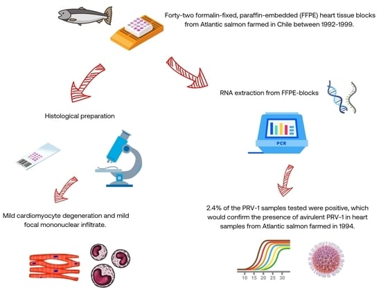
2.1. Fish and Tissue Samples

Heart samples from Atlantic salmon and coho salmon were collected from different salmon farms in the Los Lagos region of Chile between 1992 and 1999. Samples were fixed in 10% formalin and subsequently processed for standard histopathological diagnosis in the laboratory of the Instituto de Patología Animal, Facultad de Ciencias Veterinarias, Universidad Austral de Chile, Valdivia, Chile. The samples were preserved as FFPE blocks as part of the pathological material collection for about 30 years until the time of this study. Forty-two (42) FFPE blocks of cardiac tissue were used for this study.

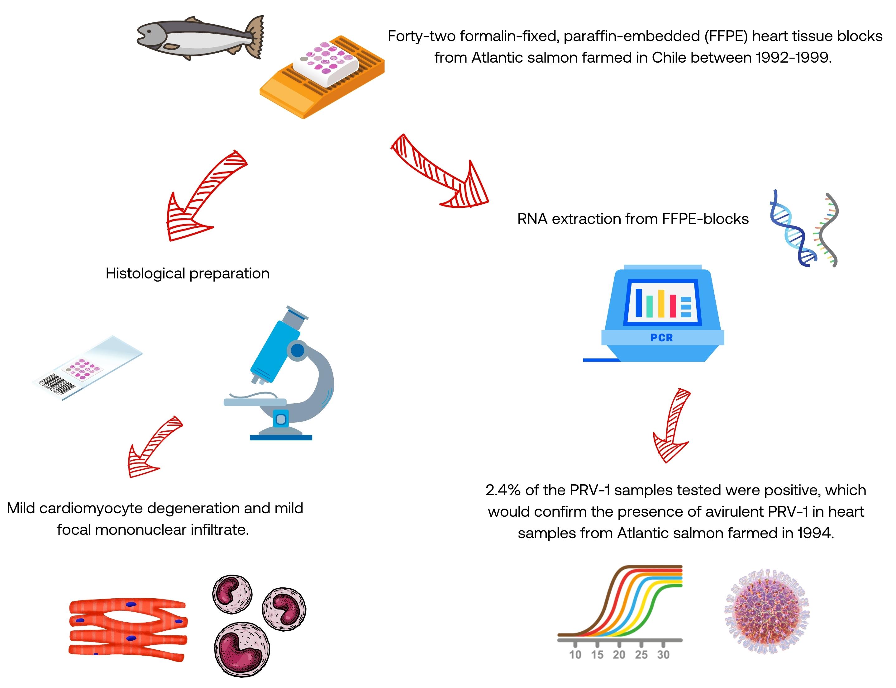
2.2. Histopathological and Immunohistochemical (IHC) Examination

Sections measuring 3 μm thick were cut from each FFPE block, stained with hematoxylin and eosin (H&E), and analyzed by light microscopy (Leica DM-2000, Hamburg, Germany). For this study, a cardiac histoscore (hsCRC) was used to semiquantify the severity of tissue changes according to the criteria described in Table 1. An hsCRC ≤ 0.9 accounts for mild cardiac lesions; hsCRC higher than 0.9, but lower than 1.8, show moderate injuries; and hsCRC > 1.8 indicates severe tissue damage. To confirm the presence or absence of PRV-antigens in cardiac tissue, an immunohistochemical (IHC) protocol was followed using an in-house developed polyclonal antibody against PRV σ1 protein based on the previously described sequences [13,14]. Atlantic salmon heart samples obtained from an HSMI outbreak in the field with histopathological lesions and confirmed as PRV-positive by RT-qPCR and IHC, were used as positive controls. Conversely, heart samples from healthy fish that were verified as negative for PRV by RT-qPCR were used as negative controls.

2.3. RNA Extraction

Each FFPE tissue block was sectioned into four or five 10 μm scrolls (total ∼40 μm per block). This volume was divided into two equivalent parts to be analyzed independently by one-step and two-step PCR. RNA was extracted from tissues using the commercial PureLink™ FFPE RNA Isolation Kit, catalog number K156002 (ThermoFisher Scientific, Carlsbad, CA, USA) based on the manufacturer’s protocol. Tissues were removed from the scrolls, placed in a fusion buffer, centrifuged at 12,000 g for 15 s, and incubated at 72 °C for 10 min. Then, 20 μL of proteinase K were added and incubated at 60 °C for 1 h, following centrifugation at 12,000 g for 1 min, and the lysate was placed into a new 1.7 mL microfuge tube. Next, 400 µL of Binding Buffer (L3) and 800 µL of absolute ethanol were mixed, and 700 µL of the resulting solution were transferred to a spin column. The solution was centrifuged at 800 g for 1 min at room temperature, discarding the flow-through. The remainder of the mixture (sample) was added to the column, centrifuged at 800 g for 1 min at room temperature, and the flow-through was discarded. A measured volume of 500 µL of Wash Buffer (W5) prepared with 100% ethanol were added, centrifuged at 12,000 g for 1 min, and the flow-through was discarded. Then, 500 µL of Wash Buffer (W5) were added again, centrifuged at 12,000 g for 1 min, and the flow-through was removed. The column was transferred to a new 1.7 mL microfuge tube, 50 µL of RNAse-free water previously heated to 65 °C was added and centrifuged at 12,000 g for 1 min at room temperature.

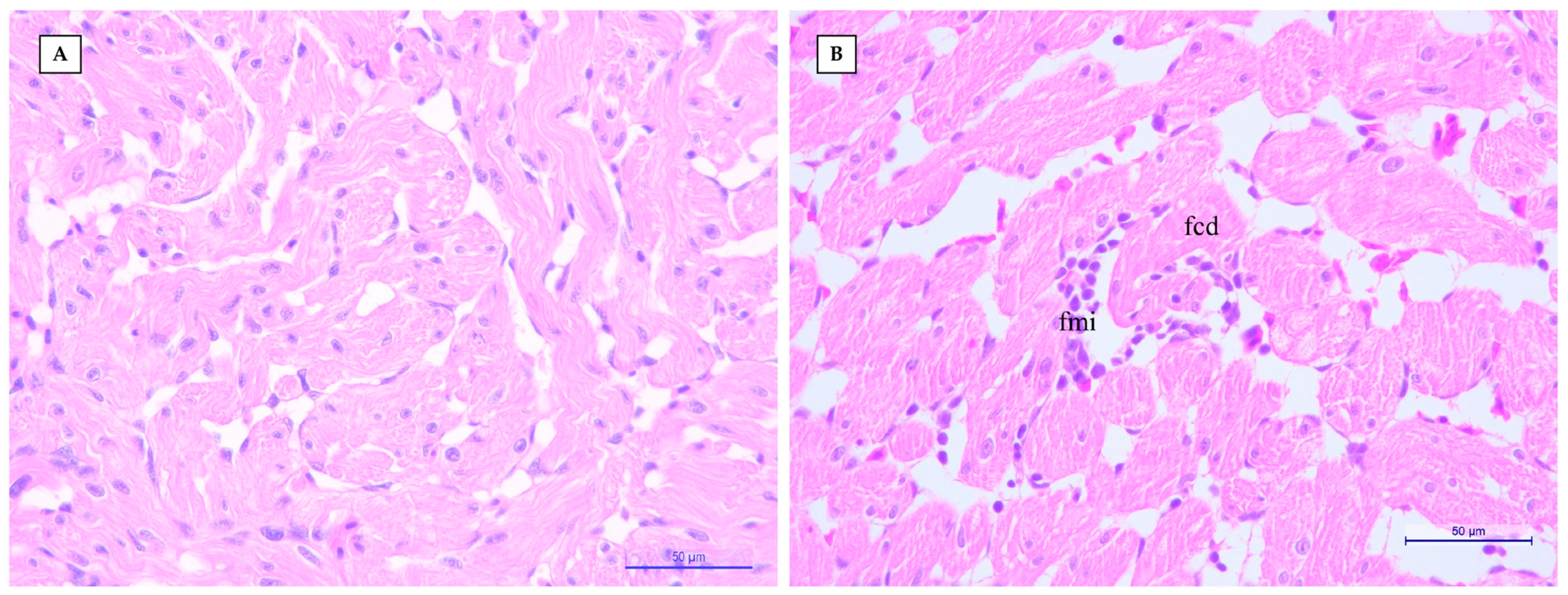
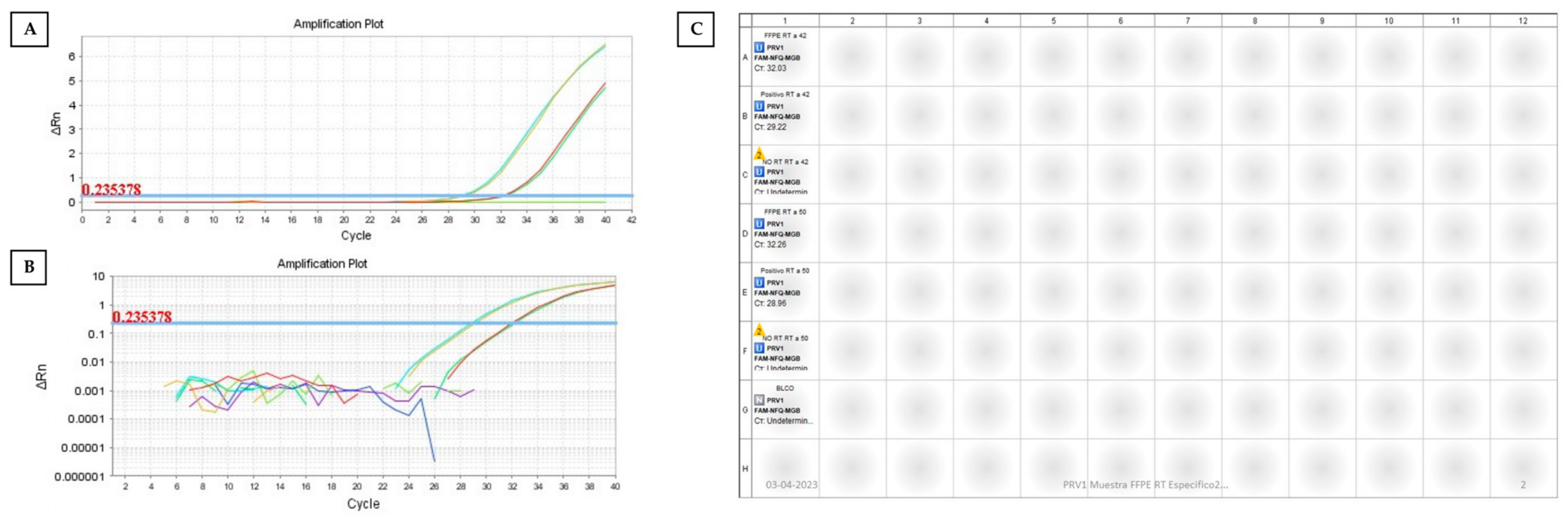
2.4. One-Step RT–qPCR

The presence or absence of PRV-1, PRV-2, PRV-3, infectious salmon Anemia virus (ISAV), and Piscirickettsia salmonis was determined by qPCR, as previously described [2,5,6,15,16], using the Brilliant III Ultrafast RT-qPCR Master Mix kit (Agilent Technologies, Santa Clara, CA, USA) in a QuantStudio 3TM Real-Time PCR system (Applied Biosystems, Life Technologies, Carlsbad, CA, USA). The RT-qPCR conditions for the detection of each pathogen consisted in adding a master mix of 7.5 µL buffer, 0.15 µL 100 mM DTT, 0.225 µL diluted ROX solution, 300 nM each primer, and 400 nM probe to 2 µL total RNA. Tubes were incubated for 10 min at 50 °C to perform reverse transcription, followed by a denaturation step of 3 min at 95 °C and 40 cycles of 3-s s at 95degC and 10 s at 60 °C. All qPCR assays were performed in duplicate. A positive control (RNA or DNA specific to the tested pathogen), a negative control without RNA or DNA, and negative extraction control were also included in every run. All qPCR runs for the pathogens tested were accompanied by the expression of the Atlantic salmon reference gene as an endogenous extraction control (EF1a). Cycling threshold (Ct) values were recorded up to a maximum of 40 Ct. A Ct below the cut-off point was considered positive, and negative (NoCt) otherwise. The cut-off points for PRV-1, -2, and -3, ISAV, and EL1a assays were 35.00, and for P. salmonis was 33.01. Allele-specific RT-qPCR to identify and segregate PRV-1a and -1b samples was carried out according to the protocol described by Siah et al. [17].

2.5. Two-Step RT–qPCR

PRV-1-positive RNA extract was used as a template for cDNA synthesis. Approximately 1 µg total RNA was reverse transcribed using the PrimeScript™ RT Reagent kit with gDNA Eraser (Takara Bio Group, San Jose, CA, USA) according to the manufacturer’s instructions. Specific primers were used for the S1 and M2 segments described by Kibenge et al. [18], and for the L1 segment described by Palacios et al. [2]. Briefly, the RT conditions consisted in the addition of a master mix of 2 µL 5× gDNA Eraser Buffer and 1 µL gDNA Eraser for each tube containing 1 µg total RNA (10 µL total volume). Tubes were incubated for 2 min at 42 °C to erase gDNA. The reverse transcription proceeded immediately on the same tubes adding a master mix of 4 µL 5× PrimeScript Buffer, 1µL PrimeScript RT Enzyme Mix I, and 4-µL-specific primers (total volume 20 µL). Tubes were incubated for 15 min at two different annealing temperatures (42 °C and 50 °C) followed by a 5-s incubation at 85 °C. The PCR products were confirmed by electrophoresis in 1% agarose gels and visualized with ethidium bromide staining. To confirm the results obtained by one-step RT-qPCR, a qPCR for the L1 segment of PRV-1 was performed using the KAPA Probe Fast qPCR kit Mastermix (2×) Universal. Briefly, a master mix consisting in 7.5 µL Buffer, 0.3 µL 50× ROX, 400 nM specific L1 primers, and 200 µL L1 probe was added to 2 µL 1:10 diluted cDNA (total volume 15 µL). The PCR cycling conditions consisted in an incubation of 3 min at 95 °C followed by 40 cycles of 3 s at 95 °C and 20 s at 60 °C. For the PCR stage of segments S1 and M2, the Platinum™ Hot Start PCR Master Mix (ThermoFisher Scientific, Carlsbad, CA, USA) (2×) was used. The liquid handling system created a master mix using 25 µL Platinum Hot Start PCR 2X Master Mix, 200 nM of each primer (either for S1 or M2, respectively), and 5 µL cDNA template to a total volume of 50 µL. The PCR cycling conditions consisted of an initial denaturation of 2 min at 94 °C followed by 40 cycles of 30 s at 94 °C, 30 s at 50 °C and 1, and at 72 °C.

3. Results and Discussion

Mild cardiomyocyte degeneration (63.3%) and mild focal mononuclear myocarditis in the ventricular stratum spongiosum (23.8%) (Figure 1) were the most frequent histopathological diagnoses in the cardiac tissues examined (Supplementary Table S1); these findings are consistent with those previously described by Kongtrop et al. [1]. No histopathological changes were recorded in the atrium of the hearts examined. Conversely, the median hsCRC was 0.13 (range 0–0.47) (Table S1), supporting that all histopathologic lesions observed were mild (hsCRC ≤ 0.9).
One of the 42 samples (2.4%) of the heart samples examined were positive for PRV-1 by one-step RT-qPCR (average Ct, 28.08, <35.00) and two-step qPCR (average Ct, 32.15, <35.00) (Figure 2). Therefore, we confirmed the presence of PRV-1 in independent qPCR reactions in different parts of the FFPE block and the loss of sensitivity of two-step PCR by about one order of magnitude (~30-fold). However, this sample was negative by IHC for PRV. Our experience indicates that to obtain an IHC-PRV-positive result usually requires a viral load (expressed in Ct) of at least 24. PCR on genetic material extracted from FFPE-blocks misses at least 4 cycles compared to running PCR on genetic material extracted from frozen or ethanol-fixed tissue [19]. Hence, we speculate that the relatively low PRV-1 load in the FFPE blocks used in this study might be the most plausible reason to explain the negative IHC results. All samples were negative for other viral and bacterial pathogens that can induce similar histological changes in the heart (Table S1).
Furthermore, it was impossible to obtain the S1 sequences of PRV-1 using the positive sample to perform a phylogenetic analysis (Figure 2). The size of the L1 segment amplified by qPCR primers and probes is small (79 bp) [2], but the fragments to amplify the S1 and M2 segments with sequencing primers are as large as 1081 and 2179 bp, respectively [18]. This situation, added with the fact that the genetic material available in the sample was probably fragmented, degraded (average Ct 33.22 for reference gene EL1a, but <35.00) and/or chemically modified after being stored for 3 decades before this analysis, could explain not achieving large amplicons in S1 and M2, and obtaining a much smaller amplicon in L1. RT-qPCR-PRV-1 positive sample was negative for the PRV-1b genotype, associated with HSMI according to Dhamotharan et al. [8], thus the sequence would most likely correspond to the low virulence PRV-1a genotype.
Taken together, our results would indicate that PRV-1 may have been present in farmed Atlantic salmon in Chile since at least 1994, some 17 years before the first outbreak of HSMI [10]. Similarly, a PRV-1 strain from Norway sampled in 1988, a decade before the emergence of HSMI in 1999, along with the low virulent HSMI cluster [8]. PRV-1 genotype causing HSMI in farmed Atlantic salmon has a recent common ancestor, and evolved prior to the rapid expansion of HSMI disease outbreaks in Norwegian Atlantic salmon farms, which spread from a specific zone of outbreaks to all aquaculture related areas within a few years after 1999, probably determined by differences in host, virus, environment, or a combination of these factors [8]; this epidemiological situation seems to correlate with the experience in Chilean salmon farming.
Chilean PRV isolates probably diverged from Norwegian isolates between 2007 and 2009 [18], ending up as pathogenic isolates causing the HSMI outbreaks recorded in 2011 [10]. Subsequently, Chilean PRV-1 isolates have shown high percentages of identity with Norwegian PRV-1 and, given that Atlantic salmon is not an endemic species in Chile, it is now not only reasonable to suggest that PRV-1 was introduced into Chile through the movement of salmon embryos [18], but that it would have occurred before 1994. FFPE tissues are a precious resource from a pathogen diagnosis perspective; therefore, archaeovirology can be a valid alternative to contribute to the epidemiological knowledge of diseases in aquaculture.

4. Conclusions

The results of our study demonstrated the presence of PRV-1 in FFPE heart samples with mild cardiomyocyte degeneration and mild focal mononuclear myocarditis in the ventricular stratum spongiosum of Atlantic salmon farmed in Chile in 1994. Consequently, our results help to better understand the biological interaction of PRV and host, and to highlight the historical epidemiology of PRV/HSMI in Chile.

Supplementary Materials

The following supporting information can be downloaded at: https://www.mdpi.com/article/10.3390/fishes8050229/s1, Table S1: Results of hsCRC, IHC PRV, and RT-qPCR for PRV-1, PRV-2, PRV-3, ISAV, and P. salmonis in each FFPE sample analyzed (individual fish). Negative (−).

Author Contributions

Conceptualization, M.R.-S.; methodology, M.R.-S.; data curation, M.R.-S., R.I., V.J., C.L. and E.P.; formal analysis and investigation, M.R.-S., R.I., V.J., C.L., L.M., A.M., D.C., S.B., E.P., A.P., C.S. and F.A.; resources, R.I., V.J., C.L., L.M., A.M., D.C., S.B., E.P., A.P., C.S. and F.A.; writing—original draft preparation, M.R.-S.; writing—review and editing, M.R.-S., A.P. and F.A.; funding acquisition, M.R.-S.; project administration, M.R.-S. All authors have read and agreed to the published version of the manuscript.

Funding

This study was funded by the Chilean Production Development Corporation (CORFO 20CVID-128114).

Institutional Review Board Statement

Not applicable.

Informed Consent Statement

Not applicable.

Data Availability Statement

The data presented in this study are available on request from the corresponding author.

Acknowledgments

We thank Enrique Paredes for sharing the biological material used in this study, which belongs to the Instituto de Patología Animal, Facultad Ciencias Veterinarias, Universidad Austral de Chile, Valdivia, Chile.

Conflicts of Interest

The authors declare no conflict of interest.

References

  1. Kongtorp, R.T.; Kjerstad, A.; Taksdal, T.; Guttvik, A.; Falk, K. Heart and skeletal muscle inflammation in Atlantic salmon, Salmo salar L.: A new infectious disease. J. Fish Dis. 2004, 27, 351–358. [Google Scholar] [CrossRef] [PubMed]
  2. Palacios, G.; Lovoll, M.; Tengs, T.; Hornig, M.; Hutchison, S.; Hui, J.; Kongtorp, R.-T.; Savji, N.; Bussetti, A.V.; Solovyov, A.; et al. Heart and Skeletal Muscle Inflammation of Farmed Salmon Is Associated with Infection with a Novel Reovirus. PLoS ONE 2010, 5, e11487. [Google Scholar] [CrossRef] [PubMed]
  3. Di Cicco, E.; Ferguson, H.W.; Schulze, A.D.; Kaukinen, K.H.; Li, S.; Vanderstichel, R.; Wessel, Ø.; Rimstad, E.; Gardner, I.A.; Hammell, K.L.; et al. Heart and skeletal muscle inflammation (HSMI) disease diagnosed on a British Columbia salmon farm through a longitudinal farm study. PLoS ONE 2017, 12, e0171471. [Google Scholar]
  4. Godoy, M.G.; Kibenge, M.J.T.; Wang, Y.; Suarez, R.; Leiva, C.; Vallejos, F.; Kibenge, F.S.B. First description of clinical presentation of piscine orthoreovirus (PRV) infections in salmonid aquaculture in Chile and identification of a second genotype (Genotype II) of PRV. Virol. J. 2016, 13, 98. [Google Scholar] [CrossRef] [PubMed]
  5. Takano, T.; Nawata, A.; Sakai, T.; Matsuyama, T.; Ito, T.; Kurita, J.; Terashima, S.; Yasuike, M.; Nakamura, Y.; Fujiwara, A.; et al. Full-Genome Sequencing and Confirmation of the Causative Agent of Erythrocytic Inclusion Body Syndrome in Coho Salmon Identifies a New Type of Piscine Orthoreovirus. PLoS ONE 2016, 11, e0165424. [Google Scholar] [CrossRef] [PubMed]
  6. Dhamotharan, K.; Vendramin, N.; Markussen, T.; Wessel, Ø.; Cuenca, A.; Nyman, I.B.; Olsen, A.B.; Tengs, T.; Dahle, M.K.; Rimstad, E. Molecular and Antigenic Characterization of Piscine orthoreovirus (PRV) from Rainbow Trout (Oncorhynchus mykiss). Viruses 2018, 10, 170. [Google Scholar] [CrossRef] [PubMed]
  7. Kuehn, R.; Stoeckle, B.C.; Young, M.; Popp, L.; Taeubert, J.-E.; Pfaffl, M.W.; Geist, J. Identification of a piscine reovirus-related pathogen in proliferative darkening syndrome (PDS) infected brown trout (Salmo trutta fario) using a next-generation technology detection pipeline. PLoS ONE 2018, 13, e0206164. [Google Scholar] [CrossRef] [PubMed]
  8. Dhamotharan, K.; Tengs, T.; Wessel, Ø.; Braaen, S.; Nyman, I.B.; Hansen, E.F.; Christiansen, D.H.; Dahle, M.K.; Rimstad, E.; Markussen, T. Evolution of the Piscine orthoreovirus Genome Linked to Emergence of Heart and Skeletal Muscle Inflammation in Farmed Atlantic Salmon (Salmo salar). Viruses 2019, 11, 465. [Google Scholar] [CrossRef] [PubMed]
  9. Wessel, Ø.; Hansen, E.F.; Dahle, M.K.; Alarcon, M.; Vatne, N.A.; Nyman, I.B.; Soleim, K.B.; Dhamotharan, K.; Timmerhaus, G.; Markussen, T.; et al. Piscine Orthoreovirus-1 Isolates Differ in Their Ability to Induce Heart and Skeletal Muscle Inflammation in Atlantic Salmon (Salmo salar). Pathogens 2020, 9, 1050. [Google Scholar] [CrossRef] [PubMed]
  10. Bustos, P.; Rozas-Serri, M.; Bohle, H.; Ildefonso, R.; Sandoval, A.; Gaete, A.; Araya, C.; Grothusen, H.; Tapia, E.; Gallardo, A.; et al. Primer reporte de piscine reovirus en salmón del Atlántico, Salmo salar, cultivado en Chile. Versión Difer. 2011, 65–69. [Google Scholar]
  11. Sernapesca. Informe Sanitario De La Salmonicultura En Centros Agua Dulce y Mar Año 2021; Animal DdS: Valparaíso, Chile, 2022; p. 52. [Google Scholar]
  12. Dahle, M.K.; Wessel, Ø.; Rimstad, E. Immune Response Against Piscine orthoreovirus (PRV) in Salmonids. Princ. Fish Immunol. 2022, 445–461. [Google Scholar] [CrossRef]
  13. Haatveit, H.M.; Nyman, I.B.; Markussen, T.; Wessel, Ø.; Dahle, M.K.; Rimstad, E. The non-structural protein μNS of piscine orthoreovirus (PRV) forms viral factory-like structures. Veter. Res. 2016, 47, 5. [Google Scholar] [CrossRef] [PubMed]
  14. Markussen, T.; Dahle, M.K.; Tengs, T.; Løvoll, M.; Finstad, Ø.W.; Wiik-Nielsen, C.R.; Grove, S.; Lauksund, S.; Robertsen, B.; Rimstad, E. Correction: Sequence Analysis of the Genome of Piscine Orthoreovirus (PRV) Associated with Heart and Skeletal Muscle Inflammation (HSMI) in Atlantic Salmon (Salmo salar). PLoS ONE 2013, 8, e70075. [Google Scholar] [CrossRef]
  15. Karatas, S.; Mikalsen, J.; Steinum, T.M.; Taksdal, T.; Bordevik, M.; Colquhoun, D.J. Real time PCR detection of Piscirickettsia salmonis from formalin-fixed paraffin-embedded tissues. J. Fish Dis. 2008, 31, 747–753. [Google Scholar] [CrossRef] [PubMed]
  16. Snow, M.; McKay, P.; McBeath, A.J.A.; Black, J.; Doig, F.; Kerr, R.; Cunningham, C.O.; Nylund, A.; Devold, M. Development, application and validation of a Taqman real-time RT-PCR assay for the detection of infectious salmon anaemia virus (ISAV) in Atlantic salmon (Salmo salar). Dev. Biol. 2006, 126, 133–145. [Google Scholar]
  17. Siah, A.; Knutsen, E.; Richmond, Z.; Mills, M.; Frisch, K.; Powell, J.F.; Brevik, Ø.; Duesund, H. Real-time RT-qPCR assay to detect sequences in the Piscine orthoreovirus-1 genome segment S1 associated with heart and skeletal muscle inflammation in Atlantic salmon. J. Fish Dis. 2020, 43, 955–962. [Google Scholar] [CrossRef] [PubMed]
  18. Kibenge, M.J.; Iwamoto, T.; Wang, Y.; Morton, A.; Godoy, M.G.; Kibenge, F.S. Whole-genome analysis of piscine reovirus (PRV) shows PRV represents a new genus in family Reoviridae and its genome segment S1 sequences group it into two separate sub-genotypes. Virol. J. 2013, 10, 230. [Google Scholar] [CrossRef] [PubMed]
  19. Kashofer, K.; Viertler, C.; Pichler, M.; Zatloukal, K. Quality Control of RNA Preservation and Extraction from Paraffin-Embedded Tissue: Implications for RT-PCR and Microarray Analysis. PLoS ONE 2013, 8, e70714. [Google Scholar] [CrossRef] [PubMed]
Figure 1. (A) Heart sample (ID #2) RT-qPCR negative for PRV-1 from 1994 with no histopathological findings (H&E, 40× bar 50 μm); and (B) heart sample (ID #40) RT-qPCR positive for PRV-1 from 1994 with histopathological changes characterized by mild focal mononuclear infiltrate (fmi) and mild focal cardiomyocyte degeneration (fcd) in the ventricular stratum spongiosum. Compared to normal cardiac tissue, PRV-1 positive fish heart tissue shows increased cardiomyocyte eosinophilia as a sign of cellular degeneration. (H&E, 40× bar 50 μm).
Fishes 08 00229 g001
Figure 2. Linear (A), and logarithmic (B) curve representations of the two-step amplification qPCR assay for the L1 gene of PRV-1 detected in a FFPE block containing heart tissue from an Atlantic salmon specimen farmed in Chile in 1994. (C) Plate array view showing a negative result for S1 and positive for L1 (Ct 32.03 at 42 °C and Ct 32.26 at 50 °C) and positive control (Ct 29.22 at 42 °C) of PRV-1. The amplification results for L1 of PRV-1 were confirmed using one-step qPCR (mean Ct 28.08). The curve in red and green in A and B represent the samples positive for the L1 segment of PRV-1; while the PRV-1 positive control curves are shown in orange and turquoise. The light blue line represents the cycle threshold.
Fishes 08 00229 g002
Table 1. Histopathological criteria and semi-quantitative weighting were used to define hsCRC in the heart. Abbreviations: NHC: no histological changes; NIC: no inflammation changes; TS: tissue surface; FCD: focal cell degeneration; DCD: diffuse cell degeneration; MiMI: mild mononuclear infiltrate; MoMI: moderate mononuclear infiltrate; SMI: severe mononuclear infiltrate. Interpretation: hsCRC ≤ 0.9 means a mild cardiac damage; hsCRC > 0.9 but ≤1.8, means a moderate damage; and hsCRC > 1.8 means a severe damage.
HistoscoreAtriumVentricle
EpicardiumStratum CompactumStratum SponsiosumEpicardium
Cell Degeneration Mononuclear Cells InfiltrateMononuclear Cells InfiltrateCell Degeneration Mononuclear Cells InfiltrateMononuclear Cells InfiltrateCell Degeneration Mononuclear Cells InfiltrateThrombosis
0NHCNICNICNICNHCNICNHCNHCNHC
1FCD (<10% TS)MiMI (<10% TS)MiMI (<10% TS)MiMI (<10% TS)FCD (<10% TS)MiMI (<10% TS)FCD (<10% TS)<10% TS<10% TS
2FCD (10–50% TS)MoMI (10–50% TS)MoMI (10–50% TS)MoMI (10–50% TS)FCD (10–50% TS)MoMI (10–50% TS)FCD (10–50% TS)10–50% TS10–50% TS
3DCD (>50% TS)SMI (>10% TS)SMI (>10% TS)SMI (>10% TS)DCD (>50% TS)SMI (>10% TS)DCD (>50% TS)>50% TS>50% TS
Relative weighting5518.313.313.313.313.316.52
Disclaimer/Publisher’s Note: The statements, opinions and data contained in all publications are solely those of the individual author(s) and contributor(s) and not of MDPI and/or the editor(s). MDPI and/or the editor(s) disclaim responsibility for any injury to people or property resulting from any ideas, methods, instructions or products referred to in the content.

