Next Issue
Volume 6, October
Previous Issue
Volume 6, June


Methods Protoc., Volume 6, Issue 4 (August 2023) – 14 articles

Cover Story (view full-size image): 
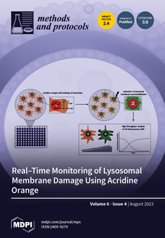
Loss of lysosomal membrane integrity affects cellular functions and can induce cell death via the release of lysosomal hydrolases. Several drugs used clinically have the ability to destabilize the lysosome, which can be utilized as a treatment strategy. The expanding interest for lysosomal function has emphasized the need for assays that follow lysosomal damage and detect minor damage that does not inevitably cause cell death. The weak base acridine orange accumulates in the acidic environment of lysosomes but is released upon lysosomal damage. In this protocol, we present a high-throughput microplate reader-based method to follow destabilization of the lysosomal membrane in real time using acridine orange. This protocol can easily be adopted for patient samples since the number of cells per sample is low and the time for analysis is short. View this paper
  • Issues are regarded as officially published after their release is announced to the table of contents alert mailing list.
  • You may sign up for e-mail alerts to receive table of contents of newly released issues.
  • PDF is the official format for papers published in both, html and pdf forms. To view the papers in pdf format, click on the "PDF Full-text" link, and use the free Adobe Reader to open them.
Order results
Result details
Section
Select all
Export citation of selected articles as:
11 pages, 2154 KiB  
Protocol
Real-Time Monitoring of Lysosomal Membrane Permeabilization Using Acridine Orange
by Ida Eriksson, Linda Vainikka, Hans Lennart Persson and Karin Öllinger
Methods Protoc. 2023, 6(4), 72; https://doi.org/10.3390/mps6040072 - 09 Aug 2023
Cited by 7 | Viewed by 2437
Abstract
Loss of lysosomal membrane integrity results in leakage of lysosomal hydrolases to the cytosol which might harm cell function and induce cell death. Destabilization of lysosomes often precede apoptotic or necrotic cell death and occur during both physiological and pathological conditions. The weak [...] Read more.
Loss of lysosomal membrane integrity results in leakage of lysosomal hydrolases to the cytosol which might harm cell function and induce cell death. Destabilization of lysosomes often precede apoptotic or necrotic cell death and occur during both physiological and pathological conditions. The weak base acridine orange readily enters cells and accumulates in the acidic environment of lysosomes. Vital staining with acridine orange is a well-proven technique to observe lysosomal destabilization using fluorescence microscopy and flow cytometry. These analyses are, however, time consuming and only adapted for discrete time points, which make them unsuitable for large-scale approaches. Therefore, we have developed a time-saving, high-throughput microplate reader-based method to follow destabilization of the lysosomal membrane in real-time using acridine orange. This protocol can easily be adopted for patient samples since the number of cells per sample is low and the time for analysis is short. Full article
(This article belongs to the Section Molecular and Cellular Biology)
Show Figures

Figure 1

12 pages, 7797 KiB  
Article
Comparative Analysis of Primers Used for 16S rRNA Gene Sequencing in Oral Microbiome Studies
by Hee Sam Na, Yuri Song, Yeuni Yu and Jin Chung
Methods Protoc. 2023, 6(4), 71; https://doi.org/10.3390/mps6040071 - 06 Aug 2023
Cited by 2 | Viewed by 3207
Abstract
Recent advances in genomic technologies have enabled more in-depth study of the oral microbiome. In this study, we compared the amplicons generated by primers targeting different sites of the 16S rRNA gene found in the Human Oral Microbiome Database (HOMD). Six sets of [...] Read more.
Recent advances in genomic technologies have enabled more in-depth study of the oral microbiome. In this study, we compared the amplicons generated by primers targeting different sites of the 16S rRNA gene found in the Human Oral Microbiome Database (HOMD). Six sets of primer targeting V1–V2, V1–V3, V3–V4, V4–V5, V5–V7 and V6–V8 regions of 16S rRNA were tested via in silico simulation. Primers targeting the V1–V2, V3–V4, and V4–V5 regions generated more than 90% of the original input sequences. Primers targeting the V1–V2 and V1–V3 regions exhibited a low number of mismatches and unclassified sequences at the taxonomic level, but there were notable discrepancies at the species level. Phylogenetic tree comparisons showed primers targeting the V1–V2 and V3–V4 regions showed performances similar to primers targeting the whole 16s RNA region in terms of separating total oral microbiomes and periodontopathogens. In an analysis of clinical oral samples, V1–V2 primers showed superior performance for identifying more taxa and had better resolution sensitivity for Streptococcus than V3–V4 primers. In conclusion, primers targeting the V1–V2 region of 16S rRNA showed the best performance for oral microbiome studies. In addition, the study demonstrates the need for careful PCR primer selections. Full article
(This article belongs to the Section Omics and High Throughput)
Show Figures

Figure 1

16 pages, 2777 KiB  
Article
Application of a Customised Franz-Type Cell Coupled with HPTLC to Monitor the Timed Release of Bioactive Components in Complex Honey Matrices
by Md Lokman Hossain, Minh Nguyen, Leah Benington, Lee Yong Lim, Katherine Hammer, Dhanushka Hettiarachchi and Cornelia Locher
Methods Protoc. 2023, 6(4), 70; https://doi.org/10.3390/mps6040070 - 03 Aug 2023
Cited by 1 | Viewed by 1318
Abstract
The aim of this study was to assess the release profile of components in five different honeys (a New Zealand Manuka and two Western Australian honeys, a Jarrah honey and a Coastal Peppermint honey) and their corresponding honey-loaded gel formulations using a custom-designed [...] Read more.
The aim of this study was to assess the release profile of components in five different honeys (a New Zealand Manuka and two Western Australian honeys, a Jarrah honey and a Coastal Peppermint honey) and their corresponding honey-loaded gel formulations using a custom-designed Franz-type diffusion cell in combination with High-Performance Thin-Layer Chromatography (HPTLC). To validate the suitability of the customised setup, release data using this new approach were compared with data obtained using a commercial Franz cell apparatus, which is an established analytical tool to monitor the release of active ingredients from topical semisolid products. The release profiles of active compounds from pure honey and honey-loaded formulations were found to be comparable in both types of Franz cells. For example, when released either from pure honey or its corresponding pre-gel formulation, the percentage release of two Jarrah honey constituents, represented by distinct bands at RF 0.21 and 0.53 and as analysed by HPTLC, was not significantly different (p = 0.9986) at 12 h with over 99% of these honey constituents being released in both apparatus. Compared to the commercial Franz diffusion cell, the customised Franz cell offers several advantages, including easy and convenient sample application, the requirement of only small sample quantities, a large diffusion surface area, an ability to analyse 20 samples in a single experiment, and lower cost compared to purchasing a commercial Franz cell. Thus, the newly developed approach coupled with HPTLC is conducive to monitor the release profile of minor honey constituents from pure honeys and honey-loaded semisolid formulations and might also be applicable to other complex natural-product-based products. Full article
(This article belongs to the Section Biochemical and Chemical Analysis & Synthesis)
Show Figures

Figure 1

13 pages, 3213 KiB  
Article
Ultrasound-Assisted Anthocyanins Extraction from Pigmented Corn: Optimization Using Response Surface Methodology
by Annisa Nurkhasanah, Titouan Fardad, Ceferino Carrera, Widiastuti Setyaningsih and Miguel Palma
Methods Protoc. 2023, 6(4), 69; https://doi.org/10.3390/mps6040069 - 30 Jul 2023
Cited by 2 | Viewed by 1499
Abstract
This study aimed to determine the optimal UAE conditions for extracting anthocyanins from pigmented corn using the Box–Behnken design (BBD). Six anthocyanins were identified in the samples and were used as response variables to evaluate the effects of the following working variables: extraction [...] Read more.
This study aimed to determine the optimal UAE conditions for extracting anthocyanins from pigmented corn using the Box–Behnken design (BBD). Six anthocyanins were identified in the samples and were used as response variables to evaluate the effects of the following working variables: extraction solvent pH (2–7), temperature (10–70 °C), solvent composition (0–50% methanol in water), and ultrasound power (20–80%). The extraction time (5–25 min) was evaluated for complete recovery. Response surface methodology suggested optimal conditions, specifically 36% methanol in water with pH 7 at 70 °C using 73% ultrasound power for 10 min. The method was validated with a high level of accuracy (>90% of recovery) and high precision (CV < 5% for both repeatability and intermediate precision). Finally, the proposed analytical extraction method was successfully applied to determine anthocyanins that covered a wide concentration range (36.47–551.92 mg kg−1) in several pigmented corn samples revealing potential varieties providing more health benefits. Full article
(This article belongs to the Special Issue Women’s Special Issue Series: Analytical Methods)
Show Figures

Figure 1

13 pages, 1430 KiB  
Protocol
Geriatric Oncology in the Instagram Era: Feasibility and Acceptability Randomised Controlled Trial on Adopting PhotoVoice to Enable Empowerment, Patient-Centred Care, and Shared Decision Making—Study Protocol
by Christopher Steer, Tshepo Rasekaba, Kylie Owen, Darren Jayasuriya, Mira Kapur, Kim Young, Nicole Webb and Irene Blackberry
Methods Protoc. 2023, 6(4), 68; https://doi.org/10.3390/mps6040068 - 26 Jul 2023
Cited by 1 | Viewed by 1185
Abstract
Geriatric assessment (GA) is fundamental to optimising cancer care in older adults, yet implementing comprehensive GA tools in real-world clinical settings remains a challenge. This study aims to assess the feasibility and acceptability of integrating information from patient-derived photographs (PhotoVoice) into enhanced supportive [...] Read more.
Geriatric assessment (GA) is fundamental to optimising cancer care in older adults, yet implementing comprehensive GA tools in real-world clinical settings remains a challenge. This study aims to assess the feasibility and acceptability of integrating information from patient-derived photographs (PhotoVoice) into enhanced supportive care (ESC) for older adults with cancer. A feasibility randomised controlled trial will be conducted at a regional cancer care centre in Australia. Participants aged 70 and above will be randomised into two groups: PhotoVoice plus ESC or usual care (ESC) alone. In the PhotoVoice group, participants will provide four photographs for deduction of representations of different aspects of their lives using photo-elicitation techniques. ESC will be conducted for both groups, incorporating PhotoVoice analysis in the intervention group. PhotoVoice may improve patient-centred care outcomes, including enhanced communication, shared decision making, and identification of patient priorities and barriers. Findings will provide insights into implementing PhotoVoice in geriatric assessment and guide future trials in cancer among older adults. Full article
(This article belongs to the Section Public Health Research)
Show Figures

Figure 1

7 pages, 537 KiB  
Protocol
Variations in Microcirculatory and Hemodynamic Parameters during Oncological Demolitive–Reconstructive Head and Neck Surgery: A Protocol for an Observational Study
by Chiara Adembri, Andrea Ungar, Iacopo Cappellini and Salvatore Mario Romano
Methods Protoc. 2023, 6(4), 67; https://doi.org/10.3390/mps6040067 - 21 Jul 2023
Viewed by 1103
Abstract
(1) Background: Oncological demolitive–reconstructive surgeries in the head and neck region cause significant stress on patients’ biohumoural, cardiac, and vascular systems, leading to disturbances in macrocirculatory and microcirculatory parameters. Traditional monitoring addresses the symptoms, but not the underlying cause. Microcirculatory assessments complement macrocirculatory [...] Read more.
(1) Background: Oncological demolitive–reconstructive surgeries in the head and neck region cause significant stress on patients’ biohumoural, cardiac, and vascular systems, leading to disturbances in macrocirculatory and microcirculatory parameters. Traditional monitoring addresses the symptoms, but not the underlying cause. Microcirculatory assessments complement macrocirculatory monitoring, and bladder-catheter-based technology offers a better representation of central microcirculation. Flap reconstruction surgeries involve demolitive and reconstructive phases, requiring optimal tissue perfusion. The literature lacks a consensus on macro–microcirculation coupling, and there is no agreement on the use of vasopressors during head and neck surgeries. Evidence-based guidelines are lacking, resulting in variations in vasopressor administration. (2) Methods: This is a 12-month observational, prospective study conducted in a single center. It aims to evaluate the impact of macro–microcirculation coupling on clinical complications in head and neck surgery. All consecutive patients undergoing oncologic surgery requiring flap reconstruction and meeting the inclusion criteria will be enrolled. The study will utilize standard hemodynamic monitoring and bladder catheterization for measuring urine output and temperature. (3) Conclusions: The study aims to evaluate the coupling of macro- and microcirculation in head and neck surgeries, assess hemodynamic parameters and microcirculatory changes, and investigate their association with postoperative complications. The results can enhance patient care and surgical outcomes. Full article
(This article belongs to the Section Biomedical Sciences and Physiology)
Show Figures

Figure 1

9 pages, 1919 KiB  
Protocol
Ex Vivo Perfusion of Porcine Pancreas and Liver Sourced from Commercial Abattoirs after Circulatory Death as a Research Resource: A Methodological Study
by Zainab L. Rai, Morenike Magbagbeola, Katie Doyle, Lukas Lindenroth, George Dwyer, Amir Gander, Agostino Stilli, Danail Stoyanov and Brian R. Davidson
Methods Protoc. 2023, 6(4), 66; https://doi.org/10.3390/mps6040066 - 12 Jul 2023
Viewed by 1261
Abstract
Background: Machine perfusion (MP) is increasingly used for human transplant organ preservation. The use of MP for research purposes is another opportunity for this technology. The porcine pancreas and liver are similar in anatomical size and function to their human counterparts, making them [...] Read more.
Background: Machine perfusion (MP) is increasingly used for human transplant organ preservation. The use of MP for research purposes is another opportunity for this technology. The porcine pancreas and liver are similar in anatomical size and function to their human counterparts, making them an excellent resource for research, but they have some important differences from human organs which can influence their research use. In this paper, we describe a technique developed and tested for the retrieval of porcine organs for use in research on perfused viable organs. Methods: Whole-organ porcine pancreata and livers were harvested at a commercial abattoir, following standard slaughterhouse processes. The standard slaughterhouse process involved a thoracotomy and mid-line laparotomy, and all the thoracoabdominal organs were removed. The pancreas, fixed in the retroperitoneum, was carefully dissected from its attachments to the surrounding structures, and tissue planes between the pancreas, spleen, duodenum, and colon were meticulously identified and dissected. Vessel exposure and division: The aorta, portal vein (PV), hepatic vein (HV), and hepatic artery (HA) were dissected and isolated, preserving the input and output channels for the liver and pancreas. A distal 3 cm of the aorta was preserved and divided and served as the input for the pancreas perfusions. The liver, PV, HV, and HA were preserved and divided to preserve the physiological channels of the input (PV and HA) and output (HV) for the liver perfusions. The porcine hepatic and pancreas anatomy shares significant resemblance with the gross anatomy found in humans, and this was taken into consideration when designing the perfusion circuitry. The porcine pancreas and spleen shared a common blood supply, with branches arising from the splenic artery. The organs were flushed with cold, heparinised normal saline and transported in a temperature-regulated receptacle maintained at a core temperature between 4 and 8 °C, in line with the standards of static cold storage (SCS), to a dedicated perfusion lab and perfused using our novel perfusion machine with autologous, heparinised porcine blood, also collected at the abattoir. Full article
(This article belongs to the Section Synthetic and Systems Biology)
Show Figures

Figure 1

9 pages, 435 KiB  
Article
Assessment of SARS-CoV-2 (COVID-19) Clinical Mouthwash Protocol and Prevalence of the Oral Pathogen Scardovia wiggsiae: A Pilot Study of Antibacterial Effects
by Melika Shayegh, Chase Sorenson, Jackson Downey, Summer Lin, Yuxin Jiang, Praneeti Sodhi, Victoria Sullivan, Katherine M. Howard and Karl Kingsley
Methods Protoc. 2023, 6(4), 65; https://doi.org/10.3390/mps6040065 - 06 Jul 2023
Cited by 1 | Viewed by 1139
Abstract
One protocol in healthcare facilities and dental offices due to the COVID-19 pandemic for reducing the amount of detectable oral SARS-CoV-2 has been gargling with mouthwash for 60 s. This protocol lasts longer than the daily routine for most patients and may have [...] Read more.
One protocol in healthcare facilities and dental offices due to the COVID-19 pandemic for reducing the amount of detectable oral SARS-CoV-2 has been gargling with mouthwash for 60 s. This protocol lasts longer than the daily routine for most patients and may have unexpected benefits in reducing oral microbes as a result. This project evaluated the prevalence of the newly identified oral pathogen Scardovia wiggsiae before and after this procedure to determine any measurable effects. Using an approved protocol, n = 36 pre-mouthwash patient samples, n = 36 matched post-mouthwash samples, and n = 36 matched recall samples were identified (total sample number n = 108). DNA was isolated from each sample (pre-, post-mouthwash, and recall). Screening using qPCR and validated primers revealed n = 10/36 or 27.8% tested positive for Scardovia among the pre-mouthwash (Sample A) isolates with n = 3/36 or 8.3% testing positive among the post-mouthwash (Sample B) isolates. Screening of the recall (Sample C) samples has revealed n = 10/36, or 27.8% once again tested positive for Scardovia, demonstrating that this pathogen was found among a significant proportion of pediatric patient samples. Moreover, the COVID-19-related procedure of requiring sustained mouth washing prior to clinical treatment appears to reduce the levels of detectable Scardovia, at least initially. However, this study found no long-term effects using this isolated protocol. Full article
(This article belongs to the Section Biomedical Sciences and Physiology)
Show Figures

Figure 1

11 pages, 2343 KiB  
Article
Development of a Wound-Healing Protocol for In Vitro Evaluation of Urothelial Cell Growth
by Christopher Foster, Todd Jensen, Christine Finck and Courtney K. Rowe
Methods Protoc. 2023, 6(4), 64; https://doi.org/10.3390/mps6040064 - 05 Jul 2023
Viewed by 1216
Abstract
Urethral healing is plagued by strictures, impacting quality of life and medical costs. Various growth factors (GFs) have shown promise as therapeutic approaches to improve healing, but there is no protocol for in vitro comparison between GFs. This study focuses the development of [...] Read more.
Urethral healing is plagued by strictures, impacting quality of life and medical costs. Various growth factors (GFs) have shown promise as therapeutic approaches to improve healing, but there is no protocol for in vitro comparison between GFs. This study focuses the development of a biomimetic in vitro urothelial healing assay designed to mimic early in vivo healing, followed by an evaluation of urothelial cell growth in response to GFs. Methods: Wound-healing assays were developed with human urothelial cells and used to compared six GFs (EGF, FGF-2, IGF-1, PDGF, TGF-β1, and VEGF) at three concentrations (1 ng/mL, 10 ng/mL, and 100 ng/mL) over a 48 h period. A commercial GF-containing medium (EGF, TGF-α, KGF, and Extract P) and a GF-free medium were used as controls. Results: There was a statistically significant increase in cell growth for IGF-1 at 10 and 100 ng/mL compared to both controls (p < 0.05). There was a statistically significant increase in cell growth for EGF at all concentrations compared to the GF-free medium control (p < 0.05). Conclusion: This study shows the development of a clinically relevant wound-healing assay to evaluate urothelial cell growth. It is the first to compare GFs for future use in reconstructive techniques to improve urethral healing. Full article
(This article belongs to the Section Biomedical Sciences and Physiology)
Show Figures

Figure 1

6 pages, 4667 KiB  
Brief Report
Precise Analysis of Nanoparticle Size Distribution in TEM Image
by Shan Zhang and Chao Wang
Methods Protoc. 2023, 6(4), 63; https://doi.org/10.3390/mps6040063 - 03 Jul 2023
Cited by 7 | Viewed by 4837
Abstract
As an essential characterization, size distribution is an important indicator for the synthesis, optimization, and application of nanoparticles. Electron microscopes such as transmission electron microscopes (TEMs) are commonly utilized to collect size information on nanoparticles. However, the current popular statistical method of manually [...] Read more.
As an essential characterization, size distribution is an important indicator for the synthesis, optimization, and application of nanoparticles. Electron microscopes such as transmission electron microscopes (TEMs) are commonly utilized to collect size information on nanoparticles. However, the current popular statistical method of manually measuring large particles one by one, using a ruler tool in the corresponding image analysis software is time-consuming and can introduce manual errors. Moreover, it is difficult to determine the measurement interval for irregularly shaped nanoparticles. Therefore, it is necessary to use an efficient and standard method to perform size distribution analysis of nanoparticles. In this work, we use basic ImageJ software (1.53 t) to analyze the size of typical silica nanoparticles in a TEM image and use Origin software to process the data, to obtain its accurate distribution quickly. Using it as a template, we believe that this work can provide a paradigm for the standardized analysis of nanoparticle size. Full article
Show Figures

Figure 1

8 pages, 839 KiB  
Protocol
A New and Profitable Protocol to DNA Extraction in Limnospira maxima
by Yirlis Yadeth Pineda-Rodriguez, Marcelo F. Pompelli, Alfredo Jarma-Orozco, Novisel Veitía Rodríguez and Luis Alfonso Rodriguez-Paez
Methods Protoc. 2023, 6(4), 62; https://doi.org/10.3390/mps6040062 - 01 Jul 2023
Cited by 3 | Viewed by 1989
Abstract
Limnospira maxima is a remarkable organism showing great potential as a versatile and sustainable food source, offering a powerful solution to address the pressing issues of malnutrition and undernourishment worldwide. L. maxima contains high amounts of proteins, vitamins, minerals, and essential fatty acids. [...] Read more.
Limnospira maxima is a remarkable organism showing great potential as a versatile and sustainable food source, offering a powerful solution to address the pressing issues of malnutrition and undernourishment worldwide. L. maxima contains high amounts of proteins, vitamins, minerals, and essential fatty acids. It can be grown in both bioreactors and open systems; however, before considering industrial production, optimization studies of the cultivation must be conducted to obtain knowledge about the ideal environmental conditions. Additionally, for the molecular typing of L. maxima strains and their industrial scaling, high-quality and large quantity DNA extraction is required. Notwithstanding, DNA extraction from L. maxima can be challenging due to the low amount of DNA in cells and the presence of difficult-to-remove substances such as polysaccharides and polyphenols. In this study, the quality and quantity of DNA extracted from two types of L. maxima samples (Limnospira maxima strain SISCA accession GenBank: OR195505.1) were evaluated using three commercially available DNA extraction kits and two types of input biological material. The results showed that Pbact-P kit had the highest quantity and quality of DNA, while CTAB-P allowed for a higher quantity and quality of RNA, making them optimal protocols for nucleic acid extraction to improve PCR, rt-PCR, and genome sequencing of L. maxima compared with other extraction methods. Full article
(This article belongs to the Section Molecular and Cellular Biology)
Show Figures

Figure 1

10 pages, 813 KiB  
Protocol
Proposed Protocol for Field Testing of Endurance Fitness of Young Labrador Retrievers
by Ella-Erika Söderlund, Heikki Kyröläinen, Outi M. Laitinen-Vapaavuori and Heli K. Hyytiäinen
Methods Protoc. 2023, 6(4), 61; https://doi.org/10.3390/mps6040061 - 28 Jun 2023
Viewed by 1262
Abstract
The number of dogs and, with it, dog sports are growing in popularity, and the training of dogs begins at an early age. Although fitness testing is an imperative part of purposeful training and sports, to our knowledge, no objective field tests are [...] Read more.
The number of dogs and, with it, dog sports are growing in popularity, and the training of dogs begins at an early age. Although fitness testing is an imperative part of purposeful training and sports, to our knowledge, no objective field tests are available for measuring young dogs’ endurance fitness. The aim of this study is to describe a simple, easy-to-repeat, and inexpensive way to test training intervention effects on endurance fitness in young Labrador Retrievers. Healthy client-owned 16-week-old Labrador Retrievers will be recruited and divided into test and control groups. The test group will have an eight-week training program followed by a four-week detraining period, while the control group will live a normal puppy life. All dogs will be tested for endurance fitness four times at four-week intervals: at baseline, one month later, two months later at the end of the training period, and one month after ending the training program. Each of the four testing sessions will be identical and will consist of four measurements of heart rate (HR) and blood lactate (BL): at baseline, after trotting 1000 m, after sprinting 200 m, and at recovery 5–8 min after the sprint. The training-induced changes in endurance fitness are evaluated by changes in HR and heart rate recovery times (HRR), BL, and running times. Full article
(This article belongs to the Section Biomedical Sciences and Physiology)
Show Figures

Figure 1

15 pages, 602 KiB  
Review
New Prospects in the Electroanalysis of Heavy Metal Ions (Cd, Pb, Zn, Cu): Development and Application of Novel Electrode Surfaces
by Vasiliki Keramari, Sophia Karastogianni and Stella Girousi
Methods Protoc. 2023, 6(4), 60; https://doi.org/10.3390/mps6040060 - 26 Jun 2023
Cited by 2 | Viewed by 1614
Abstract
The detection of toxic heavy metal ions, especially cadmium (Cd), lead (Pb), zinc (Zn), and copper (Cu), is a global problem due to ongoing pollution incidents and continuous anthropogenic and industrial activities. Therefore, it is important to develop effective detection techniques to determine [...] Read more.
The detection of toxic heavy metal ions, especially cadmium (Cd), lead (Pb), zinc (Zn), and copper (Cu), is a global problem due to ongoing pollution incidents and continuous anthropogenic and industrial activities. Therefore, it is important to develop effective detection techniques to determine the levels of pollution from heavy metal ions in various media. Electrochemical techniques, more specifically voltammetry, due to its properties, is a promising method for the simultaneous detection of heavy metal ions. This review examines the current trends related to electrode formation and analysis techniques used. In addition, there is a reference to advanced detection methods based on the nanoparticles that have been developed so far, as well as formation with bismuth and the emerging technique of screen-printed electrodes. Finally, the advantages of using these methods are highlighted, while a discussion is presented on the benefits arising from nanotechnology, as it gives researchers new ideas for integrating these technologies into devices that can be used anywhere at any time. Reference is also made to the speciation of metals and how it affects their toxicity, as it is an important subject of research. Full article
(This article belongs to the Special Issue Women’s Special Issue Series: Analytical Methods)
Show Figures

Figure 1

6 pages, 1049 KiB  
Technical Note
Optimized Protocol for Preservation of Human Platelet Samples for Fluorometric Polyphosphate Quantification
by Tomoyuki Kawase, Katsuya Suzuki, Masami Kamimura, Tomoharu Mochizuki and Takashi Ushiki
Methods Protoc. 2023, 6(4), 59; https://doi.org/10.3390/mps6040059 - 22 Jun 2023
Viewed by 1073
Abstract
Platelet polyphosphate (polyP) can be conveniently quantified by exploiting a recent methodological breakthrough using 4′,6-diamidino-2-phenylindole (DAPI). However, the preservation of these biological samples has not yet been standardized. In a preliminary study, potential protocols were screened, while accepted protocols were further tested in [...] Read more.
Platelet polyphosphate (polyP) can be conveniently quantified by exploiting a recent methodological breakthrough using 4′,6-diamidino-2-phenylindole (DAPI). However, the preservation of these biological samples has not yet been standardized. In a preliminary study, potential protocols were screened, while accepted protocols were further tested in this study. Pure-platelet-rich plasma (P-PRP) samples and washed platelet suspensions were prepared using blood obtained from non-smoking healthy male donors and were fixed with ThromboFix for 20–24 h at 4 °C. Mass polyP levels were determined using a fluorometer at wavelengths of 425 and 525 nm. Platelet polyP levels were normalized to platelet counts. Statistical analyses were performed using non-parametric tests. Platelet polyP levels significantly decreased by 20% after 7 days in the platelet suspension maintained under fixed conditions at 4 °C (control). In contrast, the platelet polyP levels in both the P-PRP and washed platelet suspensions were maintained without a significant reduction for up to 6 weeks by removing ThromboFix after fixation and subsequent freezing in pure water at −80 °C. Fluorometric polyP quantification often interferes with the low specificity of DAPI binding and the wavelength used. Our validated protocols will enable long-term preservation and high-throughput polyP quantification and can be applied to relatively large cohort studies. Full article
(This article belongs to the Section Biochemical and Chemical Analysis & Synthesis)
Show Figures

Figure 1

Previous Issue
Next Issue
Back to TopTop