
A Positive Newborn Screen for Congenital Hypothyroidism in a Clinically Euthyroid Neonate—Avoiding Unnecessary Treatment


Abstract
:1. Introduction
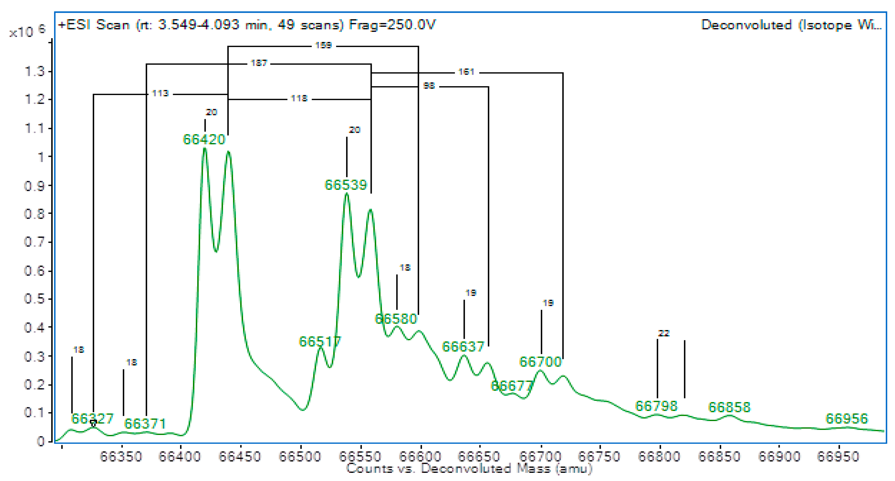
2. Case Report
3. Discussion
4. Conclusions
Author Contributions
Funding
Institutional Review Board Statement
Informed Consent Statement
Data Availability Statement
Conflicts of Interest
References
- Mimoto, M.S.; Refetoff, S. Clinical recognition and evaluation of patients with inherited serum thyroid hormone-binding protein mutations. J. Endocrinol. Investig. 2020, 43, 31–41. [Google Scholar] [CrossRef] [PubMed]
- Khoo, S.; Lyons, G.; McGowan, A.; Gurnell, M.; Oddy, S.; Visser, W.E.; Berg, S.V.D.; Halsall, D.; Taylor, K.; Chatterjee, K.; et al. Familial dysalbuminaemic hyperthyroxinaemia interferes with current free thyroid hormone immunoassay methods. Eur. J. Endocrinol. 2020, 182, 533–538. [Google Scholar] [CrossRef] [PubMed] [Green Version]
- Kragh-Hansen, U.; Galliano, M.; Minchiotti, L. Clinical, Genetic, and Protein Structural Aspects of Familial Dysalbuminemic Hyperthyroxinemia and Hypertriiodothyroninemia. Front. Endocrinol. 2017, 8, 297. [Google Scholar] [CrossRef] [PubMed] [Green Version]
- Pappa, T.; Ferrara, A.M.; Refetoff, S. Inherited defects of thyroxine-binding proteins. Best Pr. Res. Clin. Endocrinol. Metab. 2015, 29, 735–747. [Google Scholar] [CrossRef] [PubMed] [Green Version]
- Huynh, T.; Greaves, R.; Mawad, N.; Greed, L.; Wotton, T.; Wiley, V.; Ranieri, E.; Rankin, W.; Ungerer, J.; Price, R.; et al. Fifty years of newborn screening for congenital hypothyroidism: Current status in Australasia and the case for harmonisation. Clin. Chem. Lab. Med. 2022, 60, 1551–1561. [Google Scholar] [CrossRef] [PubMed]
- Heather, N.; Hofman, P. Congenital Hypothyroidism—Early Assessment and Management. Starship Child Health Clinical Guidelines. 28 April 2021. Available online: https://starship.org.nz/guidelines/congenital-hypothyroidism-early-assessment-and-management/ (accessed on 3 March 2023).
- Cho, Y.Y.; Song, J.-S.; Park, H.-D.; Kim, Y.N.; Kim, H.-I.; Kim, T.H.; Chung, J.H.; Ki, C.-S.; Kim, S.W. First Report of Familial Dysalbuminemic Hyperthyroxinemia With an ALB Variant. Ann. Lab. Med. 2017, 37, 63–65. [Google Scholar] [CrossRef] [PubMed]
- Freedman, D.B.; Halsall, D.; Marshall, W.J.; Ellervik, C. Thyroid Disorders. In Tietz Textbook of Clinical Chemistry and Molecular Diagnostics; Rifai, N.H., Ed.; Elsevier: St. Louis, MO, USA, 2018. [Google Scholar]
- Ryan, J.B.; Brennan, S.O.; Potter, H.; Wolmarans, L.; Florkowski, C.M.; George, P.M. Familial dysalbuminaemic hyperthyroxinaemia: A rapid and novel mass spectrometry approach to diagnosis. Ann. Clin. Biochem. 2016, 53 Pt 4, 504–507. [Google Scholar] [CrossRef] [PubMed] [Green Version]
- Flechner, I.; Aranoff, G.; Reifen, R.; Landau, H. Detection of albumin binding abnormalities in sera of patients with familial dysalbuminaemic hyperthyroxinaemia using isoelectric focusing. Endocr. Res. 1992, 18, 229–240. [Google Scholar] [CrossRef] [PubMed]
- Okosieme, O.E.; Agrawal, M.; Usman, D.; Evans, C. Method-dependent variation in TSH and FT4 reference intervals in pregnancy: A systematic review. Ann. Clin. Biochem. Int. J. Biochem. Lab. Med. 2021, 58, 537–546. [Google Scholar] [CrossRef] [PubMed]


| Roche Assay | Siemens Atellica Assay | Beckman Coulter Assay | |||||||
|---|---|---|---|---|---|---|---|---|---|
| TSH (mU/L) | Free T4 (pmol/L) | Free T3 (pmol/L) | TSH (mU/L) | Free T4 (pmol/L) | Free T3 (pmol/L) | Total T4 (nmol/L) | Total T3 (nmol/L) | ||
| Age-Specific Reference Interval | 0.4–16.0 | 10–40 | 3–10 | 1.0–8.0 | 15–35 | 3–10 | 76–168 | 1.4–3.96 | |
| Day 4 | Lab ‘B’ | 14.8 | 54 | ||||||
| Day 5 | Lab ‘A’ | 7.2 | 52.5 | ||||||
| Day 10 | Lab ‘A’ | 7.2 | 42.8 | 7.3 | |||||
| Lab ‘B’ | 8.1 | 47 | 7.2 | ||||||
| Lab ‘C’ | 5.7 | 34 | 9.5 | ||||||
| Day 15 | Lab ‘B’ | 8.3 | 37 | 7.1 | |||||
| Lab ‘D’ | 299 | 2.1 | |||||||
| Day 29 | Lab ‘A’ | 5 | 29.6 | 7 | |||||
Disclaimer/Publisher’s Note: The statements, opinions and data contained in all publications are solely those of the individual author(s) and contributor(s) and not of MDPI and/or the editor(s). MDPI and/or the editor(s) disclaim responsibility for any injury to people or property resulting from any ideas, methods, instructions or products referred to in the content. |
© 2023 by the authors. Licensee MDPI, Basel, Switzerland. This article is an open access article distributed under the terms and conditions of the Creative Commons Attribution (CC BY) license (https://creativecommons.org/licenses/by/4.0/).
Share and Cite
Brown, A.; Hofman, P.; Li, B.; Heron, C.; Heather, N. A Positive Newborn Screen for Congenital Hypothyroidism in a Clinically Euthyroid Neonate—Avoiding Unnecessary Treatment. Int. J. Neonatal Screen. 2023, 9, 16. https://doi.org/10.3390/ijns9020016
Brown A, Hofman P, Li B, Heron C, Heather N. A Positive Newborn Screen for Congenital Hypothyroidism in a Clinically Euthyroid Neonate—Avoiding Unnecessary Treatment. International Journal of Neonatal Screening. 2023; 9(2):16. https://doi.org/10.3390/ijns9020016
Chicago/Turabian StyleBrown, Ashleigh, Paul Hofman, Bobby Li, Campbell Heron, and Natasha Heather. 2023. "A Positive Newborn Screen for Congenital Hypothyroidism in a Clinically Euthyroid Neonate—Avoiding Unnecessary Treatment" International Journal of Neonatal Screening 9, no. 2: 16. https://doi.org/10.3390/ijns9020016