
Diagnostic Applications of Intraoral Scanners: A Systematic Review
 , , , , and
Abstract
:1. Introduction
2. Materials and Methods
2.1. Eligibility Criteria
- The use of intraoral scanner devices in the oral health diagnostics field;
- The assessment of performance in the detection of oral cavity pathologies (e.g., caries, dental wear, periodontal diseases, oral cancer, infections);
- Clear description of the diagnostic workflow;
- Comparison with a reference method or gold standard.
- Papers dealing with the technical evaluation of the accuracy and precision of intraoral scanners in reconstruction systems to achieve 3D digital mapping of the oral cavity;
- Papers exploiting digital models obtained from intraoral scanning to fabricate prostheses or to replace prostheses already implanted in the patient’s mouth.
2.2. Information Sources and Search Strategy
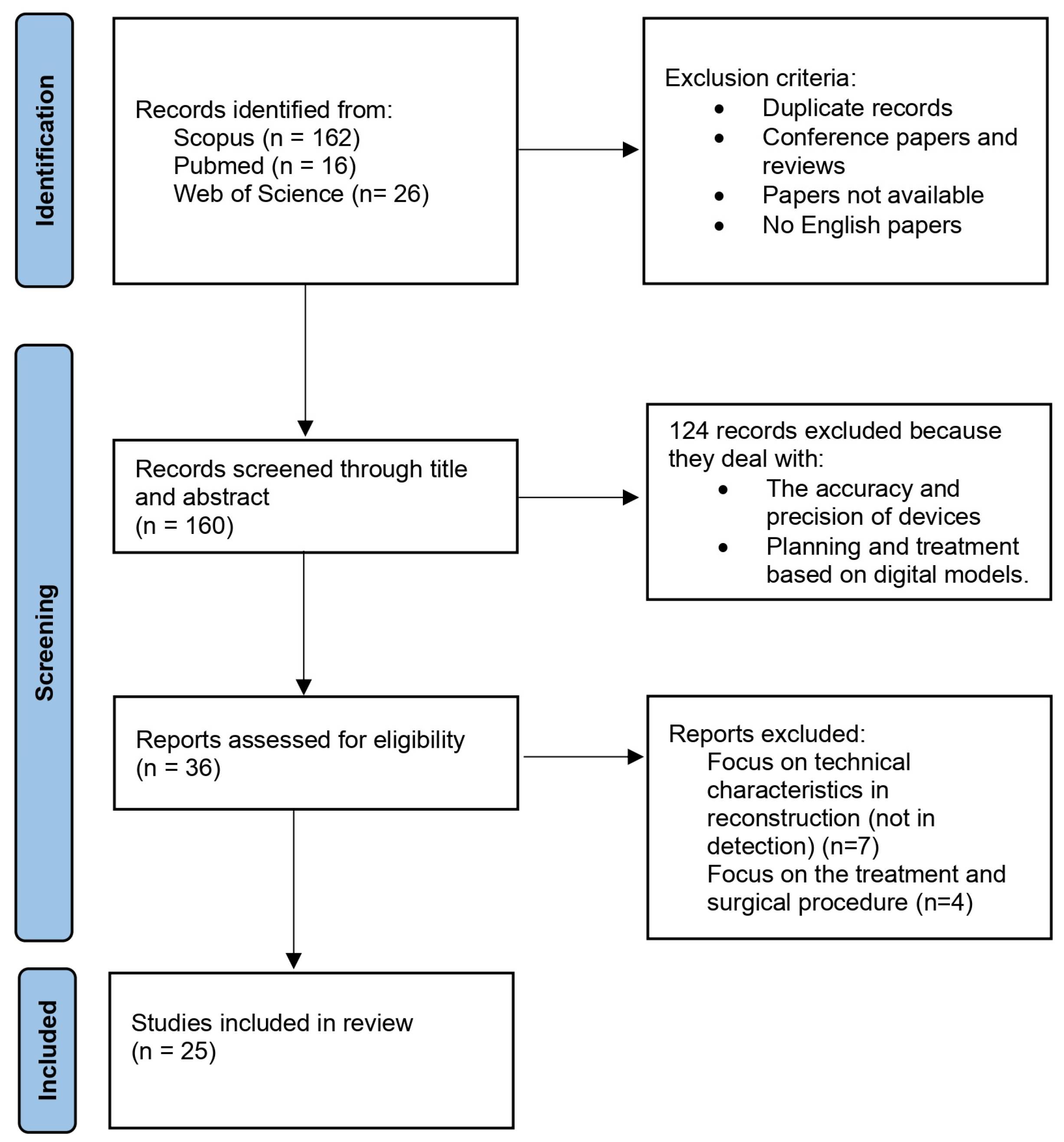
2.3. Selection Process
2.4. Data Collection
2.5. Data Item Clustering
2.6. Risk of Bias
2.7. Synthesis Methods and Analysis of the Results
3. Results
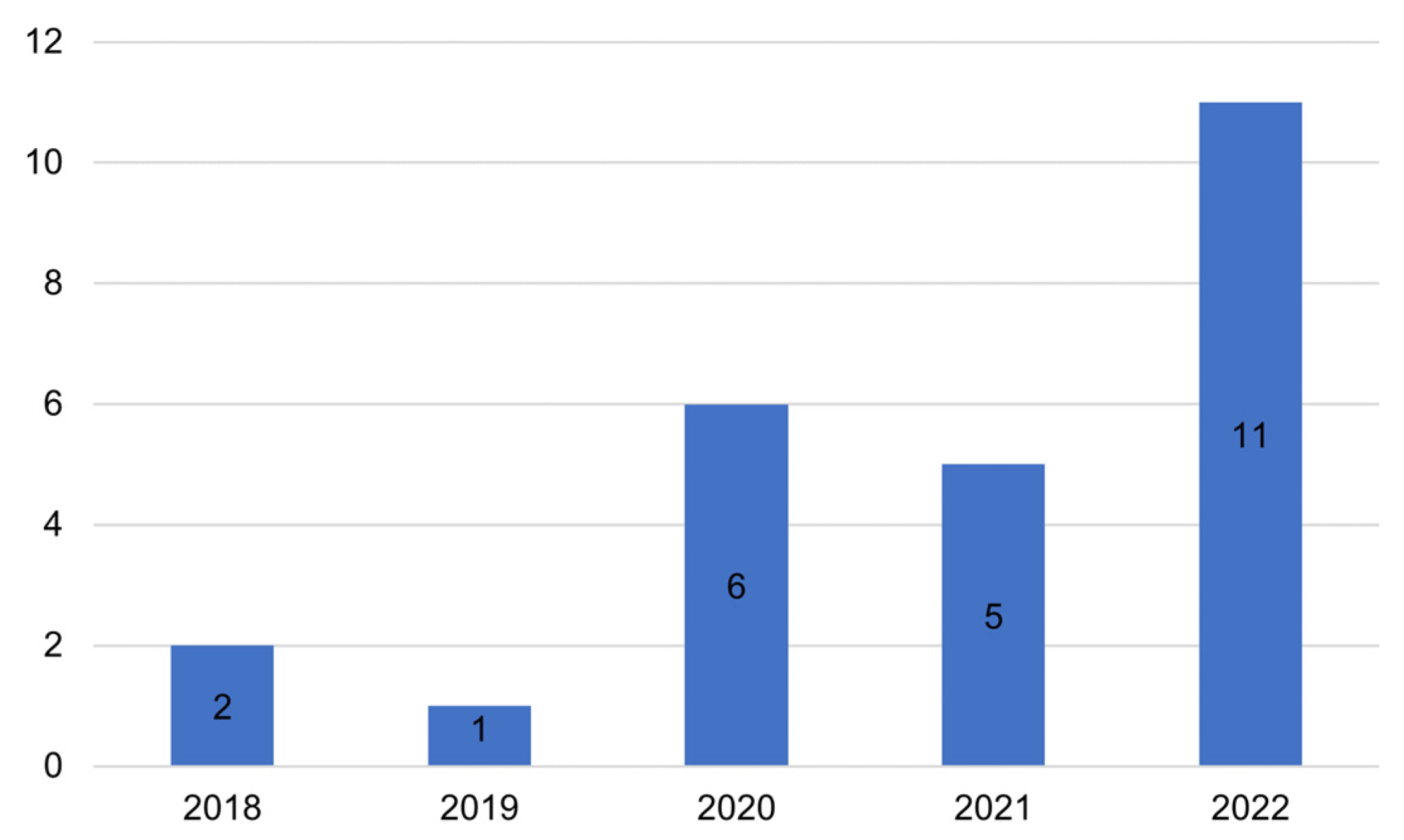
3.1. Main Findings
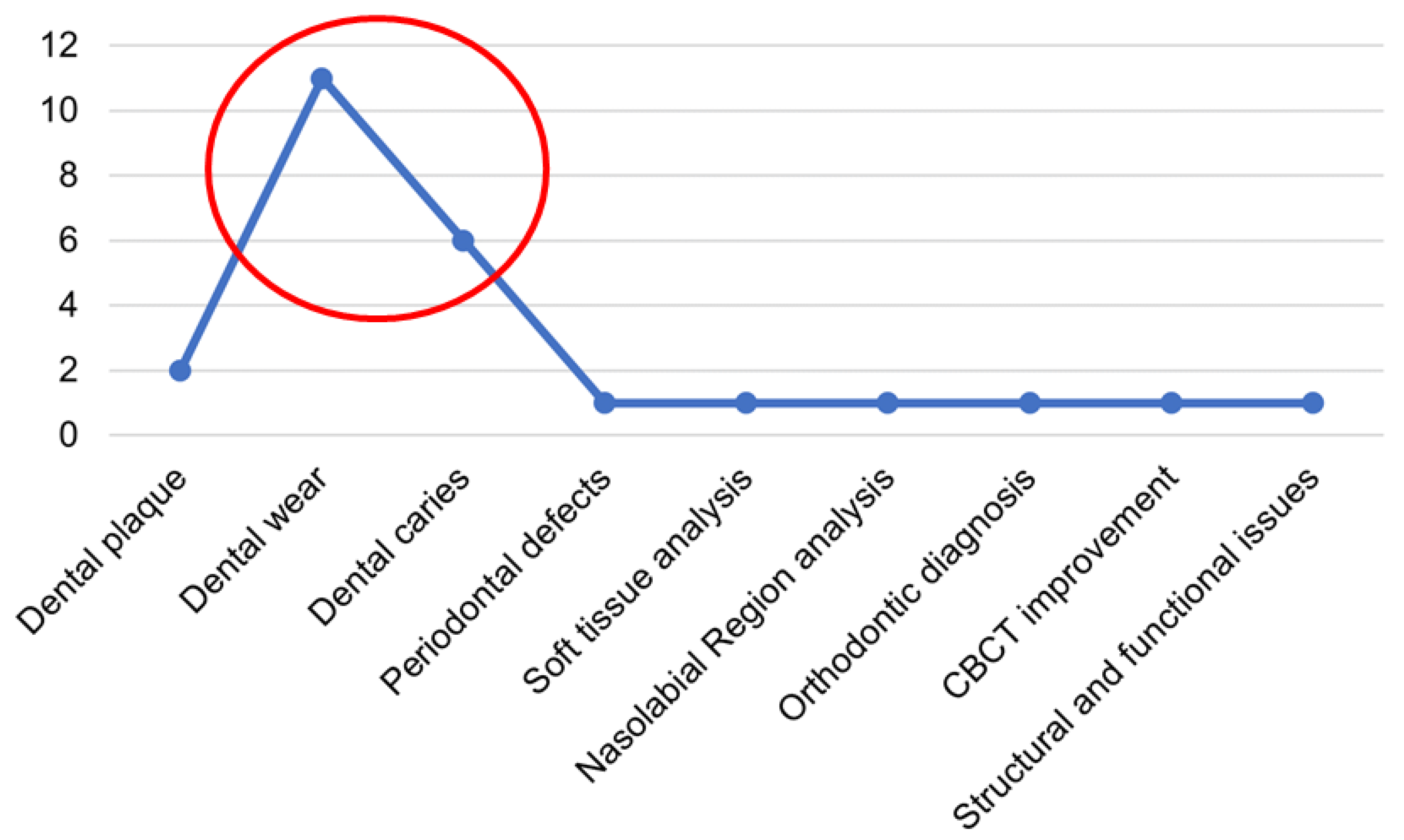
3.2. Oral Conditions Diagnosed Using an IOS
3.3. In Vivo and In Vitro Studies
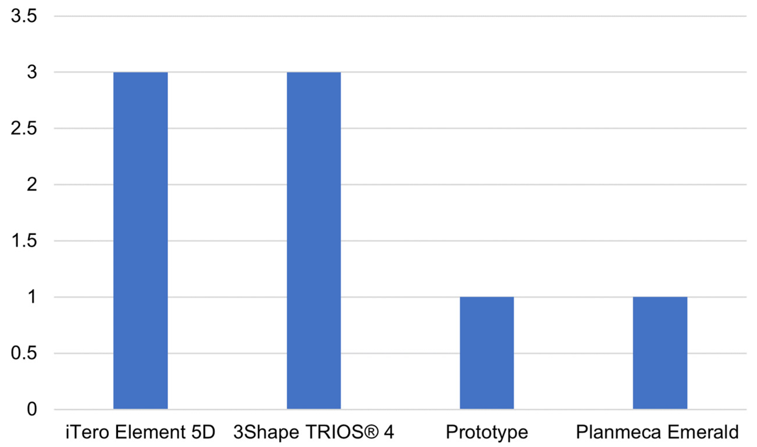
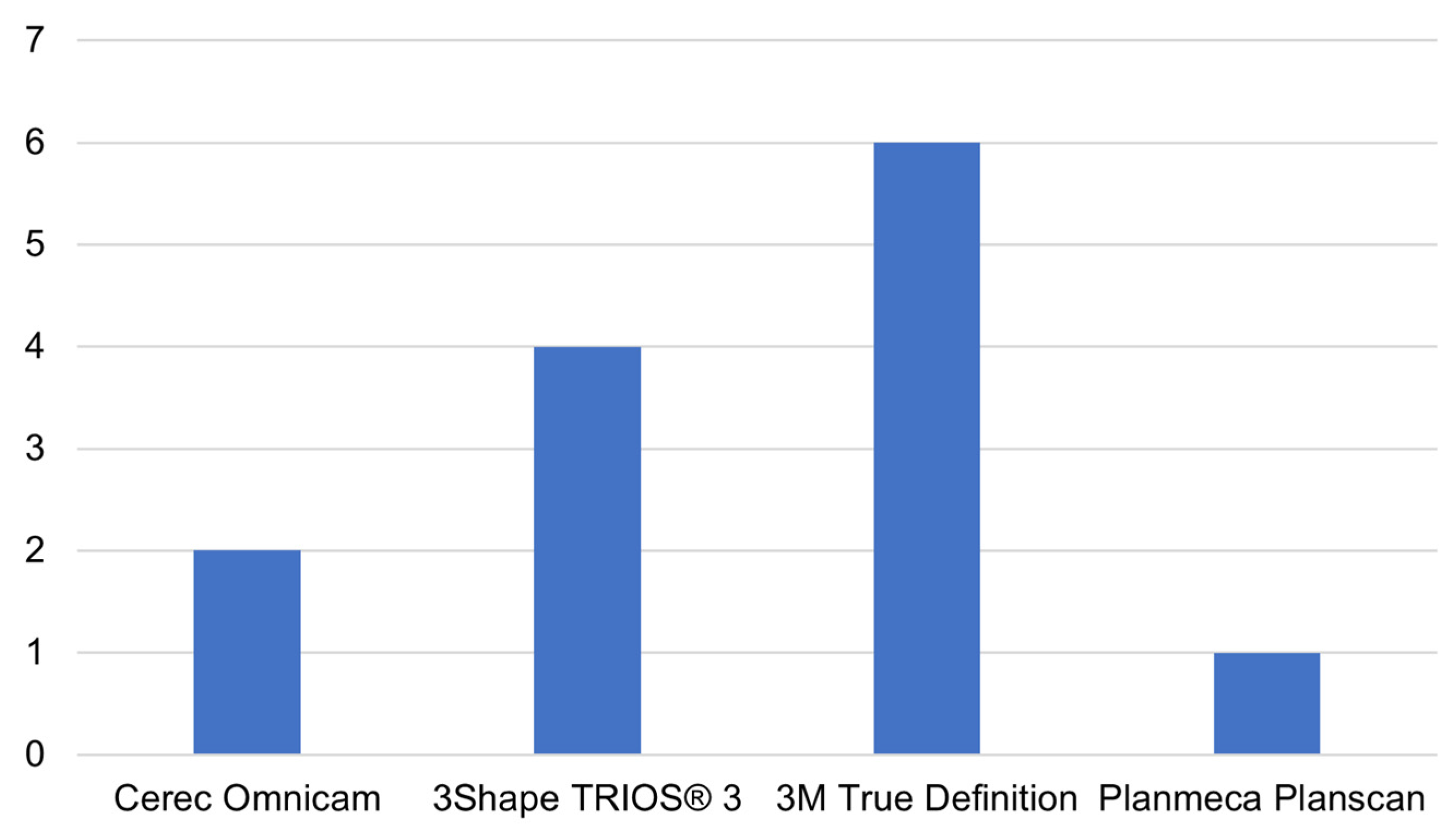
3.4. Analysis of the Main Commercial IOS
3.5. Technologies for Oral Diagnostics
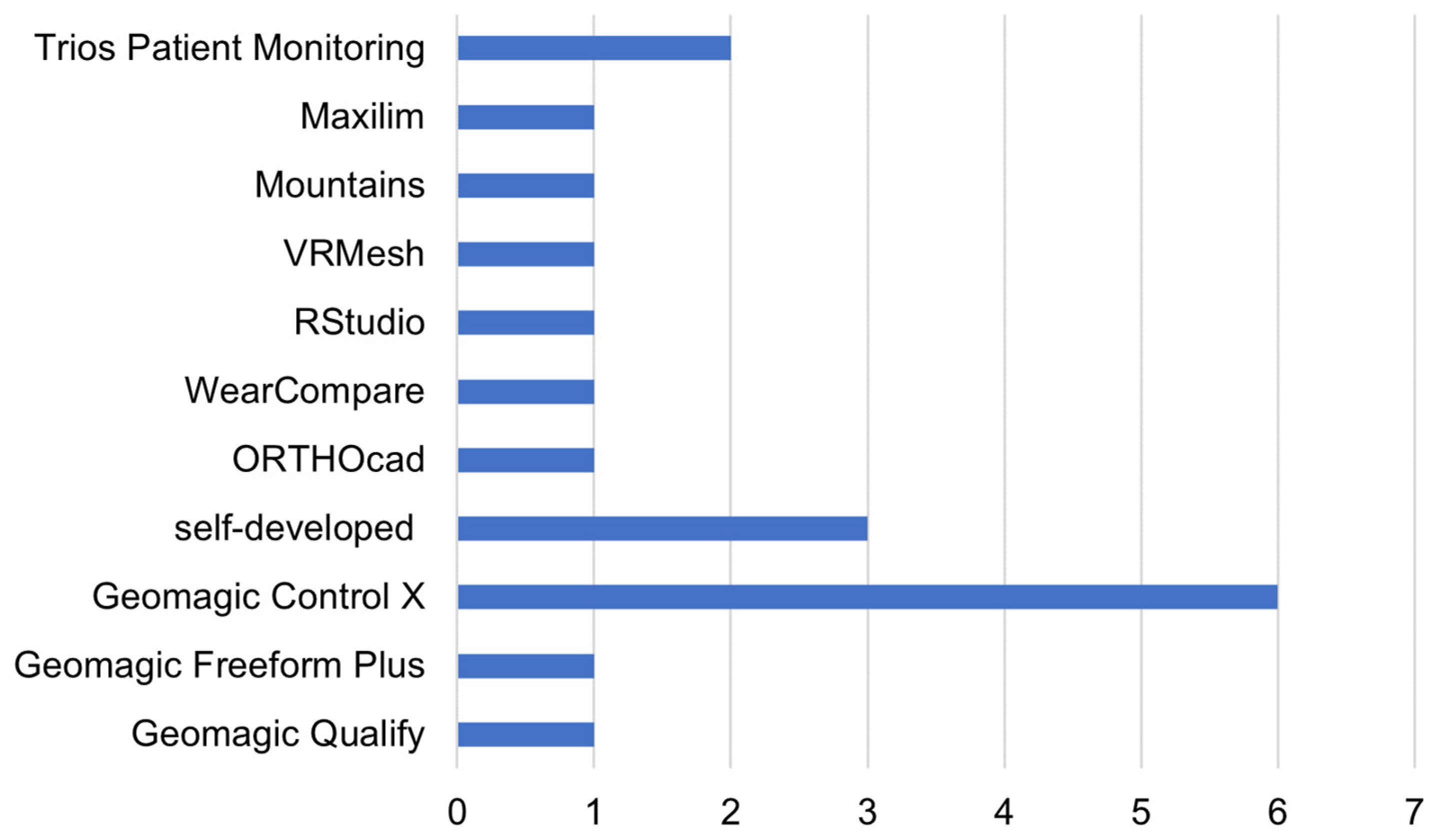
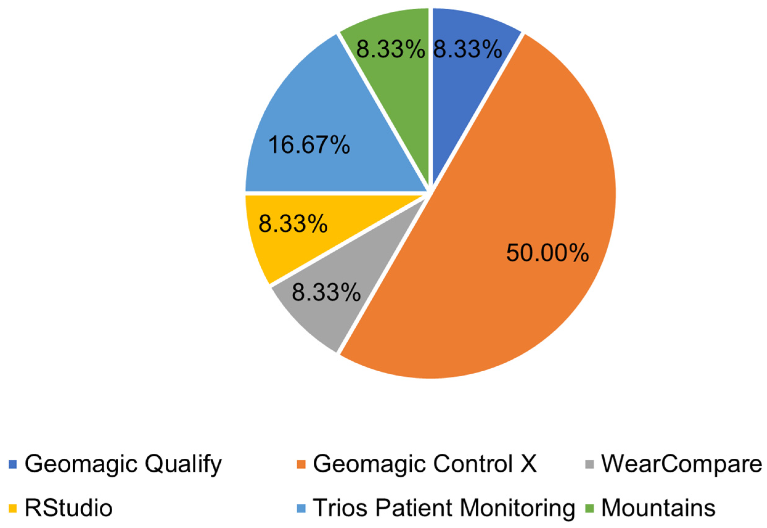
3.6. Three-Dimensional Model of Evaluation Software
4. Discussion
5. Conclusions
Author Contributions
Funding
Institutional Review Board Statement
Informed Consent Statement
Data Availability Statement
Conflicts of Interest
References
- Martin, C.B.; Chalmers, E.V.; McIntyre, G.T.; Cochrane, H.; Mossey, P.A. Orthodontic Scanners: What’s available? J. Orthod. 2015, 42, 136–143. [Google Scholar] [CrossRef] [PubMed]
- Ting-Shu, S.; Jian, S. Intraoral Digital Impression Technique: A Review. J. Prosthodont. 2014, 24, 313–321. [Google Scholar] [CrossRef] [PubMed]
- Kravitz, N.D.; Groth, C.; E Jones, P.; Graham, J.W.; Redmond, W.R. Intraoral digital scanners. J. Clin. Orthod. 2014, 48, 337–347. [Google Scholar] [PubMed]
- Mangano, F.; Gandolfi, A.; Luongo, G.; Logozzo, S. Intraoral Scanners in dentistry: A review of the current literature. BMC Oral Health 2017, 17, 149. [Google Scholar] [CrossRef] [PubMed] [Green Version]
- Mizumoto, R.M.; Yilmaz, B. Intraoral scan bodies in implant dentistry: A systematic review. J. Prosthet. Dent. 2018, 120, 343–352. [Google Scholar] [CrossRef]
- Imburgia, M.; Logozzo, S.; Hauschild, U.; Veronesi, G.; Mangano, C.; Mangano, F.G. Accuracy of four intraoral scanners in oral implantology: A comparative in vitro study. BMC Oral Health 2017, 17, 1–13. [Google Scholar] [CrossRef]
- Oral Health. Available online: https://www.who.int/health-topics/oral-health (accessed on 25 May 2023).
- Bratthall, D.; Petersen, P.E.; Stjernswärd, J.R.; Brown, L.J. Oral and craniofacial diseases and disorders. In Disease Control Priorities in Developing Countries, 2nd ed.; Oxford University Press: New York, NY, USA, 2006. [Google Scholar]
- Sulaiman, D.; Lohiya, A.; Rizwan, S.A.; Singh, A.; Dwivedi, P.; Bahuguna, P.; Dixit, J.; Verma, A.; Kumar, V. Diagnostic Accuracy of Screening of Lip and Oral Cavity Cancers or Potentially Malignant Disorders (PMD) by Frontline Workers: A Systematic Review and Meta-Analysis. Asian Pac. J. Cancer Prev. APJCP 2022, 23, 3983–3991. [Google Scholar] [CrossRef]
- Pakfetrat, A.; Falaki, F.; Delavarian, Z.; Dalirsani, Z.; Sanatkhani, M.; Zabihi Marani, M. Oral Manifestations of Human Immunodeficiency Virus-Infected Patients. Iran. J. Otorhinolaryngol. 2015, 27, 43. [Google Scholar]
- McKinney, R.; Olmo, H. Developmental Disturbances of the Teeth, Anomalies of Shape and Size. In StatPearls; StatPearls Publishing: Treasure Island, FL, USA, 2023. Available online: http://www.ncbi.nlm.nih.gov/books/NBK574555/ (accessed on 29 March 2023).
- Zou, J.; Ashley, J.W. Fluorosis. In Pathobiology of Human Disease; McManus, L.M., Mitchell, R.N., Eds.; Academic Press: San Diego, CA, USA, 2014; pp. 893–898. [Google Scholar] [CrossRef]
- Chaves, L.L.V.; Lopes Rosado, L.P.; Piccolo, S.M.; Ferreira, L.M.; Kamburoglu, K.; Junqueira, R.B.; Aquino de Castro, M.A.; Verner, F.S. Evaluation of the Maxillary Sinus of Patients with Maxillary Posterior Implants: A CBCT Cross-Sectional Study. Diagnostics 2022, 12, 3169. [Google Scholar] [CrossRef]
- Walsh, T.; Warnakulasuriya, S.; Lingen, M.W.; Kerr, A.R.; Ogden, G.R.; Glenny, A.-M.; Macey, R. Clinical assessment for the detection of oral cavity cancer and potentially malignant disorders in apparently healthy adults. Cochrane Database Syst. Rev. 2021, 12, 1465–1858. [Google Scholar] [CrossRef]
- Metzger, Z.; Colson, D.G.; Bown, P.; Weihard, T.; Baresel, I.; Nolting, T. Reflected near-infrared light versus bite-wing radiography for the detection of proximal caries: A multicenter prospective clinical study conducted in private practices. J. Dent. 2021, 116, 103861. [Google Scholar] [CrossRef]
- El-Sharkawy, Y.H.; Elbasuney, S. Non-invasive caries detection and delineation via novel laser-induced fluorescence with hyperspectral imaging. Photodiagnosis Photodyn. Ther. 2022, 40, 103186. [Google Scholar] [CrossRef]
- Chang, N.-Y.N.; Dillas, T.; Zhu, Y.; Fried, D. Assessment of the activity of secondary caries lesions with short-wavelength infrared, thermal, and optical coherence tomographic imaging. J. Biomed. Opt. 2023, 28, 094801. [Google Scholar] [CrossRef]
- Hausdörfer, T.; Harms, L.; Kanzow, P.; Hülsmann, M. Three Visual–Diagnostic Methods for the Detection of Enamel Cracks: An In Vitro Study. J. Clin. Med. 2023, 12, 973. [Google Scholar] [CrossRef]
- Liberati, A.; Altman, D.G.; Tetzlaff, J.; Mulrow, C.; Gøtzsche, P.C.; Ioannidis, J.P.; Clarke, M.; Devereaux, P.J.; Kleijnen, J.; Moher, D. The PRISMA statement for reporting systematic reviews and meta-analyses of studies that evaluate health care interventions: Explanation and elaboration. J. Clin. Epidemiol. 2009, 62, e1–e34. [Google Scholar] [CrossRef] [Green Version]
- Jung, K.; Giese-Kraft, K.; Fischer, M.; Schulze, K.; Schlueter, N.; Ganss, C. Visualization of dental plaque with a 3D-intraoral-scanner—A tool for whole mouth planimetry. PLoS ONE 2022, 17, e0276686. [Google Scholar] [CrossRef]
- von der Stück, A.; Raith, S.; Reich, S. Twenty-four months in vivo wear of enamel antagonists to lithium disilicate implant crowns—A pilot study. J. Dent. 2022, 124, 104215. [Google Scholar] [CrossRef]
- Pałka, J.; Gawda, J.; Byś, A.; Zawadka, M.; Gawda, P. Assessment of Growth Changes in the Width of Dental Arches Caused by Removable Appliances over a Period of 10 Months in Children with Malocclusion. Int. J. Environ. Res. Public Health 2022, 19, 3442. [Google Scholar] [CrossRef]
- Machado, A.C.; Phillips, T.S.; Zimmerman, R.; Scaramucci, T.; Amaechi, B.T. Monitoring erosive tooth wear with intraoral 3D scanner: A feasibility study. Am. J. Dent. 2022, 35, 49–54. [Google Scholar]
- Giese-Kraft, K.; Jung, K.; Schlueter, N.; Vach, K.; Ganss, C. Detecting and monitoring dental plaque levels with digital 2D and 3D imaging techniques. PLoS ONE 2022, 17, e0263722. [Google Scholar] [CrossRef]
- Lee, J.-H.; Byun, S.-H.; Yi, S.-M.; Park, I.-Y.; Yang, B.-E.; Lee, H.-L. Efficacy of Constructing Digital Hybrid Skull-Dentition Images Using an Intraoral Scanner and Cone-Beam Computed Tomography. Scanning 2022, 2022. [Google Scholar] [CrossRef] [PubMed]
- García, V.D.-F.; Freire, Y.; Fernández, S.D.; Murillo, B.T.; Sánchez, M.G. Application of the Intraoral Scanner in the Diagnosis of Dental Wear: An in Vivo Study of Tooth Wear Analysis. Int. J. Environ. Res. Public Health 2022, 19, 4481. [Google Scholar] [CrossRef] [PubMed]
- Sobral, A.P.T.; Gonçalves, M.L.L.; D´Annibale, A.S.; Santos, E.M.; Guedes, C.C.; Gallo, J.M.A.S.; Ferri, E.P.; Moretti, L.A.C.; Motta, L.J.; Deana, A.M.; et al. Evaluation of different methods for the diagnosis of primary caries lesions: Study protocol for a randomized controlled clinical trial. PLoS ONE 2022, 17, e0273104. [Google Scholar] [CrossRef] [PubMed]
- Michou, S.; Vannahme, C.; Bakhshandeh, A.; Ekstrand, K.R.; Benetti, A.R. Intraoral scanner featuring transillumination for proximal caries detection. An in-vitro validation study on permanent posterior teeth. J. Dent. 2022, 116, 103841. [Google Scholar] [CrossRef]
- Schlenz, M.A.; Schupp, B.; Schmidt, A.; Wöstmann, B.; Baresel, I.; Krämer, N.; Schulz-Weidner, N. New Caries Diagnostic Tools in Intraoral Scanners: A Comparative in Vitro Study to Established Methods in Permanent and Primary Teeth. Sensors 2022, 22, 2156. [Google Scholar] [CrossRef]
- Kühne, C.; Lohbauer, U.; Raith, S.; Reich, S. Measurement of Tooth Wear by Means of Digital Impressions: An In-Vitro Evaluation of Three Intraoral Scanning Systems. Appl. Sci. 2021, 11, 5161. [Google Scholar] [CrossRef]
- Bastos, R.T.D.R.M.; da Silva, P.T.; Normando, D. Reliability of qualitative occlusal tooth wear evaluation using an intraoral scanner: A pilot study. PLoS ONE 2021, 16, e0249119. [Google Scholar]
- Ayoub, A.; Khan, A.; Aldhanhani, A.; Alnaser, H.; Naudi, K.; Ju, X.; Gillgrass, T.; Mossey, P. The Validation of an Innovative Method for 3D Capture and Analysis of the Nasolabial Region in Cleft Cases. Cleft Palate-Craniofacial J. 2021, 58, 98–104. [Google Scholar] [CrossRef]
- Michou, S.; Lambach, M.S.; Ntovas, P.; Benetti, A.R.; Bakhshandeh, A.; Rahiotis, C.; Ekstrand, K.R.; Vannahme, C. Automated caries detection in vivo using a 3D intraoral scanner. Sci. Rep. 2021, 11, 21276. [Google Scholar] [CrossRef]
- Intraoral Scanning for Early Dental Erosion Assessment—An In-Vitro Study—Romanian Journal of Oral Rehabilitation. Available online: https://www.rjor.ro/intraoral-scanning-for-early-dental-erosion-assessment-an-in-vitro-study/ (accessed on 29 March 2023).
- Michou, S.; Benetti, A.R.; Vannahme, C.; Hermannsson, P.G.; Bakhshandeh, A.; Ekstrand, K.R. Development of a Fluorescence-Based Caries Scoring System for an Intraoral Scanner: An in Vitro Study. Caries Res. 2020, 54, 324–335. [Google Scholar] [CrossRef]
- Esquivel-Upshaw, J.F.; Hsu, S.-M.; Bohórquez, A.C.; Abdulhameed, N.; Scheiffele, G.W.; Kim, M.; Neal, D.; Chai, J.; Ren, F. Novel methodology for measuring intraoral wear in enamel and dental restorative materials. Clin. Exp. Dent. Res. 2020, 6, 677–685. [Google Scholar] [CrossRef]
- Alwadai, G.S.; Roberts, G.; Ungar, P.S.; González-Cabezas, C.; Lippert, F.; Diefenderfer, K.E.; Eckert, G.J.; Hara, A.T. Monitoring of simulated occlusal tooth wear by objective outcome measures. J. Dent. 2020, 102, 103467. [Google Scholar] [CrossRef]
- Michou, S.; Vannahme, C.; Ekstrand, K.R.; Benetti, A.R. Detecting Early Erosive Tooth Wear Using an Intraoral Scanner System. J. Dent. 2020, 100, 103445. [Google Scholar] [CrossRef]
- Icen, M.; Orhan, K.; Şeker, Ç.; Geduk, G.; Çakmak Özlü, F.; Cengiz, M.İ. Comparison of CBCT with Different Voxel Sizes and Intraoral Scanner for Detection of Periodontal Defects: An in Vitro Study. Dentomaxillofac. Radiol. 2020, 49, 20190197. [Google Scholar] [CrossRef]
- Kumar, S.; Keeling, A.; Osnes, C.; Bartlett, D.; O’Toole, S. The sensitivity of digital intraoral scanners at measuring early erosive wear. J. Dent. 2019, 81, 39–42. [Google Scholar] [CrossRef]
- Charalambous, P.; O’Toole, S.; Austin, R.; Bartlett, D. The threshold of an intra oral scanner to measure lesion depth on natural unpolished teeth. Dent. Mater. 2022, 38, 1354–1361. [Google Scholar] [CrossRef]
- Eom, J.B.; Ahn, J.S.; Eom, J.; Park, A. Wide field of view optical coherence tomography for structural and functional diagnoses in dentistry. J. Biomed. Opt. 2018, 23, 076008. [Google Scholar]
- Deferm, J.T.; Schreurs, R.; Baan, F.; Bruggink, R.; Merkx, M.A.; Xi, T.; Bergé, S.J.; Maal, T.J. Validation of 3D documentation of palatal soft tissue shape, color, and irregularity with intraoral scanning. Clin. Oral Investig. 2018, 22, 1303–1309. [Google Scholar] [CrossRef]
- Ferraro, J.M.; Falter, J.; Lee, S.; Watanabe, K.; Wu, T.-H.; Kim, D.-G.; Ko, C.-C.; Tanaka, E.; Deguchi, T. Accuracy of three-dimensional printed models derived from cone-beam computed tomography. Angle Orthod. 2022, 92, 722–727. [Google Scholar] [CrossRef]
- Akyalcin, S.; Cozad, B.E.; English, J.D.; Colville, C.D.; Laman, S. Diagnostic accuracy of impression-free digital models. Am. J. Orthod. Dentofac. Orthop. 2013, 144, 916–922. [Google Scholar] [CrossRef]
- Fraile, C.; Ferreiroa, A.; Romeo, M.; Alonso, R.; Pradíes, G. Clinical study comparing the accuracy of interocclusal records, digitally obtained by three different devices. Clin. Oral Investig. 2022, 26, 1957–1962. [Google Scholar] [CrossRef]
- Kirschneck, C.; Kamuf, B.; Putsch, C.; Chhatwani, S.; Bizhang, M.; Danesh, G. Conformity, reliability and validity of digital dental models created by clinical intraoral scanning and extraoral plaster model digitization workflows. Comput. Biol. Med. 2018, 100, 114–122. [Google Scholar] [CrossRef] [PubMed]
- Davidovich, E.; Shay, B.; Nuni, E.; Mijiritsky, E. An Innovative Treatment Approach Using Digital Workflow and CAD-CAM Part 1: The Restoration of Endodontically Treated Molars in Children. Int. J. Environ. Res. Public Health 2020, 17, 1364. [Google Scholar] [CrossRef] [PubMed] [Green Version]
- Davidovich, E.; Dagon, S.; Tamari, I.; Etinger, M.; Mijiritsky, E. An Innovative Treatment Approach Using Digital Workflow and CAD-CAM Part 2: The Restoration of Molar Incisor Hypomineralization in Children. Int. J. Environ. Res. Public Health 2020, 17, 1499. [Google Scholar] [CrossRef] [PubMed] [Green Version]
- Revilla-León, M.; Fountain, J.; Piedra-Cascón, W.; Zandinejad, A.; Özcan, M. Silicone Additive Manufactured Indices Performed from a Virtual Diagnostic Waxing for Direct Composite Diastema Closure Combined with Resin Infiltration Technique on White Spot Lesions: A Case Report. J. Prosthodont. 2019, 28, 855–860. [Google Scholar] [CrossRef]
- Revilla-León, M.; Raney, L.; Piedra-Cascón, W.; Barrington, J.; Zandinejad, A.; Özcan, M. Digital workflow for an esthetic rehabilitation using a facial and intraoral scanner and an additive manufactured silicone index: A dental technique. J. Prosthet. Dent. 2020, 123, 564–570. [Google Scholar] [CrossRef]
- Park, S.H.; Piedra-Cascón, W.; Zandinejad, A.; Revilla-León, M. Digitally Created 3-Piece Additive Manufactured Index for Direct Esthetic Treatment. J. Prosthodont. 2020, 29, 436–442. [Google Scholar] [CrossRef]
- DuVall, N.B. Fabricating a chairside CAD-CAM radiographic and surgical guide for dental implants: A dental technique. J. Prosthet. Dent. 2020, 125, 34–40. [Google Scholar] [CrossRef]
- Ahmed, W.M.; Hans, A.; Verhaeghe, T.V.; Nguyen, C. Managing Excessive Gingival Display Using a Digital Workflow. J. Prosthodont. 2020, 29, 443–447. [Google Scholar] [CrossRef]
- Align Technology. Available online: https://www.aligntech.com/solutions/itero_scanner (accessed on 29 March 2023).
- 3Shape TRIOS Intraoral Scanners—Compare All Models. Available online: https://www.3shape.com/en/scanners/trios (accessed on 29 March 2023).
- Virtuo VivoTM Intraoral Scanners. Available online: https://www.straumann.com/en/dental-professionals/products-and-solutions/cares-digital-solutions/equipment/io-scanners/virtuo-vivo.html (accessed on 29 March 2023).
- Abduo, J.; Elseyoufi, M. Accuracy of Intraoral Scanners: A Systematic Review of Influencing Factors. Eur. J. Prosthodont. Restor. Dent. 2018, 26, 101–121. [Google Scholar] [CrossRef]
- Chiu, A.; Chen, Y.-W.; Hayashi, J.; Sadr, A. Accuracy of CAD/CAM Digital Impressions with Different Intraoral Scanner Parameters. Sensors 2020, 20, 1157. [Google Scholar] [CrossRef] [Green Version]
- Ender, A.; Zimmermann, M.; Mehl, A. Accuracy of complete- and partial-arch impressions of actual intraoral scanning systems in vitro. Int. J. Comput. Dent. 2019, 22, 11–19. [Google Scholar]
- Hwang, H.H.M.; Chou, C.W.; Chen, Y.J.; Yao, C.C.J. An Overview of Digital Intraoral Scanners: Past, Present and Future—From an Orthodontic Perspective. Taiwan. J. Orthod. 2018, 30, 3. [Google Scholar] [CrossRef]
- Logozzo, S.; Zanetti, E.; Franceschini, G.; Kilpela, A.; Mäkynen, A. Recent advances in dental optics—Part I: 3D intraoral scanners for restorative dentistry. Opt. Lasers Eng. 2014, 54, 203–221. [Google Scholar] [CrossRef]
- Pampush, J.D.; Winchester, J.M.; Morse, P.E.; Vining, A.Q.; Boyer, D.M.; Kay, R.F. Introducing molaR: A New R Package for Quantitative Topographic Analysis of Teeth (and Other Topographic Surfaces). J. Mamm. Evol. 2016, 23, 397–412. [Google Scholar] [CrossRef]
- O’Toole, S.; Osnes, C.; Bartlett, D.; Keeling, A. Investigation into the validity of WearCompare, a purpose-built software to quantify erosive tooth wear progression. Dent. Mater. 2019, 35, 1408–1414. [Google Scholar] [CrossRef]
- Oh, W.; DeLong, R.; Anusavice, K.J. Factors affecting enamel and ceramic wear: A literature review. J. Prosthet. Dent. 2002, 87, 451–459. [Google Scholar] [CrossRef]
- Bartlett, D.; Ganss, C.; Lussi, A. Basic Erosive Wear Examination (BEWE): A new scoring system for scientific and clinical needs. Clin. Oral Investig. 2008, 12, 65–68. [Google Scholar] [CrossRef] [Green Version]


| PICO Facets | Considerations |
|---|---|
| Patient (P) | Patients with oral cavity pathologies (e.g., caries, dental wear, periodontal diseases, oral cancer, infections) |
| Intervention (I) | Detection of oral cavity pathologies by means of an intraoral scanning device |
| Comparison (C) | Traditional methods or gold standards for the diagnosis/detection of the specific oral cavity pathology (e.g., radiographic modalities) |
| Outcome (O) | Assessment of the performance of intraoral scanners in the diagnosis/detection of pathologies of the oral cavity compared with reference methods |
| First Author (Year) | Aim of the Study | Type of IOS | Product Name | Type of Study | Diagnostic Application | Software Used | Main Results |
|---|---|---|---|---|---|---|---|
| Jung et al. (2022) [20] | To investigate whether images from 3D intraoral scans are also suitable for valid planimetric plaque measurements and monitoring | Commercial | CS3600 Carestream | In vivo | Dental plaque | Self-developed software (Julia (version 1.6.28) | Planimetry, using images from the 3D intraoral scan seems to be a suitable tool for recording and monitoring dental plaques. |
| Stück et al. (2022) [21] | To evaluate the difference between the dental wear in implant/enamel and enamel/enamel contacts | Commercial | CEREC omnicam | In vivo | Dental wear | Python Software Foundation, Wilmington, Delaware, United States of America, version 3.5.2; Blender Foundation, Amsterdam, the Netherlands, version 2.7; | Intraoral scanning and computer analysis showed that the two-years wear ratios for enamel/enamel and enamel/lithium disilicate implant crowns did not differ significantly. |
| Pałka et al. (2022) [22] | To evaluate the impact of removable appliances used over a 10 month period on growth changes in children with malocclusions | Commercial | iTero | In vivo | Orthodontic diagnosis | OrthoCAD (software version not specified-https://www.itero.com/it/education-and-support/software-downloads; accessed on 26 June 2023) | The use of removable appliances in children with a narrowed maxillary transverse dimension contributes to offsetting growth changes in comparison to children with normal occlusion. |
| Machado et al. (2022) [23] | To evaluate the potential of an IOS to monitor dental wear using distinct quantitative measurement metrics | Commercial | TRIOS 3 | In vitro | Dental wear | WearCompare (software version not specified-https://leedsdigitaldentistry.com/wearcompare/; accessed on 26 June 2023); 3Shape TRIOS® Patient Monitoring (software version not specified-https://www.3shape.com/en/software/trios-patient-monitoring; accessed on 26 June 2023) | This IOS is a potential clinical tool for detecting and quantitatively monitoring early and advanced erosive tooth wear. |
| Giese-Kraft et al. (2022) [24] | To investigate whether plaque can be reliably visualized on 2D and 3D images captured with digital intraoral imaging devices | Commercial | CS3600 Carestream | In vivo | Dental plaque | N/A | Amounts of plaque can be reliably detected and monitored on 2D images from an intraoral camera and on 3D images from an intraoral scanner. |
| Lee et al. (2022) [25] | To evaluate the potential for improving dentition imaging with CBCT scans using an IOS | Commercial | CS3600 Carestream e i700 Medit | In vivo | CBCT improvement | Geomagic Freeform Plus (software version not specified-https://oqton.com/geomagic-freeform/; accessed on 26 June 2023) | The virtual skull dentition hybrid image obtained from intraoral scanners will be clinically useful. |
| Metzger et al. (2022) [15] | To compare the detection of proximal caries with NIR versus BWR | Commercial | iTero Element 5D | In vivo | Dental caries | N/A | NIR was more sensitive than BWR for detecting early caries. |
| García et al. (2022) [26] | To analyze the sensitivity and specificity of the IOS for measuring dental wear and to evaluate patients’ satisfaction | Commercial | 3M™ True Definition | In vivo | Dental wear | Geomagic™ (software version not specified-https://www.3dsystems.com/software; accessed on 26 June 2023) | The data show good levels of specificity and sensitivity, and the participants also show a high degree (40,9%) of satisfaction. |
| Sobral et al. (2022) [27] | To define a protocol to evaluate the best strategy for diagnosing primary caries lesions located in the interproximal region among visual clinical examination, IOS, and BWR | Commercial | iTero Element 5D | In vivo | Interproximal caries | N/A | Through the protocol described, clinicians will be able to assess whether there will be any difference in effectiveness between the selected diagnostic methods. |
| Michou et al. (2022) [28] | To assess the validity of an IOS system featuring NIR transillumination to aid in the detection of proximal caries lesions | Commercial | TRIOS 4 | In vitro | Interocclusal caries | N/A | IOS system featuring NIR transillumination and DIAGNOcam showed an overall good diagnostic performance. |
| Schlenz et al. (2022) [29] | To investigate new caries diagnostic tools, including three IOS, and compare them to established diagnostic methods | Commercial | 1)iTero element 5D;2)TRIOS 4;4)Planmeca Emerald S | In vitro | Proximal and occlusal caries | N/A | Caries diagnostics with intraoral scanners seem to be interesting tools that should be further investigated in clinical studies. |
| Kühne et al. (2021) [30] | To investigate whether IOSs are suitable for wear measurement compared to WLP | Commercial | Cerec Omnicam AC (OC), TRIOS 3 (Tr3) e True Definition | In vitro | Dental wear | Geomagic Qualify 2012 (V.2012_08_08_E, 64-bit, Morrisville, NC, USA) | Intraoral scanning combined with a matching software can accurately quantify clinical wear. |
| Travassos da Rosa Moreira Bastos et al. (2021) [31] | To investigate the reliability of qualitative tooth wear evaluation through 3D images captured with an IOS and compared to clinical and photographic examinations | Commercial | TRIOS POD | In vivo | Dental wear | N/A | Intraoral scanning seems to be a sound and reliable tool to evaluate tooth wear when compared to traditional methods. |
| Ayoub et al. (2021) [32] | To assess upper lip scarring and asymmetry in surgically managed unilateral cleft lip and palate (UCLP) cases through 3D images of the nasolabial region | Commercial | TRIOS 3 | In vivo | Analysis of the nasolabial region | VRMesh studio VirtualGrid (software version not specified-https://www.vrmesh.com/; accessed on 26 June 2023) | The 3D images are a reliable source for measuring lip asymmetry and the scar surface area. |
| Michou et al. (2021) [33] | To validate an automated caries scoring system for occlusal caries detection and classification, previously defined for an IOS system featuring fluorescence | Commercial | 3Shape TRIOS 4 | In vivo | Dental caries | N/A | IOS system proposed exhibits encouraging performance for clinical application on occlusal caries detection and classification. |
| Ille et al. (2021) [34] | To quantify the feasibility of using an IOS to monitor early erosive tooth wear in patients | Commercial | Planscan Planmeca | In vitro | Dental wear | Geomagic Control X (3Dsystems, Darmstadt, Germany, software version not specified-https://www.3dsystems.com/software; accessed on 26 June 2023) |
The intraoral scanner used in this experiment was capable to detect minimal dental tissue loss and could be used to monitor early erosive tooth wear. |
| Michou et al. (2020) [35] | To develop an automated fluorescence-based caries scoring system for an IOS and to test the performance of the system compared to state-of-the-art methods | Prototype | Prototype (415 nm) | In vitro | Dental caries | N/A | IOS accompanied by an automated caries scoring system may improve objective caries detection and increase the efficiency and effectiveness of oral examinations. |
| Esquivel-Upshaw et al. (2020) [36] | To test that the dental scanner combined with metrology software will measure clinical wear in vivo in agreement with measurements from X-ray computed microtomography | Commercial | 3M True Definition | In vivo | Dental wear | Geomagic Control 2014 (3Dsystems, Darmstadt, Germany-software version not specified-https://www.3dsystems.com/software; accessed on 26 June 2023) | Intraoral scanning combined with a matching software can accurately quantify clinical wear. |
| Alwadai et al. (2020) [37] | To explore quantitative outcome measures as clinical indicators of simulated occlusal tooth wear progression | Commercial | 3M True Definition | In vitro | Dental wear | Rstudio (RStudio, Inc., Boston, MA, USA-software version not specified-https://posit.co/download/rstudio-desktop/; accessed on 26 June 2023) with molaR package | IOS can serve effectively for monitoring overall tooth wear when combined with dental topographic analyses of resultant point clouds. |
| Michou et al. (2020) [38] | To assess the feasibility of detecting and monitoring early erosive tooth wear using a 3D IOS aided by specific software | Commercial | 3Shape TRIOS® 3 | In vitro | Dental wear | 3Shape TRIOS® Patient Monitoring, version 2.1.1.0 | The use of an IOS aided by specific software showed good performance for early detection and monitoring of tooth wear in vitro and has promising potential for in vivo application. |
| Icen et al. (2020) [39] | To compare the diagnostic accuracy of CBCT units with different voxel sizes with the digital intraoral scanning technique in terms of the detection of periodontal defects. | Commercial | 3D Shape TRIOS® Color P13 Shade | In vitro | Periodontal defects | N/A | Smaller voxel sizes and smaller CBCT FOV has the highest sensitivity and diagnostic accuracy. |
| Kumar et al. (2020) [40] | To investigate the sensitivity of intraoral scanners to quantitatively detect early erosive tooth wear. | Commercial | 3M True Definition | In vitro | Dental wear | Geomagic Control (3Dsystems, Darmstadt, Germany-software version not specified-https://www.3dsystems.com/software; accessed on 26 June 2023) | Precision was low, suggesting limitations for minimal changes in tooth wear |
| Charalambous et al. (2019) [41] | To investigate the threshold and accuracy of intraoral scanning in measuring freeform human enamel surfaces | Commercial | 3M True Definition Scanner | In vitro | Dental wear | Geomagic Control (3Dsystems, Darmstadt, Germany-software version not specified-https://www.3dsystems.com/software; accessed on 26 June 2023); Mountains® 8 (Digitalsurf, Besançon, France) | The intraoral scanner had a depth discrimination threshold of 73 µm on unpolished natural enamel and significant differences (p < 0.05) were observed compared to NCLP below this level. |
| Eom et al. (2018) [42] | To evaluate the performance response of a 3D intraoral scan probe based on OCT that enables structural and functional diagnoses of the human teeth | Prototype | Prototipo per OCT | In vitro | Other structural and functional issues | Self-developed multithreaded C++ software, built and compiled in Microsoft Visual Studio 2013. | The feasibility of the intraoral scan probe is demonstrated based on its ability to image and characterize the structure and function of the human teeth. |
| Deferm et al. (2018) [43] | To assess the feasibility of 3D intraoral scanning for palatal soft tissue analysis. | Commercial | POD TRIOS® 3 | In vivo | Soft tissue analysis | Maxilim software (V2.3.0, Medicim NV, Mechelen, Belgium) | IOS can perform a 3D documentation of palatal soft tissue in terms of shape, color, and curvature. |
| Authors | Aim of the Study | Type of IOS | Product Name | Type of Study | Application | Main Results |
|---|---|---|---|---|---|---|
| Ferraro et al. (2022) [44] | To compare the accuracy of 3D printed models fabricated from CBCT scans of human mandibular dry skulls with models derived from IOSs. | Commercial | TRIOS | In vitro | Comparison of 3D model technical parameters in reconstruction | Differences not clinically detectable for orthodontic applications |
| Akyalcin et al. (2018) [45] | To evaluate the accuracy of 3D digital models acquired from a chairside IOS compared with both manual and CBCT measurements of the same dental anatomy. | Commercial | Cadent iTero | In vitro | Comparison of 3D model technical parameters in reconstruction | Differences not clinically detectable for orthodontic applications |
| Fraile et al. (2022) [46] | To compare the interocclusal contact records obtained by three different digital methods with the conventional method (articulating paper). | Commercial | TRIOS Color POD | In vivo | Comparison of 3D model technical parameters in reconstruction | Reliable performance of the scanners compared to the gold standard |
| Kirschneck et al. (2018) [47] | To assess the reliability, validity, and conformity of an intraoral scanning procedure and two extraoral digitization workflows via alginate impression and plaster model scanning. | Commercial | Lythos | In vivo | Comparison of 3D model technical parameters in reconstruction | Better reliability of the extraoral 3D models. |
| Davidovich et al. (2020) [48] | To present an innovative treatment approach for endodontically treated teeth in children using a digital workflow with IOS and CAD/CAM fabrication of the restoration | Commercial | Primescan connect | In vivo | Endodontically treatment approach | The high accuracy of the scanner enables definitive restoration in young patients |
| Davidovich et al. (2020) [49] | To present an innovative treatment approach for children with MIH using a digital workflow with IOS and CAD-CAM fabrication of the restoration | Commercial | Primescan connect | In vivo | Treatment for molar incisor hypomineralization (MIH) | The digital workflow provides definitive restorations in young patients due to the high accuracy of the scanning. |
| Revilla-Leon et al. (2019) [50] | To evaluate the influence of the interocclusal space on the accuracy of the maxillomandibular relationship captured with an IOS. | Commercial | TRIOS 4 | In vitro | Fabrication of dental prostheses | The interocclusal space available when acquiring virtual bilateral occlusal records using the tested IOS impacted the accuracy of the maxillomandibular relationship. |
| Revilla-Leon et al. (2020) [51] | To describe a digital workflow for planning an esthetic treatment by using a facial and intraoral scanner, the dental and open-source software design of a facially generated diagnostic waxing, and additive manufactured (AM) clear silicone indices. | Commercial | iTero Element | In vivo | Esthetic rehabilitation | Three-piece AM clear indexes provided advantages compared with conventional procedures |
| Park et al. (2020) [52] | To describe a digital workflow protocol for treatment planning of an esthetic rehabilitation using direct composite restorations | Commercial | TRIOS 3 | In vivo | Esthetic rehabilitation | Provides several advantages compared with conventional procedures. |
| DuVall (2021) [53] | To describe a procedure that uses an intraoral scanner and milling unit to fabricate a chairside computer-aided design and computer-aided manufacturing radiographic and surgical guide for use with a Cone Beam Computed Tomography system | Commercial | CEREC Omnicam AC | In vivo | Dental implants | The procedure described is efficient compared with the traditional one. |
| Ahmed et al. (2020) [54] | To describe an approach to manage excessive gingival display by lengthening of the clinical crowns using a digital workflow. | Commercial | TRIOS 3 | In vivo | Surgical digital procedure | The procedure described is efficient compared with the traditional one. |
| Scanner Model | Year | Price | Technology of Acquisition | Illumination | Wand Dimensions [mm] | Wand Weight [g] |
|---|---|---|---|---|---|---|
| TRIOS 3 (3Shape) | 2015 | $18,950.00 | Structured light | LED | N/A | 340 |
| TRIOS 4 (3Shape) | 2019 | $30,000.00 | Structured light + AI scan | LED | 274 × 42 × 12 | 345 |
| TRIOS 5 (3Shape) | 2022 | $29,500.00 | Structured light + AI | N/A | 266 × 38 × 11 | N/A |
| True Definition (3M Espe) | 2012 | $12,000.00 | Structured Light | Blue | 254 × 16 × 14 | 233 |
| iTero Element (Align Technology) | N/A | N/A | N/A | Red laser light (680 nm) and white LED | 38.5 × 53.5 × 69.8 | 470 |
| iTero (Align Technology) | 2007 | N/A | parallel confocal | white LED light | N/A | 680.39 |
| iTero 5D Element (Align Technology) | 2021 | N/A | Structured Light + NIRI | N/A | N/A | ~500 |
| CS3800 (Carestream) | 2021 | $38,000.00 | N/A | Red, Green, Blue LEDs | 226 × 38 × 60 | 240 |
| CS3700 (CareStream) | N/A | $16,000.00 | N/A | N/A | N/A | N/A |
| CS3600 (CareStream) | 2016 | $37,000.00 | Structured Led Light | Amber, Green, Blue LEDs | 220 × 38 × 58 | 325 |
| CS3500 (CareStream) | 2017 | N/A | Parallel confocal | N/A | N/A | N/A |
| Virtuo Vivo (Dental wings) | 2019 | $19,000.00 | Multiscan Imaging Technology | Blue laser | 200 × 30 | 120 |
| DWIO (Dental Wings) | 2017 | N/A | Multiscan Imaging | Blue LED | N/A | 105 |
| IOS FastScan (Glidewell Laboratories) | N/A | $19,899.00 | Active triangulation/Scheimpflug principle | N/A | N/A | N/A |
| i700 (Medit) | 2021 | $20,000.00 | 3D-in-motion video technology | LED | 248 × 44 × 47.4 | 245 |
| i500 (Medit) | 2020 | $20,000.00 | 3D-in-motion video technology | Blue, White | 266 × 18 × 15.2 | 276 |
| 3D Progress (MHT S.p.A.) | N/A | N/A | Confocal microscope + Moireé effect detection | N/A | N/A | N/A |
| Lythos (Ormco) | 2013 | N/A | Accordion fringe interferometry | N/A | N/A | 317.51 |
| Planscan (Planmeca) | N/A | N/A | Triangulation | N/A | N/A | N/A |
| Emerald (Planmeca) | N/A | N/A | N/A | Laser | 41 × 45 × 249 | 183 |
| Aoralscan 3 (Shining 3D) | 2021 | $15,200.00 | Structured light | N/A | 281 × 33 × 46 | 240 |
| Cerec Omnicam (Dentsply Sirona) | 2012 | $35,000.00 | Optical triangulation and confocal microscopy | White LED | 228 × 16 × 16 | N/A |
| Cerec AC BlueCam (Dentsply Sirona) | 2009 | N/A | confocal microscopy/active triangulation | Blue LED | N/A | N/A |
| Cerec PrimeScan (Dentsply Sirona) | 2019 | $26,000.00 | High-frequency optical contrast analysis | N/A | N/A | 457 |
| 3Di IOS (MyRay) | N/A | $29,500.00 | Active Stereo Imaging | N/A | 256 × 45 × 45 | 150 |
| 3DISC (Heron IOS) | N/A | N/A | N/A | N/A | N/A | 145 |
| Diagnostic Application | Intraoral Scanners |
|---|---|
| Periodontal defects | 3D Shape TRIOS® |
| Soft tissue analysis | 3Shape TRIOS® 3 |
| Analysis of the Nasolabial Region | 3Shape TRIOS® 3 |
| Orthodontic diagnosis | iTero |
| CBCT improvement | Carestream CS3600/Medit i700 |
| Other structural and functional issues | prototype for OCT |
Disclaimer/Publisher’s Note: The statements, opinions and data contained in all publications are solely those of the individual author(s) and contributor(s) and not of MDPI and/or the editor(s). MDPI and/or the editor(s) disclaim responsibility for any injury to people or property resulting from any ideas, methods, instructions or products referred to in the content. |
© 2023 by the authors. Licensee MDPI, Basel, Switzerland. This article is an open access article distributed under the terms and conditions of the Creative Commons Attribution (CC BY) license (https://creativecommons.org/licenses/by/4.0/).
Share and Cite
Angelone, F.; Ponsiglione, A.M.; Ricciardi, C.; Cesarelli, G.; Sansone, M.; Amato, F. Diagnostic Applications of Intraoral Scanners: A Systematic Review. J. Imaging 2023, 9, 134. https://doi.org/10.3390/jimaging9070134
Angelone F, Ponsiglione AM, Ricciardi C, Cesarelli G, Sansone M, Amato F. Diagnostic Applications of Intraoral Scanners: A Systematic Review. Journal of Imaging. 2023; 9(7):134. https://doi.org/10.3390/jimaging9070134
Chicago/Turabian StyleAngelone, Francesca, Alfonso Maria Ponsiglione, Carlo Ricciardi, Giuseppe Cesarelli, Mario Sansone, and Francesco Amato. 2023. "Diagnostic Applications of Intraoral Scanners: A Systematic Review" Journal of Imaging 9, no. 7: 134. https://doi.org/10.3390/jimaging9070134