
Patient Dose Estimation in Computed Tomography-Guided Biopsy Procedures

 ,
,  , and
, and
Abstract
:1. Introduction
2. Materials and Methods
3. Results
4. Discussion

| Present Study (DLP mGy cm) | Other Studies (DLP mGy cm) | |||||||
|---|---|---|---|---|---|---|---|---|
| mean | median | mean | median | mean | median | mean | median | |
| Iliac bone | 672.4 | 489.4 | 793.0 [24] | 113.8 [16] | 410.0 [25] | |||
| Liver | 967.5 | 748.7 | 712.0 [24] | 652.0 [24] | 813.0 [15] 1539.2 [18] | 652.0 [17] | 710.0 [25] | |
| Lung | 894.0 | 817.2 | 507.0 [24] | 481.0 [24] | 440.0 [15] 4320.5 [18] | 481.0 [17] 113.8 [20] | 435.0 [25] | |
| Mediastinum | 744.6 | 547.2 | 549.0 [24] | 468.0 [24] | 440.0 [15] 4320.5 [18] | 481.0 [17] 113.8 [20] | 435.0 [25] | |
| Para-aortic tissue | 1182.6 | 902.8 | 781.0 [24] | 723.0 [24] | 813.0 [15] 1539.2 [18] | 652.0 [20] | 710.0 [25] | |
5. Conclusions
Author Contributions
Funding
Institutional Review Board Statement
Informed Consent Statement
Data Availability Statement
Acknowledgments
Conflicts of Interest
References
- IMV Medical Information Division. 2019 Global Imaging Market Outlook Report. Available online: https://imvinfo.com/product/imv-2019-global-imaging-market-outlook-report/ (accessed on 2 February 2022).
- Branco, I.V.; Martins, S.; Monteiro, J.P.; Rocha, D.; Pereira, T. Fluoro-Ct Guided Biopsy Of Lung Nodules: A Step By Step Revision. Port. J. Card. Thorac. Vasc. Surg. 2022, 28, 43–46. [Google Scholar] [CrossRef]
- Zhang, L.; Shi, L.; Xiao, Z.; Qiu, H.; Peng, P.; Zhang, M. Coaxial technique-promoted diagnostic accuracy of CT-guided percutaneous cutting needle biopsy for small and deep lung lesions. PLoS ONE 2018, 13, e0192920. [Google Scholar] [CrossRef] [PubMed]
- Faiella, E.; Messina, L.; Castiello, G.; Bernetti, C.; Pacella, G.; Altomare, C.; Andresciani, F.; Sarli, M.; Longo, F.; Crucitti, P.; et al. Augmented reality 3D navigation system for percutaneous CT-guided pulmonary ground-glass opacity biopsies: A comparison with the standard CT-guided technique. J. Thorac. Dis. 2022, 14, 247–256. [Google Scholar] [CrossRef] [PubMed]
- Leng, S.; Christner, J.A.; Carlson, S.K.; Jacobsen, M.; Vrieze, T.J.; Atwell, T.D.; McCollough, C.H. Radiation dose levels for interventional CT procedures. Am. J. Roentgenol. 2011, 197, W97–W103. [Google Scholar] [CrossRef] [PubMed]
- Katada, K.; Kato, R.; Anno, H.; Ogura, Y.; Koga, S.; Ida, Y.; Sato, M.; Nonomura, K. Guidance with real-time CT fluoroscopy: Early clinical experience. Radiology 1996, 200, 851–856. [Google Scholar] [CrossRef]
- Silverman, S.G.; Tuncali, K.; Adams, D.F.; Nawfel, R.D.; Zou, K.H.; Judy, P.F. CT fluoroscopy-guided abdominal interventions: Techniques, results, and radiation exposure. Radiology 1999, 212, 673–681. [Google Scholar] [CrossRef]
- International Atomic Energy Agency (IAEA). International Basic Safety Standards for Protection against Ionizing Radiation and for the Safety of Radiation Sources; Safety Series No. GSR Part 3 (Rev. 1); IAEA: Vienna, Austria, 2020. [Google Scholar]
- Official Journal of the European Union. Council Directive 2013/59/EURATOM of 5 December 2013 Laying Down Basic Safety Standards for Protection against the Dangers Arising from Exposure to Ionizing Radiation, and Repealing Directives 89/618/Euratom, 90/641/Euratom, 96/29/Euratom, 97/43/Euratom and 2003/122/Euratom. 2013. Available online: https://eur-lex.europa.eu/eli/dir/2013/59/oj (accessed on 18 October 2023).
- International Atomic Energy Agency (IAEA). Safety of Radiation Sources Used in Medicine; Safety Standards Series No. SSG-46; IAEA: Vienna, Austria, 2018. [Google Scholar]
- European Commission. European Guidelines on Diagnostic Reference Levels for Imaging; Radiation Protection No. 154; Publications Office of the European Union: Luxembourg, 2018; Available online: https://data.europa.eu/doi/10.2833/486256 (accessed on 18 October 2023).
- International Commission on Radiological Protection (ICRP). The 1990 Recommendations of the International Commission on Radiological Protection. ICRP Publication 60. Ann. ICRP 1991, 21, 1–3. [Google Scholar]
- American Association of Physicists in Medicine. The Measurement, Reporting, and Management of Radiation Dose in CT; Report of AAPM Task Group 23 of the Diagnostic Imaging Council CT Committee. AAPM Report No. 96; Medical Physics Publishing: Madison, WI, USA, 2008. [Google Scholar] [CrossRef]
- Vañó, E.; Miller, D.L.; Martin, C.J.; Rehani, M.M.; Kang, K.; Rosenstein, M.; Ortiz-López, P.; Mattsson, S.; Padovani, R.; Rogers, A. ICRP Publication 135: Diagnostic Reference Levels in Medical Imaging. Ann. ICRP 2017, 46, 1–144. [Google Scholar] [CrossRef]
- Guberina, N.; Forsting, M.; Ringelstein, A.; Suntharalingam, S.; Nassenstein, K.; Theysohn, J.; Wetter, A. Radiation exposure during CT-guided biopsies: Recent CT machines provide markedly lower doses. Eur. Radiol. 2018, 28, 3929–3935. [Google Scholar] [CrossRef]
- Greffier, J.; Pereira, F.R.; Viala, P.; Macri, F.; Beregi, J.-P.; Larbi, A. Interventional spine procedures under CT guidance: How to reduce patient radiation dose without compromising the successful outcome of the procedure? Phys. Medica 2017, 35, 88–96. [Google Scholar] [CrossRef]
- Kloeckner, R.; dos Santos, D.P.; Schneider, J.; Kara, L.; Dueber, C.; Pitton, M.B. Radiation exposure in CT-guided interventions. Eur. J. Radiol. 2013, 82, 2253–2257. [Google Scholar] [CrossRef]
- Weir, V.J.; Zhang, J.; Bruner, A.P. Impact of physician practice on patient radiation dose during CT guided biopsy procedures. J. X-ray Sci. Technol. 2014, 22, 309–319. [Google Scholar] [CrossRef] [PubMed]
- Alagic, Z.; Alagic, H.; Bujila, R.; Srivastava, S.; Jasim, S.; Lindqvist, M.; Wick, M.C. First experiences of a low-dose protocol for CT-guided musculoskeletal biopsies combining different radiation dose reduction techniques. Acta Radiol. 2019, 61, 28–36. [Google Scholar] [CrossRef]
- Kallianos, K.G.; Elicker, B.M.; Henry, T.S.; Ordovas, K.G.; Nguyen, J.; Naeger, D.M. Instituting a Low-dose CT-guided Lung Biopsy Protocol. Acad. Radiol. 2016, 23, 1130–1136. [Google Scholar] [CrossRef] [PubMed]
- Paik, N.C. Radiation Dose Reduction in CT Fluoroscopy-Guided Cervical Transforaminal Epidural Steroid Injection by Modifying Scout and Planning Steps. Cardiovasc. Interv. Radiol. 2016, 39, 591–599. [Google Scholar] [CrossRef]
- Paik, N.C. Radiation Dose Reduction in CT Fluoroscopy-Guided Lumbar Interlaminar Epidural Steroid Injection by Minimizing Preliminary Planning Imaging. Eur. Radiol. 2014, 24, 2109–2117. [Google Scholar] [CrossRef] [PubMed]
- Pieske, O.; Landersdorfer, C.; Trumm, C.; Greiner, A.; Wallmichrath, J.; Gottschalk, O.; Rubenbauer, B. CT-guided sacroiliac percutaneous screw placement in unstable posterior pelvic ring injuries: Accuracy of screw position, injury reduction and complications in 71 patients with 136 screws. Injury 2015, 46, 333–339. [Google Scholar] [CrossRef]
- Piron, L.; Le Roy, J.; Cassinotto, C.; Delicque, J.; Belgour, A.; Allimant, C.; Beregi, J.P.; Greffier, J.; Molinari, N.; Guiu, B. Radiation exposure during transarterial chemoembolisation: Angio-CT versus cone-beam CT. Cardiovasc Intervent. Radiol. 2019, 42, 1609–1618. [Google Scholar] [CrossRef]
- Greffier, J.; Ferretti, G.; Rousseau, J.; Andreani, O.; Alonso, E.; Rauch, A.; Gillet, R.; Le Roy, J.; Cabrol-Faivre, L.; Douane, F.; et al. National dose reference levels in computed tomography–guided interventional procedures—A proposal. Eur. Radiol. 2020, 30, 5690–5701. [Google Scholar] [CrossRef]
- Tsapaki, V.; Fagkrezos, D.; Triantopoulou, S.; Gourtsoyianni, S.; Lama, N.; Triantopoulou, C.; Maniatis, P. Setting ‘typical’ diagnostic reference levels for most common computed tomography guided interventional procedures. Hell J. Radiol. 2019, 4, 9–17. [Google Scholar]
- American Association of Physicists in Medicine. Size-Specific Dose Estimates (SSDE) in Pediatric and Adult Body CT Examinations: Report of AAPM Task Group 204; American Association of Physicists in Medicine: College Park, MD, USA, 2011. [Google Scholar]
- Thomas, P. National diagnostic reference levels: What they are, why we need them and what’s next. J. Med. Imaging Radiat. Oncol. 2022, 66, 208–214. [Google Scholar] [CrossRef]
- Bushberg, J.T.; Boone, J.M. The Essential Physics of Medical Imaging; Lippincott Williams & Wilkins: Philadelphia, PA, USA, 2011. [Google Scholar]
- Smith, J.C.; Jin, D.H.; Watkins, G.E.; Miller, T.R.; Karst, J.G.; Oyoyo, U.E. Ultra–low-dose Protocol for CT-guided Lung Biopsies. J. Vasc. Interv. Radiol. 2011, 22, 431–436. [Google Scholar] [CrossRef] [PubMed]
- Cahalane, A.M.; Habibollahi, S.; Staffa, S.J.; Yang, K.; Fintelmann, F.J.; Chang, C.Y. Helical CT versus intermittent CT fluoroscopic guidance for musculoskeletal needle biopsies: Impact on radiation exposure, procedure time, diagnostic yield, and adverse events. Skelet. Radiol. 2022, 52, 1119–1126. [Google Scholar] [CrossRef] [PubMed]
- Khan, M.F.; Straub, R.; Moghaddam, S.R.; Maataoui, A.; Gurung, J.; Wagner, T.O.F.; Ackermann, H.; Thalhammer, A.; Vogl, T.J.; Jacobi, V. Variables affecting the risk of pneumothorax and intrapulmonal hemorrhage in CT-guided transthoracic biopsy. Eur. Radiol. 2008, 18, 1356–1363. [Google Scholar] [CrossRef] [PubMed]
- Walsh, C.J.; Sapkota, B.H.M.; Kalra, M.K.; Hanumara, N.C.M.; Liu, B.; Shepard, J.-A.O.; Gupta, R. Smaller and Deeper Lesions Increase the Number of Acquired Scan Series in Computed Tomography-guided Lung Biopsy. J. Thorac. Imaging 2011, 26, 196–203. [Google Scholar] [CrossRef] [PubMed]
- Kim, G.R.; Hur, J.; Lee, S.M.; Lee, H.-J.; Hong, Y.J.; Nam, J.E.; Kim, H.S.; Kim, Y.J.; Choi, B.W.; Kim, T.H.; et al. CT fluoroscopy-guided lung biopsy versus conventional CT-guided lung biopsy: A prospective controlled study to assess radiation doses and diagnostic performance. Eur. Radiol. 2011, 21, 232–239. [Google Scholar] [CrossRef] [PubMed]
- Sangha, B.S.; Hague, C.J.; Jessup, J.; O’Connor, R.; Mayo, J.R. Transthoracic Computed Tomography–Guided Lung Nodule Biopsy: Comparison of Core Needle and Fine Needle Aspiration Techniques. Can. Assoc. Radiol. J. 2016, 67, 284–289. [Google Scholar] [CrossRef]
- Cardella, J.F.; Bakal, C.W.; Bertino, R.E.; Burke, D.R.; Drooz, A.; Haskal, Z.; Lewis, C.A.; Malloy, P.C.; Meranze, S.G.; Oglevie, S.B.; et al. Quality Improvement Guidelines for Image-guided Percutaneous Biopsy in Adults: Society of Cardiovascular & Interventional Radiology Standards of Practice Committee. J. Vasc. Interv. Radiol. 1996, 7, 943–946. [Google Scholar] [CrossRef]
- Fu, Y.-F.; Li, G.-C.; Xu, Q.-S.; Shi, Y.-B.; Wang, C.; Wang, T. Computed tomography-guided lung biopsy: A randomized controlled trial of low-dose versus standard-dose protocol. Eur. Radiol. 2020, 30, 1584–1592. [Google Scholar] [CrossRef]


| Iliac | Liver | Lung | Mediastinum | Para-Aortic | |
|---|---|---|---|---|---|
| Age (years) | (42–65) | (58–74) | (35–78) | (48–69) | (34–70) |
| Weight (kg) | (72–80) | (74–78) | (70–80) | (71–78) | (72–79) |
| kV | 120 | 120 | 120 | 120 | 120 |
| mAs | (50–150) | (60–180) | (65–180) | (55–250) | (70–220) |
| Collimation (mm) | (3–5) | (3–5) | (3–5) | (3–5) | (3–5) |
| N1 | 2 | 3 | 3 | 2 | 2 |
| N2 | 5 | 5 | 4 | 5 | 6 |
| Mean | SD | Min | 25th Percentile | Median | 75th Percentile | Max | |
|---|---|---|---|---|---|---|---|
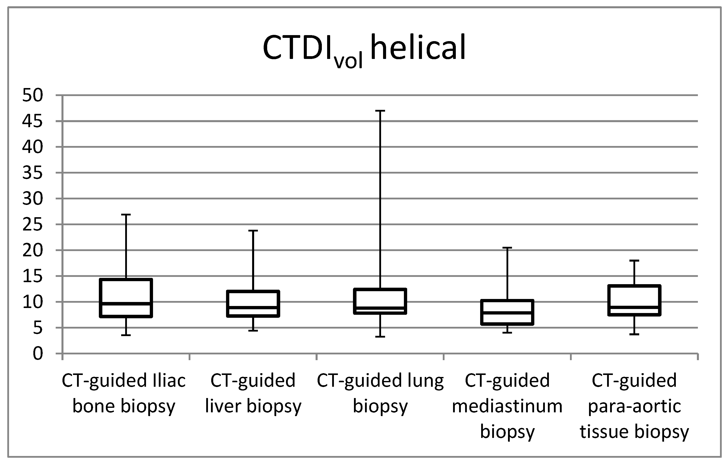
| CTDIvol helical (mGy) | 10.8 | 5.3 | 3.6 | 7.2 | 9.7 | 14.3 | 26.9 |
| CTDIvol biopsy (mGy) | 7.5 | 2.6 | 3.2 | 5.5 | 7.4 | 8.6 | 14.9 |
| DLPhelical (mGy cm) | 400.4 | 427.8 | 67.0 | 185.5 | 263.3 | 516.8 | 2743.9 |
| DLPbiopsy (mGy cm) | 272.0 | 181.6 | 75.6 | 129.6 | 224.0 | 343.2 | 905.0 |
| SSDEhelical (mGy) | 14.8 | 6.8 | 6.2 | 10.0 | 12.6 | 20.1 | 36.3 |
| SSDEbiopsy (mGy) | 9.9 | 2.9 | 5.2 | 8.0 | 9.9 | 11.1 | 17.4 |
| EDhelical (mSv) | 6.0 | 6.4 | 1.0 | 2.8 | 3.9 | 7.7 | 41.2 |
| EDbiopsy (mSv) | 4.0 | 2.7 | 1.1 | 1.8 | 3.2 | 5.0 | 13.6 |
| ESDhelical (mGy) | 13.0 | 6.4 | 4.3 | 8.6 | 11.6 | 17.2 | 32.3 |
| ESDbiopsy (mGy) | 8.9 | 3.2 | 3.9 | 6.4 | 8.8 | 10.2 | 17.9 |
| Mean | SD | Min | 25th Percentile | Median | 75th Percentile | Max | |
|---|---|---|---|---|---|---|---|
| CTDIvol helical (mGy) | 10.1 | 4.1 | 4.4 | 7.3 | 8.9 | 12.0 | 23.8 |
| CTDIvol biopsy (mGy) | 8.3 | 3.5 | 3.8 | 6.4 | 7.6 | 9.2 | 20.0 |
| DLPhelical (mGy cm) | 563.6 | 437.0 | 108.3 | 281.0 | 402.8 | 737.9 | 2417.0 |
| DLPbiopsy (mGy cm) | 403.9 | 800.5 | 44.8 | 149.9 | 246.0 | 387.0 | 6169.3 |
| SSDEhelical (mGy) | 13.2 | 4.7 | 6.4 | 9.8 | 11.8 | 16.5 | 27.6 |
| SSDEbiopsy (mGy) | 10.8 | 4.0 | 6.1 | 8.8 | 10.0 | 11.8 | 29.7 |
| EDhelical (mSv) | 8.5 | 6.6 | 1.6 | 4.2 | 6.0 | 11.1 | 36.2 |
| EDbiopsy (mSv) | 6.1 | 12.0 | 0.7 | 2.2 | 3.7 | 5.8 | 92.5 |
| ESDhelical (mGy) | 12.1 | 5.0 | 7.7 | 5.3 | 10.7 | 14.4 | 28.5 |
| ESDbiopsy (mGy) | 10.0 | 4.2 | 4.5 | 7.7 | 9.2 | 11.1 | 28.6 |
| Mean | SD | Min | 25th Percentile | Median | 75th Percentile | Max | |
|---|---|---|---|---|---|---|---|
| CTDIvol helical (mGy) | 10.5 | 5.9 | 3.2 | 7.8 | 8.8 | 12.4 | 47 |
| CTDIvol biopsy (mGy) | 6.0 | 1.8 | 2.6 | 4.7 | 5.6 | 7.5 | 12.4 |
| DLPhelical (mGy cm) | 711.2 | 374.6 | 141.5 | 452.9 | 659.4 | 849.8 | 1938.8 |
| DLPbiopsy9 (mGy cm) | 182.8 | 112.8 | 28.6 | 107.9 | 148.4 | 212.2 | 530.4 |
| SSDEhelical (mGy) | 14.7 | 11.9 | 4.9 | 10.4 | 12.0 | 16.5 | 97.8 |
| SSDEbiopsy (mGy) | 8.1 | 2.6 | 4.0 | 6.4 | 7.6 | 9.7 | 16.8 |
| EDhelical (mSv) | 10.0 | 5.2 | 2.0 | 6.3 | 9.3 | 11.9 | 27.1 |
| EDbiopsy (mSv) | 2.6 | 1.6 | 0.4 | 1.5 | 2.1 | 3.0 | 7.4 |
| ESDhelical (mGy) | 12.6 | 7.0 | 3.9 | 9.4 | 10.6 | 14.9 | 56.4 |
| ESDbiopsy (mGy) | 7.2 | 2.2 | 3.2 | 5.6 | 6.7 | 9.0 | 14.9 |
| Mean | SD | Min | 25th Percentile | Median | 75th Percentile | Max | |
|---|---|---|---|---|---|---|---|
| CTDIvol helical (mGy) | 9.1 | 4.4 | 4.0 | 5.7 | 7.9 | 10.3 | 20.5 |
| CTDIvol biopsy (mGy) | 6.4 | 2.6 | 3.7 | 4.6 | 5.6 | 7.1 | 15.1 |
| DLPhelical (mGy cm) | 489.6 | 578.6 | 102.1 | 154.4 | 388.6 | 613.7 | 3235.8 |
| DLPbiopsy (mGy cm) | 255.0 | 245.1 | 49.6 | 111.4 | 210.4 | 286.8 | 1236.5 |
| SSDEhelical (mGy) | 12.5 | 5.5 | 5.0 | 7.8 | 11.4 | 15.1 | 27.6 |
| SSDEbiopsy (mGy) | 8.7 | 2.8 | 5.0 | 6.6 | 7.8 | 10.0 | 16.3 |
| EDhelical (mSv) | 6.8 | 8.1 | 1.4 | 2.2 | 5.4 | 8.6 | 45.3 |
| EDbiopsy (mSv) | 3.6 | 3.4 | 0.7 | 1.6 | 2.9 | 4.0 | 17.3 |
| ESDhelical (mGy) | 10.9 | 5.3 | 4.8 | 6.8 | 9.5 | 12.3 | 24.6 |
| ESDbiopsy (mGy) | 7.7 | 3.1 | 4.5 | 5.5 | 6.7 | 8.5 | 18.1 |
| Mean | SD | Min | 25th Percentile | Median | 75th Percentile | Max | |
|---|---|---|---|---|---|---|---|
| CTDIvohelical (mGy) | 10.2 | 3.9 | 3.7 | 7.5 | 9.0 | 13.1 | 18.0 |
| CTDIvolbiopsy (mGy) | 9.1 | 5.0 | 3.5 | 6.3 | 7.7 | 10.4 | 25.0 |
| DLPhelical (mGy cm) | 641.3 | 592.4 | 107.5 | 254.8 | 453.4 | 797.9 | 2926.9 |
| DLPbiopsy (mGy cm) | 541.3 | 508.1 | 52.8 | 257.5 | 380.7 | 616.0 | 2546.7 |
| SSDEhelical (mGy) | 12.7 | 4.4 | 6.2 | 9.6 | 11.3 | 15.6 | 23.9 |
| SSDEbiopsy (mGy) | 11.0 | 5.0 | 5.9 | 8.2 | 9.4 | 12.5 | 24.2 |
| EDhelical (mSv) | 9.6 | 8.9 | 1.6 | 3.8 | 6.8 | 12.0 | 44.0 |
| EDbiopsy (mSv) | 8.1 | 7.6 | 0.8 | 3.9 | 5.7 | 9.2 | 38.2 |
| ESDhelical (mGy) | 12.2 | 4.7 | 4.5 | 9.0 | 10.7 | 15.7 | 21.6 |
| ESDbiopsy (mGy) | 10.9 | 6.0 | 4.2 | 7.6 | 9.2 | 12.4 | 29.9 |
Disclaimer/Publisher’s Note: The statements, opinions and data contained in all publications are solely those of the individual author(s) and contributor(s) and not of MDPI and/or the editor(s). MDPI and/or the editor(s) disclaim responsibility for any injury to people or property resulting from any ideas, methods, instructions or products referred to in the content. |
© 2023 by the authors. Licensee MDPI, Basel, Switzerland. This article is an open access article distributed under the terms and conditions of the Creative Commons Attribution (CC BY) license (https://creativecommons.org/licenses/by/4.0/).
Share and Cite
Siomou, E.; Filippiadis, D.K.; Efstathopoulos, E.P.; Antonakos, I.; Panayiotakis, G.S. Patient Dose Estimation in Computed Tomography-Guided Biopsy Procedures. J. Imaging 2023, 9, 267. https://doi.org/10.3390/jimaging9120267
Siomou E, Filippiadis DK, Efstathopoulos EP, Antonakos I, Panayiotakis GS. Patient Dose Estimation in Computed Tomography-Guided Biopsy Procedures. Journal of Imaging. 2023; 9(12):267. https://doi.org/10.3390/jimaging9120267
Chicago/Turabian StyleSiomou, Evangelia, Dimitrios K. Filippiadis, Efstathios P. Efstathopoulos, Ioannis Antonakos, and George S. Panayiotakis. 2023. "Patient Dose Estimation in Computed Tomography-Guided Biopsy Procedures" Journal of Imaging 9, no. 12: 267. https://doi.org/10.3390/jimaging9120267