Article

The Effects of Different Auxin–Cytokinin Combinations on Morphogenesis of Fritillaria meleagris Using Bulb Scale Sections In Vitro

by
Marija Marković
,
Milana Trifunović-Momčilov
,
Olga Radulović
,
Danijela M. Paunović
,
Dragana D. Antonić Reljin
,
Branka Uzelac
* and
Angelina Subotić
Department of Plant Physiology, Institute for Biological Research “Siniša Stanković”—National Institute of the Republic of Serbia, University of Belgrade, Bulevar Despota Stefana 142, 11000 Belgrade, Serbia
*
Author to whom correspondence should be addressed.
Horticulturae 2023, 9(8), 910; https://doi.org/10.3390/horticulturae9080910
Submission received: 16 July 2023 / Revised: 31 July 2023 / Accepted: 8 August 2023 / Published: 10 August 2023
(This article belongs to the Special Issue A New Decade in the Propagation of Horticultural and Medicinal Plants)

Abstract

:
Fritillaria meleagris is a horticulturally and medicinally valuable bulbous plant that requires a period of low temperatures for proper growth and flowering. Since conventional methods of propagation are ineffective and very slow, tissue culture techniques offer an integrated approach to mass production of this valuable geophyte. In this study, we investigated the effects of various auxin–cytokinin combinations on different morphogenetic pathways in bulb scale culture. Bulbs obtained in vitro were cut longitudinally, and bulb scales were cultured for four weeks at 7 °C on MS medium supplemented with 6-benzylaminopurine (BAP) in combination with 2,4-dichlorophenoxyacetic acid (2,4-D) or α-naphthaleneacetic acid (NAA) at different concentrations in order to investigate the influence of plant growth regulators (PGRs) on different morphogenetic responses. Regeneration percentage, number of shoots per explant, shoot length, number of bulbs and number of somatic embryos were monitored weekly. After chilling, bulb scales were transferred to 24 °C, and all parameters were recorded again. Low PGR concentrations were very effective for shoot multiplication, yielding up to 5.5 shoots per explant. 2,4-D (at 2 mg/L) in combination with low BAP (0.25 mg/L) produced the highest number of bulbs (11.00 ± 0.00), while PGR-free medium was extremely effective in somatic embryo formation (13.50 ± 2.90). Detached somatic embryos and bulblets continued to grow and develop on fresh PGR-free medium. We present data demonstrating that low auxin–cytokinin concentrations and PGR-free medium provide an effective method for a combined morphogenetic pathway in F. meleagris that is suitable for large-scale propagation.

1. Introduction

Fritillaria meleagris L. or snake’s head fritillary (Liliaceae) is a perennial bulbous plant, very attractive for its flowers and therefore valuable as an ornamental species. In addition to their great horticultural importance, these plants also have potential for medicinal use due to their phytochemical properties [1] thanks to different alkaloids, used for centuries as antitussive and expectorant agents, especially in Chinese medicine [2]. Most fritillary species are mainly distributed throughout the temperate climates of the northern hemisphere [3,4,5]. As most geophytes, they have a period of bulb dormancy, which enables them to survive unfavorable natural conditions in the form of dormant bulbs below the surface.
Propagation of these important plants is very slow and difficult using conventional methods (seeds and bulbs cuttings), and it takes several years for the initial seedlings to grow into mature plants [6].Tissue culture techniques can improve the regeneration potential, multiplication and large-scale production of this important plant [1,7]. Alternative propagation methods could ensure massive production of plants for continuing requests and growing demands [8]. Moreover, micropropagation requires a very small amount of plant material and facilitates the production of large numbers of homogenous plants year-round without harming the environment and natural populations of endangered plants that are difficult to propagate [9].
The success of plant tissue culture generally depends on the inclusion of plant growth regulators in the medium. The two main groups of phytohormones commonly used in tissue culture experiments include auxins and cytokinins [10]. Auxins, cytokinins and auxin–cytokinin interactions are considered to be the most important for regulating growth and development in plant tissue and organ cultures [11]. Different auxin types can generate different physiological responses in plant material, resulting in different regeneration efficiencies [12]. The most frequently used auxins in regeneration studies include 2,4-dichlorophenoxyacetic acid (2,4-D) and α-naphthaleneacetic acid (NAA) [7,12,13,14]. The most frequently used cytokinins for inducing plant regeneration are 6-benzylaminopurine (BAP) and thidiazuron (TDZ) due to the highest response in many plant species, particularly those recalcitrant to regeneration [15,16,17].
Studies have shown that various bulbous plants have great potential for regeneration from bulb scale explants [18]. The choice of plant growth regulators, their combinations and concentrations are critical for morphogenetic response (i.e., stimulating the formation of bulblets, roots, shoots, plantlets, protocorm-like bodies or somatic embryos) and plant regeneration capacity [19]. To date, numerous studies have examined the effects of various in vitro culture factors on the morphogenesis of different bulbous plants, especially bulblet multiplication [20,21,22,23,24,25,26]. Bulb scales were also a very attractive explant type for the induction of somatic embryogenesis [27]. Somatic embryogenesis often occurred simultaneously with organogenesis on the same explant under the same experimental conditions [28]. An efficient protocol for somatic embryogenesis results in rapid and effective proliferation of somatic embryos (SEs) that can be removed from the initial explant. Separated embryos have high potential for whole plant regeneration [8].
Propagation of Fritillaria by tissue culture techniques began with medically important species of the genus [29,30,31]. In most studies of Fritillaria regeneration, media with a low concentration of NAA were used in combination with BAP or KIN [27]. The most commonly used explants were bulb scales, whole bulbs and young leaves. Regeneration from bulbs and bulb scales depends on the Fritillaria species, the composition of the medium, and the combination of plant growth regulators and sugars. Bulb scales proved to be the most efficient explant type for many Fritillaria species [27], such as F. thunbergii [20], F. hupehensis [32] and F. meleagris [8]. Using parts or whole bulbs formed in vitro as starting material for initiating the regeneration process has many advantages, such as reducing contamination and destruction of natural populations [33]. The growth rate also increased with the duration of culture. For F. unibracteata cultured on medium supplemented with BAP and indole-3-acetic acid (IAA), the optimal time for bulb collection was after 50 days of culture [31]. In F. camtschatcensis, low concentrations of NAA (0.1–0.5 mg/L) had a strong effect on bulb formation (five bulbs per explant) after 40 days of culture [34]. A very large number of bulblets (14 bulblets per explant) were obtained from bulb scale sections of F. thunbergii on medium containing KIN [20].
We focused on plant regeneration from bulbs and bulb scale sections on different culture media as the most cost-effective and popular approach for bulb production [18]. Our previous studies have shown that cold treatment (at least 4 weeks) has a strong positive effect on dormancy breaking, bulb multiplication and rooting rate of F. meleagris in vitro [35,36]. Chilling treatment was also important for successful acclimatization of in vitro regenerated plantlets. Bulbs were generally stored at low temperature (4 °C) prior to regeneration experiments to improve regeneration capacity.
Previously, we investigated the potential for bulb regeneration on a culture medium supplemented with TDZ, where the maximum number of regenerated bulbs was obtained at a concentration of 0.05 mg/L [8] or with a combination of 2,4-D and kinetin (KIN) at 1 mg/L each, which produced the highest number of bulblets [14]. Moreover, somatic embryogenesis and whole plant regeneration of F. meleagris were achieved on medium containing 2,4-D (1 mg/L) or TDZ (0.1 or 0.2 mg/L) [8]. In addition to our laboratory, Kukulczanka et al. [1] reported regeneration of F. meleagris using NAA and BAP at concentrations of 1 and 2 mg/L, respectively, for optimal bulb production. Numerous procedures for in vitro regeneration, starting from various explants and using different regeneration pathways, differ in their requirements for these two classes of PGRs [13]. Therefore, PGR combinations may be further optimized for increased regeneration response.
In the present work, we explore the influence of different combinations of plant growth regulators on morphogenetic response in bulb scale segments of F. meleagris. PGRs were applied to stimulate the direct formation of bulbs and somatic embryos depending on the composition of the medium in an attempt to find a cost-effective method for a combined morphogenetic pathway that would be suitable for large-scale propagation of F. meleagris. We hope that our results will provide both practical and theoretical insights to improve the in vitro regeneration of F. meleagris, which could be useful for the production of other geophytes.
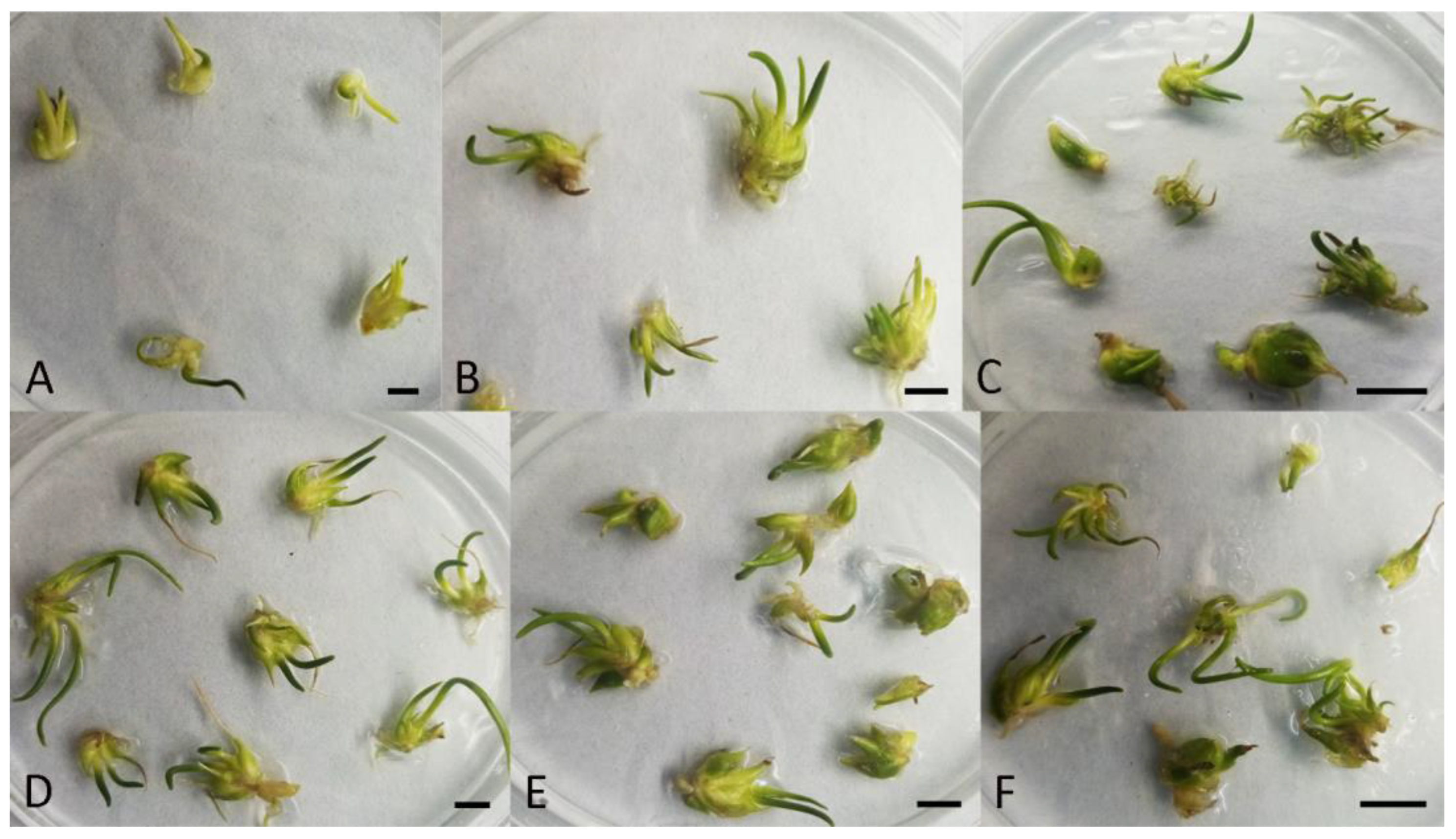
2. Materials and Methods

2.1. Plant Material, Culture Conditions and Explant Preparation

Bulb cultures of F. meleagris L. were established according to a previously published procedure [8]. Stock cultures were maintained on Murashige and Skoog (MS) medium [37] containing 30 g/L sucrose, 6 g/L agar and 1 mg/L TDZ for shoot and bulb multiplication. The pH value of the medium was adjusted to 5.8 with 1N NaOH before autoclaving at 114 °C for 25 min. Cultures were maintained under cool white fluorescent tubes with a photon flux density of 45 μmol m−2 s−1 and a 16 h photoperiod at 24 ± 2 °C. Regenerated in vitro bulblets were transferred to fresh PGR-free MS medium after two subcultures and grown for an additional two months. Newly formed bulbs (approximately 80–100 mg) were cut longitudinally into four sections (Figure 1A) and used as explants for further experiments (Figure 1B) in order to test the influence of PGRs on different morphogenetic responses.

2.2. Effect of PGRs on Explant Morphogenesis—Regeneration Procedure and Culture Conditions

Explants were cultured on MS medium supplemented with various combinations of auxins and cytokinins at different concentrations: either 6-benzylaminopurine (BAP) at 0.25 or 0.5 mg/L in combination with 2,4-dichlorophenoxyacetic acid (2,4-D) at 1, 2, 3, 4 or 5 mg/L, or α-naphthaleneacetic acid (NAA) at 0.25 or 0.5 mg/L in combination with BAP at 1, 2, 3, 4 or 5 mg/L. Basal MS medium without PGRs was used as control.
Initial explants (bulb scales) were cultured in Petri dishes (four explants per dish) in constant darkness at 7 °C for 4 weeks. During this time, detailed changes in morphogenetic potential were monitored weekly with the aid of a stereomicroscope. Any morphogenetic change in an explant (formation of shoot, bulb or SE) was considered regeneration, and each such explant was marked as responsive. At the end of each week, the number of responsive explants, the number of shoots per responsive explant and length of the longest shoot, as well as the number of bulbs and somatic embryos, were recorded for each treatment. After four weeks in the dark, bulb scales were transferred to fresh medium of the same composition and exposed to a 16 h photoperiod at 24 °C. The same morphogenetic parameters were reassessed after 4 weeks of cultivation at 24 °C.
At the end of the cold treatment, newly formed bulbs and somatic embryos (from the culture media shown to be effective for bulb/SE regeneration) were carefully separated from the initial explants, transferred to PGR-free medium and cultured at 24 °C for an additional 4 weeks to compare the development of SEs and bulbs in relation to their attachment to/detachment from the original explant.

2.3. Recordings and Data Analysis

Five replicates with four explants each were used per treatment, and the experiment was repeated (n = 40). Regeneration rates expressed as percentage of responsive explants, number of shoots per responsive explant and the longest shoot length, as well as total number of bulbs and somatic embryos per Petri dish, were evaluated weekly for each treatment. Percentage data were subjected to angular transformation prior to analysis. Statistical analyses were performed using Statistica 10 software (StatSoft, Hamburg, Germany). All results are presented as means ± standard errors. Data were subjected to analysis of variance (ANOVA), and the means of all morphogenetic parameters were separated using Fisher’s least significant difference (LSD) test at a confidence level of p ≤ 0.05.

3. Results

3.1. Effect of PGRs on Morphogenetic Response in Bulb Scales Cultured at 7 °C

The maximum regeneration percentage (100%) was recorded after only two weeks at 7 °C on medium supplemented with a combination of BAP and 2,4-D (Table 1). Within the same period, such a high regeneration percentage was not observed on a medium supplemented with the combination of BAP and NAA, where 87.50% was the highest regeneration percentage achieved (Table 2).
A very high regeneration percentage was achieved after 3 and 4 weeks at 7 °C (Table 3, Table 4, Table 5 and Table 6). After 4 weeks, the maximum regeneration percentage was reached for most of the tested combinations, while submaximal values were recorded only on media containing BAP 0.5 mg/L in combination with higher 2,4-D concentrations (3–5 mg/L, Table 5).
Shoots gradually developed on explants cultured on PGR-free medium at 7 °C. On the control medium, the number of shoots per explant increased with time and reached the maximum value at the end of the chilling experiment (Table 5 and Table 6). BAP in combination with 2,4-D was more effective in shoot multiplication, compared to BAP/NAA combination (Table 1 and Table 2). Initially, an increase in the number of shoots per explant was observed, especially at lower BAP concentrations coupled with higher 2,4-D concentrations (Table 1). However, as the chilling experiment progressed, substantially more shoots developed on explants cultured on control medium, such that the shoot proliferation recorded on BAP/2,4-D-supplemented media appeared less remarkable, with the highest concentrations of BAP (0.5 mg/L) and 2,4-D (3–5 mg/L) being the least effective (Table 3 and Table 5).
A medium devoid of PGRs also proved satisfactory for shoot elongation at 7 °C. In general, shoot length was not affected by the presence of BAP in combination with 2,4-D, except for the highest concentrations of BAP and 2,4-D, which had a diminishing effect. A statistically significant increase in shoot length was observed only in explants cultured at the highest BAP/NAA concentrations (5 mg/L BAP/0.5 mg/L NAA, Table 4 and Table 6).
After two weeks of cultivation at 7 °C, no new bulbs were induced in control explants. BAP at a concentration of 0.25 mg/L in combination with lower concentrations of 2,4-D had a stimulatory effect on bulb formation (Table 1). Among these treatments, 2,4-D at 2 mg/L was particularly beneficial, yielding the highest number of new bulbs (Table 3 and Table 5), with a maximum of 6.5 bulbs after 4 weeks at 7 °C (Table 5). However, the number of bulbs recorded for higher 2,4-D concentrations was not significantly different from the control until the end of the chilling experiment (Table 3 and Table 5). Higher concentrations of BAP in combination with NAA increased the number of induced bulbs after only 2 weeks (Table 2). This trend continued after 3 (Table 4) and 4 weeks (Table 6), although the average number of bulbs increased only slightly in the last week of these treatments.
Somatic embryos appeared at as early as 2 weeks of explant cultivation at 7 °C, both on PGR-free medium and on auxin/cytokinin-supplemented media (Table 1 and Table 2). The highest PGR concentrations of either auxin/cytokinin combination did not induce SEs throughout the 4-week period (Table 3, Table 4, Table 5 and Table 6). In general, BAP/NAA combination was more effective than BAP/2,4-D in SE induction. However, the number of somatic embryos recorded for any PGR treatment was not significantly different from the respective control.

3.2. Morphogenetic Changes Influenced by PGRs in Bulb Scales Cultured at 7 °C

Different morphogenetic pathways, affected by exogenously applied PGRs, emanated from single bulb scales after two weeks at 7 °C (Figure 2). Lower BAP/2,4-D concentrations led to regeneration of new bulbs (Figure 2A), whereas higher auxin concentrations induced formation of SEs as well as bulbs (Figure 2B). BAP below 4 mg/L in combination with NAA was not effective in bulb induction, but at the concentrations of 2 and 3 mg/L, BAP stimulated shoot proliferation and SE induction (Figure 2C–E), as did hormone-free medium (Figure 2F).
Further morphogenetic changes after three weeks for different PGR combinations are presented in Figure 3. Lower BAP/2,4-D concentrations resulted in an increase in the number of SEs (Figure 3A) and bulbs (Figure 3B) compared with two weeks of treatment. At this time point, bulbs on medium with higher 2,4-D concentrations increased in size considerably (Figure 3C,D), in contrast to BAP/NAA-treated bulbs, which developed longer sprouts (Figure 3F,G). In combination with NAA, the lower concentration of BAP (1 mg/L) did not induce SEs, but new bulbs were detected after three weeks (Figure 3E). Intermediate concentrations of BAP stimulated both shoot and SE development (Figure 3F–H). BAP at higher concentrations applied with NAA resulted in a huge increase in the number of bulbs after three weeks (Figure 3I). In control bulb-scale segments, the number of SEs continued to increase over time (Figure 3J). Prolonged cultivation in the dark resulted in a lighter color of the explants, while bulb scales showed visible necrotic changes along the edges. New bulbs had very small light-green sprouts.
After four weeks at 7 °C, low BAP/2,4-D concentrations further increased the number of bulbs and SEs (Figure 4A). Higher 2,4-D concentrations did not increase the number of induced SEs (Figure 4B). NAA combined with 2 mg/L BAP produced the highest number of SEs (Figure 4C). After four weeks in the dark, the color of new bulbs and SEs was almost white, while bulb scales had more necrotic tissue over a larger area. New bulbs had larger light-green sprouts with darker apex, and their size also increased during the time in the dark.

3.3. Effect of PGRs on Morphogenetic Response in Pre-Chilled Bulb Scales Cultured at 24 °C

Pre-chilled (4 weeks at 7 °C) bulb scales cultured at 24 °C for 4 weeks had the highest regeneration percentage for all PGR combinations, except for 2,4-D at concentrations above 3 mg/L in combination with BAP (~80%). All measured parameters were higher than under chilling conditions, as expected after a prolonged cultivation period (eight weeks in total). The number of shoots per explant followed the same pattern as during the chilling period. The highest number of shoots per explant was recorded on medium without PGRs as well as on media with low auxin/low cytokinin concentrations (Table 7 and Table 8). Lower auxin concentrations were also beneficial for shoot elongation. The highest number of bulbs was detected on media supplemented with PGRs at the lowest concentrations (0.25 mg/L BAP with 1–2 mg/L 2,4-D and 1 mg/L BAP with 0.25 mg/L NAA). However, PGR-free medium proved to be beneficial for SE induction. The highest BAP/NAA concentrations, for which no SEs were recorded throughout the chilling period, resulted in SE regeneration only at 24 °C (Table 8).

3.4. Morphogenetic Changes Influenced by PGRs in Pre-Chilled Bulb Scales Cultured at 24 °C

The combination of low cytokinin/low auxin, which stimulated the induction of new bulbs and SEs during chilling, positively affected the growth and development of these newly formed structures (Figure 5A,B). The initial explants appeared necrotic, whereas new bulbs and SEs became greener. New bulbs, even small ones, sprouted and grew after only 4 weeks at 24 °C. High auxin concentrations combined with low BAP induced regeneration of new bulbs and SEs at 24 °C (Figure 5C,D), in contrast to the same media during chilling. Sprouts were dark-green and appeared thicker than sprouts regenerated during the chilling period. Low BAP (1 mg/L) with NAA induced sprouting of new bulbs that regenerated at low temperature (Figure 5E). These bulbs were smaller, light-green and had thinner sprouts than bulbs formed at higher BAP concentrations. The size of SEs regenerated at low temperature on medium containing 2 mg/L BAP with NAA increased after 4 weeks at 24 °C (Figure 5F). In addition, the newly formed bulbs exhibited a number of green, thickened sprouts. A high concentration of BAP affected the growth of SEs and new bulbs with green elongated sprouts (Figure 5G). New SEs formed on explants after transfer to 24 °C, even those cultured on media with higher BAP concentrations (Figure 5H), which did not develop SEs during chilling. On media with the highest BAP concentration in combination with NAA, sprouts were shorter and thinner than on other PGR combinations (Figure 5I). Control bulbs cultured on PGR-free medium (Figure 5J) had elongated green sprouts similar in appearance to sprouts regenerated under a lower PGR combination.

3.5. Further Growth of Newly Formed Bulbs and SEs Excised from Pre-Chilled Bulb Scales

New bulbs and SEs, regenerated in bulb scale cultures on nutrient media of different compositions, were separated from the initial explants (bulb scales) and placed on medium without PGRs (Figure 6A). Their further development was monitored weekly during the four-week cultivation at 24 °C. Detached bulbs and SEs derived from explants previously treated with BAP/2,4-D at low concentrations developed rapidly (Figure 6B). A large number of new sprouts were observed after only one month. Similarly, prior treatment of the original explants with low BAP/NAA concentrations was beneficial for sprout induction and elongation (Figure 6C,D), as the newly formed sprouts were longer (visible to the naked eye) than those obtained with the BAP/2,4-D combination. Conversely, earlier treatment with higher BAP/NAA concentrations resulted in the formation of thicker, shorter sprouts (Figure 6E). New bulbs developed and grew faster than new SEs. SEs and bulbs excised from the original explants and cultured on PGR-free medium grew rapidly and formed the longest sprouts (Figure 6F). In general, the sprouted bulbs and developing SEs that were detached from the initial explants grew faster and formed more sprouts than those attached to the original explants.

4. Discussion

A very high percentage of regeneration (both bulblets and somatic embryos) was achieved in F. meleagris, even on PGR-free medium, after 4 weeks of cultivation at 7 °C. All explants were maintained at chilling temperature, as low temperatures have a positive effect on breaking dormancy in snake’s head fritillary [38]. Light did not affect the regeneration capacity of F. meleagris bulb scale sections, which had a very high regeneration percentage in the dark. Similar results were reported for F. thunbergii [20], in contrast to F. imperialis, where no regeneration occurred during cultivation in the dark [33]. However, Leshem et al. [21] demonstrated that light significantly affected bulblet growth, even though it did not affect regeneration capacity.
The highest regeneration rate (100% after only 2 weeks) in F. meleagris was observed on medium containing a combination of BAP and 2,4-D. Significantly lower regeneration rates were obtained with the combination of BAP and NAA, in contrast to Lilium longiflorum bulb scales grown under a combination of low BAP and low NAA for two weeks [39]. The presence of auxin and cytokinin is critical for shoot induction from bulbs, especially their optimal concentration in the culture medium [40,41]. The addition of a low concentration of auxin with a high concentration of cytokinin proved to be suitable for cell division and regeneration in vitro [42]. In F. meleagris, the number of shoots per explant was higher at low auxin/low cytokinin concentrations, which were also favorable for shoot elongation. The important role of auxins in shoot elongation has been described previously, as well as the beneficial effects of chilling and translocation of accumulated carbohydrate reserves from bulbs on this process [43,44]. Without chilling, bulbs showed no responsiveness to auxin, whereas the maximum response level increased with increasing duration of the cold treatment [43,45]. In contrast, the accumulation of cytokinins in bulbs was not temperature-dependent [45].
During chilling, PGRs accelerated the initiation of new shoots in snake’s head fritillary, especially BAP/NAA at low concentrations. NAA also affected shoot proliferation in Tulipa [46]. NAA with BAP at high concentration (4 mg/L) promoted direct shoot organogenesis in Lycoris sprengeri [47]. A similar observation regarding the initiation and development of microbulbs by the addition of PGRs was made in F. sonnikovae [48]. These authors also found that bulb scale was the best explant type for regeneration, exhibiting high morphogenetic activity on medium lacking PGRs. This is consistent with our finding that the number of shoots per explant at the end of the chilling experiment was not significantly increased by the addition of PGRs, and that the hormone-free medium proved satisfactory for shoot initiation in F. meleagris.
Bulblet multiplication capacity varies among different auxins, and their optimal concentration also varies among species. Bulblet formation in F. camtschatcensis after 40 days of culture was strongly influenced by NAA at low concentrations [34]. Organogenesis usually occurs when cytokinin concentration is higher than auxin concentration, which was exactly the case in our study on BAP/NAA-supplemented media during chilling. In our study, low NAA together with high cytokinin concentrations showed a promoting effect on bulblet regeneration in snake’s head fritillary. Such a dual, positive effect of auxin and cytokinin on in vitro bulb regeneration was also demonstrated in a number of bulbous plant species [24,26,47,49,50,51,52]. A relatively high number of bulbs in F. meleagris was also recorded at high levels of BAP, similar to L. sprengeri, where maximum bulblet proliferation was found at 4 mg/L BAP [47]. This is consistent with the findings of Nhut et al. [19], who demonstrated a direct relationship between cytokinin concentration and organ formation. However, BAP had no effect on bulblet regeneration in Lilium spp. [23] and even exerted an inhibitory effect in F. imperialis, where no bulblets formed on a medium containing BAP [53], and in Pancratium maritimum, where BA and NAA reduced the number of bulblets at any concentration [54].
The formation of new bulbs in F. meleagris at a higher concentration of BAP after only two weeks of cold treatment might be related to the effect of BAP on sucrose degradation and endogenous hormone interaction, and thus on the regeneration of new bulblets [55]. However, later, during cultivation at 24 °C, high cytokinin concentrations were no longer favorable for bulb induction, and the highest number of bulbs was obtained on a medium with a cytokinin concentration about eight-fold lower than that of auxin 2,4-D. The physiological response likely increased with increasing hormone concentration until explants reached the saturation point, as shown in Lilium ledebourii [41]. Some studies showed that increasing auxin and cytokinin concentrations above optimal levels had an inhibitory effect on the endogenous hormones of explants, resulting in a decrease in their morphogenetic response [56,57]. Exogenously applied PGRs can affect the levels of endogenous plant hormones by influencing their biosynthesis and distribution, thus altering in vitro development [58]. In F. meleagris, lower auxin/cytokinin concentrations were effective for bulb regeneration throughout the experiment, which could be attributed to adequate endogenous auxin/cytokinin content in the bulb scale explants.
In our study, medium without PGRs was very efficient for somatic embryogenesis in F. meleagris. PGR-free medium often proved to be more effective for plant morphogenesis than different PGR combinations in many Liliaceae and Amaryllidaceae species [18,25]. The high percentage of regeneration on hormone-free medium could be explained by high nutrient reserves in the scales. When propagated by scaling, these nutrient reserves strongly influenced the formation of bulblets [59]. In Lilium, the outer and middle scales tended to produce more bulblets, which researchers linked to the carbohydrate content of these scales [22]. In addition to the high nutrient reserves in the explants, the increased number of somatic embryos in F. meleagris on a medium lacking plant growth regulators could also be due to the endogenous auxin/cytokinin content, which could promote SE regeneration on the bulb scales. However, Asmita et al. [60] did not detect somatic embryos on transverse thin cell layer sections excised from in vitro bulb scales of Lilium on PGR-free medium.
In addition to PGR-free medium, low auxin/cytokinin combinations (0.25 mg/L BAP + 1 mg/L 2,4-D and 2 mg/L BAP + 0.25 mg/L NAA) had a positive effect on the number of SEs in snake’s head fritillary. A similar effect of these PGRs was observed in indirect somatic embryogenesis of African blue lily [61]. The efficacy of 2,4-D in somatic embryogenesis has already been demonstrated, as well as that of picloram and NAA, but different auxin types can generate different physiological responses in different plant species. In P. abies, 2,4-D treatment reduced embryogenic tissue proliferation, in contrast to NAA, and this reduction was related to the oxidative stress level, which was higher in the presence of 2,4-D in the proliferation medium [12]. In our study, where NAA was generally also more effective than 2,4-D in SE induction, callus formation was not observed with any auxin/cytokinin combination, suggesting direct organogenesis.
The morphogenetic pathway is strongly dependent on the genotype and the content of endogenous hormones in the explants [41]. In F. meleagris, the highest cytokinin concentrations negatively affected the formation of SEs, which could be due to the endogenous hormones in the bulbs, i.e., hormone imbalance caused by the addition of exogenous cytokinins at higher concentrations.
Somatic embryos of F. meleagris continued to develop, and future experiments will include their successful acclimatization under greenhouse conditions.

5. Conclusions

Our study has shown that the type and concentration of PGRs (auxin/cytokinin combination) in the culture media are very important for the morphogenesis of F. meleagris in bulb scale culture. For each tested auxin/cytokinin combination, the lowest auxin concentration was the most effective for shoot multiplication throughout the experiment but did not exceed the efficiency of PGR-free medium. Our results clearly indicate that in F. meleagris cultured in vitro, bulbing ability was relatively high in the absence of PGRs and for the most part unaffected by tested PGRs, regardless of auxin:cytokinin ratio. However, the lowest cytokinin concentration (0.25 mg/L BAP) with low auxin significantly increased bulb formation after transfer to a higher temperature. Medium without PGRs was extremely efficient for somatic embryogenesis throughout the experiment, as well as medium with lower PGR concentrations. The highest morphogenetic response (both bulb and SE formation) was obtained on BAP/2,4-D-containing medium with lowest PGR concentrations. Thus, we found that the use of bulb scales as initial explants and low auxin/low cytokinin concentrations as well as PGR-free medium provide an effective method for a combined morphogenetic pathway of organogenesis and somatic embryogenesis in F. meleagris that can accelerate the pace of large-scale propagation.

Author Contributions

Conceptualization, M.M. and M.T.-M.; methodology, M.M. and M.T.-M.; validation, M.M. and A.S.; formal analysis, M.M., M.T.-M., O.R. and B.U.; investigation, M.M., M.T.-M., D.M.P. and D.D.A.R.; resources, M.M. and A.S.; writing—original draft preparation, M.M.; writing—review and editing, A.S. and B.U.; visualization, M.M. and B.U.; supervision, M.M. and A.S.; project administration, M.M.; funding acquisition, A.S. All authors have read and agreed to the published version of the manuscript.

Funding

This work was supported by the Ministry of Science, Technological Development and Innovation of the Republic of Serbia, contract number 451-03-47/2023-01/200007.

Data Availability Statement

Not applicable.

Conflicts of Interest

The authors declare no conflict of interest. The funders had no role in the design of the study; in the collection, analyses, or interpretation of data; in the writing of the manuscript; or in the decision to publish the results.

References

  1. Kukulczanka, K.; Kromer, K.; Czastka, B. Propagation of Fritillaria meleagris L. through tissue culture. Acta Hortic. 1989, 251, 147–153. [Google Scholar] [CrossRef]
  2. Li, S.L.; Lin, G.; Chan, S.W.; Li, P. Determination of the major isosteroidal alkaloids in bulbs of Fritillaria by high-performance liquid chromatography coupled with evaporative light scattering detection. J. Chromatogr. A 2001, 909, 207–214. [Google Scholar] [CrossRef] [PubMed]
  3. Tatarenko, I.; Walker, K.; Dyson, M. Biological Flora of Britain and Ireland: Fritillaria meleagris. J. Ecol. 2022, 110, 1704–1726. [Google Scholar] [CrossRef]
  4. Jiang, R.; Zou, M.; Qin, Y.; Tan, G.; Huang, S.; Quan, H.; Zhou, J.; Liao, H. Modeling of the Potential Geographical Distribution of Three Fritillaria Species Under Climate Change. Front. Plant Sci. 2022, 12, 749838. [Google Scholar] [CrossRef] [PubMed]
  5. Protopopova, M.; Sandanov, D.; Pavlichenko, V.; Selyutina, I.; Stepanov, N. The Curious Case of Fritillaria sonnikovae (Liliaceae) in South Siberia: New Insights into Its Origin and Phylogeny. Diversity 2023, 15, 193. [Google Scholar] [CrossRef]
  6. Langens-Gerrits, M.; De Klerk, G.-J.; Croes, A. Phase change in lily bulblets regenerated in vitro. Physiol. Plant. 2003, 119, 590–597. [Google Scholar] [CrossRef]
  7. Subotić, A.; Trifunović, M.; Jevremović, S.; Petrić, M. Morpho-histological study of direct somatic embryogenesis in endangered species Fritillaria meleagris. Biol. Plant. 2010, 54, 592–596. [Google Scholar] [CrossRef]
  8. Petrić, M.; Subotić, A.; Jevremović, S.; Trifunović, M. Somatic embryogenesis and bulblet regeneration in snakehead fritillary (Fritillaria meleagris L.). Afr. J. Biotechnol. 2011, 10, 16181–16188. [Google Scholar] [CrossRef]
  9. Azeri, F.N.; Öztürk, G. Microbulb and plantlet formation of a native bulbous flower, Lilium monodelphum M. Bieb, var. Armenum, through tissue culture propagation. Biotechnol. Rep. 2021, 32, e00665. [Google Scholar] [CrossRef]
  10. Eudes, F.; Acharya, S.; Laroche, A.; Selinger, L.B.; Cheng, K.-J. A novel method to induce direct somatic embryogenesis, secondary embryogenesis and regeneration of fertile green cereal plants. Plant Cell Tissue Organ Cult. 2003, 73, 147–157. [Google Scholar] [CrossRef]
  11. Gaspar, T.; Kevers, C.; Penel, C.; Greppin, H.; Reid, D.M.; Thorpe, T.A. Plant hormones and plant growth regulators in plant tissue culture. In Vitro Cell. Dev. Biol.—Plant 1996, 32, 272–289. [Google Scholar] [CrossRef]
  12. Hazubska-Przybył, T.; Ratajczak, E.; Obarska, A.; Pers-Kamczyc, E. Different Roles of Auxins in Somatic Embryogenesis Efficiency in Two Picea Species. Int. J. Mol. Sci. 2020, 21, 3394. [Google Scholar] [CrossRef] [PubMed]
  13. Zdravković-Korać, S.; Milojević, J.; Belić, M.; Ćalić, D. Tissue Culture Response of Ornamental and Medicinal Aesculus Species—A Review. Plants 2022, 11, 277. [Google Scholar] [CrossRef] [PubMed]
  14. Petrić, M.; Subotić, A.; Jevremović, S.; Trifunović-Momčilov, M.; Tadić, V.; Grujić, M.; Vujčić, Z. Esterase end peroxidase isoforms in different stages of morphogenesis in Fritillaria meleagris L. in bulb scale culture. Comptes Rendus Biol. 2015, 338, 793–802. [Google Scholar] [CrossRef]
  15. Gairi, A.; Rashid, A. TDZ-induced somatic embryogenesis in non-responsive caryopses of rice using a short treatment with 2,4-D. Plant Cell Tissue Organ Cult. 2004, 76, 29–33. [Google Scholar] [CrossRef]
  16. Podwyszyńska, M.; Novák, O.; Doležal, K.; Strnad, M. Endogenous cytokinin dynamics in micropropagated tulips during bulb formation process influenced by TDZ and iP pretreatment. Plant Cell Tissue Organ Cult. 2014, 119, 331–346. [Google Scholar] [CrossRef]
  17. Banjac, N.; Vinterhalter, B.; Krstić-Milošević, D.; Milojević, J.; Tubić, L.; Ghalawenji, N.; Zdravković-Korać, S. Somatic embryogenesis and shoot organogenesis from the hypocotyl slices and free radical scavenging activity of regenerants of collard greens (Brassica oleracea L. var. acephala). Plant Cell Tissue Organ Cult. 2019, 137, 613–626. [Google Scholar] [CrossRef]
  18. Li, K.; Ren, H.; Zhao, W.; Zhao, X.; Gan, C. Factors affecting bulblet multiplication in bulbous plants. Sci. Hortic. 2023, 312, 111837. [Google Scholar] [CrossRef]
  19. Nhut, D.T.; Van Le, B.; Tran Thanh Van, K. Manipulation of the morphogenetic pathways of Lilium longiflorum transverse thin cell layer explants by auxin and cytokinin. In Vitro Cell. Dev. Biol.-Plant 2001, 37, 44–49. [Google Scholar] [CrossRef]
  20. Paek, K.Y.; Murthy, H.N. High frequency of bulblet regeneration from bulb scale section of Fritillaria thunbergii. Plant Cell Tissue Organ Cult. 2002, 68, 247–252. [Google Scholar] [CrossRef]
  21. Leshem, B.; Lilien-Kipnis, H.; Steinitz, B. The effect of light and of explant orientation on the regeneration and subsequent growth of bulblets on Lilium longiflorum Thunb, bulb-scale sections cultured in vitro. Sci. Hortic. 1982, 17, 129–136. [Google Scholar] [CrossRef]
  22. Park, N. Effect of temperature, scale position, and growth regulators on the bulblet formation and growth during scale propagation of Lilium. Acta Hortic. 1996, 414, 257–262. [Google Scholar] [CrossRef]
  23. Niimi, Y.; Nakano, M.; Isogai, N. Effects of temperature and illuminating conditions on regeneration and development of bulblets in scale culture of seven Lilium spp. J. Jpn. Soc. Hortic. Sci. 1999, 68, 28–34. [Google Scholar] [CrossRef] [Green Version]
  24. Tang, N.; Ju, X.; Hu, Y.; Jia, R.; Tang, D. Effects of Temperature and Plant Growth Regulators on the Scale Propagation of Lilium davidii var. unicolor. HortScience 2020, 55, 870–875. [Google Scholar] [CrossRef]
  25. Rice, L.J.; Finnie, J.F.; Van Staden, J. In vitro bulblet production of Brunsvigia undulata from twin-scales. S. Afr. J. Bot. 2011, 77, 305–312. [Google Scholar] [CrossRef] [Green Version]
  26. Khonakdari, M.R.; Rezadoost, H.; Heydari, R.; Mirjalili, M.H. Effect of photoperiod and plant growth regulators on in vitro mass bulblet proliferation of Narcissus tazzeta L. (Amaryllidaceae), a potential source of galantamine. Plant Cell Tissue Organ Cult. 2020, 142, 187–199. [Google Scholar] [CrossRef]
  27. Petrić, M.; Subotić, A.; Trifunović, M.; Jevremović, S. Morphogenesis in vitro of Fritillaria spp. Invited review. In Floriculture and Ornamental Biotechnology 6, Special Issue 1 Bulbous Ornamentals; Global Science Books: London, UK, 2012; pp. 78–89. [Google Scholar]
  28. Zhang, H.; Chen, Z.; Yang, L. Organ regeneration and somatic embryogenesis from young stems of Fritillaria sinica. J. West China Univ. Med. Sci. 1995, 26, 33–37. [Google Scholar]
  29. Sun, C.-S.; Chu, C.-C.; Wang, C.-C. Callus formation and organ regeneration in tissue culture of Fritillaria thunbergii Miq. J. Integr. Plant Biol. 1977, 19, 161–164. [Google Scholar]
  30. Zhao, G.F.; Cao, Y.; Wu, Y.; Fan, F.; Zhou, L.J.; Yang, W.H. Callus induction and organ regeneration in tissue culture of Fritillaria ussuriensis Maxim. Chin. Bull. Bot. 1983, 1, 40–41. [Google Scholar]
  31. Gao, S.L.; Zhu, D.N.; Cai, Z.H.; Jiang, Y.; Xu, D.R. Organ culture of a precious Chinese medical plant—Fritillaria unibracteata. Plant Cell Tissue Organ Cult. 1999, 59, 197–201. [Google Scholar] [CrossRef]
  32. Shiau, Y.-J.; Tai, C.-D.; Chen, C.-C.; Tsay, H.-S. Studies on the tissue culture of Fritillaria hupehensis Hsiao et K.C. Hsia. I. The induction of bulbscale and embryogenic callus from different source explants. J. Agric. Res. China. 2000, 49, 29–38. [Google Scholar]
  33. Witomska, M.; Lukaszewska, A. Bulblet regeneration in vitro from different explants of Fritillaria imperialis. Acta Hortic. 1997, 430, 331–338. [Google Scholar] [CrossRef]
  34. Otani, M.; Shimada, T. Micropropagation of Fritillaria camtschatcensis (L.) Ker-Gawl., “Kuroyuri”. Bull. Res. Inst. Agric. Sci. Ishikawa Agric. Coll. 1997, 5, 39–44. [Google Scholar]
  35. Nikolić, M.; Mišić, D.; Maksimović, V.; Jevremović, S.; Trifunović, M.; Subotić, A. Effect of low temperature on rooting rate and carbohydrate content of Fritillaria melegaris bulbs formed in culture in vitro. Arch. Biol. Sci. 2008, 60, 5P–6P. [Google Scholar] [CrossRef]
  36. Petrić, M.; Jevremović, S.; Trifunović, M.; Tadić, V.; Milošević, S.; Dragićević, M.; Subotić, A. The effect of low temperature and GA3 treatments on dormancy breaking and activity of antioxidant enzymes in Fritillaria meleagris bulblets cultured in vitro. Acta Physiol. Plant. 2013, 35, 3223–3236. [Google Scholar] [CrossRef]
  37. Murashige, T.; Skoog, F. A revised medium for rapid growth and bioassays with tobacco tissue cultures. Physiol. Plant. 1962, 15, 473–497. [Google Scholar] [CrossRef]
  38. Marković, M.; Trifunović-Momčilov, M.; Uzelac, B.; Jevremović, S.; Subotić, A. Bulb dormancy in vitro-Fritillaria melegaris: Initiation, release and physiological parameters. Plants 2021, 10, 902. [Google Scholar] [CrossRef]
  39. Li, L.-L.; Sun, S.-K.; Gu, H.; Chen, L.-J. Establishment of an Efficient Rapid Propagation System for Lilium × L. longiflorum Shenxiang No. 1. Res. J. Biotechnol. 2014, 9, 8–13. [Google Scholar]
  40. Jin, S.; Wang, J.; Wang, X.; Sun, D.; Li, G.; Genovesi, A.D.; Liu, S. Direct and indirect shoot and bulblet regeneration from cultured leaf explants of Lilium pumilum, an endangered species. In Vitro Cell. Dev. Biol.—Plant 2014, 50, 69–75. [Google Scholar] [CrossRef]
  41. Ghanbari, S.; Fakheri, B.A.; Naghavi, M.R.; Mahdinezhad, N. New protocol for the indirect regeneration of the Lilium ledebourii Bioss by using bulb explants. J. Plant Biotechnol. 2018, 45, 146–153. [Google Scholar] [CrossRef]
  42. Fatima, N.; Ahmad, N.; Anis, M. Enhanced in vitro regeneration and change in photosynthetic pigments, biomass and proline content in Withania somnifera L. (Dunal) induced by copper and zinc ions. Plant Physiol. Biochem. 2011, 49, 1465–1471. [Google Scholar] [CrossRef]
  43. Rietveld, P.L.; Wilkinson, C.; Franssen, H.M.; Balk, P.A.; van der Plas, L.H.V.; Weisbeek, P.J.; Douwe de Boer, A. Low temperature sensing in tulip (Tulipa gesneriana L.) is mediated through an increased response to auxin. J. Exp. Bot. 2000, 51, 587–594. [Google Scholar] [CrossRef] [Green Version]
  44. Khodorova, N.V.; Boitel-Conti, M. The Role of Temperature in the Growth and Flowering of Geophytes. Plants 2013, 2, 699–711. [Google Scholar] [CrossRef] [PubMed] [Green Version]
  45. Aung, L.H. Temperature regulation of growth and endogenous abscisic acid-like content of Tulipa gesneriana L. Plant Physiol. 1979, 63, 1111–1116. [Google Scholar] [CrossRef] [PubMed] [Green Version]
  46. Sochacki, D.; Marciniak, P.; Ciesielska, M.; Zaród, J.; Sutrisno. The Influence of Selected Plant Growth Regulators and Carbohydrates on In Vitro Shoot Multiplication and Bulbing of the Tulip (Tulipa L.). Plants 2023, 12, 1134. [Google Scholar] [CrossRef] [PubMed]
  47. Ren, Z.; Lin, Y.; Lv, X.; Zhang, J.; Zhang, D.; Gao, C.; Wu, Y.; Xia, Y. Clonal bulblet regeneration and endophytic communities profiling of Lycoris sprengeri, an economically valuable bulbous plant of pharmaceutical and ornamental value. Sci. Hortic. 2021, 279, 109856. [Google Scholar] [CrossRef]
  48. Kulkhanova, D.S.; Erst, A.A.; Novikova, T.I. In vitro Regeneration from Bulbous Scales of Fritillaria sonnikovae, an Endemic Species. Russ. J. Dev. Biol. 2015, 46, 215–221. [Google Scholar] [CrossRef]
  49. Kumar, S.; Awasthi, V.; Kanwar, J.K. Influence of growth regulators and nitrogenous compounds on in vitro bulblet formation and growth in oriental lily. Hort. Sci. 2007, 34, 77–83. [Google Scholar] [CrossRef] [Green Version]
  50. Salazar, C.; Landeros, F.; Bustos, E.; Bravo, P.; Pérez, C.; Becerra, J.; Ríos, D.; Uribe, M. Propagation and bulblet enhancement of Rhodophiala pratensis from seeds germinated in vitro. Cienc. Inv. Agr. 2019, 46, 12–22. [Google Scholar] [CrossRef]
  51. Ozel, C.A.; Khawar, K.M.; Unal, F. Factors affecting efficient in vitro micropropagation of Muscari muscarimi Medikus using twin bulb scale. Saudi J. Biol. Sci. 2015, 22, 132–138. [Google Scholar] [CrossRef] [Green Version]
  52. Kumar, S.; Chaudhary, V.; Kanwar, J.K. In vitro propagation of oriental hybrid lily from root explant. Adv. Hort. Sci. 2008, 22, 63–65. Available online: https://www.jstor.org/stable/42882613 (accessed on 7 August 2023).
  53. Hamidoghli, S.; Chamani, E.; Hamidoghli, Y.; Talei, N. Effect of different plant growth regulators on direct bulblet regeneration from scale explants of Fritillaria imperialis. J. Crop Prod. Process. 2015, 5, 211–218. [Google Scholar] [CrossRef] [Green Version]
  54. Dragassaki, M.; Economou, A.S.; Vlahos, J.C. Bulblet formation in vitro and plantlet survival extra vitrum in Pancratium maritimum L. Acta Hortic. 2003, 616, 347–352. [Google Scholar] [CrossRef]
  55. Ren, Z.; Xu, Y.; Lvy, X.; Zhang, D.; Gao, C.; Lin, Y.; Liu, Y.; Wu, Y.; Xia, Y. Early Sucrose Degradation and the Dominant Sucrose Cleavage Pattern Influence Lycoris sprengeri Bulblet Regeneration In Vitro. Int. J. Mol. Sci. 2021, 22, 11890. [Google Scholar] [CrossRef]
  56. Berrie, A.M.M. Germination and dormancy. In Advanced Plant Physiology; Wilkins, M.B., Ed.; Pitman Publishing Limited: London, UK, 1984; pp. 440–468. [Google Scholar]
  57. Bagheri, A.; Saffari, M. In Vitro Culture of Higher Plants, 4th ed.; Ferdowsi University of Mashhad: Vakilabad, Iran, 2009; p. 406. [Google Scholar]
  58. Ćosić, T.; Motyka, V.; Raspor, M.; Savić, J.; Cingel, A.; Vinterhalter, B.; Vinterhalter, D.; Trávníčková, A.; Dobrev, P.I.; Bohanec, B.; et al. In vitro shoot organogenesis and comparative analysis of endogenous phytohormones in kohlrabi (Brassica oleracea var. gongylodes): Effects of genotype, explant type and applied cytokinins. Plant Cell Tissue Organ Cult. 2015, 121, 741–760. [Google Scholar] [CrossRef]
  59. Kharrazi, M.; Tehranifar, A.; Nemati, H.; Bagheri, A. Vegetative propagation of Amaryllis (Hippeastrum× johnsonii) by different cutting methods. Hortic. Sci. Technol. 2017, 35, 373–380. [Google Scholar] [CrossRef] [Green Version]
  60. Asmita, S.; Sindhu, S.; Jayanthi, M.; Singh, M.K.; Dhiman, M.R. A novel regeneration protocol for LA Lilium. Indian J. Agric. Sci. 2020, 90, 1209–1211. [Google Scholar] [CrossRef]
  61. Yaacob, J.S.; Yussof, A.I.M.; Taha, R.M.; Mohajer, S. Somatic embryogenesis and plant regeneration from bulb, leaf and root explants of African blue lily (Agapanthus praecox ssp. minimus). Aust. J. Crop Sci. 2012, 6, 1462–1470. [Google Scholar]
Figure 1. Bulbs of F. meleagris regenerated for two months on MS medium without PGRs, cut longitudinally (A) and placed on MS medium with different combinations of PGRs (B). Scale bars = 10 mm.
Horticulturae 09 00910 g001
Figure 2. Morphogenesis of F. meleagris cultured at 7 °C for two weeks on a medium containing the following PGRs (in mg/L): (A) BAP 0.25 + 2,4-D 1; (B) BAP 0.5 + 2,4-D 4; (C) BAP 2 + NAA 0.25; (D) BAP 2 + NAA 0.5; (E) BAP 3 + NAA 0.25; (F) control bulb segments cultured on PGR-free medium. Note that explants and shoots are dark-green, while new bulbs and SEs appear lighter. Scale bars = 10 mm.
Horticulturae 09 00910 g002
Figure 3. Morphogenesis of F. meleagris cultured at 7 °C for three weeks on a medium containing the following PGRs (in mg/L): (A) BAP 0.25 + 2,4-D 1; (B) BAP 0.25 + 2,4-D 2; (C) BAP 0.5 + 2,4-D 4; (D) BAP 0.5 + 2,4-D 5; (E) BAP 1 + NAA 0.5; (F) BAP 2 + NAA 0.25; (G) BAP 2 + NAA 0.5; (H) BAP 3 + NAA 0.25; (I) BAP 4 + NAA 0.25; (J) Control bulb segments cultured on PGR-free medium. Scale bars = 10 mm.
Horticulturae 09 00910 g003
Figure 4. Morphogenesis of F. meleagris cultured at 7 °C for four weeks on a medium containing the following PGRs (in mg/L): (A) BAP 0.25 + 2,4-D 1; (B) BAP 0.5 + 2,4-D 4; (C) BAP 2 + NAA 0.25. Scale bars = 10 mm.
Horticulturae 09 00910 g004
Figure 5. Morphogenesis of pre-chilled F. meleagris bulb scales cultured at 24 °C for four weeks on a medium containing the following PGRs (in mg/L): (A) BAP 0.25 + 2,4-D 1; (B) BAP 0.25 + 2,4-D 2; (C) BAP 0.5 + 2,4-D 3; (D) BAP 0.5 + 2,4-D 4; (E) BAP 1 + NAA 0.5; (F) BAP 2 + NAA 0.25; (G) BAP 4 + NAA 0.25; (H) BAP 4 + NAA 0.5; (I) BAP 5 + NAA 0.5; (J) control bulb segments cultured on PGR-free medium. Scale bars = 10 mm.
Horticulturae 09 00910 g005
Figure 6. Bulbs and SEs removed from bulb scale sections, immediately upon excision (A) and after four weeks of cultivation on PGR-free medium at 24 °C (BF). Bulb scale sections were previously cultured at 7 °C (4 weeks) on a medium containing the following PGRs (in mg/L): (B) BAP 0.25 + 2,4-D 1; (C) BAP 1 + NAA 0.5; (D) BAP 2 + NAA 0.25; (E) BAP 4 + NAA 0.25; (F) PGR-free medium. Scale bars = 10 mm.
Horticulturae 09 00910 g006
Table 1. Effect of BAP and 2,4-D on morphogenesis of F. meleagris induced on bulb scale segments after two weeks at 7 °C.
Plant Growth Regulators (mg/L)Regeneration Percentage (%)Number of Shoots per ExplantShoot Length (mm)Number of BulbsNumber of SEs
BAP2,4-D
--56.25 ± 6.25 abc0.50 ± 0.00 ab5.75 ± 0.75 cd0.00 ± 0.00 a2.75 ± 1.03 c
0.25 181.25 ± 6.25 cde1.04 ± 0.14 abcd6.00 ± 1.35 cd1.50 ± 0.28 bc2.50 ± 0.86 bc
0.25 281.25 ± 6.25 cde0.53 ± 0.32 ab1.75 ± 1.18 a3.25 ± 0.25 d0.25 ± 0.25 a
0.25388.75 ± 6.57 de1.35 ± 0.41 cd3.25 ± 0.47 ab1.50 ± 0.28 bc1.25 ± 0.62 ab
0.25 475.00 ± 10.2 cde1.41 ± 0.21 cd4.50 ± 0.50 bc0.00 ± 0.00 a0.00 ± 0.00 a
0.25 5100.00 ± 0.00 e1.43 ± 0.25 cd6.00 ± 0.70 cd2.00 ± 0.40 c0.00 ± 0.00 a
0.51100.00 ± 0.00 e1.52 ± 0.22 d7.00 ± 0.70 d0.75 ± 0.47 ab0.25 ± 0.25 a
0.5 268.75 ± 15.72 bcd0.77 ± 0.17 abcd4.25 ± 0.85 bc0.00 ± 0.00 a0.00 ± 0.00 a
0.5 345.00 ± 2.88 ab1.29 ± 0.49 bcd2.50 ± 0.28 ab0.00 ± 0.00 a0.00 ± 0.00 a
0.5 431.25 ± 18.75 a0.64 ± 0.37 abc1.75 ± 1.03 a0.75 ± 0.47 ab1.25 ± 0.75 ab
0.5531.25 ± 6.25 a0.31 ± 0.06 a1.75 ± 0.25 a1.00 ± 0.57 b0.00 ± 0.00 a
* Values represent mean ± S.E. Different letters within columns indicate significant differences between treatments at p ≤ 0.05. Maximum values are in bold.
Table 2. Effect of BAP and NAA on morphogenesis of F. meleagris induced on bulb scale segments after two weeks at 7 °C.
Plant Growth Regulators (mg/L)Regeneration Percentage (%)Number of Shoots per ExplantShoot Length (mm)Number of BulbsNumber of SEs
BAPNAA
--56.25 ± 6.25 abc0.50 ± 0.00 ab5.75 ± 0.75 cd0.00 ± 0.00 a2.75 ± 1.03 bcd
10.2548.75 ± 18.52 abc1.27 ± 0.51 b2.25 ± 1.03 ab0.00 ± 0.00 a0.00 ± 0.00 a
10.523.75 ± 10.28 a0.44 ± 0.29 ab1.25 ± 0.62 a0.00 ± 0.00 a0.00 ± 0.00 a
20.2570.00 ± 23.80 bc0.64 ± 0.33 ab5.50 ± 2.06 cd0.00 ± 0.00 a4.50 ± 2.32 d
20.540.00 ± 20.00 ab1.33 ± 0.45 b3.00 ± 1.08 abc0.25 ± 0.25 a1.50 ± 0.95 ab
30.2541.25 ± 13.59 ab1.32 ± 0.46 b4.75 ± 1.03 bcd0.00 ± 0.00 a1.75 ± 1.18 abc
30.587.50 ± 7.51 c1.21 ± 0.12 b5.00 ± 0.40 bcd2.25 ± 0.25 b4.25 ± 0.25 cd
40.2522.50 ± 13.14 a0.17 ± 0.11 a1.75 ± 1.18 a1.50 ± 0.95 b1.25 ± 0.75 ab
40.525.00 ± 14.43 a0.45 ± 0.31 ab1.25 ± 0.75 a0.00 ± 0.00 a0.00 ± 0.00 a
50.2539.50 ± 6.27 ab1.16 ± 0.22 b3.00 ± 0.70 abc1.75 ± 0.25 b0.00 ± 0.00 a
50.573.75 ± 9.43 bc1.10 ± 0.24 b6.50 ± 0.50 d1.50 ± 0.50 b0.00 ± 0.00 a
* Values represent mean ± S.E. Different letters within a column indicate significant differences between treatments at p ≤ 0.05. Maximum values are in bold.
Table 3. Effect of BAP and 2,4-D on morphogenesis of F. meleagris induced on bulb scale segments after three weeks at 7 °C.
Plant Growth Regulators (mg/L)Regeneration Percentage (%)Number of Shoots per ExplantShoot Length (mm)Number of BulbsNumber of SEs
BAP2,4-D
--93.75 ± 6.25 c1.81 ± 0.25 cd12.75 ± 1.11 cde2.25 ± 1.03 abcd5.75 ± 1.54 d
0.251100.00 ± 0.00 c1.75 ± 0.25 cd15.33 ± 3.17 de3.00 ± 1.15 abcd4.66 ± 1.45 cd
0.25291.66 ± 8.33 bc1.33 ± 0.51 bcd7.00 ± 2.00 abc4.66 ± 0.33 d3.33 ± 1.20 c
0.25393.75 ± 6.25 c1.55 ± 0.21 cd15.25 ± 4.80 de3.50 ± 095 bcd2.75 ± 0.75 bc
0.254100.00 ± 0.00 c1.25 ± 0.10 bc7.50 ± 0.87 abc1.00 ± 0.41 a1.00 ± 0.00 ab
0.255100.00 ± 0.00 c1.74 ± 0.32 cd9.50 ± 2.90 bcd4.00 ± 0.71 cd0.00 ± 0.00 a
0.51100.00 ± 0.00 c2.00 ± 0.20 d17.00 ± 2.85 e2.00 ± 0.57 abc1.00 ± 0.71 ab
0.5293.75 ± 6.25 c1.43 ± 0.32 bcd9.50 ± 2.90 bcd1.25 ± 0.48 ab0.75 ± 0.25 ab
0.5372.50 ± 11.08 ab0.78 ± 0.08 ab3.75 ± 0.47 ab1.50 ± 0.28 ab0.00 ± 0.00 a
0.5460.75 ± 14.22 a0.78 ± 0.25 ab3.75 ± 0.75 ab2.25 ± 1.44 abcd0.50 ± 0.28 a
0.5556.25 ± 6.25 a0.43 ± 0.06 a3.00 ± 0.71 a2.50 ± 0.50 abcd0.00 ± 0.00 a
* Values represent mean ± S.E. Different letters within a column indicate significant differences between treatments at p ≤ 0.05. Maximum values are in bold.
Table 4. Effect of BAP and NAA on morphogenesis of F. meleagris induced on bulb scale segments after three weeks at 7 °C.
Plant Growth Regulators (mg/L)Regeneration Percentage (%)Number of Shoots per ExplantShoot Length (mm)Number of BulbsNumber of SEs
BAPNAA
--93.75 ± 6.25 b1.81 ± 0.25 cd12.75 ± 1.11 de2.25 ± 1.03 ab5.75 ± 1.54 c
10.2581.25 ± 11.96 ab0.92 ± 0.14 ab6.25 ± 2.01 abc2.75 ± 0.75 abc0.00 ± 0.00 a
10.565.00 ± 15.67 a0.65 ± 0.15 a3.50 ± 0.50 a2.00 ± 1.08 ab0.00 ± 0.00 a
20.25100.00 ± 0.00 b2.61 ± 0.38 d10.33 ± 0.33 bcde0.00 ± 0.00 a5.66 ± 1.66 c
20.586.66 ± 6.66 ab1.68 ± 0.78 bc12.33 ± 6.35 cde1.66 ± 0.33 ab4.66 ± 1.33 bc
30.2595.00 ± 5.00 b1.80 ± 0.28 cd8.00 ± 0.71 abcde2.75 ± 0.47 abc4.75 ± 1.11 c
3 0.5100.00 ± 0.00 b1.53 ± 0.24 bc14.00 ± 2.48 e3.75 ± 0.47 bc1.75 ± 0.75 a
4 0.2585.00 ± 9.57 ab0.90 ± 0.11 ab5.75 ± 1.44 abc5.50 ± 1.32 c2.25 ± 0.47 ab
40.587.50 ± 12.50 ab1.14 ± 0.08 abc4.00 ± 0.41 ab4.00 ± 1.22 bc0.00 ± 0.00 a
5 0.2579.00 ± 7.31 ab0.87 ± 0.15 ab6.75 ± 2.78 abcd3.75 ± 1.11 bc0.00 ± 0.00 a
50.583.75 ± 5.54 ab0.97 ± 0.18 ab20.25 ± 1.93 f5.25 ± 1.31 c0.00 ± 0.00 a
* Values represent mean ± S.E. Different letters within a column indicate significant differences between treatments at p ≤ 0.05. Maximum values are in bold.
Table 5. Effect of BAP and 2,4-D on morphogenesis of F. meleagris induced on bulb scale segments after four weeks at 7 °C.
Plant Growth Regulators (mg/L)Regeneration Percentage (%)Number of Shoots per ExplantShoot Length (mm)Number of BulbsNumber of SEs
BAP2,4-D
--93.75 ± 6.25 b2.31 ± 0.21 d15.25 ± 1.89 de3.00 ± 0.81 a7.00 ± 1.78 de
0.251100.00 ± 00.00 b2.58 ± 0.51 d19.66 ± 1.45 e6.33 ± 1.45 b8.66 ± 1.66 e
0.252100.00 ± 00.00 b1.62 ± 0.62 abcd12.50 ± 2.50 bcd6.51 ± 0.51 b5.00 ± 1.00 cd
0.253100.00 ± 00.00 b1.77 ± 0.10 bcd16.00 ± 4.41 de5.00 ± 1.47 ab4.00 ± 0.71 c
0.254100.00 ± 00.00 b1.45 ± 0.04 abc8.25 ± 1.11 abc2.25 ± 0.25 a2.00 ± 0.00 abc
0.255100.00 ± 00.00 b2.08 ± 0.40 cd13.00 ± 2.85 cde4.75 ± 1.03 ab1.25 ± 0.25 ab
0.51100.00 ± 00.00 b2.37 ± 0.29 d19.00 ± 2.04 e2.75 ± 0.25 a3.00 ± 1.68 bc
0.52100.00 ± 00.00 b2.06 ± 0.41 cd12.50 ± 3.22 cde2.75 ± 1.25 a1.75 ± 0.63 abc
0.53 77.50 ± 10.31 a0.83 ± 0.05 a5.00 ± 0.41 ab2.50 ± 0.64 a0.00 ± 0.00 a
0.5469.00 ± 10.84 a1.10 ± 0.31 ab5.00 ± 0.81 ab3.50 ± 1.50 ab1.25 ± 0.63 ab
0.5562.51 ± 7.21 a0.68 ± 0.12 a3.75 ± 0.75 a3.50 ± 0.64 ab0.00 ± 0.00 a
* Values represent mean ± S.E. Different letters within a column indicate significant differences between treatments at p ≤ 0.05. Maximum values are in bold.
Table 6. Effect of BAP and NAA on morphogenesis of F. meleagris induced on bulb scale segments after four weeks at 7 °C.
Plant Growth Regulators (mg/L)Regeneration Percentage (%)Number of Shoots per ExplantShoot Length (mm)Number of BulbsNumber of SEs
BAP NAA
--93.75 ± 6.25 bc2.31 ± 0.21 de15.25 ± 1.88 d3.00 ± 0.81 ab7.00 ± 1.78 d
10.2593.75 ± 6.25 bc1.29 ± 0.12 abc7.25 ± 2.62 abc5.50 ± 1.19 bc0.00 ± 0.00 a
10.576.25 ± 10.28 a1.01 ± 0.27 ab4.25 ± 0.62 a4.25 ± 0.85 abc0.00 ± 0.00 a
20.25100.00 ± 0.00 c3.00 ± 0.57 e13.66 ± 1.33 bcd1.66 ± 0.33 a7.66 ± 2.18 d
20.586.66 ± 6.66 abc1.83 ± 0.83 abcd14.00 ± 5.56 cd3.00 ± 0.57 ab7.00 ± 1.52 d
30.25100.00 ± 0.00 c2.17 ± 0.35 cde12.50 ± 3.01 bcd4.50 ± 0.64 bc6.50 ± 1.44 cd
30.5100.00 ± 0.00 c1.81 ± 0.37 bcd15.00 ± 2.91 d4.50 ± 0.64 bc3.25 ± 0.75 b
40.2590.00 ± 5.77 abc1.23 ± 0.08 ab7.25 ± 1.03 abc5.75 ± 1.11 c4.00 ± 0.40 bc
40.593.75 ± 6.25 bc1.21 ± 0.07 ab5.00 ± 0.00 a4.75 ± 0.85 bc0.00 ± 0.00 a
50.2579.00 ± 7.31 ab0.87 ± 0.15 a6.75 ± 2.75 ab3.75 ± 1.11 abc0.00 ± 0.00 a
50.593.75 ± 6.25 bc1.08 ± 0.23 ab22.25 ± 1.31 e5.50 ± 1.19 bc0.00 ± 0.00 a
* Values represent mean ± S.E. Different letters within a column indicate significant differences between treatments at p ≤ 0.05. Maximum values are in bold.
Table 7. Effect of BAP and 2,4-D on morphogenesis of F. meleagris induced in pre-chilled bulb scales cultured at 24 °C.
Plant Growth Regulators (mg/L)Number of Shoots per ExplantShoot Length (mm)Number of BulbsNumber of SEs
BAP2,4-D
--4.87 ± 0.38 e19.25 ± 1.49 bcd5.75 ± 1.03 abc13.50 ± 2.90 e
0.25 13.9 ± 0.73 de37.33 ± 6.22 e10.00 ± 2.88 de13.00 ± 2.64 de
0.25 22.25 ± 0.5 abc20.5 ± 9.5 cbd11.00 ± 0.00 e6.50 ± 1.50 bc
0.2532.73 ± 0.30 bcd16.66 ± 4.41 bcd6.66 ± 1.33 cd8.33 ± 0.88 cd
0.2542.04 ± 0.24 ab12.5 ± 2.32 abc2.75 ± 0.47 a4.00 ± 1.08 abc
0.2552.52 ± 0.53 bc20.25 ± 4.58 cd5.00 ± 1.08 abc2.50 ± 0.28 ab
0.514.12 ± 0.54 e23.25 ± 1.98 d3.25 ± 0.47 ab5.50 ± 0.55 bc
0.523.5 ± 1.00 cde23.00 ± 8.00 cd6.50 ± 0.50 bcd5.00 ± 1.00 abc
0.531.26 ± 0.26 a10.25 ± 1.79 ab3.50 ± 0.28 abc1.25 ± 0.94 a
0.541.52 ± 0.23 ab10.66 ± 2.96 abc6.66 ± 1.33 cd3.66 ± 1.33 abc
0.550.87 ± 0.16 a5.25 ± 1.37 a4.75 ± 0.75 abc0.00 ± 0.00 a
* Values represent mean ± S.E. Different letters within a column indicate significant differences between treatments at p ≤ 0.05. Maximum values are in bold.
Table 8. Effect of BAP and NAA on morphogenesis of F. meleagris induced in pre-chilled bulb scales cultured at 24 °C.
Plant Growth Regulators (mg/L)Number of Shoots per ExplantShoot Length (mm)Number of BulbsNumber of SEs
BAPNAA
--4.87 ± 0.38 c19.25 ± 1.49 bc5.75 ± 1.03 abc13.50 ± 2.90 e
10.252.15 ± 0.19 ab12.50 ± 3.21 ab8.50 ± 0.64 c0.00 ± 0.00 a
10.51.41 ± 0.21 a9.50 ± 0.95 a5.50 ± 0.95 ab2.75 ± 0.47 ab
20.255.47 ± 0.59 c23.33 ± 4.17 c3.00 ± 0.57 a15.00 ± 2.31 e
20.52.48 ± 0.91 ab19.00 ± 6.11 bc4.66 ± 1.20 ab9.00 ± 3.05 d
30.252.87 ± 0.37 b23.50 ± 3.71 c6.75 ± 1.11 bc7.50 ± 0.95 cd
30.52.82 ± 0.51 b19.75 ± 4.11 bc6.00 ± 0.57 abc5.50 ± 0.86 bcd
40.251.87 ± 0.21 ab10.00 ± 0.71 a6.50 ± 0.95 bc5.25 ± 0.75 bcd
40.51.60 ± 0.11 a8.25 ± 1.03 a6.25 ± 1.11 bc4.25 ± 1.03 bc
50.251.71 ± 0.17 a8.50 ± 2.32 a4.75 ± 1.31 ab3.75 ± 0.62 abc
50.51.90 ± 0.31 ab27.00 ± 1.22 c6.25 ± 1.11 bc4.25 ± 0.85 bc
* Values represent mean ± S.E. Different letters within a column indicate significant differences between treatments at p ≤ 0.05. Maximum values are in bold.
Disclaimer/Publisher’s Note: The statements, opinions and data contained in all publications are solely those of the individual author(s) and contributor(s) and not of MDPI and/or the editor(s). MDPI and/or the editor(s) disclaim responsibility for any injury to people or property resulting from any ideas, methods, instructions or products referred to in the content.

