Article

Bioefficacy of Lecanoric Acid Produced by Parmotrema austrosinense (Zahlbr.) Hale against Tea Fungal Pathogens

by
Kalidoss Rajendran
1,
Ponnusamy Ponmurugan
2,*,
Balasubramanian Mythili Gnanamangai
3,
Ponmurugan Karuppiah
4,*,
Mohammed Rafi Shaik
5,*,
Mujeeb Khan
5,
Merajuddin Khan
5 and
Baji Shaik
6
1
Department of Education, St. Joseph University in Tanzania, Dar Es Salaam P.O. Box 11007, Tanzania
2
Biomedical Research Lab, Department of Botany, Bharathiar University, Coimbatore 641046, Tamil Nadu, India
3
Department of Biotechnology, K.S. Rangasamy College of Technology, Tiruchengode 637215, Tamil Nadu, India
4
Department of Botany and Microbiology, College of Science, King Saud University, Riyadh 11451, Saudi Arabia
5
Department of Chemistry, College of Science, King Saud University, P.O. Box. 2455, Riyadh 11451, Saudi Arabia
6
School of Chemical Engineering, Yeungnam University, Gyeongsan 38541, Republic of Korea
*
Authors to whom correspondence should be addressed.
Horticulturae 2023, 9(6), 705; https://doi.org/10.3390/horticulturae9060705
Submission received: 10 May 2023 / Revised: 11 June 2023 / Accepted: 14 June 2023 / Published: 16 June 2023
(This article belongs to the Special Issue Mycorrhizal Roles in Horticultural Plants)

Abstract

:
Lichens are symbiotic organisms that are composed of fungal partners and photosynthetic algal partners. During the symbiotic process in lichen thallus, the fungus synthesizes certain secondary metabolites in which lecanoric acid is very important in terms of antibiotic properties. Considering the vital importance of lecanoric acid, the present study aimed to produce lecanoric acid from the thallus of Parmotrema austrosinense lichen using Modified Bold’s basal salt medium and evaluate the bio-efficacy against tea fungal pathogens. Lecanoric acid was purified and confirmed by micro-crystallization method and subsequently bioassayed against tea fungal pathogens. The results revealed that lecanoric acid registered a significant antifungal activity in terms of the growth inhibition of test pathogens. Companion systemic and botanical fungicides were found to be inferior to lecanoric acid in the percentage of growth inhibition. The inhibition rate varied among tea pathogens. Of the tea pathogens tested, tea leaf disease-causing pathogens including Cercospora theae (C. theae), Glomerella cingulata (G. cingulate), and Phomopsis theae (P. theae) showed the highest percentage of growth inhibition followed by stem and root rot diseases. The present study suggests that lecanoric acid showed an inhibitory effect against tea pathogens, which might be due to antibiotic properties and fungicidal action of lecanoric acid.

1. Introduction

Lichens are remarkable self-sustaining symbiotic partnerships between photoautotrophic algae partners known as phycobionts and fungal partners known as mycobionts. There are over 20,000 different species of lichens in the globe, with about 2300 of them found in India. Of these, Tamil Nadu has the most lichens (785 taxa), followed by Andhra Pradesh (656 taxa), and Karnataka (612 taxa). This is a result of the fact that the Western and Eastern Ghats, which cover vast areas, contain several biodiversity hotspots [1]. According to reports, lichens can flourish in habitats that include rock (saxicolous), soil (terricolous), and bark (corticolous). Lichens belong to the category of the slowest growing plants, which possess an extraordinary tolerance towards adverse atmospheric conditions [2]. Lichens are known to produce a variety of unique secondary metabolites which amount to a total of approximately 1050 various bioactive compounds. Among these bioactive compounds, over 550 compounds are unique to lichens and are not present in any other species [3]. Bioactive substances obtained from lichen thallus exhibit a variety of biological functions, and these substances demonstrate excellent antimicrobial properties and thus can be considered as potential antibiotic substitutes. Lichen bioactive substances are found to provide a wide range of therapeutic benefits. More than 50% of the known lichen species exhibited medicinal values [4]. The large percentage of lichen components have a broad range of biological activities, including anti-cancer, anti-inflammatory, anti-arthritis, anti-analgesic, anti-pyretic, and anti-proliferative activity [5,6]. Sati and Joshi [7] determined the antibiotic and antifungal activities of Indian lichens against plant pathogens. The bioactive secondary metabolites of lichens include depsides and dibenzofuran, which are mostly phenolic compounds such as orcinol and β-orcinol derivatives, usnic acid, barbatic acid, salazinic acid, picrolichenic acid, protolichesterinic acid, parietin, vulpinic acid, etc. Apart from these, the depsides of lichens from other resources also include atranorin, evernic acid, and lecanoric acid, which have been known to possess excellent bioactivities including antioxidant, antimicrobial, and anticancer properties [8,9]. Particularly, lecanoric acid, which belongs to the compounds of the orcinol series, has been used as an antioxidant and antimicrobial agent [10]. For instance, in a recent study, lecanoric acid isolated from the lichen of Parmelia cetrata induced 100% growth inhibition of A. fischeri at a concentration of 100 µM [11,12].
The young shoots of commercially grown tea plants (Camellia sinensis (L.) O. Kuntz) are used to make tea, which is the most consumed and cheapest hot beverage in the world. More than 50 countries around the world have established plantations where tea is grown. They favor warm, humid weather with evenly distributed rainfall and lengthy periods of sunshine. It provides a stable environment for housing a variety of microorganisms in the rhizosphere of tea bushes because it is a monocultural and perennial crop. Among the tea pathogenic organisms, stem diseases such as wood rot, branch canker, collar canker, and thorny stem blight disease, leaf diseases such as bird’s eye spot, brown blight and grey blight, and red root rot disease are very important because they affect the tea bush health and green leaf yield [13]. These diseases are a major issue in all regions of the world where tea is grown, resulting difficulties in replanting and fresh clearings [14]. According to reports, the majority of tea infections are fungi, and more than 300 fungi species have been identified in tea bushes. Crop loss due to stem and root diseases is substantial as it leads to capital losses in the tea industry. Leaf diseases are very important due to tea plants being being cultivated for harvesting young succulent leaves, which in turn affects the overall quality of tea powder [15].
Tea diseases can be effectively controlled through antimicrobial compounds secreted by normal soil microorganisms and medicinal plants. This is because antimicrobial compounds can control the growth of phytopathogens very effectively by means of antibiosis and hyper-parasitism mechanisms. However, only meager information is available on the biological control of tea pathogens rather than using antimicrobial novel compounds [16]. It has been reported that interaction between several tea pathogens and antagonists of bacterial, fungal, and actinomycete origin were studied in vitro through dual culture and antibiosis techniques, which revealed the pathogen growth was suppressed considerably [17].
The emergence of deadly plant pathogens and the persistent development of pesticide-resistant strains have adversely affected global agricultural production. To overcome these problems, the development of novel antifungal agents with effective bioactivities and unique modes of action for the inhibition of targeted plant pathogens is highly desirable. In this context of the present study, attempts were made to control leaf, stem, and root diseases caused by a variety of different fungal pathogens including Hypoxylon serpens, Macrophoma theicola, Phomopsis theae, Tunstallia aculeate, Cercospora theae, Glomerella cingulate, Pestalotiopsis theae, and Poria hypolateritia by using purified lecanoric acid extracted from the thallus of the P. austrosinense lichen. All the experiments were performed under in vitro conditions in triplicate.

2. Results

The thallus of P. austrosinense lichen is pale grey in color, foliose type, corticated on both upper and lower surfaces, and has brownish unbranched rhizines (root-like extension, attachment structure) and sorediate margins without apothecia in the thallus (Figure 1A). The lichen thallus was identified by the spot color test which showed K, C+, and KC+ medulla. The mycobiont of P. austrosinense was able to grow well in MBBS solid and liquid media (Figure 1B,D). The colony margin was regular and hyaline initially and later changed to white with an irregular margin.
Lecanoric acid was produced by mycobionts of P. austrosinense in laboratory conditions cultured on MBBS broth that had been amended with sucrose, nitrate, and potassium as sources of carbon, nitrogen, and potassium, respectively. Produced lecanoric acid was recognized by the micro-crystallization method. The lecanoric acid from the mycobionts of P. austrosinense showed long needles with slightly curved crystals in nature (Figure 1L). The extract of Parmotrema austrosinense mycobionts was purified by silica gel column chromatography using 5% ethyl acetate in benzene with the molecular formula of C16H14O7.

2.1. Antifungal Activity of Lecanoric Acid

The antifungal activity of lecanoric acid, which was isolated from the mycobiont culture filtrate of P. austrosinense, was examined against selected tea fungal pathogens. The results showed that lecanoric acid exhibited significant antifungal activity in terms of the growth inhibition of test pathogens rather than the companion and botanical fungicides. The results further revealed that a prominent inhibition zone was formed around the well loaded with different concentrations of lecanoric acid. Among the different tea pathogens tested for bioefficacy of lecanoric acid, tea leaf disease-causing pathogens such as C. theae, G. cingulate, and P. theae showed the highest percentage of growth inhibition followed by stem pathogens such as H. serpens, M. theicola, and P. theae. Whereas, tea root rot-causing pathogen P. hypolateritia was had the least growth inhibition (Figure 2). Of the tested tea leaf disease-causing pathogens, C. theae was noticed with a maximum percentage of inhibition growth followed by P. theae and G. cingulate. On the other hand, there was no significant difference among tea stem disease-causing pathogens tested in terms of growth inhibition. The fungicides such as expel, nimbicidine, and nimbidoxin were found to be less effective than lecanoric acid for controlling fungal pathogen growth inhibition (Figure 2).
The antifungal activity of different concentrations of lecanoric acid extracted from P. austrosinense lichen on the growth inhibition of tea fungal pathogens was studied (Table 1). The results indicated that a 15 μL concentration of lecanoric acid was an efficient concentration for tea fungal pathogen growth inhibition. Growth inhibition of tea pathogens was exponentially increased from 5 μL up to 15 μL concentration of lecanoric acid, which then declined at 20 μL concentrations. C. theae registered the maximum percentage of growth inhibition at about 91.5% followed by 90% with P. theae at a 15 μL concentration. On the other hand, only a marginal difference was noticed with stem and root disease-causing pathogens between various concentrations of lecanoric acid tested. The effect of different solvents on the dissolving of lecanoric acid on growth inhibition of tea fungal pathogens was studied (Table 2). The results indicated that methanol was better than ethanol, acetone, and petroleum, but water showed the least performance in dissolving lecanoric acid when bioassay against tea fungal pathogens.

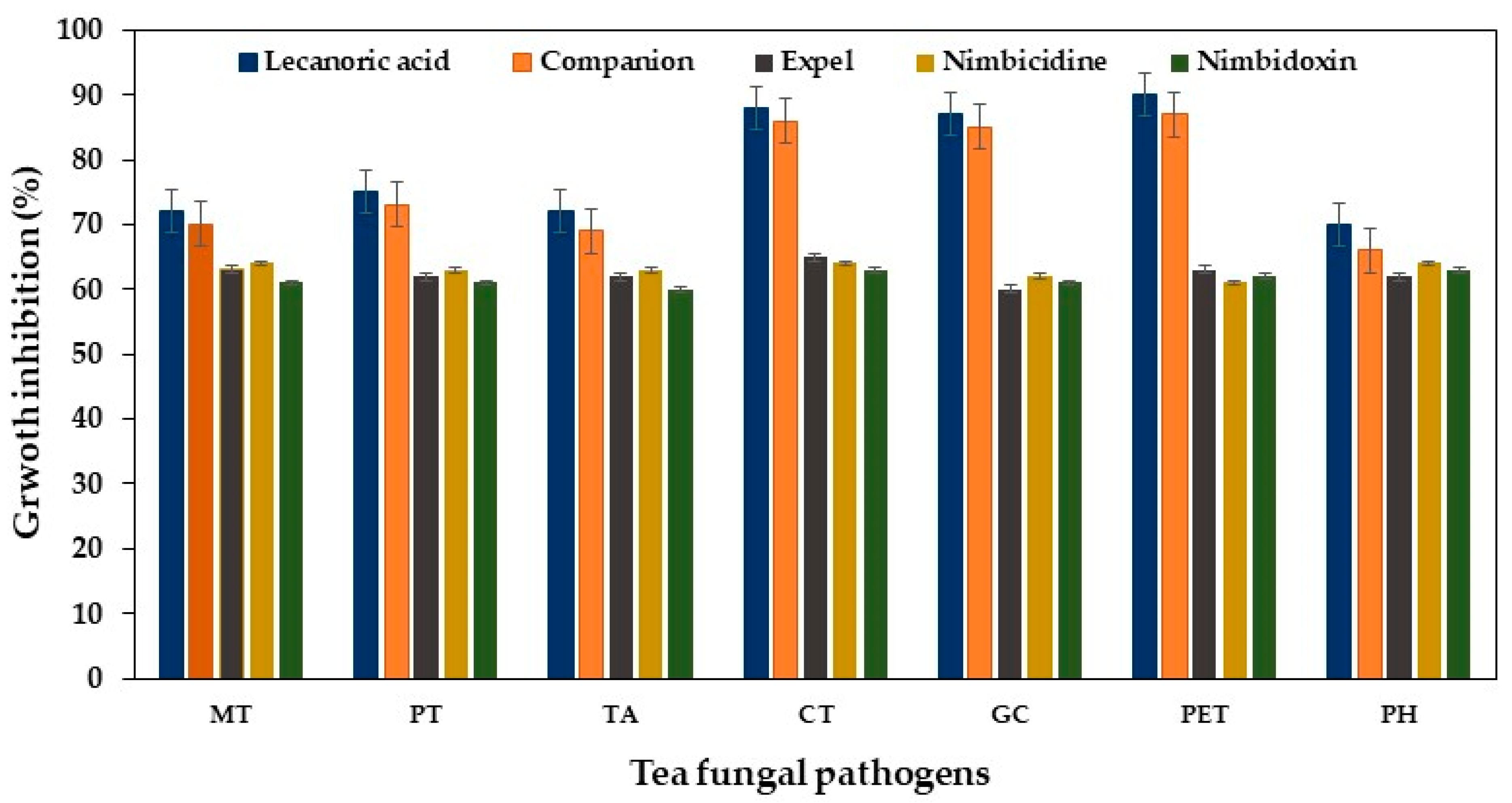
2.2. Suppressive Effect of Systemic and Botanical Fungicide

Similarly, various concentrations of companion systemic fungicide were evaluated to find the optimum concentration on the percentage of growth inhibition of test pathogens (Table 3). The results showed that a 20 μL concentration of companion was found to be optimum in inhibiting the growth of tea pathogens. Similar to that of growth inhibition due to lecanoric acid, C. theae registered the maximum percentage of growth inhibition at about 85.7% followed by 83.5% with P. theae at 20 μL concentration. P. hypolateritia recorded the least percentage of growth inhibition about 68.5% at 20 μL concentration. A progressive increase in the growth inhibition percent of the pathogens was observed with an increase in the concentration of the fungicide (Table 3). Amongst the botanical fungicides evaluated, nimbicidine was highly effective against pathogens, followed by expel and nimbidoxin. However, they were found to be inferior to lecanoric acid and companion in terms of the percentage of growth inhibition of tea fungal pathogens (Figure 2).

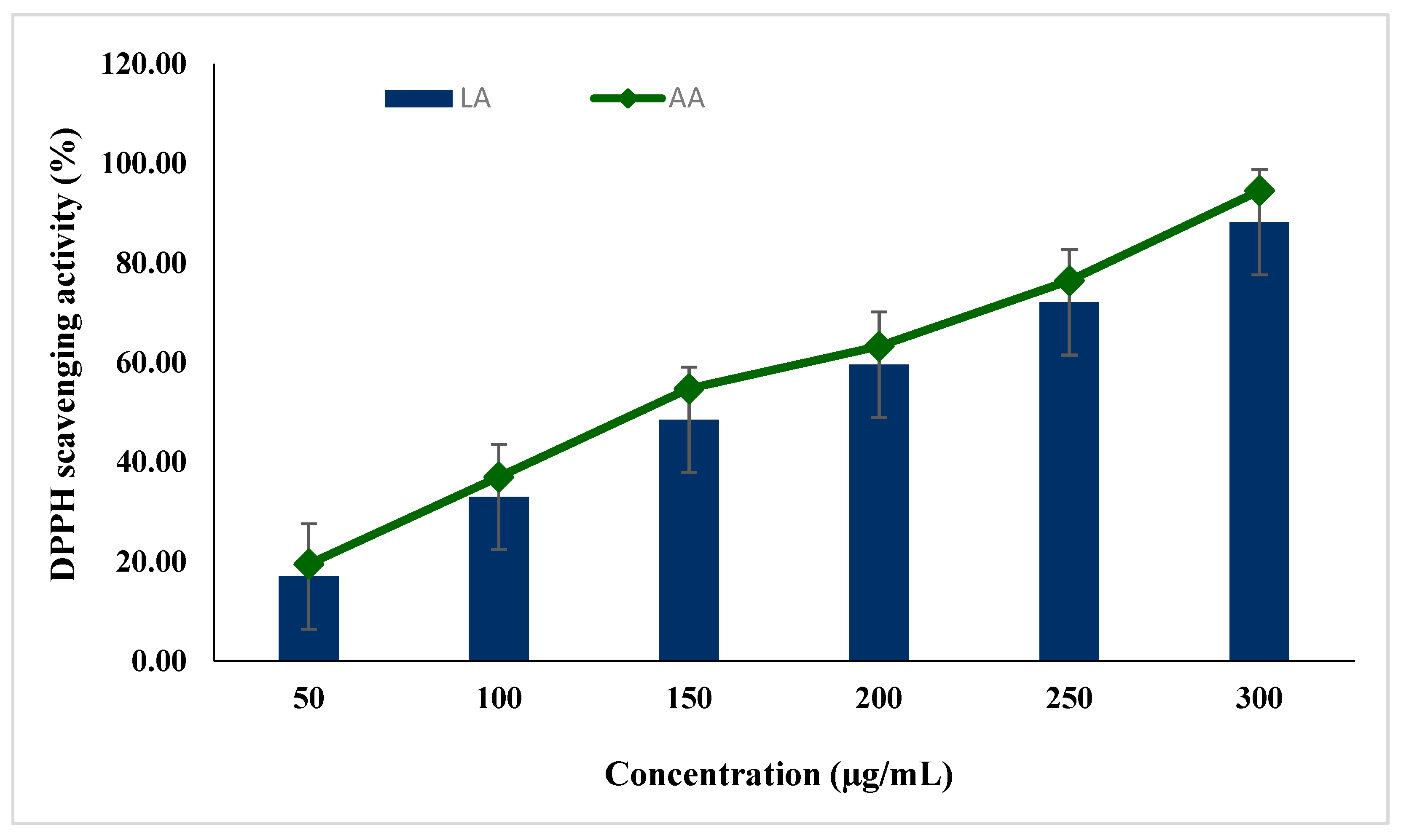
2.3. Free Radical Scavenging Activity of Lecanoric Acid

Lecanoric acid (LA) exhibited free radical scavenging activity, as presented in Figure 3. The IC50 % was attained at 54.75% at the concentration of 150 µg/mL compared to standard ascorbic acid (AA). The study also revealed free radical scavenging activity was dose-dependent for both standard and lecanoric acid.

3. Discussion

Since the first discovery of the antimicrobial properties of lichen extract (thalli), which was reported by Burkholder et al. (1944), a significant number of studies have been published which have highlighted the biological potential of purified lichen substances as well as lichen extracts [10]. However, most of the published studies have mainly focused on crude lichen extracts for comparative study on antimicrobial activities; thus, there is a growing demand for the investigation of the biological properties of purified lichen substances against specific organisms including plant pathogens [18].
Lichens are successful alliances of dual organisms with the combinations of mycobionts (fungal partner) and phycobionts (algal partner) coexisting together to form a single thallus [1]. The major role of an algal partner is to synthesize food materials for the growth of fungal partners. The fungal partners are crucial in the production of a variety of intracellular and extracellular antimicrobial bioactive compounds. P. austrosinense belonging to the Parmeliaceae family has diverse pharmacological activity. They are most commonly found on rocks and trees in open habitats at Kodaikanal hills of Tamil Nadu, India [19]. P. austrosinense lichen grew well in MBBS solid and liquid media (Figure 1B,D). The mycobiont of P. austrosinense produced lecanoric acid in MBBS broth amended with sucrose and potassium nitrate sources. Lecanoric acid was identified through the micro-crystallization method, which exhibited needle-like and slightly curved crystals (Figure 1C). A naturally occurring depside antibiotic substance called lecanoric acid was produced and extracted from a range of foliose and fruticose lichens. It is an effective antioxidant that outperforms ascorbic acid in the DPPH free radical scavenging experiment [20].
The antifungal activity of lecanoric acid extracted from P. austrosinense was evaluated against tea fungal pathogens such as H. serpens, M. theicola, P. theae, and T. aculeate belonging to stem disease, C. theae, G. cingulate, and P. theae belonging to leaf disease and P. hypolateritia of red root rot disease (Figure 2). Lecanoric acid inhibited the growth of tea fungal pathogens significantly when compared to companion and botanical fungicides. All the three biofungicides including expel, nimbicidine, and nimbidoxin were found to be inferior to lecanoric acid and companion systemic fungicides with respect to the percentage of growth inhibition. The results showed that various concentrations of lecanoric acid, companion systemic fungicides, and botanical fungicides inhibited the growth of tea pathogens in varying degrees (Table 1 and Table 3). The zone of inhibition formed around the fungal pathogens was purely due to lecanoric acid and its antifungal properties. Similar studies were conducted by Nepolean et al. [21], which revealed that expel botanical fungicide showed good results against wood rot pathogen. The most widely accepted hypothesis, according to Demain and Fang (1995), is that pathogenic microbes compete with antimicrobial chemicals in environments where nutrients are limited. The bio-efficacy of botanical fungicides against the wood rot disease of tea was evaluated, as they are safe and eco-friendly [21].
Lichen metabolites have relatively low molecular weights and are crystallized on the hyphal cell walls of plant pathogenic microorganisms. The superior efficacy of lecanoric acid in suppressing pathogens by inhibiting mitosis processes such as carbendazim fungicide [22] and other metabolic processes was reported earlier [23]. The growth inhibition caused by lecanoric acid may be directly related to their inhibitory effects in several signaling cascades involved in the pathophysiology and progression of the disease, which are still poorly understood. Plant compounds as antimicrobial agents exhibit positive features, as they are of natural origin and are safer for the environment and they can reduce the development of disease resistance by pathogenic microorganisms [24]. Due to their significant efficiency over synthetic compounds against pathogenic infections, lichen metabolites are gaining popularity over previously used agrochemicals such as bio-fungicides (Huneck 1999). Lecanoric and orsellinic acid, which are renowned for their antifungal characteristics, were found to be responsible for the P. tinctorum methanol extract’s substantially superior efficiency against plant pathogenic fungi according to Tiwari et al. [25].
The antioxidant activity of lecanoric acid showed moderate activity, while, comparing the IC50 = 150 µg/mL of the earlier study, it was in a significant concentration (IC50 = 50.00 µg/mL) compared with other reported lichen species extracts [26,27]. The free radical is generated endogenously by a physiological process; it decreases the absorbance and indicated the uptake of superoxide anion in the test solution. Thus, it exhibits a concentration-dependent increase in the radical scavenging activity and needs of the detoxification process. Finally, the current results specified that the lecanoric acid extracted from lichens would be a significant source of food ingredients for oxidative stress for human health.

4. Materials and Methods

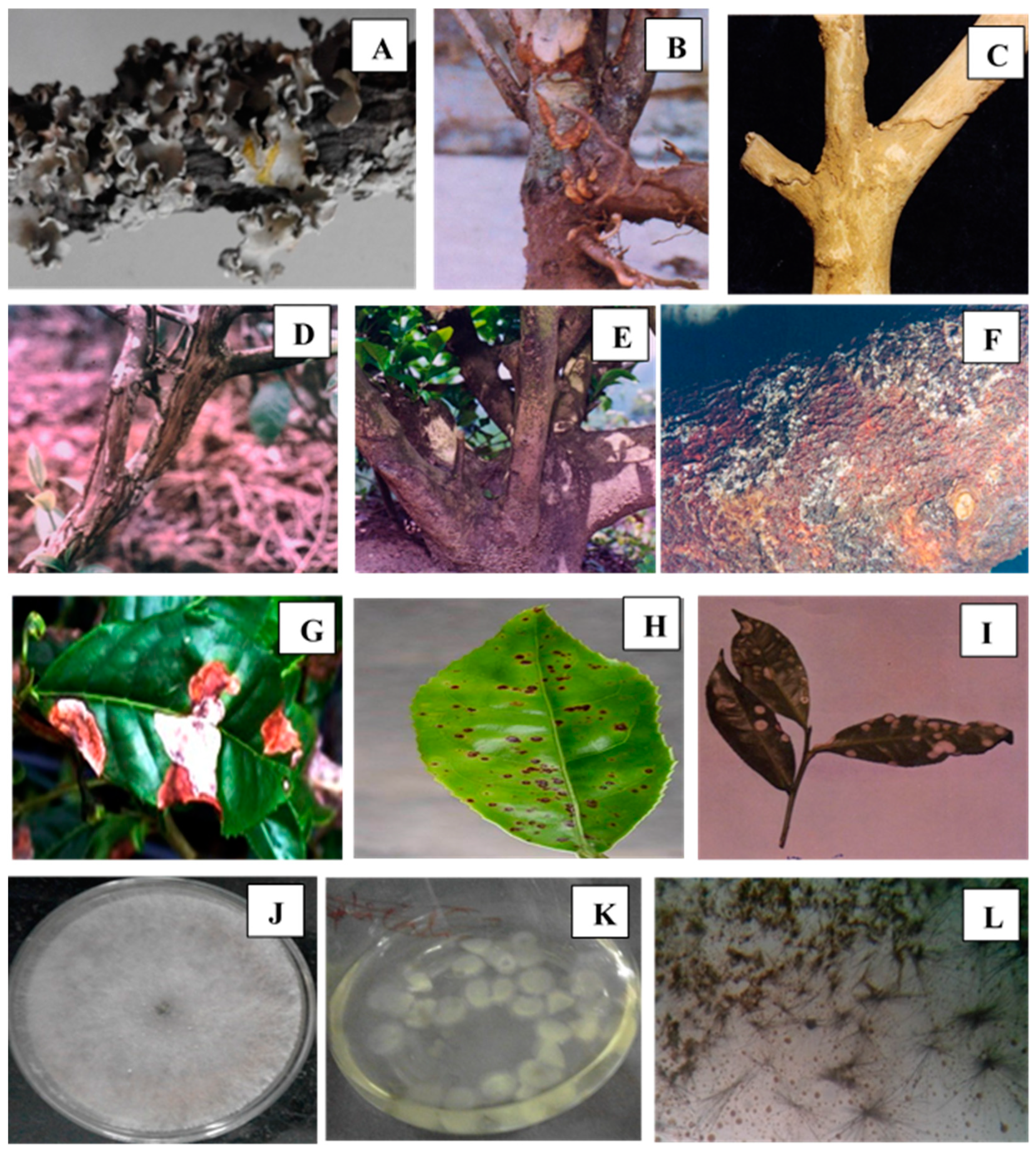
4.1. Lichen Sample Collection and Identification

For the present research, lichen samples were collected in Kodaikanal Hills (2130 m above sea level), Tamil Nadu, India. They were identified by morphological, anatomical, and spot color tests [28]. Using the pertinent key and the monographs of Divakar and Upreti [29], it was determined that the lichen specimen was P. austrosinense. The identified lichen sample was further validated and the voucher specimen was placed with Accession No. 35618 at National Botanical Research Institute (NBRI), Lucknow, Uttar Pradesh, India for future reference.

4.2. Isolation of Mycobionts from P. austrosinense

P. austrosinense lichen thalli were rinsed in running water before being surface sterilized by soaking for 30 s in 95% C2H5OH, 1 min in 0.1% mercuric chloride, and 30 s in 75% C2H5OH. The sterilized thalli were air dried and fungal filaments were gently teased using sterilized forceps and then placed in sterile Petri dishes containing Modified Bold’s Basal Salt (MBBS) medium. They were incubated at 25 °C for eight weeks to allow the growth of lichen mycobionts [30].

4.3. Production and Confirmation of Lecanoric Acid from Mycobionts

MBBS broth supplemented with 4% sucrose and 1% potassium nitrate was inoculated with P. austrosinense mycobiont into 250 mL Erlenmeyer flasks. The culture was shaken constantly at 150 rpm for a period of 15 days for lecanoric acid production. Lecanoric acid was confirmed by the micro-crystallization method [31]. A few drops of culture filtrate of P. austrosinense mycobiont were placed on a microscopic glass slide to which a glycerol/ethanol/water (1:1:1) solvent mixture was added and covered with a cover slip. The glass slide was gently heated over a tiny flame for 3 min to observe the appearance of micro-crystals. Upon cooling, lecanoric acid appeared as needle-like, long, but slightly curved crystals under a 40 × objective lens of a light microscope.

4.4. Purification of Lecanoric Acid by Column Chromatography

Hexane was used to make silica gel slurry, which was then packed into a 60 cm by 18 mm column. Lecanoric acid containing mycobionts culture filtrate was dissolved in a minimum quantity of acetone, impregnated onto the silica gel, and loaded in the column. The compounds were eluted recurrently by increasing the polarity of the eluent. Lecanoric acid was eluted with absolute benzene and 5% ethyl acetate in benzene. Lecanoric acid was purified by re-crystallizing in 50% acetone after the elute solvents had been evaporated in a vacuum [32].

4.5. Antifungal Activity of Lecanoric Acid

Tea fungal pathogens such as Hypoxylon serpens cause wood rot disease, Macrophoma theicola, causes branch canker disease, Phomopsis theae causes collar canker, Tunstallia aculeate causes thorny stem blight, Cercospora theae is a causative agent of bird’s eye spot disease, Glomerella cingulate causes brown blight disease, Pestalotiopsis theae is a causative agent of grey blight disease, and Poria hypolateritia is a causative fungus of red root rot disease, all of which were obtained from Microbial Type Culture Collection (MTCC) Centre, Chandigarh and UPASI Tea Research Institute, Valparai, Tamil Nadu, India.
The antifungal activity of lecanoric acid against these tea fungal pathogens was found by the poisoned food technique using potato dextrose agar in vitro [33]. PDA culture plates were prepared with the requisite quantity of lecanoric acid, a companion systemic fungicide, and botanical fungicides were incorporated and then poured into Petri plates. The PDA plates were inoculated with a 5 mm mycelial disc of the test pathogens from a 5 day--old culture with their mycelial side down. A control plate devoid of lecanoric acid, companion, and botanical fungicides was taken for comparison. The growth of the pathogen was measured after 7 days of incubation. The growth inhibition percentage of tea pathogenic microorganisms was calculated by the Bell scale method [34].
Different solvents such as ethanol, methanol, acetone, petroleum ether, and water were selected to find the best solvent (100 mL) for lecanoric acid extractions and bioassayed against the selected tea fungal pathogens. Companion systemic fungicide (0.5% solution) at various concentrations such as 10, 20, 30, and 40 μL was prepared in sterile distilled water and was mixed with molten, cooled 20 mL of PDA medium, and dispensed uniformly into Petri plates. Since companion fungicide is being recommended for the control of various stem, leaf, and root diseases in tea plantations, it was selected for the study to compare the bio-efficacy over lecanoric acid. In addition, some of the recommended botanical fungicides such as expel, nimbicidine, and nimbidoxin (1.0% solution) were also evaluated for comparison purposes [35].

4.6. Determination of Free Radical Scavenging Activity by DPPH Assay

The free radical scavenging activity of the lichen lecanoric acid was assessed with the 1-diphenyl-2-picryl-hydrazil (DPPH) method described by Blois [33]. The lecanoric acid (0.1 mL, gradient concentration from 10 to 300 µg/mL) was added into a 0.1 mM of 1, 1-DPPH solution. It was incubated at room temperature, and after 30 min, the absorbance was read at 517 nm. A solution mixture without lecanoric acid and ascorbic acid was used as a negative and positive control, respectively. The free radical scavenging activity of lecanoric acid was designed as IC50 (50% inhibition concentration), evaluated by the following equation:
Free radical scavenging activity (%) = (100 − A(sample)/A(blank) × 100

4.7. Statistical Analysis

Using the statistical software SPSS 17.0 (SPSS, Inc., Chicago, IL, USA), all of the data were statistically analyzed. Additionally, standard deviations (SA) and analysis of variance (ANOVA) were applied to the data that were acquired from the experiments. Critical difference (CD) was used to separate the significant means at various degrees of significance [36].

5. Conclusions

In this study, we conducted a detailed analysis of the biological properties of purified lichens compound, such as lecanoric acid. This purified compound was isolated from the thallus of P. austrosinense, which was used to evaluate its antimicrobial activities against a variety of fungal plant pathogens which are notorious for their disease-causing abilities to crops and trees of agricultural and horticultural importance. In this case, tea fungal pathogens were selected for the study, against which the lecanoric acid demonstrated excellent growth inhibition properties at a concentration 15 μL. According to these results, it might be established that lecanoric acid is an effective antifungal compound to control tea fungal pathogens and could be used as a potential antifungal compound in tea-cultivating areas. This study also supports to development of an appropriate strategy and integrated control disease management program for tea diseases, which in turn might reduce the agrochemical residues and hazards to the environment.

Author Contributions

Conceptualization, P.P.; methodology, B.M.G.; validation, P.K. formal analysis, M.R.S., M.K. (Mujeeb Khan), M.K. (Merajuddin Khan) and B.S.; investigation, K.R.; resources, P.P.; data curation, K.R., M.K. (Mujeeb Khan), M.K. (Merajuddin Khan) and B.S.; writing—original draft preparation, K.R.; writing—review and editing, P.K. and M.R.S.; supervision, P.P.; project administration, P.P.; funding acquisition, M.R.S. All authors have read and agreed to the published version of the manuscript.

Funding

The authors acknowledge the funding from Researchers Supporting Project number (RSPD2023R665), King Saud University, Riyadh, Saudi Arabia.

Data Availability Statement

Data contained within the Article.

Acknowledgments

The authors acknowledge the funding from Researchers Supporting Project number (RSPD2023R665), King Saud University, Riyadh, Saudi Arabia.

Conflicts of Interest

The authors declare no conflict of interest.

References

  1. Ponmurugan, P.; Ayyappadasan, G.; Verma, R.S.; Nayaka, S. Survey, distribution pattern and elemental composition of lichens in Yercaud hills of Eastern Ghats in southern India. J. Environ. Biol. 2016, 37, 407. [Google Scholar]
  2. Shukla, V.; Joshi, G.P.; Rawat, M. Lichens as a potential natural source of bioactive compounds: A review. Phytochem. Rev. 2010, 9, 303–314. [Google Scholar] [CrossRef]
  3. Ganesan, A.; Thangapandian, M.; Ponnusamy, P.; Sundaraj, J. Antibacterial and antioxidant activity of Parmotrema reticulatum obtained from Eastern Ghats, Southern India. Biomed. Res. 2017, 28, 1593–1597. [Google Scholar]
  4. Vartia, K.O. Antibiotics in lichens. In The Lichens; Elsevier: Amsterdam, The Netherlands, 1973; pp. 547–561. [Google Scholar]
  5. Tanas, S.; Odabasoglu, F.; Halici, Z.; Cakir, A.; Aygun, H.; Aslan, A.; Suleyman, H. Evaluation of anti-inflammatory and antioxidant activities of Peltigera rufescens lichen species in acute and chronic inflammation models. J. Nat. Med. 2010, 64, 42–49. [Google Scholar] [CrossRef] [PubMed]
  6. Shrestha, G.; El-Naggar, A.M.; St. Clair, L.L.; O’Neill, K.L. Anticancer activities of selected species of North American lichen extracts. Phytother. Res. 2015, 29, 100–107. [Google Scholar] [CrossRef]
  7. Sati, S.; Joshi, S. Antibacterial activity of the Himalayan lichen Parmotrema nilgherrense extracts. Br. Microbiol. Res. J. 2011, 1, 26. [Google Scholar] [CrossRef]
  8. Roser, L.A.; Erkoc, P.; Ingelfinger, R.; Henke, M.; Ulshöfer, T.; Schneider, A.-K.; Laux, V.; Geisslinger, G.; Schmitt, I.; Fürst, R. Lecanoric acid mediates anti-proliferative effects by an M phase arrest in colon cancer cells. Biomed. Pharmacother. 2022, 148, 112734. [Google Scholar] [CrossRef] [PubMed]
  9. Yamazaki, M.; Matsuo, M.; Shibata, S. Biosynthesis of lichen depsides, lecanoric acid and atranorin. Chem. Pharm. Bull. 1965, 13, 1015–1017. [Google Scholar] [CrossRef] [Green Version]
  10. Paguirigan, J.A.; Liu, R.; Im, S.M.; Hur, J.-S.; Kim, W. Evaluation of Antimicrobial Properties of Lichen Substances against Plant Pathogens. Plant Pathol. J. 2022, 38, 25. [Google Scholar] [CrossRef]
  11. Nugraha, A.S.; Untari, L.F.; Laub, A.; Porzel, A.; Franke, K.; Wessjohann, L.A. Anthelmintic and antimicrobial activities of three new depsides and ten known depsides and phenols from Indonesian lichen: Parmelia cetrata Ach. Nat. Prod. Res. 2021, 35, 5001–5010. [Google Scholar] [CrossRef]
  12. Umezawa, H.; Shibamoto, N.; Naganawa, H.; Ayukawa, S.; Matsuzaki, M.; Takeuchi, T.; Kono, K.; Sakamoto, T. Isolation of lecanoric acid, an inhibitor of histidine decarboxylase from a fungus. J. Antibiot. 1974, 27, 587–596. [Google Scholar] [CrossRef] [PubMed] [Green Version]
  13. Ponmurugan, P.; Manjukarunambika, K.; Gnanamangai, B.M. Impact of various foliar diseases on the biochemical, volatile and quality constituents of green and black teas. Australas. Plant Pathol. 2016, 45, 175–185. [Google Scholar] [CrossRef]
  14. Ponmurugan, P.; Gnanamangai, B.M.; Baby, U. Incidence, prevalence and clonal susceptibility of Phomopsis canker disease in southern Indian tea plantations. Indian Phytopathol. 2013, 66, 46–52. [Google Scholar]
  15. Ramya, M.; Ponmurugan, P.; Saravanan, D. Management of Cephaleuros parasiticaus Karst (Trentepohliales: Trentepohliaceae), an algal pathogen of tea plant, Camellia sinsensis (L.) (O. Kuntze). Crop Prot. 2013, 44, 66–74. [Google Scholar] [CrossRef]
  16. Baby, U.; Chandramouli, B. Biological antagonism of Trichoderma and Gliocladium spp. against certain primary root pathogens of tea. J. Plant. Crops 1996, 24, 249–255. [Google Scholar]
  17. Mareeswaran, J.; Nepolean, P.; Jayanthi, R.; Premkumar, S.; Radhakrishnan, B. In vitro studies on branch canker pathogen (Macrophoma sp.) infecting tea. J. Plant Pathol. Microbiol. 2015, 6, 284. [Google Scholar]
  18. König, G.M.; Wright, A.D. 1H and 13C-NMR and biological activity investigations of four lichen-derived compounds. Phytochem. Anal. 1999, 10, 279–284. [Google Scholar] [CrossRef]
  19. Kalidoss, R.; Ayyappadasan, G.; Gnanamangai, B.M.; Ponmurugan, P. Antibacterial activity of lichen Parmotrema spp. Inter. J Pharmaceut. Biol. Sci. 2020, 8, 7–11. [Google Scholar]
  20. Wei, A.H.; Zhou, D.N.; Ruan, J.L.; Cai, Y.L.; Xiong, C.M.; Li, M.X. Characterisation of phenols and antioxidant and hypolipidaemic activities of Lethariella cladonioides. J. Sci. Food Agric. 2012, 92, 373–379. [Google Scholar] [CrossRef]
  21. Nepolean, P.; Balamurugan, A.; Jayanthi, R.; Mareeswaran, J.; Premkumar, R. Bioefficacy of certain chemical and biofungicides against Hypoxylon spp. causing wood rot disease in tea. J. Plant Crops 2014, 42, 341–347. [Google Scholar]
  22. Kalim, S.; Luthra, Y.; Gandhi, S. Influence of Bavistin Seed Treatment on Morpho-physiological and Biochemical Parameters of Cowpea Roots Susceptible to Rhizoctonia Species. J. Mycol. Plant Pathol. 2000, 30, 375–379. [Google Scholar]
  23. Vyas, C.S. Hand Book of Systemic Fungicides; General Aspects; Mcgraw-Hill Education: New Delhi, India, 1993. [Google Scholar]
  24. Basile, A.; Conte, B.; Rigano, D.; Senatore, F.; Sorbo, S. Antibacterial and antifungal properties of acetonic extract of Feijoa sellowiana fruits and its effect on Helicobacter pylori growth. J. Med. Food 2010, 13, 189–195. [Google Scholar] [CrossRef] [PubMed]
  25. Tiwari, P.; Rai, H.; Upreti, D.; Trivedi, S.; Shukla, P. Antifungal activity of a common Himalayan foliose lichen Parmotrema tinctorum (Despr. ex Nyl.) Hale. Nat. Sci. 2011, 9, 167–171. [Google Scholar]
  26. Gulluce, M.; Aslan, A.; Sokmen, M.; Sahin, F.; Adiguzel, A.; Agar, G.; Sokmen, A. Screening the antioxidant and antimicrobial properties of the lichens Parmelia saxatilis, Platismatia glauca, Ramalina pollinaria, Ramalina polymorpha and Umbilicaria nylanderiana. Phytomedicine 2006, 13, 515–521. [Google Scholar] [CrossRef] [PubMed]
  27. Fernández-Moriano, C.; Gómez-Serranillos, M.P.; Crespo, A. Antioxidant potential of lichen species and their secondary metabolites. A systematic review. Pharm. Biol. 2016, 54, 1–17. [Google Scholar] [CrossRef]
  28. Wolseley, P. A Compendium of the Macrolichens from India, Nepal and Sri Lanka. By Dharani Dhar, Bishen Singh Mahendra Pal Singh, Dehra Dun, India. Lichenologist 2008, 40, 267–268. [Google Scholar] [CrossRef]
  29. Divakar, P.K.; Upreti, D.K. Parmelioid Lichens in India: A Revisionary Study; Bishen Singh Mahendra Pal Sin: Dehra Dun, India, 2005. [Google Scholar]
  30. Verma, N.; Behera, B.C. In Vitro culture of lichen partners: Need and Implications. Recent Adv. Lichenol. 2015, 2, 147–159. [Google Scholar]
  31. Orange, A.; James, P.W.; White, F. Microchemical Methods for the Identification of Lichens; British Lichen Society: London, UK, 2001. [Google Scholar]
  32. Yoshimura, I.; Kinoshita, Y.; Yamamoto, Y.; Huneck, S.; Yamada, Y. Analysis of secondary metabolites from lichen by high performance liquid chromatography with a photodiode array detector. Phytochem. Anal. 1994, 5, 197–205. [Google Scholar] [CrossRef]
  33. Adams, P.; Wong, J. The effect of chemical pesticides on the infection of sclerotia of Sclerotinia minor by the biocontrol agent Sporidesmium sclerotivorum. Phytopathology 1991, 81, 1340–1343. [Google Scholar] [CrossRef]
  34. Cappuccino, J.; Sherman, N. Microbiology, a Laboratory Manual; Benjamin, Cummings Pub. Co.: New York, NY, USA, 1992. [Google Scholar]
  35. Mareeswaran, J.; Nepolean, P. In vitro screening of chemical and organic fungicides against Branch canker disease in tea. J. Mycopathol. Res. 2016, 54, 299–301. [Google Scholar]
  36. Gomez, K.A.; Gomez, A.A. Statistical Procedures for Agricultural Research; John Wiley & Sons: Hoboken, NJ, USA, 1984. [Google Scholar]
Figure 1. P. austrosinense lichen thallus, nature of tea plant diseases and production of lecanoric acid under in vitro conditions. (A) Thallus of P. austrosinense, (B) Collar canker, (C) Thorny stem blight, (D) Branch canker, (E) Wood rot, (F) Red root rot, (G) Grey blight, (H) Bird’s eye spot, (I) Brown blight, (J) Growth of P. austrosinense in MBBS solid medium, (K) Growth of P. austrosinense in MBBS broth, and (L) Lecanoric acid with microcrystals.
Horticulturae 09 00705 g001
Figure 2. Antifungal activity of lecanoric acid extracted from P. austrosinense lichen on growth inhibition of a different variety of tea fungal pathogens. MT—Macrophoma theicola, PT—Phomopsis theae, TA—Tunstallia aculeate, CT—Cercospora theae, GC—Glomerella cingulate, PET—Pestalotiopsis theae, PH—Poria hypolateritia.
Horticulturae 09 00705 g002
Figure 3. Free radical scavenging activity of lecanoric acid by DPPH free radical scavenging activity assay.
Horticulturae 09 00705 g003
Table 1. Antifungal activity of different concentrations of lecanoric acid extracted from P. austrosinense lichen on the growth inhibition of different varieties of tea fungal pathogens.
Tea Fungal Pathogens #Concentration of Lecanoric Acid (μL)/Growth Inhibition (%)
5101520
Hypoxylon serpens53.5 ± 1.375.3 ± 1.377.7 ± 1.375.0 ± 1.5
Macrophoma theicola53.3 ± 1.573.0 ± 1.776.5 ± 1.570.7 ± 1.8
Phomopsistheae60.0 ± 1.776.0 ± 2.080.5 ± 1.373.0 ± 2.4
Tunstallia aculeate52.5 ± 1.672.0 ± 1.575.0 ± 1.469.5 ± 2.0
Cercosporatheae71.3 ± 2.588.0 ± 1.191.5 ± 2.085.5 ± 2.5
Glomerella cingulata65.5 ± 2.184.0 ± 2.386.5 ± 2.180.7 ± 2.3
Pestalotiopsis theae68.7 ± 2.090.7 ± 1.590.0 ± 2.383.3 ± 1.8
Poria hypolateritia50.3 ± 1.571.5 ± 1.373.3 ± 1.568.5 ± 1.6
* CD at p = 0.055.46.55.34.6
* CD—critical difference, #, all the fungal strains were procured from Microbial Type Culture Collection (MTCC).
Table 2. Effect of different solvents for lecanoric acid amendment on growth inhibition of different varieties of tea fungal pathogens.
Tea Fungal PathogensSolvents (mg mL)/Growth Inhibition
EthanolMethanolAcetonePetroleum EtherWater
Hypoxylon serpens72.5 ± 1.274.0 ± 1.772.0 ± 1.573.0 ± 1.652.5 ± 1.3
Macrophoma theicola73.0 ± 1.374.7 ± 1.074.3 ± 1.573.3 ± 1.855.7 ± 1.3
Phomopsis theae74.3 ± 0.976.0 ± 1.575.3 ± 1.375.0 ± 1.555.3 ± 1.1
Tunstallia aculeate71.3 ± 1.173.0 ± 1.372.3 ± 1.072.5 ± 1.252.0 ± 1.3
Cercospora theae84.0 ± 2.087.5 ± 2.085.0 ± 1.785.0 ± 1.964.0 ± 2.1
Glomerella cingulata80.7 ± 1.585.3 ± 2.082.3 ± 2.581.5 ± 1.553.0 ± 2.1
Pestalotiopsis theae81.5 ± 1.385.7 ± 1.882.0 ± 2.083.5 ± 1.554.5 ± 2.0
Poria hypolateritia70.7 ± 1.173.0 ± 1.671.5 ± 1.670.7 ± 1.453.3 ± 1.7
* CD at p = 0.054.55.34.03.83.3
* CD—critical difference.
Table 3. Antifungal activity of different concentrations of companion systemic fungicide on the growth inhibition of various tea fungal pathogens.
Tea Fungal PathogensConcentration of Companion Fungicide (μL)/Growth Inhbition (%)
10203040
Hypoxylon serpens63.0 ± 1.571.0 ± 1.770.3 ± 1.168.3 ± 1.3
Macrophoma theicola65.3 ± 1.670.3 ± 1.570.7 ± 1.367.0 ± 1.4
Phomopsis theae66.0 ± 1.571.5 ± 1.770.3 ± 0.870.3 ± 1.5
Tunstallia aculeate63.0 ± 1.369.3 ± 1.369.5 ± 0.967.5 ± 1.7
Cercospora theae72.3 ± 1.685.7 ± 1.581.5 ± 0.880.7 ± 1.1
Glomerella cingulata75.5 ± 1.580.7 ± 1.780.3 ± 1.678.0 ± 1.6
Pestalotiopsis theae75.3 ± 1.683.5 ± 1.883.0 ± 1.980.0 ± 1.5
Poria hypolateritia63.7 ± 0.968.5 ± 1.565.7 ± 1.765.0 ± 1.1
* CD at p = 0.054.74.23.35.0
* CD—critical difference.
Disclaimer/Publisher’s Note: The statements, opinions and data contained in all publications are solely those of the individual author(s) and contributor(s) and not of MDPI and/or the editor(s). MDPI and/or the editor(s) disclaim responsibility for any injury to people or property resulting from any ideas, methods, instructions or products referred to in the content.

