Article

In Vitro Propagation of Three Populations of the Endangered, Greek Endemic Cerastium candidissimum and Short-Term Storability of Alginate-Encapsulated Shoot Explants for Exploitation and Conservation

by
Konstantinos Bertsouklis
* and
Stella Tsopela
Laboratory of Floriculture and Landscape Architecture, Department of Crop Science, School of Plant Sciences, Agricultural University of Athens, 75 Iera Odos Street, GR-11855 Athens, Greece
*
Author to whom correspondence should be addressed.
Horticulturae 2023, 9(2), 273; https://doi.org/10.3390/horticulturae9020273
Submission received: 10 January 2023 / Revised: 11 February 2023 / Accepted: 14 February 2023 / Published: 17 February 2023
(This article belongs to the Special Issue In Vitro Propagation and Biotechnology of Horticultural Plants)

Abstract

:
Cerastium candidissimum Correns is an endangered species. Nevertheless, its ornamental characteristics make it an ideal candidate for commercial use as a new ornamental and landscape plant. As there are no references to the in vitro propagation of this species, the present research aims to comparatively investigate the potential that in vitro propagation may have of three different populations of the plant, from Mounts Parnassos (PS), Parnitha (PR), and Hymettus (HM), respectively. The establishment of the initial cultures took place by cotyledonary- and stem-node explants, excised from in vitro grown seedlings, on Murashige and Skoog (MS) media, without hormone (Hf), or with 0.5 mg L−1 6-benzyladenine (BA). The shooting percentage was higher in the case of the PS population (97.6%). It was also higher for stem-node explants (93.9%). The multiplication phase took place on Hf MS, or on MS containing BA (0.5 and 1.0 mg L−1), with or without 0.1 mg L−1 1-Naphthaleneacetic acid. The higher shoot production was observed in the PS and PR populations (88.0% and 76.9% shooting, with 3.1 and 2.7 shoots/explant, respectively). Micro-shoots rooted at high percentages on half-strength MS (80.0–100.0%). Successful acclimatisation (95.0%) was established on peat: perlite (1:1 v/v). Moreover, an efficient alginate-encapsulation procedure was developed from PS stem-node explants, using a 3.0% w/v sodium alginate and 14.7 g L−1 calcium chloride solution. Alginate beads exhibited a higher shoot proliferation (3.8 shoots) after 60 days of storage at 4.0 °C and a 50-day growth on Hf MS than non-encapsulated in vitro node-explants did. The presented protocol of in vitro propagation of C. candidissimum could facilitate the exploitation of the species by the floricultural industry and contribute to conservation strategies.

Graphical Abstract

1. Introduction

Cerastium candidissimum Correns (family: Caryophyllaceae) is a mountainous species, endemic to central and southern Greece. It grows in the middle-altitude zone, usually on dry, rocky sites in woodland clearings and alpine meadows. Depending on the altitude and degree of exposure, this caespitose perennial, commonly known as the “Greek Snow-in-Summer”, flowers from late spring to late summer [1,2].
The plant possesses certain remarkable ornamental characteristics; specifically dainty, snowy-white, star-shaped blooms as well as pearly-white foliage and stems (a unique pairing of features within the genus). Thus, it is undeniably evident that its marketable value shows promise not only in the potted plant production, but also if embraced by the cut flower industry, especially as a novel alternative in the filler flower palette. It is worth noting that ‘special’ cut flowers have gained tremendous ground over the last 15 years. Additionally, they favour sustainability since their production, ideally, necessitates minimum energy and/or agrochemical inputs [3]. Consequently, C. candidissimum could be introduced as an eco-friendly, more offbeat alternative to traditional ceremony flowers, all the while remaining in the spirit of sustainable solutions for the floral industry.
In addition to its potential value for floristry, the plant is equally suitable for use by landscape architecture, particularly as a groundcover on rock and in naturalistic garden designs. Apart from the plant’s carpeting habit, which allows it to form quite dense procumbent mats in a profusion of diminutive flowers, its greatest asset is the fact that it is not as “aggressive” [4] as its widespread congener C. tomentosum. The practice of intensive seed harvesting for commercial purposes [5] should be viewed as an incentive for keen interest in and demand for the plant.
Overall, there is little documentation in the literature regarding environmental, medicinal, or other aspects of the species [6]. Despite the fact that the matter of documentation in the literature has not been thoroughly researched, there is evidence that the essential oil of C. candidissimum holds potential value for pharmaceutical applications by virtue of its rare compounds, namely damascone isomers, and the complete lack of monoterpene hydrocarbons [7,8]. What is more, the herbal extract of the plant has been distinguished among some of the most active Greek traditional herbal medicines due to its moderate inhibitory effect against Helicobacter pylori, a bacterium linked to various chronic infections of the gastrointestinal system, including peptic ulceration [9]. Additionally, the fact that the species has been spotted among other endemics to be thriving on its own, forming colonies in disused mining facilities, indicates that, in all likelihood, C. candidissimum is a first-rate, native candidate for deployment in rehabilitation projects.
Until recently, the species was considered to be widespread throughout its native range of habitats. Today, it appears that an increasing number of local populations have been gradually declining as a result of their natural habitat’s partial or complete devastation. There are concerns that, in the near future, the distressing after-effects arising in the wake of pernicious anthropogenic interference, including the ongoing climate change, may be rendering the species’ long-term survival questionable. It was with those concerns in mind that C. candidissimum was selected as an important regional endemic species [10] for inclusion in the Balkan Botanic Garden of Kroussia in Northern Greece. Following the extensive work carried out by Kougioumoutzis’ research team, and the performance of an extinction risk assessment test run in compliance with the regulations of the International Union for Conservation of Nature (IUCN), the species is now listed among the Endangered (EN) Greek Endemics [11].
For the purposes of this study, three distinct populations from different geographical mountainous sites, i.e., Hymettus (HM), Parnitha (PR), and Parnassos (PS), were studied concurrently in order to establish an efficient in vitro propagation protocol. In vitro-grown seedlings were used as the primary source of cotyledonary- and stem-node explants (CN and NE explants, respectively). The use of explants of seedling origin promotes genetic diversity and a high proliferation rate for in vitro culture establishment as in the case of C. banaticum [12] and other mountainous plants [13,14], while the use of cotyledonary nodes has been proved efficient for other species [15,16,17]. Based on the data collected, in vitro-grown plantlets of one (PS) out of the three populations selected was isolated and chosen as the one that would proceed to the protocol’s second part: the establishment of an encapsulation method. ‘Capsule’—the diminutive of the Latin word ‘capsa’—is used when referring to a small container. Thus, an encapsulation method involves the complete envelopment of each explant in a calcium alginate bead made from a nutritive and protective gel matrix. Encapsulation technology is a method that has been applied to many species in the past for the short-, medium- and long-term conservation of buds and somatic embryos. It has undoubtedly proved most efficient in improving the micropropagation dynamic and in conserving a series of medicinal and ornamental plants on a large scale [18,19]. The synthetic seed technology—which, in essence, combines the benefits of clonal propagation along with those of seed propagation and storage [20]—is also widely used for the conservation of endangered species genome [21] such as Tylophora indica [22], Eryngium alpinum [23], Decalepis salicifolia [24], and Satureja khuzistanica [25]. In view of the ornamental value of C. candidissimum and its endangered status, the present research chose to develop an efficient in vitro propagation protocol for that species. Additionally, an encapsulation of propagules into alginate beads was performed, in order to determine the effect of different durations of short-term storage of the plant at a low temperature. To the best of our knowledge, there are no previous reports in the literature on in vitro propagation of this species of the genus Cerastium, except for two references to other species of the genus, i.e., C. transilvanicum and C. banaticum. For the in vitro propagation of C. transilvanicum, Paunescu [26] used nodal explants from in vitro germinated plantlets cultured on Murashige and Skoog (MS) [27] containing 1 mg L−1 6-benzyladenine (BA) and 0.1 mg L−1 1-naphthaleneacetic acid (NAA); use of the same media has been made for the in vitro propagation of C. banaticum, a rare and endemic species from Romania [12]. The present research did not concentrate merely on establishing an efficient and commercially applicable micropropagation protocol for C. candidissimum: it also focused on comparatively evaluating the in vitro propagation potential among different populations of the plant, and assessing the possibility of prolonging, through an effective encapsulation process, the storage time for C. candidissimum in terms of the plant’s post-storage survival rate and regrowth.

2. Materials and Methods
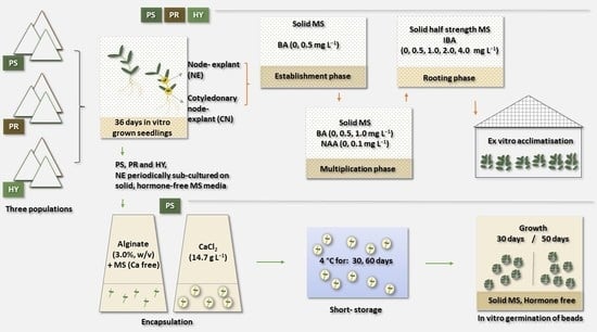
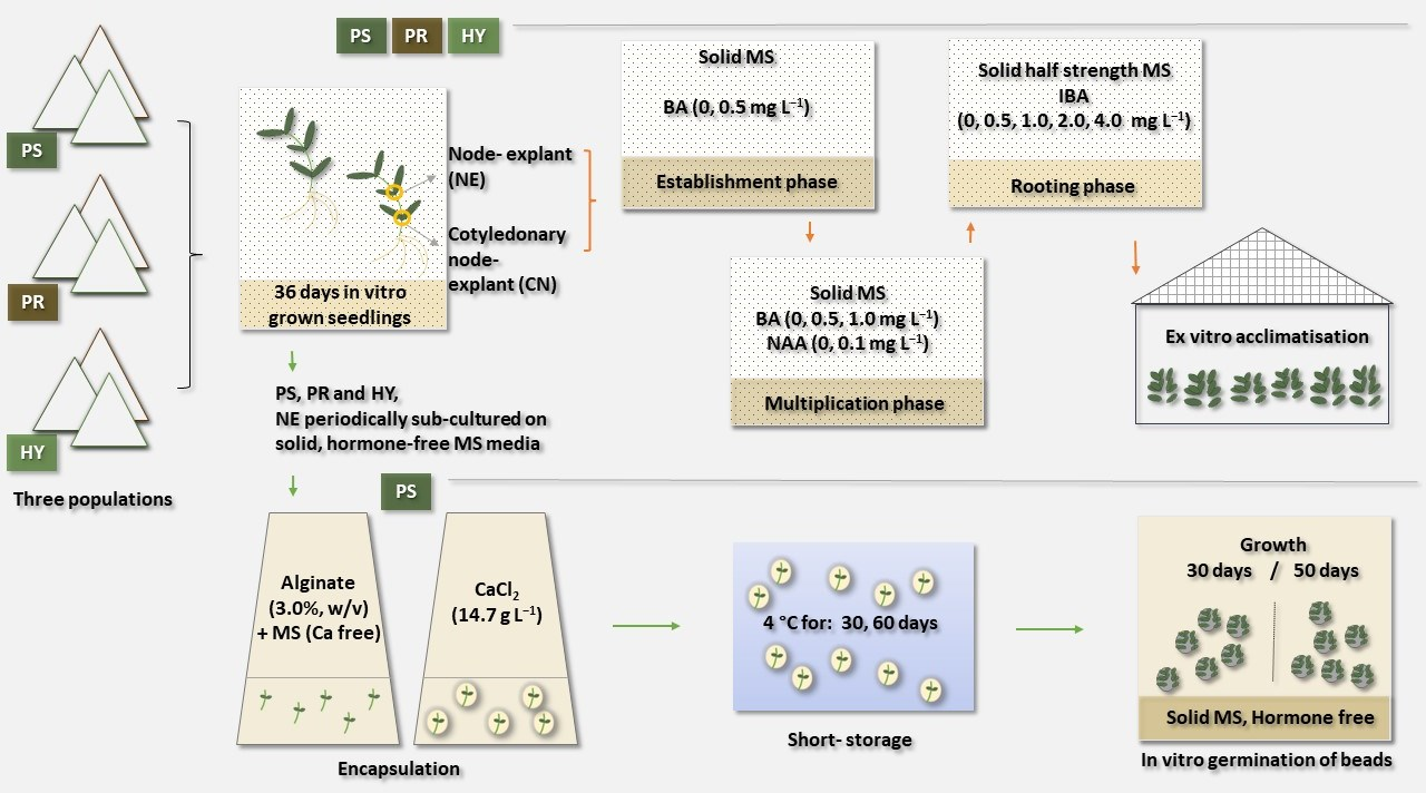
2.1. Plant Material

Seeds were harvested fully mature, in late July 2021, from three C. candidissimum populations, growing at the following three, discrete geographical sites in Greece: Mount Hymettus (37°57′23.0″ N, 23°49′03.6″ E); Mount Parnitha (38°09′55.3″ N, 23°42′59.4″ E); and Mount Parnassos (38°33′06.2″ N, 22°34′46.8″ E). The seed pericarp was separated manually and the seeds were left to dry spread out on a laboratory bench for 15 days. Following the 15-day period, the seeds were stored dry, in the dark, in 9-cm, unsealed, plastic Petri dishes between two layers of filter paper under room conditions (relative humidity 30–40%, room temperature 23 ± 2 °C) until the time of the germination experiments. In vitro germination took place after six months of collection.
Prior to the germination treatments, the seeds were surface-sterilized with 20% (v/v) commercial bleach (4.6% w/v sodium hypochlorite) for 10 min and rinsed three times (3 min each) with sterile, distilled water. They were then sown in 9-cm plastic Petri dishes with sucrose- and hormone-free, half-strength MS medium. Actively grown seedlings coming from in vitro germinated seeds at 15 °C and 20 °C were used as contamination-free stock planting material for in vitro propagation studies.

2.2. Establishment of Initial Cultures

Two discrete origin explants, namely, (a) cotyledonary-node and (b) stem-node explants, with two opposite axillary buds, 0.5–0.6 cm in length, were excised from 36-day-old seedlings taken from each Cerastium population. The explants were cultured onto MS medium either hormone-free (Hf) or supplemented with BA at 0.5 mg L−1. A completely randomised three-factorial design was used, i.e., three populations (PS, PR, HY) × two explant types (CN, NE) × two media (Hf MS or MS containing 0.5 mg L−1 (BA). As there are no previous data on in vitro propagation of C. candidissimum, the type of nutrient medium and plant growth regulators used were based on the results of previous studies on in vitro propagation of C. transilvanicum [26].

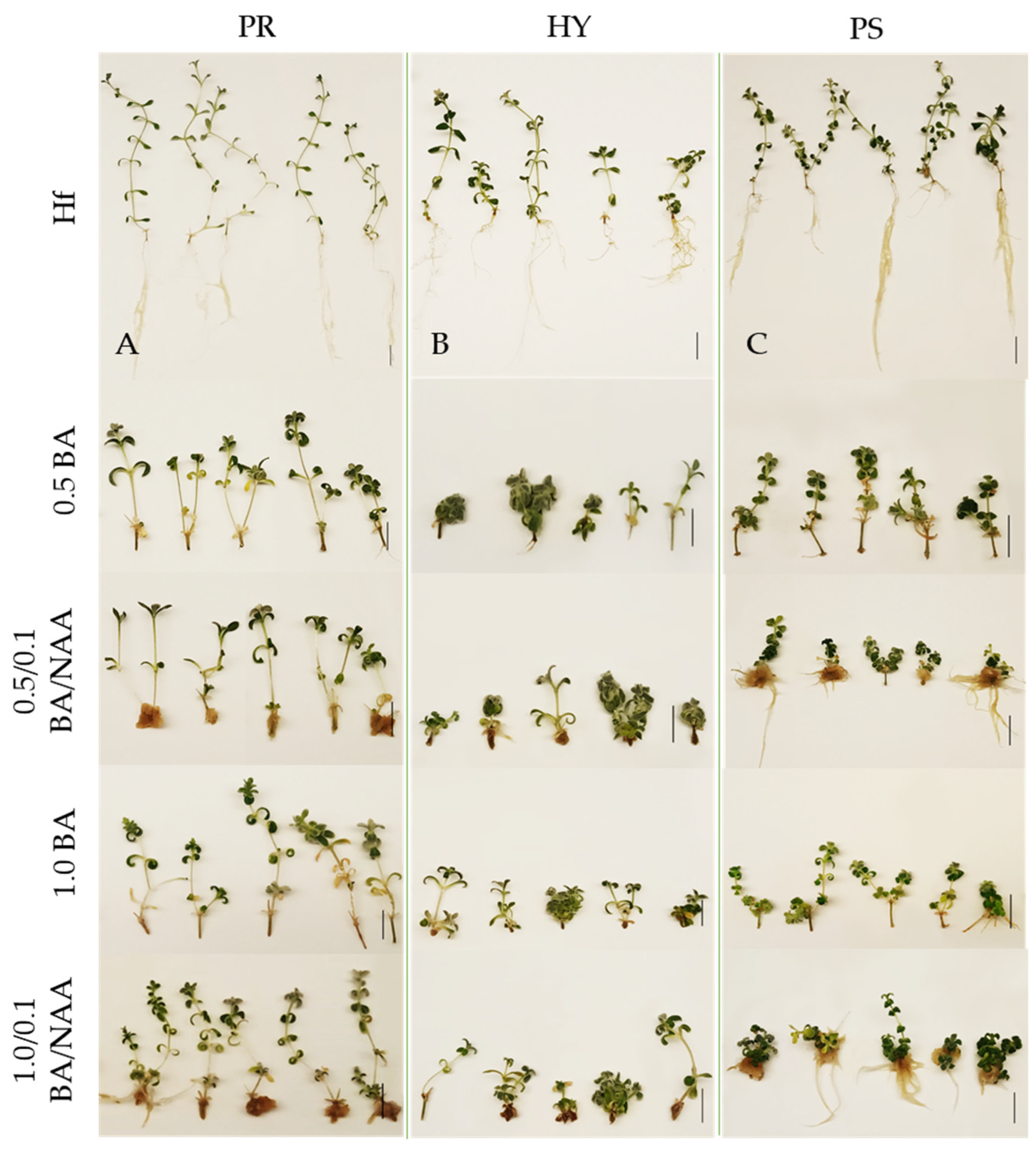
2.3. Multiplication Phase

Nodal explants were excised from micro-shoots produced on MS medium either Hf or containing BA during the establishment phase. The multiplication phase consisted of two subcultures. Each subculture lasted 50 days. The first subculture was carried out on either Hf MS media or media containing BA at 0.5 mg L−1 (with or without 0.1 mg L−1 NAA). Nodal explants for the establishment of the subsequent (i.e., second) subculture were derived from micro-shoots that had formed on Hf MS media during the first subculture. The second subculture was carried out on media that were either Hf MS or containing BA at 1.0 mg L−1 (with or without 0.1 mg L−1 NAA). All explants were 0.5–0.6 cm in length with two opposite axillary buds. The ensuing data were pooled for statistical analysis. A completely randomised, two-factorial design was used, i.e., three populations (PS, PR, HY) × five BA/NAA (BA/NAA: 0/0, 0.5/0, 0.5/0.1, 1.0/0, 1.0/0.1) concentrations.

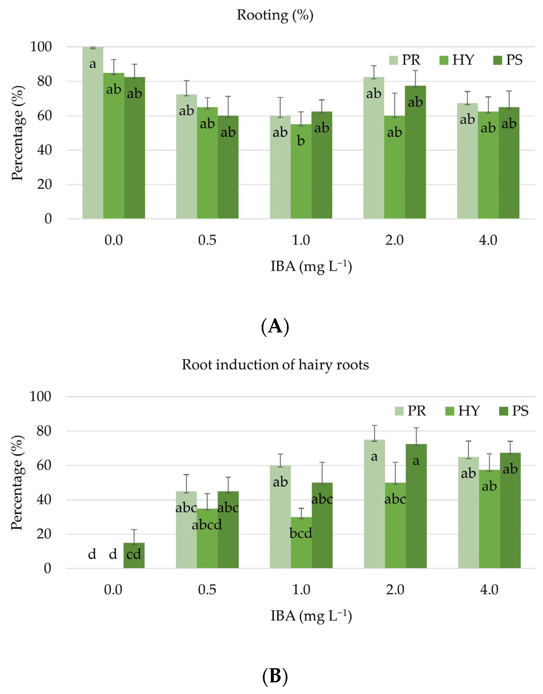
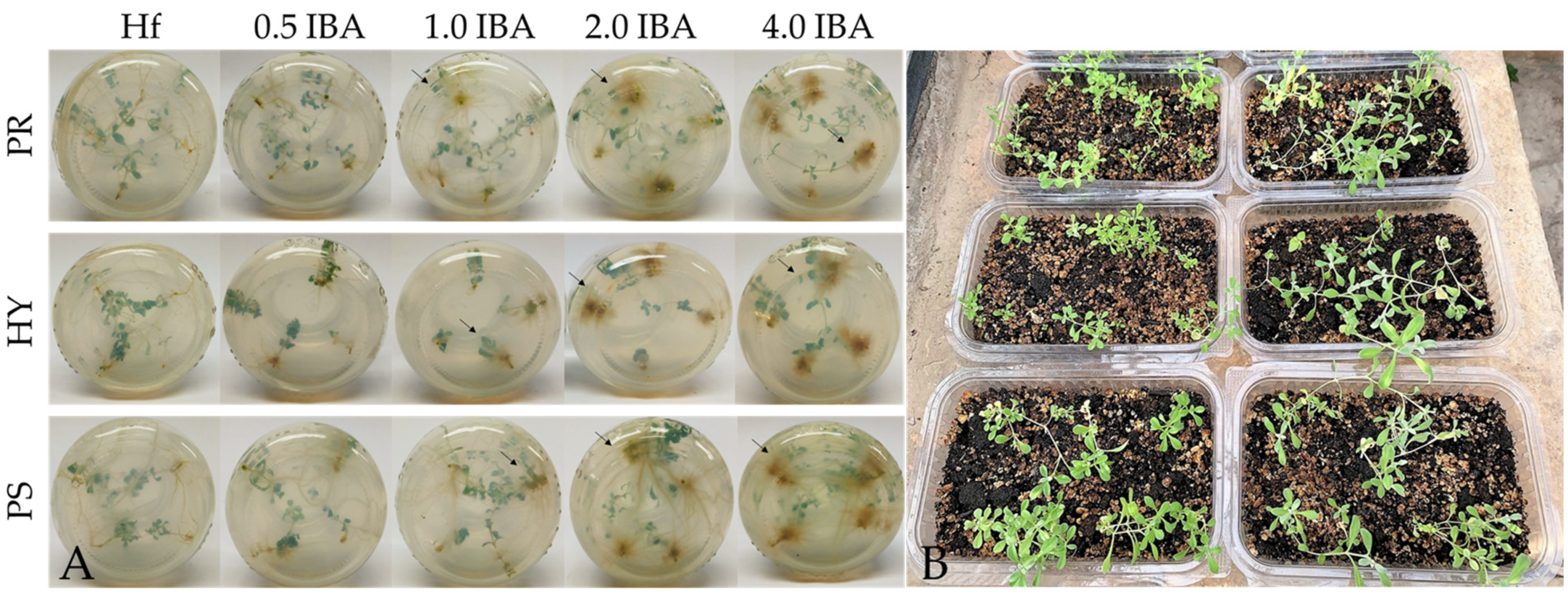
2.4. In Vitro Rooting and Acclimatisation
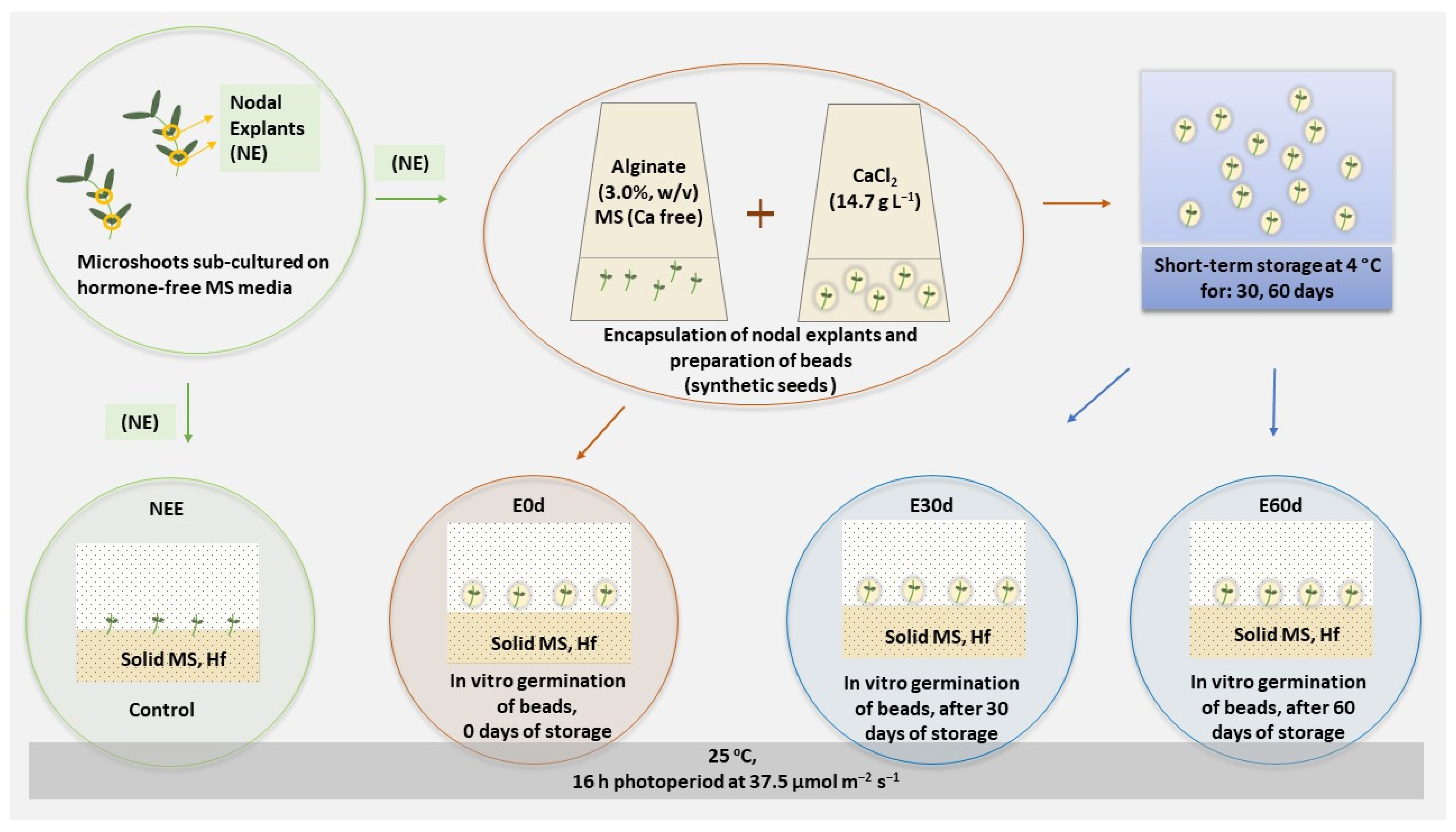
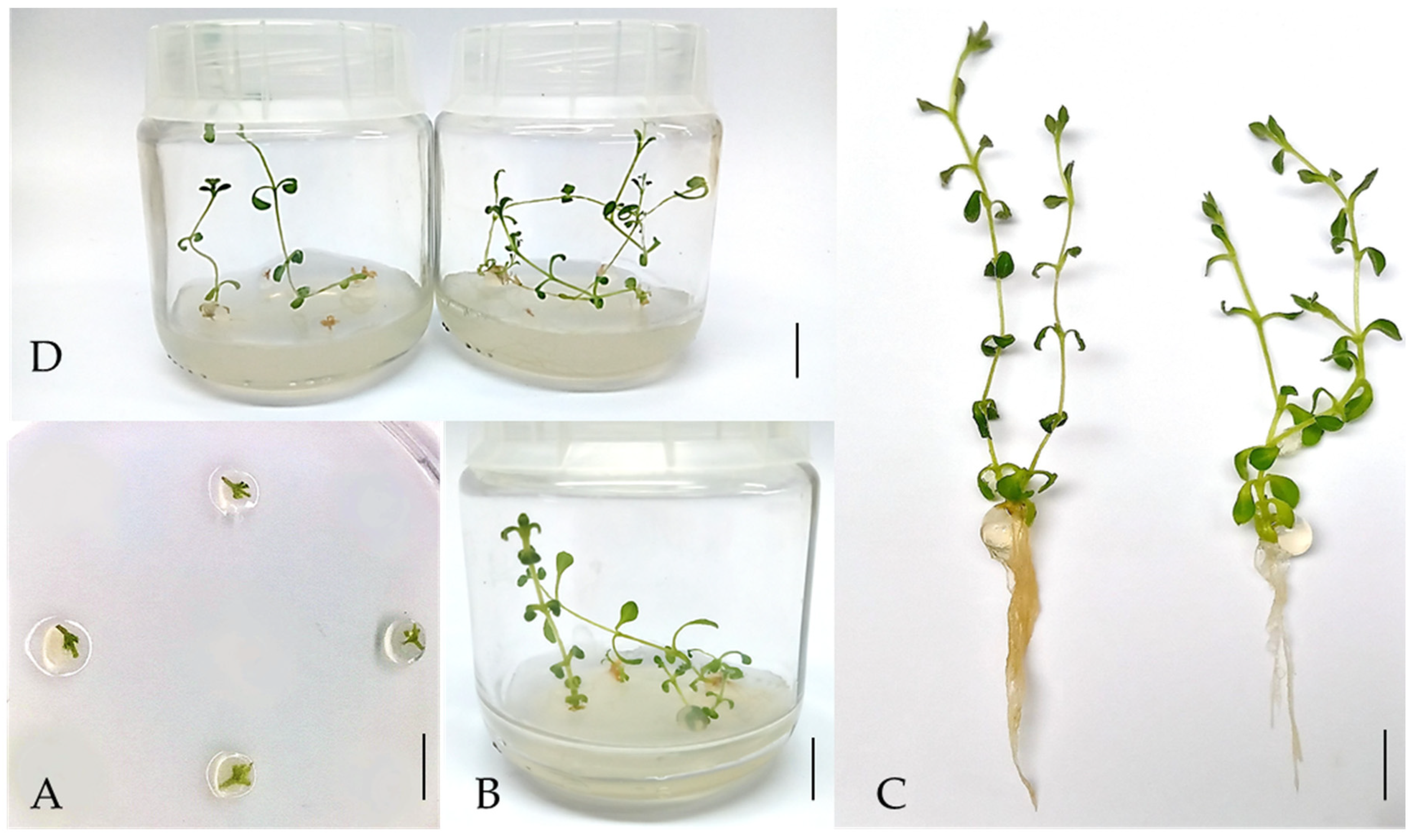
Micro-shoots taken from 50-day-old shoots, which were 2.0–2.5 cm long and produced during the multiplication stage, were cultured onto rooting media consisting of Hf, half-strength MS (MS/2) or supplemented with 0.5, 1.0, 2.0 or 4.0 mg L–1 indole-3-butyric acid (IBA). The experimentation that took place on the media described above was based on other studies on species originating in the same habitats, i.e., Sideritis raeseri subsp. Attica [14] and Sideritis scardica [28]. A completely randomised, two-factorial design was used, i.e., three populations (PS, PR, HY) × five IBA (0, 0.5, 1.0, 2.0, 4.0) concentrations.
The first clear signs of rooting formation were noticed after 10–15 days of culture, whereas data were collected 15 days later (i.e., after 30 days of culture). That data consisted of the number of roots and the mean length of the roots formed per rooted micro-shoot. Micro-shoots with roots longer than 1 cm were thoroughly rinsed under running tap water. Next, they were planted into containers (500 mL, eight plantlets/pot) with peat (pH 5.5–6.5, Klasmann-Delimann GmbH, Geeste, Germany); and perlite (particles diameter 1–5 mm, Perloflor, Isocon S.A., Athens, Greece) (1:1, v/v). Containers were covered with transparent plastic wrap (Sanitas; Sarantis S.A., Athens, Greece) in order to maintain their humidity and were transferred into a growth chamber. Seven days later and once the transparent film was removed, the containers were transferred onto a heated glasshouse bench (37°58′58.0″ N, 23°42′19.2″ E). The acclimatisation phase was followed by transplantation into 500 mL plastic pots containing peat: perlite (1/1, v/v). The plants were fertilised weekly with 2 g L−1 fully water-soluble fertilizer (Nutrileaf 60, 20-20-20; Miller Chemical and Fertilizer Corp., Hanover, PA, USA).

2.5. Encapsulation

Prior to encapsulation, nodal explants with two opposite axillary buds were excised from in vitro grown micro-shoots which had been previously sub-cultured periodically but at regular intervals, on fresh MS media, throughout the multiplication phase. Various biometric data on the three populations’ in vitro responses were accumulated in order to reveal the population with the highest in vitro propagation potential. In order to arrive at the production of synthetic seeds, the following encapsulation protocol was carried out: (1) the nodal segments were mixed for a few minutes in a sodium alginate (3%, w/v) medium, containing the MS components (without Ca) supplemented with 30 g L−1 sucrose (coating); (2) next, they were dropped and held for 30 min into a 14.7 g L−1 calcium chloride (CaCl2·2H2O) solution (polymerization of sodium alginate and complexation) [29]; (3) last, the synthetic seeds containing the coated explants were collected, rinsed thoroughly with liquid MS/2 medium in order to remove the excess amount of calcium chloride, and laid on sterile filter paper for 10 min to dry. The entire process took place under aseptic conditions in a Laminar Air Flow cabin. The sodium alginate gel matrix, the calcium chloride solution, and the liquid MS/2 medium had been autoclaved at 121 °C for 20 min. A total of four different treatments (NEE, E0d, E30d, E60d) were carried out to study the effect of encapsulation and storage duration. Non-encapsulated explants and encapsulated node explants were cultured onto Magenta B-cap vessels containing solid Hf MS medium (treatments NEE and E0d, respectively). Additionally, another set of beads (4 beads/dish), placed onto Petri dishes containing solid (8 g L–1 agar), Hf, sucrose-free MS/2, were stored at 4 °C, in the dark, for 30 and 60 days (treatments E30d and E60d, respectively) (Figure 1). Following their respective 30 and 60 days in storage, the beads were then transplanted onto Magenta B-cap vessels and were placed in a growth chamber. During the post-transfer period, data were recorded after 30 and 50 days, respectively. A completely randomised, two-factorial design was used, i.e., there were four treatments (NEE, E0d, E30d, E60d) × two growth (30-, 50-day) periods.

2.6. In Vitro Culture Conditions and Data Collection

In vitro cultures (establishment, multiplication, and rooting phases) were carried out in Magenta B-cap vessels (100 mL, Sigma-Aldrich, Steinheim, Germany), with four explants per vessel (40 explants per treatment). They were maintained in a growth chamber at 25 °C with cool-white, fluorescent lamps providing a 16-h photoperiod at 37.5 μmol m–2 s–1, as were the containers with the rooted micro-shoots. Each treatment in the rooting phase necessitated ten replicates of four micro-shoots. Regarding the encapsulation phase, a set of four alginate beads were placed in each vessel (10 vessels per treatment). All media were supplemented with 30 g L−1 sucrose and solidified with 8 g L–1 agar. The pH of the medium had been adjusted to 5.7 to 5.8 before autoclaving at 121 °C min for 20 min. Data were collected after 50 days of culture. In the case of the rooting experiments, data were recorded after 30 days of culture. The number of roots was categorised as follows: categories 1, 2, and 3 corresponded to 0–5, 6–10, and ≥11 roots, respectively. The experiment of acclimatisation used fifteen replicates of eight rooted micro-shoots. Data on acclimatisation were recorded 21 days after ex vitro transplantation. The plants’ survival rate was calculated 30 days after their transplantation into pots. For the purposes of the encapsulation experiment, data were collected after 30 and 50 days of culture.
Data were recorded on the: shoot formation percentage; total shoot number per explant; high shoot number (HS, shoots longer that 0.5 cm); length of shoots; number of nodes per shoot; percentage of rooting; root number; and root length. The percentage (%) of hairy root formation was also recorded in the experiment during the final rooting induction stage. The “multiplication index” (MI) was calculated by multiplying the percentage of shoot-producing explants by the mean number of shoots per responding explant; and then dividing the ensuing number by 0.6 to obtain the culture’s proliferation potential.

2.7. Statistical Analysis

The statistical analysis of the experiments used a completely randomized design method. The treatment means were compared using Tukey’s test at p < 0.05 (JMP 14.0 software, SAS Institute Inc., Cary, NC, USA, 2013). Data on percentages were arcsine-transformed prior to the statistical analysis to ensure the homogeneity of variance. As shown in the data tables, the number of replicates per treatment differed between experiments. Principal coordinate analysis (PCA) was also carried out so that it could assist with data visualisation. PCA analysed the related variables during in vitro morphogenesis at the establishment, multiplication, and rooting phase of each of the three C. candidissimum populations.

3. Results

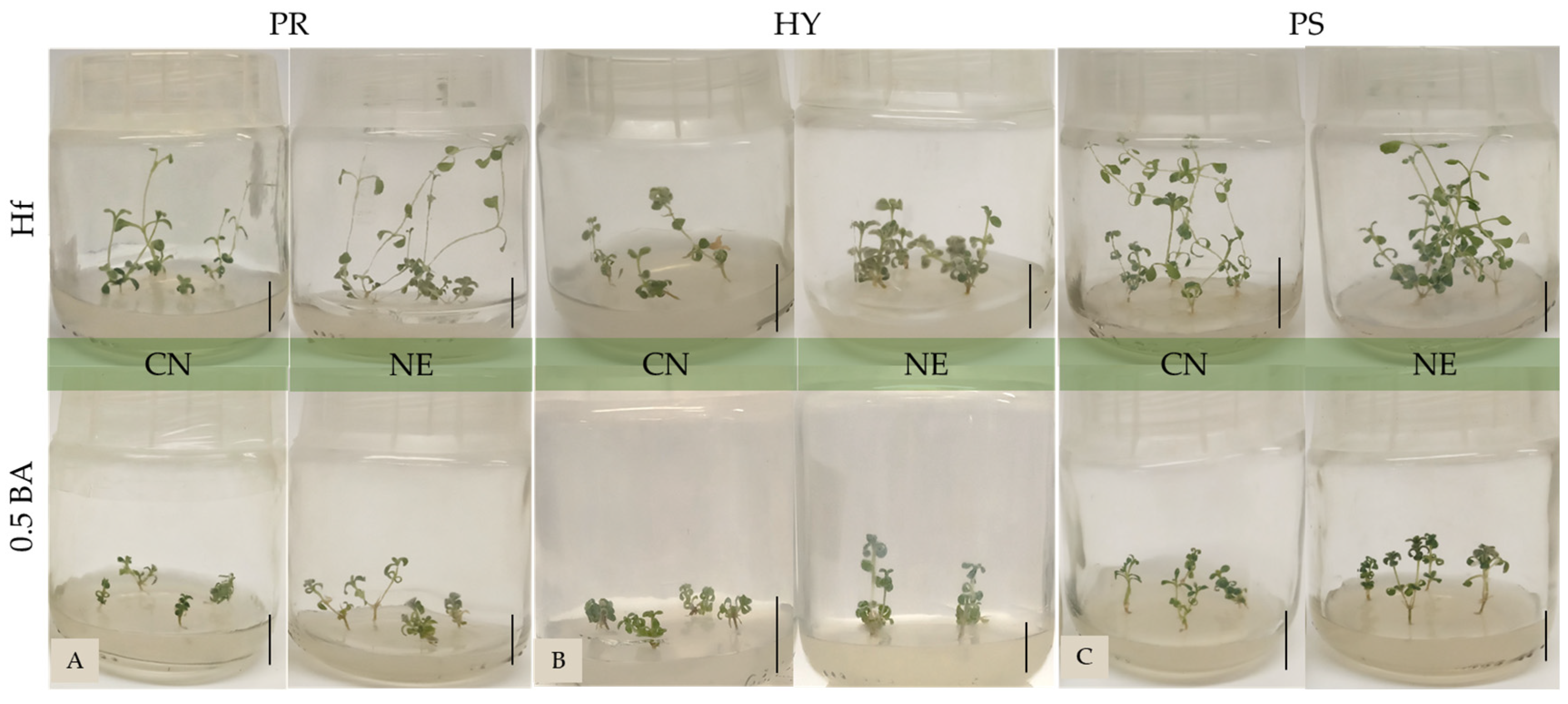
3.1. Establishment of Initial Cultures

Three-way analysis showed that the interaction of the main factors, i.e., population (PR, HY, PS), explant type (CN, NE) and medium (Hf, 0.5 mg L−1 BA), was significant during the establishment phase for the total shoot number, high shoot number (HS), shoot length, node number, MI, root number, and root length. Regarding the shooting and rooting percentages, three-way analysis revealed the significance of population and explant origin (Table 1 and Figure 2). The shooting percentage for the PS population was higher (97.6%) than those recorded for the PR and HY populations. The shooting percentage of NE explants was higher (93.9%) than that of CN explants. NE explants, which were cultured on MS containing 0.5 mg L−1 BA, produced the higher shoot number (2.4–2.8 shoots/explant), while the longer shoots (3.4 cm) were produced by NE explants cultured on Hf medium and originating in Mount Parnitha. HS production (1.8) was the highest for NE from Mount Parnitha on MS media supplemented with 0.5 mg L−1 BA. The MI was higher for PS and PR node-explants, and for PS cotyledonary-node explants cultured on Hf media.
Spontaneous rooting was observed during the establishment phase. Three-way analysis showed that PS micro-shoots rooted at a higher percentage (74.0%), followed by PR micro-shoots (57.7%), while the presence of BA reduced the rooting percentage to 34.7% in comparison to a percentage of 79.2% for rooting on Hf MS (Table 1). The longest roots (3.4 cm) belonged to plantlets rooted on Hf MS and produced by CN explants from the PS population.

3.2. Multiplication

The multiplication phase comprised two subcultures as it was important to ascertain whether growth would be optimized on MS media either without hormones or supplemented with BA at 0.5 or 1.0 mg L−1, with or without NAA at 0.1 mg L−1. Two-way analysis showed the significance of a population’s origin on the shooting percentage: the mean shooting percentage of PS explants was the highest (88.0%) and presented no difference from the percentage of 76.9% PR explants presented (Table 2). Population origin had a significant effect on the total production of shoots as well, and registered higher for the PS and PR populations (3.1 and 2.7, respectively). The BA/NAA concentration also registered a significant impact: the BA media, with or without NAA, produced the highest mean total shoot number (2.4–3.1 shoots/explants) (Table 2 and Figure 3). HS production was higher for PR population (1.3). Media supplemented with 1.0/0.1 BA/NAA (mg L−1) produced the lowest mean HS number (0.6) (Table 2). The BA/NAA concentration also had a significant effect on the total number of nodes: explants on Hf media produced 3.8 nodes/explant (Table 2). Two-way ANOVA showed that the interaction between two main factors, i.e., population and BA/NAA concentration, was significant during the multiplication phase when it came to shoot length and the MI. The highest shoot length (5.3 cm) was derived from explants coming from the PR population on Hf media (Table 2). The MI was the highest for PR explants cultured on media supplemented with 1.0/0.1 BA/NAA (mg L−1).
Spontaneous rooting was observed during the multiplication phase (Figure 3). The two main factors, i.e., population and BA/NAA, had a significant effect on the rooting percentage: PS and PR micro-shoots rooted at higher percentages (37.0% and 39.0%, respectively). The highest percentage of rooting (64.4%) was recorded by explants rooted on Hf media (Table 2).

3.3. Rooting Phase and Acclimatisation

The presence of IBA during the main rooting phase led to the formation of hairy roots. The two-way analysis using two main factors, i.e., population and IBA concentration, showed that populations of different origin shared the same rooting performance. The rooting percentage registered higher on Hf media (Figure 4A and Figure 5A). On the other hand, a significant effect the two main factors had was observed during the induction of hairy roots: PS and PR micro-shoots produced a higher percentage of hairy roots (50.0% and 49.0%, respectively). The IBA concentration also showed significant impact on the induction of hairy roots (Figure 4B and Figure 5A). The optimum concentration of IBA stood at 2.0 mg L−1 and led to higher percentages. There was significant interaction between each population and the IBA concentration on the average length of roots which was in the range of 0.5–2.2 cm (data not shown). The acclimatisation percentage was 92.0% for rooted micro-shoots from all three populations (Figure 5B). The final survival percentage was 100.0%, 30 days after accomplishing acclimatisation.
PCA analysis (Table 3 and Figure 6) of the original data of establishment, multiplication and rooting phase transformed the variables into a set of uncorrelated new variables (principal components including eigen values > 1). Three components were produced by PCA, in declining order of importance. They explained 86.9% of the total variability of data regarding shoot-formation and shoot-rooting during the in vitro process accomplished. The first PC component (PC1) accounted for 54.0% of the total variation and was defined by shoot length, node number, rooting percentage, root number, and root length. The second PC component (PC2) explained another 20.1% of the total variation and was defined by the shooting percentage, total shoot number, and multiplication index. Last, the third component (PC3) and high shoot number accounted for another 12.4% and was defined by the latter.

3.4. Shoot Growth from Beads

Two-way analysis showed that the encapsulated node-explants formed shoots no different than non-encapsulated explants (NEE), after a storage time of 0, 30, and 60 days (E0d, E30d, and E60d, respectively). Nor was the micro-shoots’ growth significantly different after a post-storage time of 30 or 50-day (Table 4 and Figure 7). In the case of the E60d treatment, the shoot length was higher (3.4 cm). A 50-day growth led to a higher shoot length (3.4 cm) as well. The MI was higher for the E30d and E60d treatments (3.3 and 3.8, respectively), after a 50-day growth in media (Table 4). The interaction between the treatment and the growth stage proved significant for the total shoot number, high-shoot number, and node number. The highest shoot number (3.8) was produced on the E60d treatment’s media, after a 50-day growth (Table 4). In the case of the E30d treatment, the same growth period led to a greater production of high shoots (2.1). Moreover, the E60d treatment presented the highest node number (4.1) for encapsulated nodes when cultured 50 days after storage. It was observed that the treatment affected spontaneous rooting (Table 4). The treatment also had a significant effect on the root number and length. In total, 72.0% of non-encapsulated explants (NEE) rooted without presenting any difference from the E60d treatment’s encapsulated explants (62.5%).

4. Discussion

The main aim of the present research was to establish an efficient micropropagation protocol of the endangered, endemic C. candidissimum by comparatively studying the in vitro performance of three different populations and by recommending an easy and productive encapsulation method. In vitro techniques and biotechnology methods have proved of the utmost importance towards facilitating the introduction of new species as ornamental plants, and are widely used in conservation strategies. C. candidissimum has been propagated through root division (100.0% success) and, according to Krigas et al. [30], seed germination percentage reached 80.0–100.0%. However, to the best of our knowledge, there are no data on the in vitro propagation of C. candidissimum. Nevertheless, the use of young tissues of in vitro grown seedlings has proved quite an efficient method in the case of a number of endangered species showing promising ornamental value, such as Carlina diae [31]; Aloe peglerae [32]; S. raeseri subsp. Attica [14]; and Dyckia brevifolia [33].
The in vitro establishment of the initial culture of the three populations (PR, PS and HY) onto MS media, with or without BA at 0.5 mg L−1, produced high shooting percentages that stood over 85.0% (Table 1). The PS population showed the highest shooting performance in terms of shooting percentage (97.6%, Table 1) and also in terms of node explants (93.9%). Those high percentages were followed by the high ones of the PR population. Moreover, during the same initial culture, it was observed that the cotyledonary nodes, which contain primary germ cells, can lead to a high proliferation rate in accordance with Paz et al. [34] and Behera et al. [35]. They have been successfully used in in vitro studies of other species, such as Anthyllis barba-jovis [16]; Tectona grandis [36]; and Toona ciliata [17]. In the present study and regarding the formation of shoots, there was significant interaction between the three main factors (population, explant type, and BA concentration), possibly due to the variation among different populations. The performance of the CN and NE explants was similar to and in line with previous results concerning other species and, more specifically, the cotyledonary node transformation of 28 diverse, soybean cultivars [37]; Camelia oleifera [38]; and Passiflora caerulea [39].
During the multiplication phase, the subcultures were successfully accomplished onto MS media. The PS and PR populations exhibited higher shooting and spontaneous rooting percentages than the HY population did. In terms of the total shoot number and with regard to the proliferation rate, those two populations (PS and PR) proliferated at a higher rate. However, in terms of the high-shoot (HS) number, it was the PR population that doubled. The MI revealed that neither the PS nor the PR population gained any particular significance due to their interaction with the media they were planted on. Be that as it may, whenever the MI was significantly higher, it involved a PR or PS population (Table 1 and Table 2). Cytokinins are effective in the promotion of shooting formation and the sprouting of lateral buds [40]. In agreement with our results, the use of MS supplemented with low concentrations of BA (0.1–1.0 mg L−1) has also proved effective in the case of C. transsilvanicum [41]. On the other hand, in this current study, the media containing BA and NAA at low concentrations did increase the total shoot number albeit gradually (Table 2). The high shoot number did not follow the total shoot number variation. That is in line with the PCA analysis which confirmed the following: the high shoot number alone, as the variation’s only PC3 component, explained 12.4% of that variation’s total (Table 3 and Figure 6).
An efficient micropropagation protocol presupposes that rooting will take place successfully, while a successful acclimatisation phase is equally desirable. In the present study, spontaneous rooting took place during the establishment and multiplication phases in all media (Table 1 and Table 2), in a similar manner to that of other native species, i.e., Aconitum chasmanthum [42]; S. raeseri subsp. attica [14]; and Salix acmophylla [43]. CN explants led to a higher percentage of spontaneous root induction (Table 1). That result had previously been confirmed in the study on the rooting capacity of different walnut tissues [44]; and the rooting capacity of Echinacea purpurea [45]. The PS and PR populations rooted spontaneously two to four times more than the HY population did, during both the establishment and the multiplication phases (Table 1 and Table 2). It is worth noting that our findings are corroborated by the observation that different S. acmophylla ecotypes had significant variation in their spontaneous rooting ability [43]. During the rooting phase, the performance of the micro-shoots of all three populations was the same across the board and the micro-shoots of all three populations rooted over 80.0%, onto Hf MS/2 medium. The rooting percentage during the main rooting phase presented no difference between and among the three populations (Figure 5A), a finding that is supported by the observation regarding the in vitro rooting capacity of another rare species, Rhodiola rosea from various habitats [46], while a differentiation in rooting ability has been found for various ecotypes of the S. acmophylla [43] and Argania spinosa [47]. The only variation exhibited by the three populations in our study was in regards to the hairy root percentage (%), which was higher in the cases of the PR and PS populations (Figure 6). Significant interaction between population and medium has also been found to exist by previous studies on other species and, more specifically, in the performance of the in vitro culture of Argania spinosa [47]; Allium hirtifolium [48]; and willows [43].
Well-formed, rooted plantlets were obtained seven months after the completion of seed germination (publication of studies on vitro germination of the three populations is forthcoming). The percentage of survival after 51 days reached 100.0% for all three populations. An important finding of the present study was the induction of hairy roots, which are usually produced by incubating Rhizobium rhizogenes (previously known as Agrobacterium rhizogenes) bacteria [49,50]. The percentage of hairy roots gradually increased when we increased IBA up to 2.0 mg L−1, in line with Ho et al. [51]. Higher concentrations did not appear to further positively affect the induction of hairy roots. Hairy root cultures are very useful for research and industrial purposes. They are characterized by a high growth rate and spontaneous shoot regeneration [52], producing high concentrations of valuable secondary metabolites [53].
In order to protect endangered species and introduce new species into floriculture, it is necessary that modern biotechnology methods be employed, which can assist in the sustainable use of plant genetic resources. In doing so, they contribute to in vitro conservation. In turn, in vitro conservation enhances the employment of rare plant material [21,54]. The presented encapsulation method revealed an efficient procedure requiring alginate beads that could be used not only for exploitation of the species as a novel ornamental plant, but also by in vitro conservation strategies. The encapsulation procedure was successful and its establishment began with the PS population. It was critical for the node-explant to have a length of no more than 0.5 cm so that a uniform and isodiametric firm bead could be produced through the present procedure (Figure 7A). That was in line with West et al. [55] who also reported that longer explants protruding out of the beads decreased the latter’s survival rate. Beads had high shooting percentages with no differences after a 30- or a 50-day growth. Moreover, increasing the storage duration led not only to a higher MI but to higher rooting percentages as well (Table 4). The improved development of the beads, in terms of shoot length, when compared to that of naked node explants (Table 4) could be due to the ingredients of the alginate beads: they have proved effective in terms of nutrients in the case of the medicinal Spilanthes acmella [56] and other ornamental species [19].
It is well known that, in most cases, the genetic uniformity among regenerated plantlets and mother plant donors of the encapsulated explants is maintained. However, it should be noted that there have been times when the plantlets derived from in vitro cultures may present genetic variations, due to cold storage and slow growth techniques [20,57]. Hence, assessment of ploidy stability and clonal fidelity of the plants derived from synthetic seeds could be attempted in further studies by means of techniques such as flow cytometry and RAPD [57]. Individual genotypes with special morphological and ornamental characteristics could also be preserved under cold storage conditions and be regenerated for commercial use [57,58]. Once the genetic uniformity among mother plants and regenerated plantlets has been verified, the encapsulation technique could contribute through short- and medium-term storage to large scale propagation in nurseries and ex-situ conservation [21,22]. Botanic gardens are also known for their contribution to ex-situ conservation as they account for stewardship of at least 105,634 species (30% of all plant species diversity) and the conservation of over 41% of all known threatened species [59,60,61]. Thus, an initiative proposing the establishment of C. candidissimum population collections maintained by a number of botanical gardens may prove of crucial importance in the near future.
In our research and in terms of explant protection against tissue dehydration, the effectiveness of alginate covering was confirmed, providing nutritive elements essential to maintaining explant viability while in short storage. The present study indicated clearly that axillary buds, artificially encapsulated with 3% sodium alginate and 14.7 g L−1 calcium chloride, could be stored as synthetic seeds at 4 °C for 2 months without losing any of their viability and, on the contrary, showing high regeneration potential. Indeed, the present method could lead to the establishment of a combined encapsulation-vitrification technique [62]. The current achievement could also contribute to the development of a cryopreservation method with a view to storing long-term. The use of the encapsulation technique for the purposes of long-term storage for ornamental plants unsurprisingly enjoys great popularity, since it comprises the first step towards developing a successful cryopreservation protocol—a highly suitable micropropagation strategy especially for the conservation of endangered species genome. Considering that breeding and the protection of endangered species present a challenge for the floricultural industry, micropropagation and synseeds are promising methods for germplasm conservation of valuable and uniform raw material. Regarding the cost of an efficient cryopreservation, it must be mentioned that micropropagation and acclimatization of a single shoot explant stands approximately at ¼ of the cost of establishing and maintaining a single plant ex situ [63,64].

5. Conclusions

The study carried out revealed an efficient micropropagation method for the endangered, Greek endemic C. candidissimum starting from in vitro grown seedlings, and their promising exploitation as ornamental plants. The establishment of initial cultures was successfully accomplished by CN and NE explants on Hf MS medium. This medium proved suitable during the multiplication phase as well. The present study also revealed a low-cost method for rooting onto MS/2 without the use of hormones. The PS population showed a high in vitro morphogenetic potential and was closely followed by the PR one. As Mount Hymettus is closer to the metropolitan area of Athens and, consequently, more susceptible to anthropogenic pressure, its population’s lower in vitro morphogenetic dynamic reveals the signal importance of the present study: economic exploitation of the species aside, the study showcases the necessity for development of suitable conservation strategies. It is promising that the rooting phase and the acclimatisation one were both successfully implemented in the case of the HY population, just as it did for the PS and PR ones.
The impact of the present study also merits consideration in terms of the simple and efficient procedure established here for encapsulation of nodal explants. The presented protocol may prove valuable for the short-term storage of alginate beads that could be of use to the floricultural industry and conservation strategies alike. The induction of a high percentage of hairy roots may become an additional tool towards exploitation of the species. Future studies could evaluate the optimization of the micropropagation method and the development of a medium- and long-term preservation. The storage of the synthetic seeds could meet conservation goals and assist in the improvement of the species’ propagation.

Author Contributions

Conceptualization, K.B.; methodology, K.B.; validation, K.B.; formal analysis, K.B.; investigation, K.B. and S.T.; resources, K.B.; data curation, K.B. and S.T.; writing—original draft preparation, K.B. and S.T.; writing—review and editing, K.B.; visualization, K.B.; supervision, K.B.; project administration, K.B.; funding acquisition, K.B. All authors have read and agreed to the published version of the manuscript.

Funding

This research received no external funding.

Acknowledgments

The authors gratefully acknowledge Apostolos-Emmanouil Bazanis, agronomist-researcher, for the collection of the seed material and his kindly offering them to us.

Conflicts of Interest

The authors declare no conflict of interest.

References

  1. Strid, A. Mountain Flora of Greece; Cambridge University Press: Cambridge, UK, 1986; Volume 1, pp. 110–122. [Google Scholar]
  2. Khalaf, M.K. Biosystematic studies in Cerastium tomentosum group (Caryophyllaceae). Ph.D. Thesis, University of Leicester, Leicester, UK, 1993. Available online: https://hdl.handle.net/2381/35321 (accessed on 23 December 2022).
  3. Darras, A. Overview of the Dynamic Role of Specialty Cut Flowers in the International Cut Flower Market. Horticulturae 2021, 7, 51. [Google Scholar] [CrossRef]
  4. Klett, J.E.; Wilson, C.R.; Feucht, J.R. Xeriscaping: Ground Cover Plants. Gardening Series. Yard 2007, no. 7.230. Available online: https://extension.colostate.edu/docs/pubs/garden/07230.pdf (accessed on 2 December 2022).
  5. Krigas, N.; Menteli, V.; Vokou, D. The electronic trade in Greek endemic plants: Biodiversity, commercial and legal aspects. Econ. Bot. 2014, 68, 85–95. [Google Scholar] [CrossRef]
  6. Cheminal, A.; Kokkoris, I.P.; Zotos, A.; Strid, A.; Dimopoulos, P. Assessing the Ecosystem Services Potential of Endemic Floras: A Systematic Review on the Greek Endemics of Peloponnese. Sustainability 2022, 14, 5926. [Google Scholar] [CrossRef]
  7. Couladis, M.; Tzakou, O. Volatile constituents of Cerastium candidissimum Corr. from Greece. J. Essent. Oil Res. 2000, 12, 691–692. [Google Scholar] [CrossRef]
  8. Lazari, D.M.; Skaltsa, H.D.; Constantinidis, T. Volatile constituents of Cerastium candidissimum, a Greek endemic species. Flavour Fragr. J. 2000, 15, 174–176. [Google Scholar] [CrossRef]
  9. Stamatis, G.; Kyriazopoulos, P.; Golegou, S.; Basayiannis, A.; Skaltsas, S.; Skaltsa, H. In vitro anti-Helicobacter pylori activity of Greek herbal medicines. J. Ethnopharmacol. 2003, 88, 175–179. [Google Scholar] [CrossRef]
  10. Maloupa, E.; Krigas, N.; Grigoriadou, K.; Lazari, D.; Tsoktouridis, G. Conservation strategies for native plant species and their sustainable exploitation: Case of the Balkan Botanic Garden of Kroussia, N. Greece. In Floriculture Ornamental Plant Biotechnology: Advances and Topical Issues, 1st ed.; Teixeira da Silva, J.A., Ed.; Global Science Books: Isleworth, UK, 2008; Volume V, pp. 37–56. [Google Scholar]
  11. Kougioumoutzis, K.; Kokkoris, I.P.; Panitsa, M.; Strid, A.; Dimopoulos, P. Extinction risk assessment of the Greek endemic flora. Biology 2021, 10, 195. [Google Scholar] [CrossRef]
  12. Cristea, V.; Besenyei, E.; Jarda, L.; Farkas, A.; Marcu, D.; Clapa, D.; Halmagyl, A.; Butiuc-Keul, A. In-situ genetic variability and micropropagation of Cerastium banaticum (Rochel) Heuff. (Caryophyllaceae)—A rare and endemic species from Romania. Acta Biol. Crac. Ser. Bot. 2019, 61, 65–74. [Google Scholar] [CrossRef]
  13. Papafotiou, M.; Bertsouklis, K.F.; Trigka, M. Micropropagation of Arbutus unedo, A. andrachne, and their natural hybrid, A. x andrachnoides from seedling explants. J. Hortic. Sci. Biotechnol. 2013, 88, 768–775. [Google Scholar] [CrossRef]
  14. Bertsouklis, K.; Theodorou, P.; Aretaki, P.-E. In vitro Propagation of the Mount Parnitha Endangered Species Sideritis raeseri subsp. Attica. Horticulturae 2022, 8, 1114. [Google Scholar] [CrossRef]
  15. Dalal, N.V.; Rai, V.R. In vitro propagation of Oroxylum indicum Vent. a medicinally important forest tree. J. For. Res. 2004, 9, 61–65. [Google Scholar] [CrossRef]
  16. Trigka, M.; Papafotiou, M. In vitro propagation of Anthyllis barba-jovis from seedling tissues. Acta Hortic. 2017, 1189, 473–748. [Google Scholar] [CrossRef]
  17. Song, H.; Mao, W.; Shang, Y.; Zhou, W.; Li, P.; Chen, X. A regeneration system using cotyledons and cotyledonary node explants of Toona ciliata. J. For. Res. 2021, 32, 967–974. [Google Scholar] [CrossRef]
  18. Micheli, M.; Regni, L.; da Silva, D.F. Encapsulation in Calcium Alginate of Nodes from Stolons of Mentha spicata L. Horticulturae 2022, 8, 456. [Google Scholar] [CrossRef]
  19. Benelli, C.; Micheli, M.; De Carlo, A. An improved encapsulation protocol for regrowth and conservation of four ornamental species. Acta Soc. Bot. Pol. 2017, 86, 3559. [Google Scholar] [CrossRef] [Green Version]
  20. Kamińska, M.; Gołębiewski, M.; Tretyn, A.; Trejgell, A. Efficient long-term conservation of Taraxacum pieninicum synthetic seeds in slow growth conditions. Plant Cell Tissue Organ Cult. 2018, 132, 469–478. [Google Scholar] [CrossRef] [Green Version]
  21. Chokheli, V.A.; Dmitriev, P.A.; Rajput, V.D.; Bakulin, S.D.; Azarov, A.S.; Varduni, T.V.; Stepanenko, V.V.; Tarigholizadeh, S.; Singh, R.K.; Verma, K.K.; et al. Recent Development in Micropropagation Techniques for Rare Plant Species. Plants 2020, 9, 1733. [Google Scholar] [CrossRef]
  22. Faisal, M.; Anis, M. Regeneration of plants from alginate-encapsulated shoots of Tylophora indica (Burm. f.) Merrill, an endangered medicinal plant. J. Hortic. Sci. Biotechnol. 2007, 82, 351–354. [Google Scholar] [CrossRef]
  23. Kikowska, M.; Sliwinska, E.; Thiem, B. Micropropagation and Production of Somatic Seeds for Short-Term Storage of the Endangered Species Eryngium alpinum L. Plants 2020, 9, 498. [Google Scholar] [CrossRef] [Green Version]
  24. Rodrigues, V.; Kumar, A.; Gokul, S.; Verma, R.S.; Rahman, L.; Sundaresan, V. Micropropagation, encapsulation, and conservation of Decalepis salicifolia, a vanillin isomer containing medicinal and aromatic plant In Vitro. Cell. Dev. Biol. Plant. 2020, 56, 526–537. [Google Scholar] [CrossRef]
  25. Asadi, R.; Abdollahi, M.R.; Moosavi, S.S.; Mirzaie-Asl, A. Alginate encapsulation of micro-cuttings in endangered Satureja khuzistanica species: A promising method for obtaining genetically stable plants with high rosmarinic acid content. Plant Cell Tissue Organ Cult. 2022, 151, 307–320. [Google Scholar] [CrossRef]
  26. Paunescu, A. Biotechnology for endangered plant conservation: A critical overview. Rom. Biotechnol. Lett. 2009, 14, 4095–4103. [Google Scholar]
  27. Murashige, T.; Skoog, F. A revised medium for rapid growth and bioassays with tobacco tissue cultures. Physiol. Plant. 1962, 15, 473–497. [Google Scholar] [CrossRef]
  28. Shtereva, L.A.; Vassilevska-Ivanova, R.D.; Kraptchev, B.V. In vitro cultures for micropropagation, mass multiplication and preservation of an endangered medicinal plant Sideritis scardica Griseb. Bot. Serbica 2015, 39, 11–120. Available online: https://botanicaserbica.bio.bg.ac.rs/arhiva/pdf/2015_39_2_633_full.pdf (accessed on 10 October 2022).
  29. Dixon, R.A.; Gonzales, R.A. (Eds.) Plant Cell Culture: A Practical Approach, 2nd ed.; IRL Press: Oxford, UK, 1996; pp. 154–155. [Google Scholar]
  30. Krigas, N.; Mouflis, G.; Grigoriadou, K.; Maloupa, E. Conservation of important plants from the Ionian Islands at the Balkan Botanic Garden of Kroussia, N Greece: Using GIS to link the in-situ collection data with plant propagation and ex situ cultivation. Biodivers. Conserv. 2010, 19, 3583–3603. [Google Scholar] [CrossRef]
  31. Grigoriadou, K.; Sarropoulou, V.; Krigas, N.; Maloupa, E.; Tsoktouridis, G. GIS-Facilitated Effective Propagation Protocols of the Endangered Local Endemic of Crete Carlina diae (Rech. f.) Meusel and A. Kástner (Asteraceae): Serving Ex Situ Conservation Needs and Its Future Sustainable Utilization as an Ornamental. Plants 2020, 9, 1465. [Google Scholar] [CrossRef]
  32. Hlatshwayo, N.A.; Amoo, S.O.; Olowoyo, J.O.; Doležal, K. Efficient Micropropagation Protocol for the Conservation of the Endangered Aloe peglerae, an Ornamental and Medicinal Species. Plants 2020, 9, 506. [Google Scholar] [CrossRef] [Green Version]
  33. Bertsouklis, K.; Panagaki, K.-P. In vitro Germination and Propagation of Dyckia brevifolia, An Ornamental and Endangered Bromeliad. Horticulturae 2022, 8, 390. [Google Scholar] [CrossRef]
  34. Paz, M.M.; Martinez, J.C.; Kalvig, A.B.; Fonger, T.M.; Wang, K. Improved cotyledonary node method using an alternative explant derived from mature seed for efficient Agrobacterium-mediated soybean transformation. Plant Cell Rep. 2006, 25, 206–213. [Google Scholar] [CrossRef] [PubMed]
  35. Behera, S.; Kar, S.K.; Rout, K.K.; Barik, D.P.; Panda, P.C.; Naik, S.K. Assessment of genetic and biochemical fidelity of field established Hedychium coronarium J. Koenig regenerated from axenic cotyledonary node on meta-topolin supplemented medium. Ind. Crop Prod. 2019, 134, 206–215. [Google Scholar] [CrossRef]
  36. Tambarussi, E.V.; Rogalski, M.; Galeano, E.; Brondani, G.E.; Martin, V.D.; Silva, L.A.; Carrer, H. Efficient and new method for Tectona grandis in vitro regeneration. Crop Breed. Appl. Biotechnol. 2017, 17, 124–132. [Google Scholar] [CrossRef] [Green Version]
  37. Meurer, C.; Dinkins, R.; Collins, G. Factors affecting soybean cotyledonary node transformation. Plant Cell Rep. 1998, 18, 180–186. [Google Scholar] [CrossRef]
  38. Li, Z.; Tan, X.; Liu, Z.; Lin, Q.; Zhang, L.; Yuan, J.; Zeng, Y.; Wu, L. In vitro propagation of Camellia oleifera Abel. using hypocotyl, cotyledonary node, and radicle explants. HortScience 2016, 51, 416–421. [Google Scholar] [CrossRef] [Green Version]
  39. Jafari, M.; Daneshvar, M.H.; Lotfi, A. In vitro shoot proliferation of Passiflora caerulea L. via cotyledonary node and shoot tip explants. BioTechnologia 2017, 98, 113–119. [Google Scholar] [CrossRef] [Green Version]
  40. Thorpem, T.A. In vitro organogenesis and somatic embryogenesis: Physiological and biochemical aspects. In Morphogenesis in Plants; Roubelakis-Angelakis, K.A., Tram Thanh Van, K., Eds.; Plenum Press: New York, NY, USA, 1993; pp. 19–38. [Google Scholar]
  41. Holobiuc, I.; Paunescu, A.; Blindu, R. Ex situ conservation using in vitro methods in some Caryophyllaceae plant species from the Red List of vascular plants in Romania. Rom. J. Biol. Plant Biol. 2007, 49, 3–16. [Google Scholar]
  42. Rafiq, S.; Wagay, N.A.; Bhat, I.A.; Kaloo, Z.A.; Rashid, S.; Lin, F.; El-Abedin, T.K.Z.; Wani, S.H.; Mahmoud, E.A.; Almutairi, K.F.; et al. In vitro propagation of Aconitum chasmanthum Stapf Ex Holmes: An endemic and critically endangered plant species of the western Himalaya. Horticulturae 2021, 7, 586. [Google Scholar] [CrossRef]
  43. Capuana, M.; Nissim, W.G.; Klein, J.D. Protocol for In vitro Propagation of Salix acmophylla (Boiss.). Studies on Three Ecotypes. Forests 2022, 13, 1124. [Google Scholar] [CrossRef]
  44. Feito, I.; Gea, M.A.; Fernández, B.; Rodríguez, R. Endogenous plant growth regulators and rooting capacity of different walnut tissues. J. Plant Growth Regul. 1996, 19, 101–108. [Google Scholar] [CrossRef]
  45. Choffe, K.L.; Murch, S.J.; Saxena, P.K. Regeneration of Echinacea purpurea: Induction of root organogenesis from hypocotyl and cotyledon explants. Plant Cell Tissue Organ Cult. 2000, 62, 227–234. [Google Scholar] [CrossRef]
  46. Erst, A.A.; Yakubov, V.V. Regenerative In Vitro Capacity of Rare Species Rhodiola rosea L. from Various Habitats. Contemp. Probl. Ecol. 2019, 12, 368–376. [Google Scholar] [CrossRef]
  47. Nouaim, R.; Mangin, G.; Breuil, M.C.; Chaussod, R. The argan tree (Argania spinosa) in Morocco: Propagation by seeds, cuttings and in-vitro techniques. Agrofor. Syst. 2002, 54, 71–81. [Google Scholar] [CrossRef]
  48. Panahandeh, J.; Farhadi, N.; Motallebi-Azar, A.R.; Alizadeh, S. Improved in vitro culture and multiplication of different Allium hirtifolium Bioss. ecotypes. Acta Hortic. 2016, 1143, 105–110. [Google Scholar] [CrossRef]
  49. Kumari, P.; Mehta, A. In vitro root proliferation from different explants of Solanum nigrum L. Vegetos 2022, 1–7. [Google Scholar] [CrossRef]
  50. He, J.; Plácido, J.P.A.; Pateraki, I.; Kampranis, S.; Favero, B.T.; Lütken, H. Hairy Root Induction of Taxus baccata L. by Natural Transformation with Rhizobium rhizogenes. Horticulturae 2023, 9, 4. [Google Scholar] [CrossRef]
  51. Ho, T.T.; Le, K.C.; Kim, S.W.; Park, S.Y. Culture condition optimization and FT-IR analysis of Polygonum multiflorum Thunbadventitious root cultures grown in an air-lift bioreactor system. Plant Cell Tissue Organ Cult. 2021, 144, 371–381. [Google Scholar] [CrossRef]
  52. Mehrotra, S.; Mishra, S.; Srivastava, V. Hairy Roots Biotechnology Unzipped: A Journey of Reality and Promises. In Hairy Root Cultures Based Applications; Springer: Berlin/Heidelberg, Germany, 2020; pp. 1–10. [Google Scholar]
  53. Hussain, M.J.; Abbas, Y.; Nazli, N.; Fatima, S.; Drouet, S.; Hano, C.; Abbasi, B.H. Root Cultures, a Boon for the Production of Valuable Compounds: A Comparative Review. Plants 2022, 11, 439. [Google Scholar] [CrossRef]
  54. Coelho, N.; Gonçalves, S.; Romano, A. Endemic Plant Species Conservation: Biotechnological Approaches. Plants 2020, 9, 345. [Google Scholar] [CrossRef] [Green Version]
  55. West, T.P.; Ravindra, M.B.; Preece, J.E. Encapsulation, cold storage, and growth of Hibiscus moscheutos nodal segments. Plant Cell Tissue Organ Cult. 2006, 87, 223–231. [Google Scholar] [CrossRef]
  56. Singh, S.K.; Rai, M.K.; Asthana, P.; Pandey, S.; Jaiswal, V.S.; Jaiswal, U. Plant regeneration from alginate-encapsulated shoot tips of Spilanthes acmella (L.) Murr., a medicinally important and herbal pesticidal plant species. Acta Physiol. Plant. 2009, 31, 649–653. [Google Scholar] [CrossRef]
  57. Kundu, S.; Salma, U.; Ali, M.N.; Mandal, N. Conservation, ex vitro direct regeneration, and genetic uniformity assessment of alginate-encapsulated nodal cuttings of Sphagneticola calendulacea (L.) Pruski. Acta Physiol. Plant. 2018, 40, 53–63. [Google Scholar] [CrossRef]
  58. Kulus, D. Effect of Bead Composition, PVS Type, and Recovery Medium in Cryopreservation of Bleeding Heart ‘Valentine’—Preliminary Study. Agronomy 2020, 10, 891. [Google Scholar] [CrossRef]
  59. Mounce, R.; Smith, P.; Brockington, S. Ex situ conservation of plant diversity in the world’s botanic gardens. Nat. Plants 2017, 3, 795–802. [Google Scholar] [CrossRef] [PubMed] [Green Version]
  60. Reed, B.M.; Sarasan, V.; Kane, M.; Bunn, E.; Pence, V.C. Biodiversity conservation and conservation biotechnology tools In Vitro. Cell. Dev. Biol.-Plant 2011, 47, 1–4. [Google Scholar] [CrossRef]
  61. Engelmann, F. Use of biotechnologies for the conservation of plant biodiversity In Vitro. Cell. Dev. Biol.-Plant 2011, 47, 5–16. [Google Scholar] [CrossRef]
  62. Kulus, D.; Zalewska, M. In vitro plant recovery from alginate encapsulated Chrysanthemum grandiflorum /Ramat./ Kitam. shoot tips. Prop. Ornam. Plants 2014, 14, 3–12. [Google Scholar]
  63. Hummer, K.E.; Reed, B.M. Establishment and operation of a temperate clonal field genebank. In Proceedings of the Management of Field and In Vitro Germplasm Collections: Proceedings of a Consultation Meeting, Cali, Colombia, 15–20 January 1996; Engelmann, F., Ed.; IPGRI: Rome, Italy, 2000; pp. 29–31. [Google Scholar]
  64. Keller, E.R.J.; Kaczmarczyk, A.; Senula, A. Cryopreservation for plant genebanks: A matter between high expectations and cautious reservation. CryoLetters 2008, 29, 53–62. [Google Scholar]
Figure 1. Schematic representation of C. candidissimum in vitro encapsulation of nodal explants and procedure starting from micro-shoots sub-cultured on hormone-free Murashige and Skoog (MS, Hf) medium. NEE: Non-encapsulated explants (control); E0d: encapsulated node explants; E30d: encapsulated node explants stored at 4 °C, in the dark, for 30 days; E60d: encapsulated node explants stored at 4 °C, in the dark, for 60 days.
Horticulturae 09 00273 g001
Figure 2. Establishment phase—in vitro regeneration of three C. candidissimum populations by cotyledonary-node (CN) and stem-node (NE) explants, after 50 days, on Murashige and Skoog media either hormone-free (Hf) or supplemented with 6-benzyladenine (BA) at 0.5 mg L−1: (A) Parnitha population (PR); (B) Hymettus population (HY); (C) Parnassos population (PS). Bars represent a length of 1.0 cm.
Horticulturae 09 00273 g002
Figure 3. In vitro regeneration of three C. candidissimum populations after 50 days on different Murashige and Skoog (MS) media, during the multiplication phase: (A) Parnitha population (PR); (B) Hymettus population (HY); (C) Parnassos population (PS). MS media either were hormone-free (Hf) or contained 6-benzyladenine (BA) at 0.5 or 1.0 mg L−1, without or with 1-naphthaleneacetic acid (NAA) at 0.1 mg L−1. Bars represent a length of 1.0 cm.
Horticulturae 09 00273 g003
Figure 4. Effect of indole−3-butyric acid (IBA) concentration on root induction of micro-shoots from three C. candidissimum populations—Parnitha (PR), Hymettus (HY) and Parnassos (PS)—on half-strength Murashige and Skoog media: (A) rooting percentage (%); (B) induction of hairy roots (%). Two-way ANOVA results for root induction, comparison of LS means with Tukey’s HSD: Fpopulation×IBA ns, Fpopulation ns, FIBA ***; two-way ANOVA results for induction of hairy roots: Fpopulation×IBA ns, Fpopulation **, FIBA ***; mean values followed by the same letter are not significantly different at p < 0.05; ns: non-significant at p ≤ 0.05; **, ***: significant at p ≤ 0.01, p ≤ 0.001, respectively; n = 40; standard errors of the means are shown in each bar graph.
Horticulturae 09 00273 g004
Figure 5. (A) In vitro rooting of three C. candidissimum populations: Parnitha (PR), Hymettus (HY) and Parnassos (PS). Induction of roots took place on half strength Murashige and Skoog media, either hormone-free (Hf) or supplemented with indole-3-butyric acid (IBA) at 0.5, 1.0, 2.0 and 4.0 mg L−1; arrows show hairy roots on various IBA concentrations. (B) Acclimatisation phase: 21 day old plants showing vigorous growth in 500 mL containers.
Horticulturae 09 00273 g005
Figure 6. Principal components analysis (PCA) of variables related to in vitro morphogenetic dynamic during establishment, multiplication, and rooting phase of three populations of C. candidissimum: Parnitha (PR), Hymettus (HY) and Parnassos (PS). Shoot length (SL); node number (NO); rooting percentage (RO); mean root number (RN); mean root length (RL); shooting percentage (SH); total shoot number (TS); multiplication index (MI); high shoot number (HS).
Horticulturae 09 00273 g006
Figure 7. In vitro culture of C. Candidissimum encapsulated nodes (Parnassos population) on hormone-free (Hf) Murashige and Skoog media (MS): (A) encapsulated nodes; (B) a 50-day growth (E0d treatment); (C) rooted plantlets converted during a 50-day growth (E30d treatment); (D) a 50-day growth (E60d treatment). Bars represent a length of 1.0 cm. E0d: encapsulated node storage for 0 days at 4 °C; E30d: encapsulated node storage for 30 days at 4 °C; E60d: encapsulated node storage for 60 days at 4 °C.
Horticulturae 09 00273 g007
Table 1. Establishment of initial cultures of three populations (Parnitha, Hymettus, Parnassos: PR, HY and PS respectively) of C. candidissimum, by cotyledonary-node (CN) and stem-node (NE) explants, excised from in vitro grown seedlings, on Murashige and Skoog media, hormone-free (Hf) or supplemented with 6-benzyladenine (BA) at 0.5 mg L−1.
PopulationExplantMediumShooting (%)Total Shoot NumberHS
Number
Shoot Length (cm)Node NumberMI ††Rooting (%)Root Number
Ranking †††
Root
Length (cm)
PRCN-(Hf)87.5 2.0 b0.9 bcd2.0 bc2.6 bcd5.8 bc87.51.4 bc1.8 cd
BA66.62.3 ab0.3 d1.3 abc2.3 abcd3.3 d29.21.0 bc0.4 d
NE-(Hf)92.82.0 b1.7 ab3.4 a3.6 abc10.5 a85.71.3 bc2.8 ab
BA92.82.8 a1.8 a1.1 c1.9 d4.7 bcd28.61.0 bc0.7 d
HYCN-(Hf)75.01.6 b0.6 cd1.6 bc2.6 abcd3.2 d66.71.3 bc1.8 bcd
BA85.01.8 b 0.9 bcd1.2 c2.3 cd3.0 d30.01.0 bc0.8 d
NE-(Hf)85.71.8 bc1.3 abc1.5 c2.7 bcd3.9 cd39.31.0 c0.9 d
BA91.62.8 a1.5 abc1.5 c2.8 bcd6.4 b20.81.0 bc0.3 d
PSCN-(Hf)95.61.9 b1.6 abc3.1 ab3.9 ab9.4 a95.82.0 a3.4 a
BA95.02.0 b1.1 abcd0.9 c2.1 d2.9 d70.01.0 c0.7 d
NE-(Hf)100.01.9 b 1.6 abc2.9 ab4.5 a9.1 a96.41.6 ab2.4 bc
BA100.02.4 ab0.9 bcd1.1 c2.0 d4.4 bcd35.71.0 bc0.9 d
Significance of three-way ANOVA
Population* ******
Mediumns *******
Population × Mediumnsns**********ns****
Explant type * *** ***ns *
Population × Explant typensns***nsnsnsnsns*
Medium × Explant typens*nsnsns***nsnsns
Population × Explant type × Mediumnsnsnsns ns***nsns*
Mean values of factors ††††
Shooting (%)Rooting (%)
PopulationMeanExplantMean PopulationMeanExplantMeanMediumMean
PS97.6 aNE93.9 a PS74.0 aCN63.7 a0.079.2 a
PR85.0 bCN84.4 b PR57.7 bNE51.8 a0.5 34.7 b
HY85.0 b HY35.7 c
Mean values displayed in columns according to Tukey’s test at p < 0.05. ns, *, ***: non-significant or significant at p < 0.05 and p < 0.001, respectively; mean values followed by the same letter are not significantly different at p < 0.05; n = 40. HS: high shoot number (longer than 0.5 cm); †† MI: multiplication index = shooting (%) × mean shoot number × mean shoot length/0.6; ††† root number ranking: 1 (0–5 roots), 2 (6–10 roots), 3 (≥11 roots); †††† when interactions are not significant, mean values of factors are shown. When interactions are significant, mean values of the three factor combinations and their significance are shown.
Table 2. Multiplication phase of three populations (Parnitha, Hymettus, Parnassos: PR, HY and PS respectively): shoot proliferation of C. candidissimum explants excised from shoots produced during the establishment phase, on Murashige and Skoog media without hormone (Hf), or containing 6-benzyladenine (BA) at 0.5 or 1.0 mg L−1, with or without 0.1 mg L−1 1-naphthaleneacetic acid (NAA).
PopulationBA/NAA
(mg L−1)
Shooting (%)Total Shoot NumberHS
Number
Shoot Length (cm)Node NumberMI ††Rooting (%)Root Number
Ranking †††
Root Length (cm)
PR0/078.01.41.45.3 a4.09.7 ab78.02.6 a4.0 a
0.5/081.53.31.52.1 bcd1.99.4 ab46.91.0 c0.9 c
0.5/0.169.02.41.52.0 bcd2.05.5 cde34.31.0 c0.7 c
1.0/072.02.81.11.8 bcd2.06.0 bcd6.3 1.0 c2.0 bc
1.0/0.184.03.40.92.3 bcd3.110.9 a31.31.0 c1.0 c
HY0/080.01.60.93.1 b3.76.6 bcd50.0 1.0c0.4 c
0.5/070.01.80.60.9 d1.92.0 e0.00.0 d0.0 d
0.5/0.162.51.80.81.3 cd2.52.0 e4.01.5 bc ††††2.7 b ††
1.0/055.02.41.11.6 bcd1.93.5 de0.00.0 d0.0 d
1.0/0.150.02.40.31.7 bcd1.94.3 cde0.00.0 d0.0 d
PS0/085.02.10.92.5 bc3.77.4 bcd65.02.0 b2.9 b
0.5/090.02.80.81.2 d1.94.9 cde20.01.0 c0.9 c
0.5/0.190.03.10.71.4 cd2.36.3 bcd37.51.2 c0.9 c
1.0/087.53.60.61.4 cd1.87.5 abc25.01.1 c0.8 c
1.0/0.187.53.60.51.3 cd1.77.0 bcd37.51.2 c0.9 c
Significance of two-way ANOVA
Population************ns******--
BA/NAAns******************--
Population × BA/NAAnsnsns***ns***ns--
Mean values of factors †††††
Shooting (%)Total shoot numberRooting (%)
PopulationMean PopulationMeanBA/NAAMeanPopulationMeanBA/NAAMean
PS88.0 a PS3.1 a0/01.7 bPS37.0 a0/064.4 a
PR76.9 ab PR2.7 a0.5/0.12.4 abPR39.0 a0.5/0.125.3 b
HY63.5 b HY2.0 b1.0/0.13.1 aHY10.2 b1.0/0.1 22.9 b
0.5/02.6 a 0.5/022.3 b
1.0/02.9 a 1.0/010.4 b
High shoot numberNode number
PopulationMeanBA/NAAMeanBA/NAAMean
PS0.7 b 0/01.1 a0/03.8 a
PR1.3 a0.5/0.11.0 a0.5/0.12.2 b
HY0.8 b1.0/0.10.6 b1.0/0.12.2 b
0.5/01.0 a0.5/01.9 b
1.0/0 1.0 ab1.0/01.9 b
Mean values displayed in columns according to Tukey’s test at p < 0.05. ns, ***: non-significant or significant at p < 0.001, respectively; mean values followed by the same letter are not significantly different at p < 0.05; n = 40; HS: high shoot number (longer than 0.5 cm); †† MI: multiplication index = shooting (%) × mean shoot number × mean shoot length/0.6; ††† root number ranking: 1 (0–5 roots), 2 (6–10 roots), 3 (≥11 roots); †††† two-way analysis of root number and root length did not take place, due to the small number of replications for rooting micro-shoots on 0.5/0.1 BA/NAA; ††††† when interactions are not significant, mean values of factors are shown. When interactions are significant, mean values of the two factor combinations and their significance are shown.
Table 3. Results of principal components analysis (PCA) of variation on total response data.
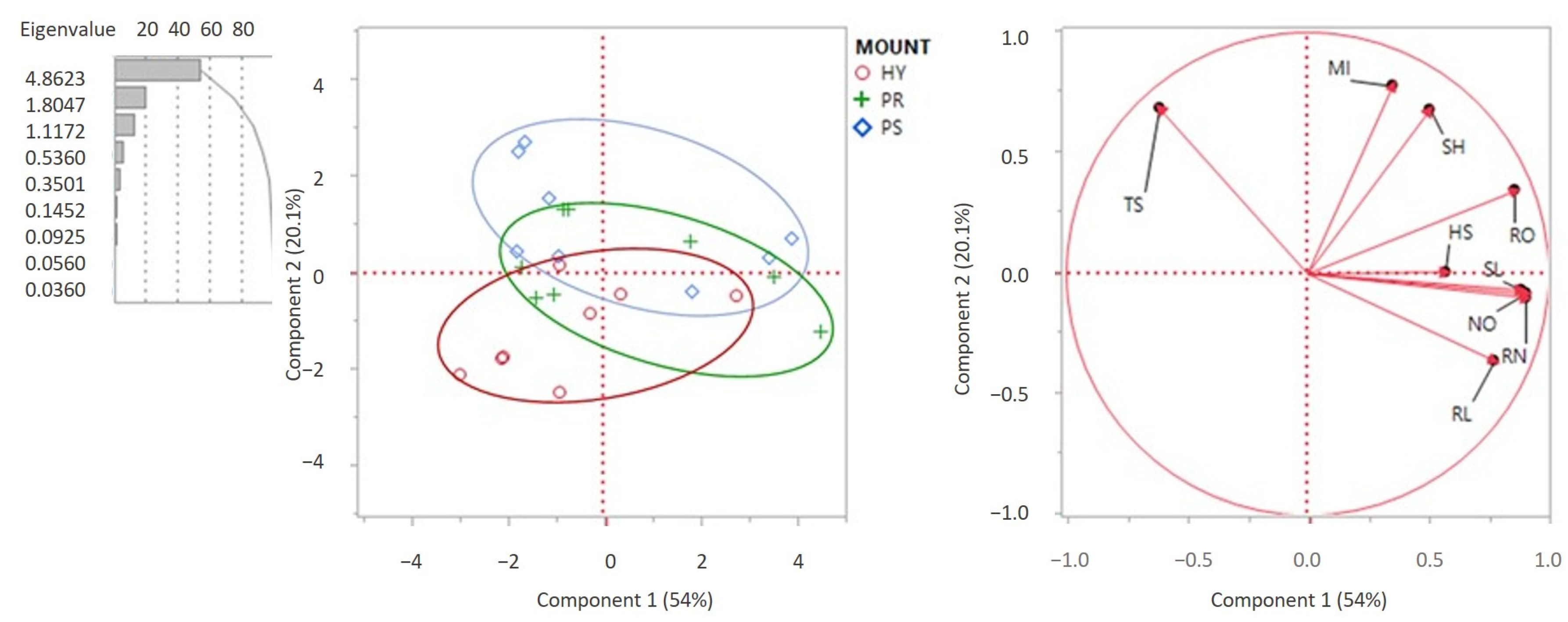
Principal Components (PC)
PC1PC2PC3
% Contribution of variability
54.020.112.4
Related variables
SLSHHS
NOTS
ROMI
RN
RL
Shoot length (SL); node number (NO); rooting percentage (RO); mean root number (RN); mean root length (RL); shooting percentage (SH); total shoot number (TS); multiplication index (MI); high shoot number (HS).
Table 4. Shoot regeneration of non-encapsulated (NEE) and encapsulated nodal explants when cultured on hormone-free Murashige and Skoog media, after 0, 30, and 60 days’ storage of the beads (E0d, E30d, and E60d, respectively) at 5 °C, and cultured for 30 or 50 days on media at 25 °C.
TreatmentGrowth
(Days)
Shooting (%)Total Shoot NumberHS NumberShoot Length (cm)Node NumberMI ††Rooting
(%)
Root Number
Ranking †††
Root Length (cm)
NEE30 84.02.0 c1.1 b2.52.7 b2.868.01.21.2
E0d 58.02.0 c1.1 b2.42.3 b1.933.01.91.9
E30d 78.01.9 c1.1 b2.12.3 b2.542.01.91.9
E60d 75.02.3 bc1.2 b2.62.9 b2.955.01.61.6
NEE5084.02.0 c1.2 b2.52.7 b2.872.01.41.4
E0d 58.02.3 bc1.5 ab3.43.4 ab2.239.02.42.4
E30d 72.03.4 ab2.1 a3.33.4 ab4.145.02.22.2
E60d 72.03.8 a1.2 b4.34.1 a4.670.02.32.3
Significance of two-way ANOVA
Treatmentns *** ***********
Growthns *** **ns******
Treatment × Growthns******ns***nsnsnsns
Mean values of factors ††††
Shooting (%)Shoot length (cm)
TreatmentMeanGrowthMeanTreatmentMeanGrowthMean
NEE87.0 a30 days72.5 aNEE2.5 b30 days2.4 b
E0d58.0 a50 days70.0 aE0d2.9 ab50 days3.4 a
E30d75.0 a E30d2.7 ab
E60d73.5 a E60d3.4 a
MIRoot number ranking
TreatmentMeanGrowthMeanTreatmentMeanGrowthMean
NEE2.8 ab30 days2.5 bNEE1.3 b30 days1.6 b
E0d2.1 b50 days3.4 aE0d2.2 a50 days2.1 a
E30d3.3 a E30d
E60d3.8 a E60d
Rooting (%) Root length (cm)
TreatmentMeanGrowthMeanTreatmentMeanGrowthMean
NEE72.0 a30 days50.5 aNEE2.1 b 30 days2.3 b
E0d36.0 b50 days56.0 aE0d3.9 a50 days2.7 a
E30d43.0 b E30d2.3 b
E60d62.5 a E60d2.5 b
Mean presented in columns according to Tukey’ s test at p < 0.05. ns, **, ***: non-significant or significant at p < 0.01 or p < 0.001, respectively; mean values followed by the same letter are not significantly different at p < 0.05; n = 40; HS: high shoot number (longer than 0.5 cm); †† MI: multiplication index = shooting (%) × mean shoot number × mean shoot length/0.6; ††† root number ranking: 1 (0–5 roots), 2 (6–10 roots), 3 (≥11 roots); †††† when interactions are not significant, mean values of factors are shown. When interactions are significant, mean values of the two factor combinations and their significance are shown.
Disclaimer/Publisher’s Note: The statements, opinions and data contained in all publications are solely those of the individual author(s) and contributor(s) and not of MDPI and/or the editor(s). MDPI and/or the editor(s) disclaim responsibility for any injury to people or property resulting from any ideas, methods, instructions or products referred to in the content.

