Article

Dominated “Inheritance” of Endophytes in Grapevines from Stock Plants via In Vitro-Cultured Plantlets: The Dawn of Plant Endophytic Modifications

1
School of Ecology and Environmental Science, Yunnan University, Kunming 650504, China
2
Institute of Forestry, Meizhou Academy of Agriculture and Forestry Sciences, Meizhou 514000, China
3
School of Life Science, Yunnan University, Kunming 650504, China
4
School of Chemistry and Environment, Yunnan MinZu University, Kunming 650504, China
5
College of Plant Protection, Yunnan Agricultural University, Kunming 650201, China
*
Authors to whom correspondence should be addressed.
Horticulturae 2023, 9(2), 180; https://doi.org/10.3390/horticulturae9020180
Submission received: 4 January 2023 / Revised: 18 January 2023 / Accepted: 24 January 2023 / Published: 1 February 2023
(This article belongs to the Special Issue Primary Production and Processing in Viticulture)

Abstract

:
We hypothesize herein the “inheritance” of endophytes in grapevines through in vitro-culture plantlets (IVCPs) from a stock plant to established plants, and, subsequently, that endophytes can be modified at the IVCP stage to emphasize one or more of those “heritable” endophytes in later-developed plants. Using a DNA amplicon sequencing approach, we investigated the dynamic successions of endophytic communities in two taxonomically different varieties of grapevines from IVCPs (stage 1) to plants of later growth stages (stages 2–4). Despite the great alterations of endophytic amplicon sequence variants (ASVs) during the development of grapevines, our results demonstrated the dominant preservation of earlier-stage-acquired endophytic ASVs in grapevines of later stages. More importantly, we detected the dominant “inheritances” of the IVCP-borne ASVs, which succeeded from the stock grapevine throughout all growth stages of grapevines, with a few of these ASVs accounting for the major relative abundance (RA: 35–76%) in later-established grapevines. Notably, most of these dominantly “inherited” IVCP-borne endophytes belong to genera from which species have been frequently reported to have great agricultural and horticultural importance. In addition, horizontally transmitted endophytic (HTE) ASVs are able to dominate in later-developed grapevines. This work illustrates the evolution of endophytes from IVCPs to plants of later-growth stages. The results suggest a strategy to “breed” plant lines with certain beneficial endophytes at the IVCP stage, which has been termed herein as “plant endophytic modification”.

1. Introduction

Naturally occurring plants, as well as aseptically cultured plantlets, organs and tissues are all hosts of microbial endophytes [1,2]. The intimate relationship between plants and endophytes makes the system a crucial entity when plants are challenged with environmental stresses [3]. As a result, the colonization of plants by endophytes greatly enhances the genomic and metabolic characteristics of plants, provides host plants with a range of essential life-support functions or enhances the survival strategies of host plants [4]. The host plants, in turn, provide nutrients and habitats that support the survival of the endophytes [5]. Because of beneficial plant–endophyte interactions, endophytes are of great interest and are expected to be applied in agricultural and horticultural practices [6]. However, despite increasing documentation, fundamental knowledge of plant–endophyte interactions and the underlying origination, assemblage, and function of endophytes remains to be elucidated before these endophytes can be efficiently applied [7].
Endophytes in host plants can originate horizontally or vertically [8,9]. Soil- or rhizosphere-inhabiting microorganisms are considered to be the primary horizontally transmitted endophytes (HTEs) via roots, followed by airborne microbes via plant leaves or stems [10]. On the other hand, successfully colonized endophytes can be transmitted across plant generations from parents to progenies via seeds or other propagules, and represent vertically-transmitted endophytes (VTEs) [8,11]. Seed-borne endophytes are VTEs of great concern, owing to their possible presence in emerging plants, and play significant roles in these plants during seed germination and early seedling establishment [8]. Generally, vegetative propagules (e.g., shoot tips or cuttings) carrying endophytes can also serve as VTEs, and confer host plants with equal or more functions compared with seed-borne ones, owing to the relatively larger volume and lower threshold for accommodating a greater endophytic diversity. It was revealed that shoot VTEs play significant roles in the assembly of root-associated microbiomes and the hyperaccumulation of heavy metals in Sedum alfredii plants [11]. Compared to HTEs, cross-generational VTEs could be looked upon as a kind of “acquired inheritable trait” of plants [3] which can be modified to create plant lines that are colonized with certain functional VTEs. In addition to the traditional strategy of plant genetic modification, we conceptualized this strategy as “plant endophytic modification”. However, to effectively apply the strategy of “plant endophytic modification” in practice, it is necessary to clarify the presence and duration of linkage colonization of VTEs between maternal plants and their progenies.
Endophytes may be obligate or facultative with their host plants [1]. Obligate endophytes require plant tissues complete their life cycle [12]. Obviously, endophytes that colonize in vitro-cultured plantlets (IVCPs) or plant calli [2] are more inclined to be obligate endophytes with higher host dependency. These endophytes were undoubtedly inherited from the maternal plants that shoot tips were taken from for IVCP induction. Because of host dependency, plants conserve these endophytes in IVCPs and calli, which may imply their functional importance; however, this is still poorly understood. In this study, the dynamic succession of amplicon sequence variants (ASVs) of IVCP-derived bacterial and fungal endophytes in two different varieties of grapevine were investigated at subsequent development stages, and the roles of these IVCP-derived endophytes in assembling the endophytic microbiota of grapevines at later stages are discussed.

2. Materials and Methods

2.1. Preparation of IVCPs

Vine shoot tips of two taxonomically different varieties of grapevine, Cabernet sauvignon (CS; Vitis vinifera L.) and rose honey (RH; V. vinifera L. × V. labrusca L.), that were used to induce grapevine IVCPs were harvested separately in 2013 from two different local vineyards in Yunnan Province, China. Single-bud clones of IVCPs were then subcultured under aseptic conditions for 7 years (more than 30 generations) and propagated into sufficient plantlets for subsequent studies. The IVCPs were cultured at 25 °C under a 12 h/12 h light/dark period. Prior to experimentation, the IVCPs of both cultivars of grapevines were randomly harvested (24 plantlets for each cultivar, with every 8 plantlets pooled as one sample) under aseptic conditions as stage 1 samples (labelled as C1 and R1 for CS and RH cultivars, respectively). The samples (leaves and tender stems of rootless IVCPs) were cut into 0.5 cm fragments and directly placed in 10 mL aseptic plastic tubes, rapidly frozen in liquid nitrogen, and subsequently stored at −80 °C for further analyses.

2.2. Cultivation and Sampling of Grapevines at Different Growth Stages

The rooted IVCPs were cultivated by using standard refining procedures in February 2020. Briefly, the lids of the culture bottles were first opened in the tissue culture room, and subsequently transported to the training room for transplantation into pots containing culture materials (humus soil: perlite = 3:2). The potted vine plantlets were irrigated once every 3 days with tap water and kept at 25 °C under a 12 h/12 h light/dark period. After 30 days of transplantation, the plantlets were randomly harvested (15 plantlets for each cultivar, with every 5 plantlets pooled as one sample) as stage 2 samples (C2 and R2). The samples were surface-sterilized, frozen with liquid nitrogen, and stored at −80 °C for further analyses. Vine plantlets were surface-sterilized with 75% alcohol for 30 s and 3% sodium hypochlorite (NaClO) for 20 min, followed by three washes with sterilized water. Surface sterilization was confirmed by smearing the water from the third wash over prepared potato dextrose agar (PDA) and yeast extract peptone dextrose (YEPD) plates. After hardening, the vine plants were transplanted into pots (30 cm in diameter) containing soil (field soil with 30% humus soil), and transferred to a greenhouse illuminated with natural sunlight (14 h /10 h = light/dark) at temperatures ranging between 16 °C and 28 °C. The plants in each pot were irrigated with 0.5 L of tap water every 3 days. The growth of one shoot was maintained in each of the vine plants by removing all of the other emerging lateral buds. When vine shoots reached a height of approximately 0.8 m with 5–7 expanded blades in June 2020, grapevines were harvested (9 plantlets for each cultivar and every 3 plantlets were pooled as one sample) as stage 3 samples (C3 and R3). The stage 3 plants were divided into sections and sampled as stems (C3S and R3S), leaves (C3L and R3L), and roots (C3R and R3R), after which the samples were surface sterilized and stored at −80 °C for subsequent analyses. The remaining plants were transplanted to the open field and grown under natural conditions until October 2020. The plants had stopped growing at this stage (stage 4) and reached a height of approximately 1.2 m. The plants were harvested (9 plantlets for each cultivar and every 3 plantlets were pooled as one sample) by sections (C4L, R4L, C4S, R4L, C4R and R4R). The leaves and stems of the upper (C4uL, C4uS, R4uL and R4uS) and lower (C4lL, C4lS, R4lL and R4lS) sections of stage 4 plants were separately harvested for both the cultivars. The samples were surface-sterilized and stored at −80 °C for subsequent analyses. The information for all harvested samples above is listed in Table 1, and 3 biological replicates for each of the samples were prepared in the experiment.

2.3. Profiling Endophytic Microbiota of Grapevine Tissues

Genomic DNA was extracted from all the samples using a FastDNA® Spin kit (MP Biomedicals, Solon, OH, USA), according to the manufacturer’s protocol. The size and quality of the extracted DNA were verified by electrophoresis using 1% (w/v) agarose gels. The concentration and purity of the extracted DNA were spectroscopically estimated using a NanoDrop™ 2000 spectrophotometer (Thermo Fisher Scientific, Wilmington, DE, USA). Bacterial 16S rRNA and fungal ITS regions were amplified from all the samples using the ITS1-FI2 (5′-GTGARTCATCGAATCTTTG-3′) and ITS2 (5′-TCCTCCGCTTATTGATATGC-3′) primers for the endophytic fungal microbiomes, and the 341F (5′-CCTACGGGNGGCWGCAG-3′) and 805R (5′-GACTACHVGGGTATCTAATC1C-3′) primers for the endophytic bacterial microbiomes. Polymerase chain reaction (PCR) was performed for each sample, which included 35 cycles of denaturation at 98 °C for 10 s, annealing at 54 °C for 30 s, extension at 72 °C for 45 s, and a final step at 72 °C for 10 min. The 25 μL PCR mixture contained 12.5 μL of Phusion® Hot Start Flex 2× Master Mix (New England Biolabs Inc., Beverly, MA, USA), 2.5 μL of each primer (1 μM), and 50 ng of template DNA. The PCR products were collected and purified using an agarose gel DNA purification kit (TaKaRa Bio Inc., Shiga, Japan). The amplicon pools were prepared for sequencing, and the size and quantity of the amplicon library were assessed using an Agilent 2100 Bioanalyzer (Agilent, Santa Clara, CA, USA) and a Library Quantification Kit for Illumina (Kapa Biosciences, Woburn, MA, USA), respectively. The libraries were sequenced using an Illumina MiSeq PE300 Sequencer (Illumina, Inc., San Diego, CA, USA) at LC-BioTechnologies Co., Ltd. (Hangzhou, China). The fungal and bacterial sequences were deposited in NCBI under accession number PRJNA846512.

2.4. Computational Analyses

Sequence analyses were performed using the Quantitative Insights Into Microbial Ecology 2 (QIIME2) program, version 2019.1, and necessary plugins [13]. For constructing the amplicon sequence variants (ASVs), denoising and quality control (including removal of chimera) were performed with the deficiency of adenosine deaminase type 2 (DADA2) plugin, using the qiime dada2 denoise-paired command, and the reads were truncated to avoid low-quality scores (N235 bp for forward reads and N142 bp for reverse reads; truncQ = 2, maxEE = 2). Microbial taxonomy was assigned to the ASVs using the UNITE dynamic database [14] (version 2019.2) at 97–99% similarity, using the qiime feature-classifier classify-sklearn command. Unidentified microbes at the kingdom or phylum levels, reads of poor quality, chloroplast DNA, mitochondrial DNA, and other sequences were removed from the dataset.
All the statistical analyses were performed using specific packages in R, version 3.5.1 (The R Foundation for Statistical Computing, Vienna, Austria), unless otherwise stated. The rarefaction curves based on the ASV tables were calculated using USEARCH, version 10.0.240. The alpha diversities (Shannon indices) between sample groups were compared using boxplots. Kruskal–Wallis and pairwise Wilcoxon tests were performed to verify the significant differences among sample groups. Principal coordinate analysis (PCoA) of the ASVs detected in different stages and sections was performed to determine the beta diversity at the ASV level [15]. The codistribution of endophytic ASVs in different stages and sections was determined by Venn analysis. The endophytic abundance of a sample was evaluated by the detected total clean reads of the sample, while the relative abundance (RA) of certain endophytic ASVs (or at other taxonomy levels such as genus, phylum) was calculated as percentages.
The different packages in R and the tools in Excel and PowerPoint were used for performing most of the statistical analyses and graphical representation of the results.

3. Results

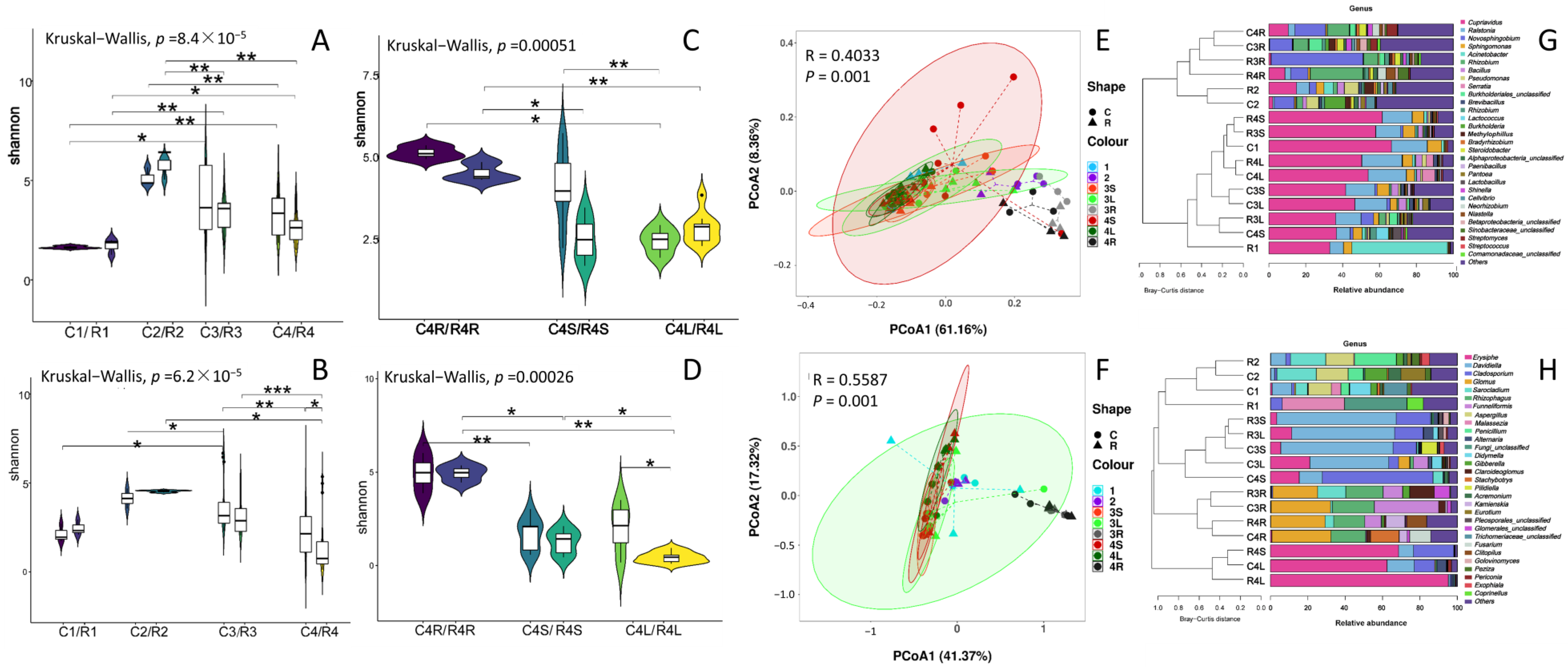
3.1. IVCPs and Later-Developed Grapevines Established Endophytic Microbiomes in a Stage-, Variety- and Tissue-Specific Manner

The profiling of the endophytic microbiomes of all grapevine samples was validated (Supplementary Tables S1–S4; Supplementary Figure S1). A total of 2,116,102 16S rRNA clean reads were obtained (1,619,664 and 496,438 from CS and RH cultivars, respectively), and 5993 ASVs were generated (4529 and 2999 from CS and RH cultivars, respectively) from the 80 tissue samples. Additionally, a total of 4,974,454 clean ITS reads (CS: 3,468,260; and RH: 1,506,194) and 2051 fungal endophytic ASVs (CS: 1497; and RH: 1162) were obtained.
Of the four growth stages of grapevine studied herein, both the abundance and ASV numbers of bacterial endophytes in grapevines were maximum at stage 2 in both the CS and RH cultivars, and then declined at stages 3 and 4 (Figure 1A,B). The abundance of fungal endophytes was greatly increased from stage 1 to stage 2 grapevines, while the ASV numbers of fungal endophytes increased along growth stages in grapevines of both variants, and peaked at stage 3 (Figure 1C,D). In stage 4 grapevines, the roots of both cultivars had the highest number of sectionally detected ASVs, which declined along sections of roots, stems and leaves (Figure 1E–H). Accordingly, the roots of both vine cultivars were also colonized by the most abundant bacterial endophytes (Figure 1E,F), whereas the abundance of fungal endophytes did not show obvious differences among sections in stage 4 grapevines of both variants (Figure 1G,H).
With only one exception, the majority (47~72%) of the detected endophytic ASVs in each grapevine stage showed specificity in different variants, but these varietal-specific endophytic ASVs contributed quite low relative abundances (RA = 3~22%) in the grapevines (Figure 1I,J).
Of the four stages, IVCP (stage 1) of both vine variants showed a lower diversity (Shannon index = 1.1–2.0) of bacterial and fungal endophytes (Figure 2). The endophytic diversity rapidly increased and peaked at stage 2 (4.60–5.98), and subsequently decreased in stage 3 and 4 grapevines (1.75–5.80) of both variants (Figure 2A,B). Of the 3 vine sections (root, stem and leaf), root had the highest endophytic diversity (4.04–4.78; Figure 2C,D). In most cases, the diversity between different growth stages and sections reached a significant difference (p < 0.05) (Figure 2).
In the principal coordinate analysis (PCoA), endophytes of stage 1 grapevines, as well as the root endophytes (both bacterial and fungal) of stage 3 and 4 grapevines, were isolated from other vine stages and vine parts in both variants. However, endophytes of different vine cultivars were not well resolved by PCoA (Figure 2E,F). The bacterial endophytes of stage 2 vines closely clustered with root bacterial endophytes of stage 3 and 4 grapevines (Figure 2E). In addition, endophytes of other grapevine samples of both vine variants could hardly be resolved in the PCoA plots (Figure 2E,F).
The bacterial endophytes detected in IVCPs of both the CS and RH cultivars mainly belonged to the phylum Proteobacteria (RA ≥ 99%; Supplementary Figure S2). In total, 66 and 74 bacterial genera were detected in CS and RH IVCPs, respectively. Among these, Cupriavidus, Ralstonia, Sphingomonas and Acinetobacter were the most dominant genera in both the CS and RH variants (Figure 2G; Supplementary Figure S3). Accordingly, the detected endophytic fungi in the IVCPs of the CS cultivarwere primarily of the Ascomycota (RA = 78.7%), Basidiomycota (RA = 19.9%), and Glomeromycota (RA = 1.3%) phyla, of which 37 genera exhibited a dominant distribution pattern (RA > 0.1%). Aspergillus, Didymella, Davidiella and an unclassified Trichomeriaceae were the most dominant genera (RA > 10%). Notably, arbuscular mycorrhizal fungi (AMF), including Glomus, Funneliformis and Rhizophagus were also the dominant genera (RA > 0.1) in IVCPs of the CS cultivar (Figure 2H; Supplementary Figures S2 and S3).
During the growth of the grapevine from IVCP to different stages, the endophytic microbiota in grapevines were differentially established with a certain degree of variety-, stage-, and compartment-specificity (Figure 2G,H; Supplementary Figures S2 and S3). For bacterial endophytes, the generic composition of endophytes of stage 2 grapevines and the roots of stage 3 and 4 clustered together as one group, and the remaining samples formed another group (Figure 2G). Roots were dominated mainly by the bacterial genera Novosphingobium and Rhizobium, while other parts and stage 1 grapevines were dominated mainly by the genera Cupriavidus, Ralstonia and Sphingomonas of both cultivars (Figure 2G). Of the fungal genera, in addition to the root parts of stages 3 and 4, endophytes of stage 1 and 2 vines also separately clustered as one group. The root parts were mostly dominated by the AMF-forming genera Glomus and Rhizophagus, while the stem and leaf parts of stage 3 and 4 grapevines were dominantly colonized by Davidiella and Cladosporium in both cultivars (Figure 2H). In addition to the aforementioned fungal genera, stem and leaf parts of both varieties were also dominantly colonized by the fungal genus Erysiphe, a disease causing (powdery mildew) agent (Figure 2H).

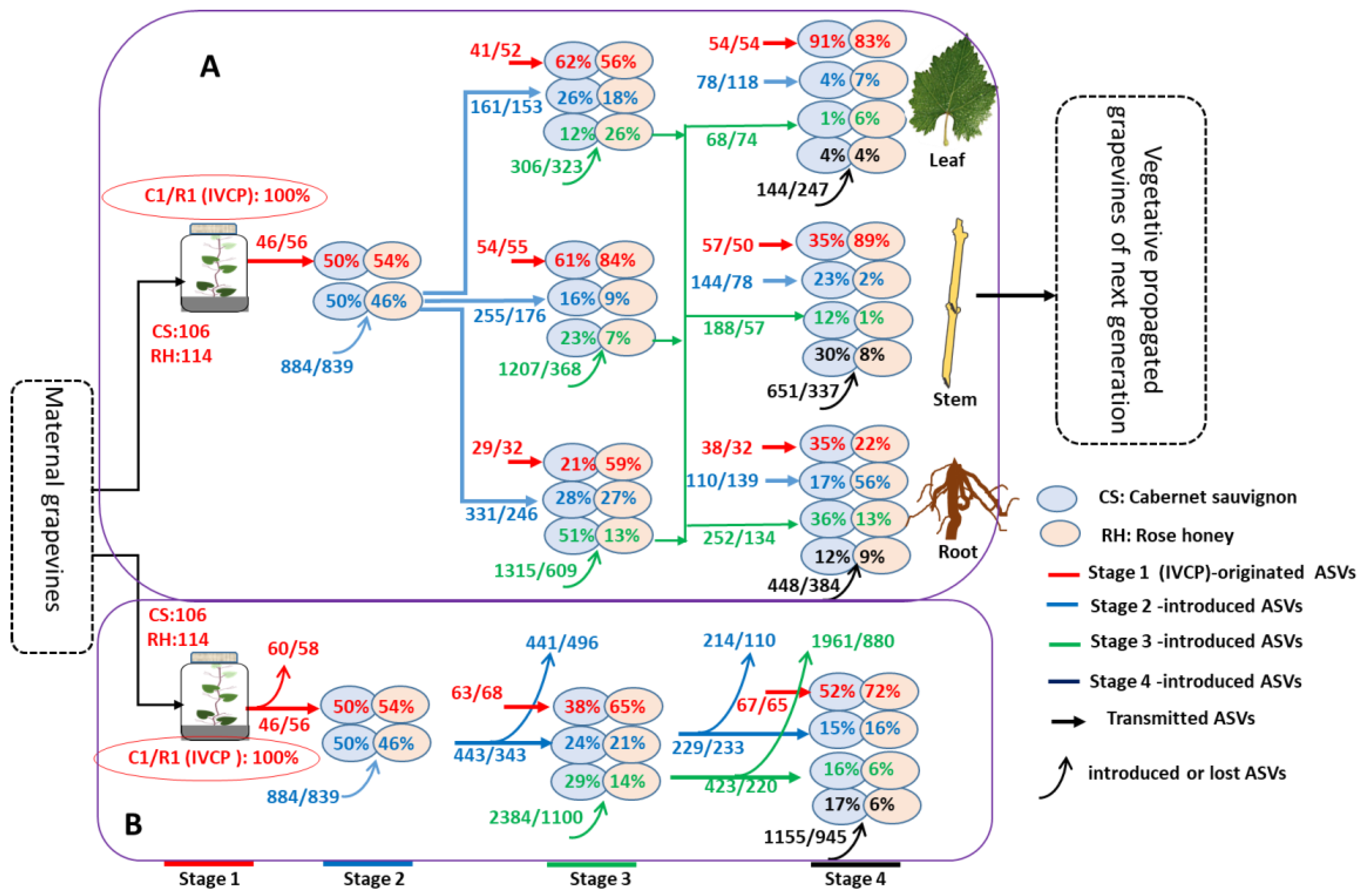
3.2. Dynamic Succession of Endophytic ASVs in Grapevines along Growth Stages

A total of 106 and 114 endophytic bacterial ASVs colonized in the CS and RH IVCPs, respectively (Figure 3A,B). Approximately half of those IVCP-derived endophytic bacterial ASVs were detected and remained in high relative abundance (RA: 38–72%) in grapevines of later-growth stages (Figure 3A,B; Supplementary Figure S4). The highest RA of IVCP-derived bacterial ASVs was detected in the blades (56–91%), followed by the stems (35–89%), and roots (21–59%) in stage 3 and 4 vine plants of both cultivars (Figure 3A). The number and RA of IVCP-derived bacterial ASVs varied according to the cultivar, stage of growth, and plant section (Figure 3A).
In addition to the IVCP-sourced endophytes, large numbers (839–2384) of horizontally-transmitted endophytic bacterial ASVs (HTE-ASVs) were detected in every later-stage grapevine, whereas only a small proportion of these HTE-ASVs could be detected in grapevines of subsequent growth stages (Figure 3B). However, a reduction in the number of bacterial HTE-ASVs in later-stage grapevines did not indicate a reduction in their RA (Figure 3A). For instance, 255 successful bacterial HTE-ASVs derived from stage 2 were detected in 16% of stage 3 CS vine stems, while 144 stage 2-derived bacterial HTE-ASVs accounted for 23% of the RA in stage 4 CS vine stems. The number of preserved bacterial HTE-ASVs in later-stage grapevines varied by vine growth stage, variety, and compartment (Figure 3B). Additionally, the newly colonized bacterial HTE-ASVs were mainly distributed in the roots, with a higher RA of 56% and 36% in the RH and CS cultivars, respectively, followed by stems and blades, with a few exceptions (Figure 3A).
The succession of endophytic fungi in CS grapevines of different stages and compartments was generally similar to that of endophytic bacteria (Figure 4). The IVCP-derived endophytic fungal ASVs tended to accumulate specifically in the blades and stems of stage 3 and 4 plants, while the fungal HTE -ASVs mainly dominated in the roots (Figure 4A). In contrast to the bacteria, fewer fungal ASVs were horizontally transmitted into grapevines during stages 2, 3 and 4, and most of these fungal HTE-ASVs were not detected in vines of the subsequent growth stages (Figure 4B).
The dynamic alterations of the 10 most dominant (Top10) endophytic ASVs in different-staged grapevines are depicted (Figure 5). In this study, more than half of the Top10 distribution endophytic ASVs (both the fungal and bacterial endophytes) in grapevines of later growth stages succeeded from IVCPs (Figure 6). Among these, three of the top 10 bacterial ASVs [Bc01 (Cupriavidus sp.), Bc02 (Ralstonia sp.), Bc03 (Sphingomonas sp.)] in later-staged CS vines were also the top 10 ASVs in IVCPs of the cultivar. Similarly, three bacterial ASVs in IVCPs of RH grapevines were preserved through all later growth stages. Interestingly, these Top10 distribution bacterial ASVs in both the IVCPs and later-stage grapevines were commonly shared between CS and RH cultivars (e.g., Br02/Bc01 (representing that the Br02 in RH grapevines is the same ASV as that of the Bc01 in CS vines); Br03/Bc02; Br04/Bc03). In addition, another low-abundance distribution bacterial endophytic ASV which was detected in CS (Bc32) and RH (Br48) IVCPs showed the top 10 distribution in all later-staged grapevines, and the ASV (Bc32/Br48), which belongs to genus Novosphingobium, was also commonly shared in grapevines of both variants (Figure 5A,B; Supplementary Table S5). In contrast, other low-abundance endophytic bacterial ASVs in IVCPs exhibited a top 10 distribution in grapevines at stages 2 or 3, whereas none of these ASVs maintained the top 10 distribution in stage 4 grapevines (Figure 5A,B).
Accordingly, ten, eight and seven IVCP-derived endophytic fungal ASVs were separately detected among the top 10 distributions in stage 2, 3 and 4 grapevines of the CS cultivar. Endophytic fungal ASVs Fc02 (Davidiella), Fc07 (Sarocladium), Fc09 (Aspergillus) and Fc10 (Davidiella) were detected among the top 10 distributions in both the IVCP and later-staged vines of the CS variant (Figure 5C). Fc07 and Fc09 were only detected in the top 10 distribution in stage 2 grapevines, while Fc02 and Fc07, which belong to the same genus Davidiella, maintained the top 10 distribution in stages 3 and 4 CS grapevines (Figure 5C). In addition, other top 10 distribution fungal ASVs in later-staged grapevines of the CS cultivar were derived from those low-abundant distribution ASVs in IVCPs (Figure 5C). A few fungal endophytic ASVs (16) were detected from the IVCPs of the RH cultivar, and two of these ASVs (Fr05 and Fr15) maintained the top 10 distribution in RH grapevines of later development stages. These top 10 distribution fungal ASVs were also commonly shared in both cultivars (Fr05/Fc16; Fr15/Fc10) (Figure 5D).
Some HTE-ASVs showed the top 10 distribution in grapevines at the stages in which they were introduced or the subsequent stages (Figure 5).
In stage 4 grapevines of the both variants, four bacterial HTE-ASVs derived from grapevines of later growth stages showed a dominant distribution (Top10) in stage 4 CS plants (Figure 5A). Accordingly, six and one bacterial HTE-ASVs that originated from stage 2 and stage 3 grapevines, respectively, showed a Top10 distribution in stage 4 RH plants (Figure 5B). One fungal HTE-ASV from stage 2 plants, and two fungal HTE-ASVs from stage 3 plants exhibited a dominant distribution in stage 4 CS vines (Figure 5C).
The 3 most abundant bacterial endophytic ASVs in grapevines of later growth stages were inherited from IVCPs of both the cultivars. These endophytic ASVs belong to the genera Cupriavidus (Bc01/Br02), Ralstonia (Bc02/Br03), Sphingomonas (Bc03/Br04), Novosphingobium (Bc32/Br48), Burkholderia (Bc59) and Pseudomonas (Br52) (Figure 5A,B, Supplementary Table S5). Accordingly, the three most dominant fungal endophytic ASVs in stage 2, 3, and 4 grapevines were also inherited from IVCP, and these fungal ASVs belong to genera Sarocladium (Fc07), Aspergillus (Fc09), Davidiella (Fc10/Fr15/Fc34), Gibberella (Fc12), Cladosporium (Fc16/Fr05), Erysiphe (Fc20) and Eurotium (Fc22) (Figure 5C,D; Supplementary Table S5).

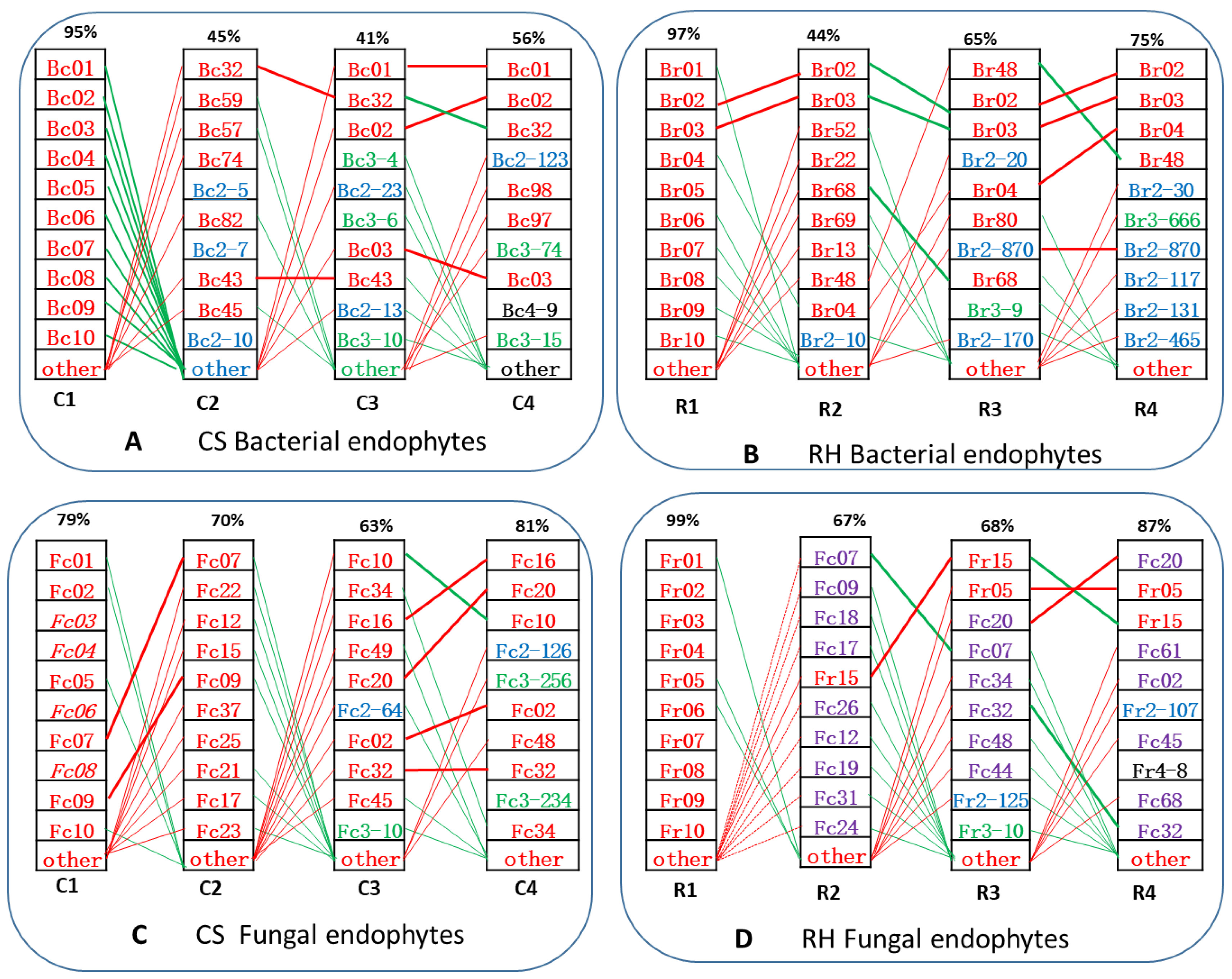
3.3. IVCP-Derived Core Endophytic ASVs (c-ASVs) Dominated in the Grapevines of Later Growth Stages

Despite the alternation of large quantities of endophytic ASVs, certain numbers of IVCP-derived c-ASVs remained constant in grapevines throughout all growth stages (Figure 6A–D). A total of 42 bacterial c-ASVs were detected with RAs of 39%, 36%, and 44% in stage 2, 3, and 4 CS grapevines, respectively (Figure 6A). Interestingly, the percentages of these dominantly distributed bacterial c-ASVs in stage 2, 3, and 4 CS grapevines were below 5% (Figure 6E). Fewer than 5% (45) of the bacterial c-ASVs also exhibited a dominant distribution (with RAs of 47%, 64%, and 71% in stages 2, 3, and 4, respectively) in RH vines of later growth stages (Figure 6B,F). A total of 24 IVCP-derived fungal c-ASVs maintained a linkage endophytism in all growth stages of the CS grapevines, and a small percentages (<10%) of these fungal ASV numbers also dominated (with the RAs of 70%, 56%, and 76% in stages 2, 3, and 4, respectively) in the later-stage grapevines (Figure 6C,G). Three endophytic fungal c-ASVs were detected in RH vines, and these three fungal ASVs also showed dominant distribution (RA = 45%) in stage 3 vines of the cultivar (Figure 6D,H).
Of the total bacterial c-ASVs detected in the CS and RH grapevines, 27 were commonly shared by the two cultivars (Figure 6I). The numbers of bacterial c-ASVs varied from different compartments of stage 4 plants (Figure 6J–L). An average 20 bacterial c-ASVs exhibited higher RAs in the vine leaves (CS: 83%; RH: 83%) and stems (CS: 53%; RH: 89%); however, a similar number of endophytic bacterial c-ASVs exhibited lower dominance in the roots (CS: 35%; RH: 24%) (Figure 6J,K). Accordingly, an average number of 9 fungal c-ASVs showed a dominant distribution pattern in the leaves (RA = 91%) and stems (RA = 82%) of the CS cultivar, while the RA of similar numbers of CS-c-ASVs was lower in the roots (RA = 10%) of this cultivar (Figure 6L).
The endophytism of c-ASVs exhibited a tissue-specific distribution pattern (Figure 6M,N). Of the endophytic bacterial c-ASVs, Bc01/Br02, Bc02/Br03 and Bc03/Br04 tended to dominate in the blade and stem sections, while Bc32/Br48 showed a dominant distribution pattern specifically in the roots (Figure 6M). The endophytic fungal c-ASVs Fc20, Fc16, Fc10, Fc48 (Alternaria sp.) and Fc02 (Davidiella sp.) showed a dominant distribution pattern specifically in the leaves and stems, while Fc32 (Glomus sp.), F44 (Claroideoglomus sp.), and F58 (Rhizophagus sp.) were specifically distributed in the roots, with higher RA values (Figure 6N).

4. Discussion

4.1. Grapevine IVCPs Inherit Diverse Endophytes from Maternal Plants

In addition to naturally growing plants, long-term, actively maintained healthy grape calli harbor diverse bacterial endophytes [2]. Our present study further confirmed the diversity distribution of both the bacterial and fungal endophytes in IVCPs of grapevine. Bacterial endophytic genera that were detected in IVCPs of our present experiment, such as Lactobacillus, Streptococcus, Pseudomonas, Acinetobacter, Bacillus and others, were also detected in grapevine calli [2].
The genotype specificity of endophytic communities is widely recognized, and partially explains the optimal inhabited niche hypothesis [4,16,17,18]. Of the total 177 IVCP-borne endophytic bacterial ASVs, 134 ASVs were specifically distributed separately in CS and RH IVCPs (Figure 1). Interestingly, more than 60% of these vine variant-specific endophytic bacterial ASV numbers showed extremely low relative abundances (RA ≤ 3%) in the IVCPs of both cultivars (Figure 2I). On the other hand, these results could be attributed to the fact that the IVCP of different grapevine cultivars tends to selectively preserve a higher abundance of commonly shared bacterial endophytes for their benefits. In contrast to the bacterial endophytes, fungal endophytic ASVs preserved in vine IVCPs of the two variants showed a different pattern in this assay; the two varieties only shared 5 fungal ASVs and their RAs varied greatly in CS and RH IVCPs, being 3% and 47%, respectively (Figure 2I). Together with the results that fungal endophytes in vine IVCP samples compositionally differed from one replicate to another (Figure 2F; Supplementary Figure S2), we can conjecture the uneven distribution of fungal endophytes in plant tissues. As a result, the fungal endophytic ASVs were differently preserved in different individual IVCPs during the subculture manipulations.
Obviously, the endophytes that colonized inside IVCPs were vertically transmitted from a maternal grapevine, where vine shoot tips were taken to induce IVCPs, similar to seed-borne VTEs [8]. We could also speculate that some of these IVCP-borne endophytes had been “inherited” from a far-ancestor mother plant via generations of vegetative propagations. Our results that the IVCP-borne endophytic ASVs dominated in stems of later-stage grapevines ensured that these endophytes could be passed to the next vegetative generations via cut propagations. However, it remains to be elucidated how these endophytes originated and were preserved in IVCPs long-term in subcultivated generations.

4.2. Competition- and Succession-Driven Evolution of Endophytic Microbiota during Plant Growth and Development

The endophytism of microorganisms in plants is influenced by numerous biotic and abiotic factors [4]. Endophytes in vine plants processed dynamic changes during the growth of grapevines from the IVCP stage, through the seedling hardening stage, and finally to the mature grapevine stage in the fields (Figure 3 and Figure 4). The diversity and abundant distribution of endophytes in stage 2 vine plantlets (Figure 1 and Figure 2), implies a window stage of plants for horizontally transmitted endophytes (HTEs). The extremely high out-rates of endophytic HTE-ASVs in plants along growth stages (Figure 3 and Figure 4) illustrates the severe competition among colonized endophytes, and the effects of host selectivity. However, the underlying mechanisms of environment–host–endophyte interactions remain largely unelucidated.
HTEs exhibited sectional distribution in the blades, stems, and roots of stage 3 and stage 4 plants. The roots showed a higher tendency of being colonized by HTE-ASVs, and their RAs gradually decreased from the roots to stems to blades (Figure 3 and Figure 4). This confirmed that soil-borne microbes introduced into the roots primarily account for the HTEs in roots and certain aboveground tissues [17,19]. However, the successful colonization of HTEs in plants became increasingly difficult during plant growth and development owing to the declining numbers and RAs of both the introduced and preserved HTE-ASVs in vine plants along the growth stages (Figure 3 and Figure 4), which could be explained as eco-niche occupation by earlier colonizers.

4.3. Grapevines Dominantly “Inherit” IVCP-Borne Endophytes Which Are Inferred to Be of Great Agricultural and Horticultural Importance

IVCP-borne bacterial endophytic ASVs belong to the genera Cupriavidus (Bc01/Br02), Ralstonia (BC02/Br03), Sphingomonas (Bc03/Br04), Brevibacillus (Bc08/Br08), Novosphingobium (Bc32/Br48), Methylophilus (Bc43/Br22), Pseudomonas (Br52), Burkholderia (Bc59), Aquabacterium (Bc57) and Cellvibrio (Bc74), which were detected to be have dominant distribution in grapevines of later development stages (Figure 5 and Figure 6; Supplementary Table S5). Bacterial species belonging to the genera Cupriavidus, Ralstonia, Brevibacillus and Burkholderia, are rhizobia with nitrogen-fixation functions [20,21]. Cupriavidus and Ralstonia also serve as model organisms for heavy metal detoxification [22]. Endophytic bacterial ASVs from Pseudomonas, Sphingomonas, Bacillus, Acinetobacter and other genera exhibited a dominant distribution pattern in IVCP and later growth stages (Figure 6), and bacteria belonging to these genera conferred host resistance to pathogens via induced systemic resistance (ISR) and alleviated drought damage in host plants via their root/shoot growth-promoting activities [23,24,25,26]. Pseudomonas, Sphingomonas, and other genera represent the signature taxa of the aboveground vascular system, and some members of these genera have roles in phytohormone production and defence against vascular pathogens by occupying the same niche [27,28,29]. Methylophilus sp. are capable of using methanol as the sole carbon and energy source during growth, and are assumed to potentially dominate the phyllospheric environment [30]. A study demonstrated that the inoculation of strawberry plants with a Methylophilus sp. strain influenced the biosynthesis of flavor compounds, including furanones, in the host plants [31], which suggested a potential role of these bacteria in regulating the sensory qualities of grapes and resulting wines.
The most dominantly distributed IVCP-borne fungal endophytic ASVs in CS vines of later growth stages included Fc07, Fc22 (Eurotium sp.), Fc12 (Gibberella sp.), Fc15 (Acremonium sp.), Fc09, Fc10, Fc34, Fc16, Fc49 (Pilidiella sp.) and Fc20 (Erysiphe sp.) (Figure 5C and Figure 6N). With the one exception of the fungal genus Erysiphe (a disease-causing agent for powdery mildew), other fungal genera, such as Gibberella, Acremonium, Aspergillus, Davidiella, Penicillium, and Sarocladium, are reported to have multiple beneficial effects on host plants, including growth promotion, stress tolerance, and pathogen resistance, owing to their ability to produce phytohormones, antibiotics, and other secondary metabolites [1,11]. The fungal genera Glomus and Rhizophagus are known as AMF, which have undeniable ecological and economic importance and play crucial roles in plant water and nutrient absorption [32,33]. It is interesting that AMF as endophytes colonized in some IVCP samples (Figure 2H), which were reported mainly in soils and roots [34]. However, the endophytism of these fungal genera in vine stems and leaves of later developed grapevines in this experiment may explain why stem-colonized AMF might have opportunities to be transmitted into subsequent vegetative progeny vines. Owing to the nature of cultivation-recalcitrance, the precise functions of all these IVCP-borne endophytes in grapevines require further clarification.

4.4. Grapevines “Inherit” IVCP-Borne Endophytes That Govern the Assemblage of Endophytic Microbiota in Later-Developed Plants

Plant pathogens or endophytes are known to spread via vegetative propagations [35]. One may hypothesize that fractions of these maternal endophytes can be transmitted to commercial plants via cuttings [17], which could possibly be developed to “breed” plant lines associated with certain beneficial endophytes. To validate this hypothesis, the present study tracked IVCP-borne endophytes in later stages of developed grapevines. We tracked the endophytes at the ASV level instead of other taxonomical levels (such as genus or species) to ensure that the detected endophytes in later-staged grapevines were IVCP-derived, rather than HTEs of the same genera or species. Additionally, the place where the experiments were performed was hundreds of kilometers away from the vineyard where vine shoot tips were used to induce IVCPs, further minimizing the possible occurrence of the same environmental ASVs as the IVCP-borne ASVs. Our results that the dominant “inheritance” of IVCP-borne endophytes and a few IVCP-derived endophytic ASVs contributed to the major RAs in later-staged grapevines (Figure 5 and Figure 6), illustrates the dominant role of IVCP-borne endophytes in governing the assemblage of endophytic microbiota in later stages of grapevines. These findings imply that the modification of endophytes at the IVCP stage may result in a differently shaped endophytic microbiota in later-developed grapevines. This suggests a possible strategy to purposely “breed” plant lines that are dominated by certain beneficial “heritable endophytes” which we have termed here as “plant endophytic modification”. Nonetheless, there is still a long way to go before this strategy can be successfully applied in practice.

Supplementary Materials

The following supporting information can be downloaded at: https://www.mdpi.com/article/10.3390/horticulturae9020180/s1, Table S1. Statistics of 16S rRNA amplicon sequences in grapevine samples of variety Cabernet sauvignon. Table S2. Statistics of 16S rRNA amplicon sequences in grapevine samples of variety Rose honey. Table S3. Statistics of ITS amplicon sequences in grapevine samples of variety Cabernet sauvignon. Table S4. Statistics of ITS amplicon sequences in grapevine samples of variety Rose honey. Table S5. List of the 5 most dominantly distributed (top 5) endophytic ASVs in different-staged grapevines. Figure S1. The Goods coverage (A–C) and Chao 1 (D–E) rarefaction curves for 16r rRNA (A, B, D and E) and ITS DNA sequencing in grapevine samples of the cultivar CS (A, C, D, E) and RH (B and E). Figure S2. The cluster and composition of the top 20 dominant endophytic phyla in different-staged and compartmentalized grapevines. C1-C2 and R1-R2 separately represent the stage 1 to stage 2 grapevine samples of the two cultivars. Grapevines of stages 3 and 4 of both grapevine cultivars were further divided sectionally into stems (C3S, C4S, R3S and R4S), leaves (C3L, C4L, R3L and R4L) and roots (C3R, C4R, R3R and R4R) as summarized in Table S1. Replicates of all samples are shown in the plots. Figure S3. The cluster and composition of the top 20 dominant endophytic genera in different-staged and compartmentalized grapevines. C1-C2 and R1-R2 separately represent the stage 1 to stage 2 grapevines of the two cultivars. Grapevines of stages 3 and 4 of both grapevine cultivars were further divided sectionally into stems (C3S, C4S, R3S and R4S), leaves (C3L, C4L, R3L and R4L) and roots (C3R, C4R, R3R and R4R) as summarized in Supplementary Table S1. Replicates of all samples are shown in the plots. Figure S4. Dynamic changes in endophytic ASV numbers and relative abundances along staged grapevines. The succeeded numbers (N) of endophytic ASVs (16S: bacterial; ITS: fungal) and the relative abundances (D) in different-staged grapevines are listed in the figure. The total detected numbers of endophytic ASVs in different-staged vines and variants are listed accordingly inside the ellipses. C1-C4 and R1-R4 separately represent the stage 1 to stage 4 grapevines of the two cultivars Cabernet sauvignon (CS) and rose honey (RH).

Author Contributions

M.-Z.Y. and S.-S.Z. designed the research and wrote the main manuscript text. S.-Y.X. performed the majority of the experiments and data analysis. Y.-T.W., C.-X.C., C.-M.L. and T.L. participated in the lab work and data analysis. X.-X.P., participated in the figure preparation and manuscript organization. All authors have read and agreed to the published version of the manuscript.

Funding

This work was financially supported by the National Natural Science Foundation of China (NSFC: 31560538); the joint foundation of the Yunnan Provincial Department of Science and Technology and Yunnan University (No. 2019FY003024); and the Yunnan provincial key S&T Special Project (202102AE090042-02-04).

Data Availability Statement

All data and materials are available in the manuscript and supporting information, and in NCBI under the accession number PRJNA846512.

Conflicts of Interest

The authors declare that the research was conducted in the absence of any commercial or financial relationships that could be construed as a potential conflict of interest.

Abbreviations

IVCPin vitro-cultured plantlets
ASVsamplicon sequence variants
VTEvertically transmitted endophytes
HTEhorizontally transmitted endophytes
c-ASVscommonly shared core ASVs
RArelative abundance
PCoAPrincipal coordinate analysis

References

  1. Hardoim, P.R.; van Overbeek, L.S.; Berg, G.; Pirttila, A.M.; Compant, S.; Campisano, A.; Doring, M.; Sessitsch, A. The HiddenWorld within Plants: Ecological and Evolutionary Considerations for Defining Functioning of Microbial Endophytes. Microbiol. Mol. Biol. Rev. 2015, 79, 293–320. [Google Scholar] [PubMed]
  2. Thomas, P.; Franco, C. Intracellular Bacteria in Plants: Elucidation of abundant and diverse cytoplasmic bacteria in healthy plant cells using in vitro cell and callus cultures. Microorganisms 2021, 9, 269. [Google Scholar] [CrossRef] [PubMed]
  3. Rosenberg, E.; Sharon, G.; Zilber-Rosenberg, I. The hologenome theory of evolution contains Lamarckian aspects within a Darwinian framework. Environ. Microbiol. 2009, 11, 2959–2962. [Google Scholar] [PubMed]
  4. Cordovez, V.; Dini-Andreote, F.; Carrion, V.; Raaijimakers, J.M. Ecology and evolution of plant microbiomes. Annu. Rev. Microbiol. 2019, 73, 69–88. [Google Scholar] [PubMed]
  5. Schulz, B.; Römmert, A.; Dammann, U.; Aust, H.; Strack, D. The endophyte-host interaction: A balanced antagonism? Mycol. Res. 1999, 103, 1275–1283. [Google Scholar]
  6. Pacifico, D.; Squartini, A.; Crucitti, D.; Barizza, E.; Schiavo, F.L.; Muresu, R.; Carimi, F.; Zottini, M. The role of the endophytic microbiome in the grapevine response to environmental triggers. Front. Plant Sci. 2019, 10, 1256. [Google Scholar] [PubMed]
  7. De Vilhena Araújo, É.; de Moraes Pontes, J.G.; da Silva, S.N.; da Silva Amaral, L.; Fill, T.P. The chemical warfare involved in endophytic microorganisms-plant associations. In Microbial Endophytes; Elsevier: Amsterdam, The Netherlands, 2020; pp. 125–159. [Google Scholar]
  8. Truyens, S.; Weyens, N.; Cuypers, A.; Vangronsveld, J. Bacterial seed endophytes: Genera, vertical transmission and interaction with plants. Environ. Microbiol. Rep. 2015, 7, 40–50. [Google Scholar]
  9. Redman, R.S. Thermotolerance Generated by Plant/Fungal Symbiosis. Science 2002, 298, 1581. [Google Scholar] [CrossRef]
  10. Tadych, M.; Bergen, M.; Dugan, F.M.; White, J.F. Evaluation of the potential role of water in spread of conidia of the Neotyphodium endophyte of Poa ampla. Mycol. Res. 2007, 111, 466–472. [Google Scholar] [CrossRef] [PubMed]
  11. Luo, J.; Tao, Q.; Jupa, R.; Liu, Y.; Li, T. The Role of Vertical Transmission of shoot endophytes in root-associated microbiome assembly and heavy metal hyperaccumulation in Sedum alfredii. Environ. Sci. Technol. 2019, 53, 6954–6963. [Google Scholar] [PubMed]
  12. Hardoim, P.R.; Overbeek, L.; Elsas, J. Properties of bacterial endophytes and their proposed role in plant growth. Trends Microbiol. 2008, 16, 463–471. [Google Scholar] [CrossRef] [PubMed]
  13. Caporaso, J.G.; Kuczynski, J.; Stombaugh, J.; Bittinger, K.; Knight, R. QIIME allows analysis of high-throughput community sequencing data. Nat. Methods 2010, 7, 335–336. [Google Scholar] [CrossRef] [PubMed] [Green Version]
  14. Abarenkov, K.; Nilsson, R.H.; Larsson, K.H.; Alexander, I.J.; Eberhardt, U.; Erland, S.; Høiland, K.; Kjøller, R.; Larsson, E.; Pennanen, T.; et al. The UNITE database for molecular identification of fungi–recent updates and future perspectives. New Phytol. 2010, 186, 281–285. [Google Scholar] [PubMed]
  15. Sabaghnia, N.; Mohammadi, M.; Karimizadeh, R. Principal coordinate analysis of genotype× environment interaction for grain yield of bread wheat in the semi-arid regions. Genetika 2013, 45, 691–701. [Google Scholar] [CrossRef]
  16. Arnold, A.E.; Lutzoni, F. Diversity and host range of foliar fungal endophytes: Are tropical leaves biodiversity hotspots? Ecology 2007, 88, 541–549. [Google Scholar] [CrossRef]
  17. Deyett, E.; Rolshausen, P.E. Endophytic microbial assemblage in grapevine. FEMS Microbiol. Ecol. 2020, 96, fiaa053. [Google Scholar] [CrossRef]
  18. Wearn, J.; Sutton, B.C.; Morley, N.J.; Gange, A.C. Species and organ specificity of fungal endophytes in herbaceous grassland plants. J. Ecol. 2012, 100, 1085–1092. [Google Scholar]
  19. Cregger, M.A.; Veach, A.M.; Yang, Z.K.; Crouch, M.J.; Vilgalys, R.; Tuskan, G.A.; Schadt, C.W. The Populus holobiont: Dissecting the effects of plant niches and genotype on the microbiome. Microbiome 2018, 6, 31. [Google Scholar]
  20. Amadou, C.; Pascal, G.; Mangenot, S.; Glew, M.; Bontemps, C.; Capela, D.; Cruveiller, S.; Dossat, C.; Lajus, A.; Marchetti, M.; et al. Genome sequence of the beta-rhizobium Cupriavidus taiwanensis and comparative genomics of rhizobia. Genome Res. 2008, 18, 1472–1483. [Google Scholar] [CrossRef]
  21. Moulin, L.; Munive, A.; Dreyfus, B.; Boivin-Masson, C. Nodulation of legumes by members of the beta-subclass of Proteobacteria. Nature 2001, 411, 948–950. [Google Scholar] [CrossRef] [PubMed]
  22. Von Rozycki, T.; Nies, D.H. Cupriavidus metallidurans: Evolution of a metal-resistant bacterium. Antonie Van Leeuwenhoek 2009, 96, 115–139. [Google Scholar]
  23. Zamioudis, C.; Pieterse, C.M. Modulation of host immunity by beneficial microbes. Mol. Plant Microbe Inter. 2012, 25, 139–150. [Google Scholar] [CrossRef]
  24. Robert-Seilaniantz, A.; Grant, M.; Jones, J.D. Hormone crosstalk in plant disease and defense: More than just jasmonate-salicylate antagonism. Annu. Rev. Phytopathol. 2011, 49, 317–343. [Google Scholar]
  25. Rolli, E.; Marasco, R.; Vigani, G.; Ettoumi, B.; Mapelli, F.; Deangelis, M.L.; Gandolfi, C.; Casati, E.; Previtali, F. Improved plant resistance to drought is promoted by the root-associated microbiome as a water stress-dependent trait. Environ. Microbiol. 2015, 17, 316–331. [Google Scholar]
  26. Rolli, E.; Marasco, R.; Saderi, S.; Corretto, E.; Mapelli, F.; Cherif, A.; Borin, S.; Valenti, L.; Sorlini, C.; Daffonchio, D. Root-associated bacteria promote grapevine growth: From the laboratory to the field. Plant Soil. 2017, 410, 369–382. [Google Scholar]
  27. Zarraonaindia, I.; Owens, S.M.; Weisenhorn, P.; West, K.; Gilbert, J.A. The Soil Microbiome Influences Grapevine-Associated Microbiota. mBio 2015, 6, e02527-14. [Google Scholar] [CrossRef] [PubMed]
  28. Asaf, S.; Khan, M.A.; Khan, A.L.; Waqas, M.; Shahzad, R.; Kim, A.Y.; Kang, S.M.; Lee, I.J. Bacterial endophytes from arid land plants regulate endogenous hormone content and promote growth in crop plants: An example of Sphingomonas sp. and Serratia marcescens. J. Plant Inter. 2017, 12, 31–38. [Google Scholar] [CrossRef]
  29. Deyett, E.; Roper, C.; Ruegger, P.; Yang, J.I.; Rolshausen, P.E. Microbial landscape of the grapevine endosphere in the context of pierce’s disease. Phytobiomes J. 2017, 1, 138–149. [Google Scholar] [CrossRef] [Green Version]
  30. Sy, A.; Timmers, A.; Knief, C.; Vorholt, J.A. Methylotrophic metabolism is advantageous for methylobacterium extorquens during colonization of medicago truncatula under competitive conditions. Appl. Environ. Microbiol. 2005, 71, 7245. [Google Scholar] [PubMed]
  31. Nasopoulou, C.; Pohjanen, J.; KoskimaKi, J.J.; Zabetakis, I.; Pirttila, A.M. Localization of strawberry (Fragaria x ananassa) and Methylobacterium extorquens genes of strawberry flavor biosynthesis in strawberry tissue by in situ hybridization. J. Plant Physiol. 2014, 171, 1099–1105. [Google Scholar]
  32. Jeffries, P.; Gianinazzi, S.; Perotto, S.; Turnau, K.; Barea, J.M. The contribution of arbuscular mycorrhizal fungi in sustainable maintenance of plant health and soil fertility. Biol. Fert. Soils. 2003, 37, 1–16. [Google Scholar] [CrossRef]
  33. Casieri, L.; Lahmidi, N.; Doidy, J.; Veneault-Fourrey, C.; Migeon, A.; Bonneau, L.; Courty, P.E.; Garcia, K.; Charbonnier, M.; Delteli, A.; et al. Biotrophic transportome in mutualistic plant-fungal interactions. Mycorrhiza 2013, 23, 597–625. [Google Scholar]
  34. Liu, H.L.; Tan, Y.; Nell, M.; Zitter-Eglseer, K.; Wawscrah, C.; Kopp, B.; Wang, S.M.; Novak, J.; College, N.; University, S. Arbuscular mycorrhizal fungal colonization of Glycyrrhiza glabra roots enhances plant biomass, phosphorus uptake and concentration of root secondary metabolites. J. Arid Land 2014, 6, 186–194. [Google Scholar] [CrossRef] [Green Version]
  35. Waite, H.; Gramaje, D.; Whitelaw-Weckert, M.; Torley, P.; Hardie, W.J. Soaking grapevine cuttings in water: A potential source of cross contamination by micro-organisms. Phytopathol. Mediterr. 2012, 52, 359–368. [Google Scholar]
Figure 1. Comparative analysis of the endophytic ASVs in number and abundance in grapevines of different stages, sections and cultivars. (AD) Dynamic changes in bacterial (A,B) and fungal (C,D) endophytes in number and abundance in different grapevine stages of the variants CS (A,C) and RH (B,D); (EF) sectional distribution of bacterial (E,F) and fungal (G,H) endophytic ASVs in number and abundance in stage 4 grapevines of both the variants CS (E,G) and RH (F,H). (I,J) Venn diagrams displaying the specific and commonly shared endophytic ASV ((I) bacteria; and (J) fungi) between CS and RH cultivars at the same growth stages. The number percentage and the relative abundance (RA) of the specifically distributed ASVs are listed in the plots. The total abundance of endophytes in a sample was measured by the detected total clean reads of the sample. The significances of abundance differences between stages and sections are indicated by different letters (p < 0.05).
Horticulturae 09 00180 g001
Figure 2. Diversity distribution of the endophytes in different stages and compartments of grapevine plants. (A,B) show the Shannon diversities of the bacterial (A) and fungal (B) endophytes in different-staged grapevines of both the CS and RH variants; (C,D) display the Shannon diversities of the bacterial (C) and fungal (D) endophytes in different parts of stage 4 grapevines of the CS and RH cultivars; (E,F) Principal coordinate analysis (PCoA) of the bacterial (E) and fungal (F) endophytes in grapevines of different growth stages and sections. (G,H) show the compositional and clustering analysis of the 30 most dominant endophytic bacteria (G) and fungi (H) at the genus level in grapevines of different stages and sections. In A-D, ‘*’: significance at 0.05 (p < 0.05); ‘**’: significance at 0.01 (p < 0.01); ‘***’: significance at 0.001 (p < 0.001).
Horticulturae 09 00180 g002
Figure 3. Diagram of the dynamic changes in the number and relative dominance of bacterial endophytic ASVs in grapevines of different growth stages and sections. (A) Changes in bacterial endophytic ASVs in grapevines of different stages and sections; (B) evolution of bacterial endophytic ASVs along different-staged grapevines; The transmitted number of ASVs (outside the circles) and the RA of these ASVs (inside the circles) in the corresponding stages and sections of grapevines are presented in the diagram.
Horticulturae 09 00180 g003
Figure 4. Diagram of the dynamic changes in number and relative dominance of fungal endophytic ASVs in grapevines of different growth stages and sections. (A) Changes in fungal endophytic ASVs in grapevines of different stages and sections; (B) evolution of fungal endophytic ASVs along different-staged grapevines; The transmitted numbers of ASVs (outside the circles) and the RA (inside the circles) of these ASVs in the corresponding stages and sections of grapevines are presented in the diagram.
Horticulturae 09 00180 g004
Figure 5. Evolutionary dynamics of the 10 most dominantly distributed (Top10) bacterial (A,B) and fungal (C,D) endophytic ASVs in different-staged grapevines of cultivars CS (A,C) and RH (B,D). The ASVs that originated at different stages are indicated by different colors (stage 1: red; stage 2: blue; stage 3: green; and stage 4: black). The ASVs in each bar are ordered by declining RA from top to bottom. The serial number within an ASV ID indicates the dominance order of the ASV in the staged vines, where the endophytic ASV originated. The genera of the five most dominant ASVs in each developmental stage are listed in Supplementary Table S5.
Horticulturae 09 00180 g005
Figure 6. Distribution of c-ASVs in grapevines of different growth stages and sections. (AD) Venn diagrams exhibited the numbers of coshared bacterial (A,B) and fungal (C,D) endophytic ASVs in all staged grapevines of the variants CS (A,C) and RH (B,D); (EH) the number percentages and the RA of the coshared bacterial (E,F) and fungal (G,H) endophytic ASVs in different-staged grapevines of cultivars CS (E,F) and RH (F,G); (I), Venn diagrams exhibited the coshared bacterial ASVs between CS and RH grapevines; (J,K) sectional distribution in numbers and the RA of coshared endophytic bacterial ASVs in stage 4 CS (I) and RH (K) grapevines; (L) sectional distribution in numbers and RAs of coshared endophytic fungal ASVs in stage 4 CS grapevines; (M) heatmap depicting the sectional distribution of coshared endophytic bacterial ASVs in stage 4 CS and RH vines; (N) heatmap depicting the sectional distribution of coshared endophytic fungal ASVs in stage 4 CS grapevines.
Horticulturae 09 00180 g006
Table 1. The sampling scheme for the two grapevine cultivars Cabernet sauvignon (CS) and rose honey (RH) in the experiment.
Vine StagesSample InformationSample ID
CSRH
1In vitro-culture plantletsC1R1
2Plantlets after hardeningC2R2
3Roots from stage 3 plantsC3RR3R
Stems of stage 3 plantsC3SR3S
Leaves of stage 3 plantsC3LR3L
4Roots from stage 4 plantsC4RR4R
Upper section stems from stage 4 plantsC4uSR4uS
Middle section stems from stage 4 plantsC4mSR4mS
Lower section stems from stage 4 plantsC4lSR4lS
Upper section leaves from stage 4 plantsC4uLR4uL
Middle section leaves from stage 4 plantsC4mLR4mL
Lower section leaves from stage 4 plantsC4lLR4lL
Disclaimer/Publisher’s Note: The statements, opinions and data contained in all publications are solely those of the individual author(s) and contributor(s) and not of MDPI and/or the editor(s). MDPI and/or the editor(s) disclaim responsibility for any injury to people or property resulting from any ideas, methods, instructions or products referred to in the content.

