Article

Microbial Communities on Samples of Commercially Available Fresh-Consumed Leafy Vegetables and Small Berries

1
Research Centre Agrobiotech, Slovak University of Agriculture in Nitra, Tr. A. Hlinku 2, 94976 Nitra, Slovakia
2
Institute of Biotechnology, Faculty of Biotechnology and Food Sciences, Slovak University of Agriculture in Nitra, Tr. A. Hlinku 2, 94976 Nitra, Slovakia
3
Geneton Ltd., 84104 Bratislava, Slovakia
4
Faculty of Natural Sciences and Informatics, Constantine the Philosopher University in Nitra, Tr. A. Hlinku 1, 94901 Nitra, Slovakia
*
Author to whom correspondence should be addressed.
Horticulturae 2023, 9(2), 150; https://doi.org/10.3390/horticulturae9020150
Submission received: 19 December 2022 / Revised: 16 January 2023 / Accepted: 19 January 2023 / Published: 23 January 2023
(This article belongs to the Special Issue Interaction between Microorganisms and Horticultural Crops)

Abstract

:
Microbial communities on fresh-consumed plant products are an important predictor of quality and safety for the consumer. Totally, 45 samples of berry fruits (8 blackberries, 9 blueberries, 8 strawberries, 8 raspberries, 12 currants) and 40 samples of leafy vegetables (20 lettuce, 6 cornsalad, 8 rocket, 8 spinach) were analyzed using cultivation and DNA-depended methods. Total aerobic count, coliforms, and yeasts were significantly lower in fruits while counts of filamentous fungi were similar. Pantoea, Enterobacter, and Klebsiella were the most common colonies grown on VRBL agar. Salmonella was detected in single sample of cornsalad using qPCR but no sample contained Escherichia coli harboring stx1, stx2 and intimin genes. Sequencing of V4 region of bacteria 16S rRNA and ITS2 region of fungi amplified from plant tissue-extracted DNA confirmed different composition of fruit and vegetable microbiome. Pre-enrichment of bacteria in phosphate buffered water allowed deeper analysis of Enterobacteriaceae using V4–V5 region of 16S rRNA while differences among communities were described similarly. Pantoea, Klebsiella, or Staphylococcus were more frequent in berries while Pseudomonas, Flavobacterium, or Sphingobacterium in leafy vegetables. Comparison of inner and outer leaves of head-forming lettuces (6 iceberg, 5 romain) showed that outer leaves are colonized by more bacteria with higher diversity. Microbiological safety of fresh production requires more attention as the potentially pathogenic bacteria were detected, particularly in leafy vegetables. However, the true pathogenicity of such bacteria needs further research.

1. Introduction

Fresh vegetables and fruits are an important part of a human diet. Their consumption has a significant positive impact on health [1]. Fresh plant food is an important source of dietary fiber, vitamins, minerals, antioxidants and other beneficial substances [2]. The worldwide consumption of fresh vegetables and fruits is increasing. Their production is therefore also increasing and improved cultivation harvesting, transportation and storage technologies allow their availability in different locations over a wide range of time periods [3].
In the past, microbial contamination of fresh vegetables and fruits was not considered significant. However, in recent decades, it has been recognized that these foods can also be a possible source of microorganisms pathogenic to humans [4]. In particular, high attention has been paid to contamination of plant foods after the outbreaks of Escherichia coli in Japan in 1996 (source: radish) [5] and Germany in 2012 (probable source: sprouts) [6]. Dozens of findings of pathogenic bacteria in fresh or frozen vegetable products can be identified in Rapid Alert System for Food and Feed (RASFF) in EU [7] or in outbreak database of Centers for Disease Control and Prevention in USA [8].Leafy vegetables such as lettuce or rocket are much more frequently associated with contamination than berries. The most common pathogens found in vegetables include bacteria such as Salmonella, E. coli [9], Campylobacter [10], and Listeria [11] whereas contamination with viruses such as Norovirus [12] is mainly reported in berry fruits.
Plants are naturally colonized by various microorganisms (plant microbiome). The abundance and diversity of microorganisms are highest in the root zone where many interactions between microorganisms and the plant take place [13]. Many microorganisms are taken up by the plant into the roots and translocation can take place throughout the plant [14]. Their abundance and diversity are determined by the microbial composition of the soil, possible influences such as fertilizers agrochemicals or microbial inoculants [14,15]. Microorganisms in the phyllosphere (leaves) and carposphere (fruits) are also dependent on environmental factors and agronomic interventions. Their abundance as well as diversity is lower than in roots [16]. Here, microorganisms can live epiphytically on the surface or in an internalized form as endophytes [17].
The plant microbiome often contains species that are positively related to the plant. Certain microorganisms increase nutrient availability, promote growth through the production of plant hormones, and/or increase the plant’s resistance to biotic and abiotic stress [18]. On the contrary, other microorganisms act as pathogens and induce plant diseases. From a human perspective, important microorganisms are capable of causing health problems after consuming food produced from infected plants [19]. Among the bacteria, the most important are the groups mentioned above but also other opportunistic pathogens from the Enterobacteriaceae group such as Enterobacter, Klebsiella, Pantoea or also species from the genera Pseudomonas, Burkholderia, and Bacillus [20]. Many of them are able to colonize the plant without any negative effect and even potentially benefit the plant [21]. Although the presence of significant pathogens in plants is not frequent their visual or other sensory detection is impossible, which poses a potential risk. Microscopic fungi are usually not directly pathogenic, but they can produce mycotoxins that remain in the food even after cooking [22]. Fungi also complicate the storage of plant products especially fruits because they are able to spread rapidly to damage even previously healthy pieces [23].
Contamination of plants with human pathogens is usually associated with the use of untreated or inadequately treated organic fertilizers [24] or irrigation water [25]. In this case, pathogens are unintentionally applied to the soil or directly to the plant where conditions are suitable for their growth. Their subsequent growth and internalization cause contamination of the plant that is not removable by normal processing, such as washing [26]. Contamination can also occur from uncontrollable events such as contamination by wild animal excrements (birds, rodents, other wild animals) [27] or as a result of translocation of contaminated water following flooding.
Recently, the microbiome of leafy vegetables has been widely analyzed. Studies have been conducted on the natural bacterial colonization of different species of lettuce, rocket, cornsalad as well as other species [28,29]. Most studies confirm the high numbers of bacteria naturally occurring on these vegetables. The influence of cultivation methods, packaging or processing has also been analyzed, where the possibility of cross-contamination was found to be high and the potential for reducing the overall microbial load relatively low. In the case of head lettuces, it is assumed that the amount of microorganisms decreases towards the inside of the head [30], however, the inner leaves provide better conditions for the development of pathogenic microorganisms [31]. How the species composition of microorganisms changes along this gradient remains unknown.
Compared to leafy vegetables, berry fruits are relatively understudied. This is probably due to the lower risk of contamination by bacteria. Studies on colonization of strawberries or raspberries have been conducted [32,33]. Only a few sources report contamination of blackberries and blueberries [34,35], and information is very sparse for currants (Ribes spp.).
The aim of this study is to describe the microbiome of leafy vegetables and small fruits in the commercial network in the Slovak Republic. Samples of lettuce, rocket, corn salad, strawberries, blueberries, blackberries, raspberries and currants were analyzed to assess the overall microbiome composition and the occurrence of pathogenic microorganisms. The abundance and diversity of microorganisms on the inner and outer leaves of lettuce were also compared.

2. Materials and Methods

Samples for this study were obtained in grocery stores and fresh market in 4 different cities of Slovakia (Nitra, Šaľa, Bratislava, Topoľčany). All samples were obtained fresh, no wilted, without any visible signs of damage or decay. Samples were analyzed within 4 h after purchase. Information about all analyzed samples is summarized in Supplementary Table S1.
Totally 45 samples of berry fruits were analyzed; namely 8 Blackberries (Rubus fruticosus L.) 9 Blueberries (Vaccinium corymbosum L). 8 strawberries (Fragaria × ananassa) 8 raspberries (Rubus ideaus L.) and 12 Currants (Ribes nigrum L, and Ribes rubrum L.).
Among leafy vegetables, 8 samples of wild rocket (Diplotaxis tenuifolia L.), 6 samples of cornsalad (Valerianella locusta L.), 6 samples of spinach (Spinacia oleracea L.), and 20 samples of lettuce (Laccuca sativa L.) were analyzed. Out of lettuces, 15 were head forming (4 little gem, 6 iceberg, 5 romaine) and 5 were leaf types. In all lettuces, the oldest covering leaves were discarded. In iceberg and romaine types of lettuce, one sample was taken from the outer layer of remaining head (middle aged leaves, further referred as outer leaves). Another sample (internal leaves) was obtained by cutting the lettuce head with a sterile knife and removing the most internal leaves. Considering the inner and outer parts of lettuces as a separate samples, together 96 samples were analyzed.
Small pieces of the sample (i.e., fresh plant tissue), trimmed with a scalpel from several leaves or berries, were collected in a 50 mL tube so that the total amount was at least 5 g. This mixture was homogenized using a sterile pestle in a mortar to reduce the heterogeneity of the material. 250 mg of this homogenate was used for DNA extraction for complex metabarcoding analysis of bacterial and fungal microbiomes.
To pre-enrich bacterial population, 25 g of sample was inoculated in 225 mL of phosphate-buffered peptone water (Himedia, Maharashtra India) in Seward BA6041/STR (Seward, Worthing, UK) bags with internal mesh. Pre-enrichment of samples was used for PCR detection of Salmonella and E. coli, and 16S metabarcoding. Sample was homogenized in peristaltic homogenizer AES easyMIX (AES, Marcy-l’Étoile, France) and cultivated for 24 h at 37 °C. Then, 1 mL of pre-enriched culture was centrifuged at 10,000× g for 5 min and used for DNA extraction.
For quantification of microbial load, another 25 g was homogenized in saline peptone solution and then serially diluted. Samples were inoculated on PCA media for total aerobic microbial counts (cultivation 30 °C for 3 days), on VRBL media for total coliform counts (37 °C for 24 h), and on DRBC media for total counts of filamentous fungi (molds) and yeasts (25 °C for 5 days). Each type of colony grown on VRBL was confirmed in Brila broth (all media Himedia, Maharashtra India). Bacteria were then re-inoculated on PCA and resulting cultures were used for MALDI TOF identification. Single loop of culture was suspended in 300 µL of water, 900 µL of ethanol was added, homogenized and centrifuged and supernatant was discarded. Dried pellet was re-suspended in 50 µL of 70% formic acid and 50 µL of acetonitrile. After centrifugation, 1 µL of supernatant was loaded into MALDI plate and covered by HCCA matrix (all chemicals Merck KGaA, Germany). Identification was made on MicroFlex LT (Bruker Daltonics, Germany) instrument using MALDI BioTyper internal database.

2.1. DNA Extraction, Amplification, and Sequencing

DNA from fresh tissue homogenate and pre-enriched culture was extracted using the EZ 10 Plant DNA extraction kit (Biobasics, Canada). Prior the extraction, plant tissue or microbial culture was homogenized using 2 mm diameter zirconium oxide beads in BeadBug homogenizer (Benchmark scientific, Sayreville, NJ, USA).
qPCR detection of Salmonella and E. coli was conducted only in DNA extracted from pre-enriched samples. Specific primers and taqman probes (Supplement Table S2) were used to amplify genes encoding shiga toxin 1 (stx1), shiga toxin 2 (stx2) and intimin (eaeA) in E. coli [36] and Invasion A (invA) protein gene for Salmonella [37]. qPCR mixture contained 10 µL of KAPA PROBE FAST qPCR Master Mix 2X (Roche, Basel, Switzerland), 300 nM of each primer and probe and 2 µL of DNA. The amplification was carried out in the Agilent mx3005p thermal cycler (Agilent, Santa Clara, USA).
For DNA extracted from plant tissue, general bacterial primer 515F [38] in combination with chloroplast excluding primer 799R [39] were used for amplification of V4 region of bacterial 16S rRNA For analysis of fungal community, primers gITS7 [40] and ITS4 [41] were used in amplification of ITS2 region (Supplement Table S1). General bacterial primers 515F [38] and 907R [42] were used for amplification of V4-V5 region of 16S rRNA gene in pre-enriched samples. All primers were enhanced by a 6 bp identification sequence (tag). Following composition of the PCR mixture was used: 15 µL Q5 High Fidelity polymerase (New England BioLabs, Ipswich, MA, USA), 4 µL of each primer (2.5 µM), and 2 µL of extracted DNA. The amplification was carried out in the Agilent mx3005p thermal cycler (Agilent, Santa Clara, CA, USA) with the configuration as follows. Initial denaturation for 90 s at 98 °C was followed by 35 cycles of denaturation for 15 s at 98 °C, annealing for 15 s at 62 °C, and by extension for 15 s at 72 °C. The PCR products were purified using PCR purification kit (Jena Bioscience, Jena, Germany) and quantified by Qubit fluorometer (Thermo Scientifics, Waltham, MA, USA). PCR amplicons were diluted to the same concentration and pooled together. Sequencing library was prepared by TruSeq LT PCR free kit (Illumina, San Diego, CA, USA) with omitted DNA fragmentation and size selection steps. The library was quantified by qPCR using Kapa Illumina Library Quantification kit (Roche, Basel, Switzerland), diluted to 4 nM concentration, and denatured. Library was sequenced on Illumina MiSeq sequencer using MiSeq Reagent Kit v3 (600-cycle).

2.2. Sequences Analysis

Acquired data was processed in SEED2 environment (version 2.12) [43]. Forward and reverse readings were joined and all sequences with overall quality less than Q30 were discarded. The samples were demultiplexed according to their tag sequences and primers were removed. Vsearch [44] algorithm was used to detect chimeras, which were also removed from further analysis. Obtained sequences were clustered to operational taxonomic units (OTUs) using Vsearch set at 97% similarity level. The most abundant sequence was found in each cluster (OTU), and such sequences for each OTU were identified using the RDP classifier [45]. Sequences of chloroplasts, mitochondria and ITS region of plants were removed from further analysis. Most abundant sequences in each OTU were aligned with MAAFT [46], and a phylogenetic tree was constructed using PhyML [47]. Weighted Unifrac [48] distance matrix was calculated using the phylogenetic tree and OTU table in the R statistical environment [49]. Non-metric multidimensional scaling (NMDS) analysis, permutational multivariate analysis of variance (PERMANOVA), and multivariate homogeneity of group dispersions (betadisper) statistics based on the Unifrac matrix were obtained with the package Vegan [50]. Heatmaps were made using Heatmap3 package [51] in R. Linear discriminant analysis effect size (LefSe) [52] was used for comparison of taxa abundance between samples and biomarker discovery, and sparCC was used for analysis of microbiome networks [53]. Both LefSe and sparCC was conducted through MicrobiomeAnalyst environment [54].
Tables of OTUs were rarefied to the lowest sequence count among samples for alpha diversity assessment. Alpha diversity was described by OTU Richness, Shannon’s index, and Pielou’s corrected evenness. The indices were statistically evaluated using the one-way ANOVA or paired t-test in R. Normality of residuals was checked using Shapiro-Wilk test.

3. Results

Using cultivation methods, we recorded the amount of total aerobic microorganisms in the range of 2.5–7.9 logs. The number of colonies was significantly higher in leafy vegetables than in small fruits (7.0 to 4.9; ANOVA, p < 0.001). When only older leaves of lettuce were considered, there was no significant difference between the leafy vegetables species (Figure 1). In the case of small fruits, the difference was significant. Currants had the highest number of CFU and blueberries the lowest (Figure 1a).
In the case of coliform bacteria, we observed demonstrably higher counts in leafy vegetables than in small fruits (5.4 to 3.8; ANOVA, p < 0.001). In fruits, the abundance was often below the detection limit (10 CFU/g). There was no difference between leafy vegetables or between fruit species (Figure 1b). We isolated a total of 213 isolates from VRBL agar which were identified by MALDI TOF. The species occurrence in each sample type is provided in the table (Table 1). The most frequently detected species were Pantoea agglomerans and Klebsiella oxytoca, followed by various species from the genera Enterobacter, Citrobacter, Serratia, and Raoultela. The amount of isolates as well as the amount of species detected from small fruits was lower. We cannot confirm the presence of specific microbial species in any of the sample types according to results of MALDI-TOF identification.
Using cultivation analysis on DRBC agar, we found a higher abundance of yeasts in vegetables (5.7 to 5.0; p = 0.002) with the lowest averages on raspberries and strawberries and the highest one in rocket (Figure 1c). Filamentous microscopic fungi were similarly abundant on vegetable and fruit samples (4.1 vs. 4.4; p = 0.103). The lowest (3.8; strawberries) as well as the highest average (4.7; currants) were recorded on fruits (Figure 1d). Colonies of filamentous fungi with dark mycelia such as Cladosporium or Alternaria were predominant on DRBC plates.
Using qPCR, the presence of Salmonella containing the invA gene for was detected in a single sample of cornsalad (CS03). The presence of E. coli carrying the stx1, stx2, or eaeA genes was not confirmed.

3.1. Diversity and Structure of Microbial Communities

In the analysis of the community of bacteria isolated from the pre-enriched medium, 1,896,485 sequences (16,286–38,910, mean 19,775) were obtained from which 3981 OTUs were generated. Five phyla were detected and the majority of sequences were from the phylum Proteobacteria (Gammaproteobacteria) and Firmicutes (Bacilli and Clostridia). The distribution of genera (Figure 2) among the samples was highly variable with frequent dominance of only one or a few genera in a single sample.
When the bacterial community was analyzed from fresh samples using chloroplast-excluding primers, 2,572,212 sequences (10,354–43,365, mean 26,793) were recorded of which some portion (less than 20%) was lost due to the presence of plant mitochondrial sequences in several samples. A total of 2719 OTUs derived from 21 phyla were detected of which Proteobacteria, Firmicutes, Bacteroidetes and Actinobacteria were the most frequent. Compared to the pre-enriched samples, other Proteobacteria genera were more abundant (Figure 3) however, the dominance of only a few OTUs in the sample was still frequent.
Analysis of the ITS2 region in DNA isolated from fresh samples yielded 2,472,863 sequences (10,784–39,192, mean 25,759) of which a substantial fraction (more than 50%) were identified as plant OTUs and thus the number of fungal sequences per sample was lower. 845 fungal OTUs were from only two phyla, Ascomycota and Basidiomycota. Consistent with the culture analysis, the most frequent genera were Cladosporium, Botrytis, Aureomyces and Alternaria (Figure 4).
All alpha diversity indices of vegetable samples were higher than indices of fruit samples in pre-enriched bacteria (Table 2). Only OTU richness was lower in bacteria community from fresh plant tissues. Diversity indices were not different between sample types in the fungal community. There was a difference in diversity between sample groups only for pre-enriched bacteria (Supplementary Table S3). Lowest richness was found in currants, blackberries, and blueberries, while the highest was in cornsalad and spinach. Similar differences were found also for Shannon’s index.
NMDS and PERMANOVA analyses of beta diversity (Figure 5) showed the specific colonization of vegetables and fruits in all three categories evaluated in this study. Bacterial community in fresh tissues showed also different dispersion (betadisper p = 0.001) of samples which was wider in leafy vegetables. Differences between fruit and vegetable community composition were strong for pre-enriched bacteria as well as for bacteria from fresh tissue but less for fungi (Supplementary Table S4).
Analysis of differential abundance of microbial groups using LefSe brought differential abundant features on all taxonomic levels. Orders Enterobacterales, Lactobacillales Neisseriales, Bacteroidales, Mycobacteriales were found more abundant in fruit samples for both pre-enriched and tissue extracted bacterial communities (Figure 6). On the other side Pseudomonadales, Flavobacteriales, Xantomonadales, Sphingobacteriales and Oceanospirillales were associated with leafy vegetables. For each type of samples, biomarker discovery on genus level resulted to different results for each type of samples which was inconsistent between fresh and pre-enriched samples (Supplementary Figures S1 and S2). Despite discrepancies there were consistently associated to fruits or vegetables (e. g. Pantoea—strawberry or currants, Pseudomonas—spinach or lettuce).
Co-occurrence network of bacteria on the level of order inferred using the SparCC method with a significance level of p < 0.05 and correlation coefficient R > 0.3 or R < −0.3 revealed complex co-operative and competing interaction among the bacterial communities in fresh bacteria samples (Supplementary Figure S3). The network was divided into the three components. The first component included 49 bacterial orders. The bacteria inferring with other bacterial genera most often, so called hubs, were Sphingomonadales, Lactobacillales, Mycobacteriales, Clostridiales, Bacteroidales, and Neisseriales. Considering correlations with coefficients R > 0.5 or R < −0.5 (68 correlations, 62 with R > 0 and 6 R < 0) there can be identified two big clusters within this component. Pseudomonadeles, Xanthomonadeles, Oceanospirillales, Flavobaceterias and Sphingobacteriales prevalent mostly in berry fruit samples inducted the first of the clusters. All the correlations between the listed species were positive, what suggest similar conditions needed for the bacteria. The first cluster was connected by the articulation node Pasteurellales with the second cluster including genera typical for leafy vegetables samples, Lactobacillales, Bacteroidales, Neisseriales, Mycobacteriales, Aeromonadales, Clostridiales and Bacillales, again with only positive correlations between the listed species.
The co-occurrence network for pre-enriched bacteria (Supplementary Figure S4) was much sparser comparing to fresh bacteria with only 115 correlations (88 with R > 0) with |R| > 0.3 compared to 342 for samples obtained from fresh material. The network was a connected graph without forming any clusters. Pseudomonadales, Burkholderiales, Xanthomonadales, Mycobacteriales, and Sphingobacteriales were the hub orders with the most correlation with other bacteria orders.
The SparCC analysis revealed only 16 correlations (only two with R < 0) with significance level of p < 0.05 and correlation coefficient R > 0.3 or R < -0.3 for community of filamentous fungi in the order level therefore we focus on genera level (Supplementary Figure S5). The network was divided into 10 components, some of which consisted of two or three vertices. It contrasts with the complex and cooperative structure of bacterial communities. The most prevalent fungal genera, Cladosporium and Alternaria were in the same component. Hub nodes Cadophora, Curvibasidium, Parastagonospora, Pilidium, Rhodotorula, and Sampaiozyma were included in single component and correlated positively.

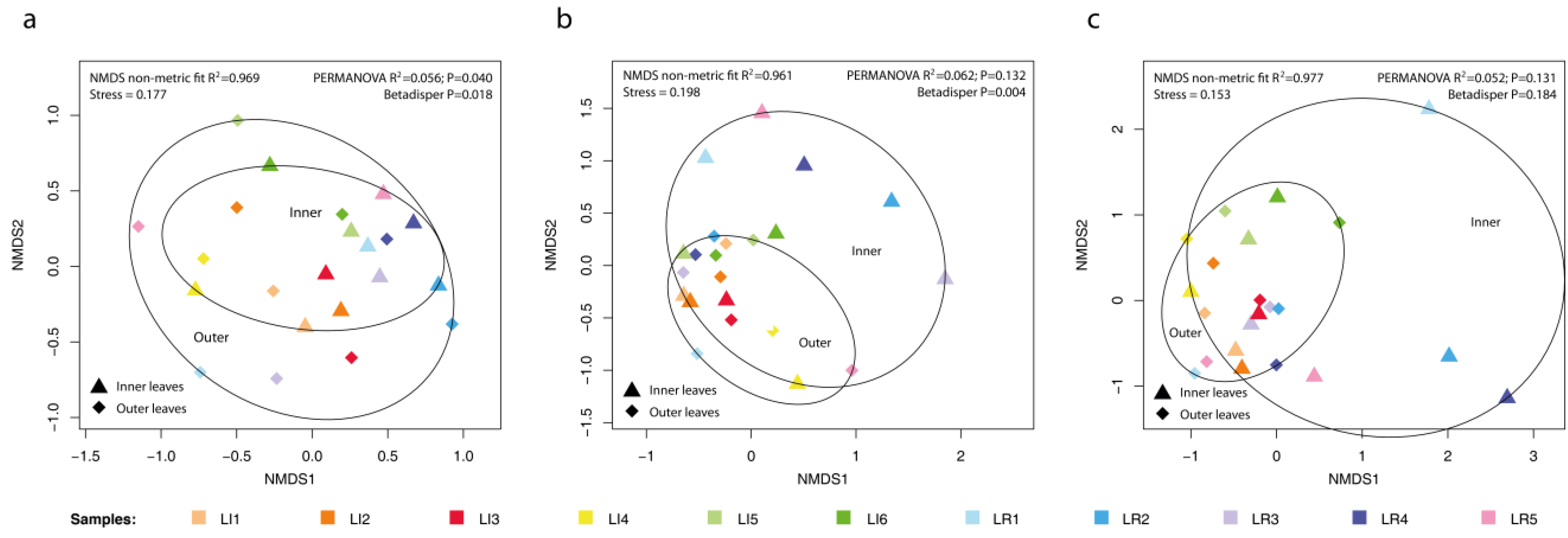
3.2. Comparison of Microbial Colonization Inside/Outside Lettuce Rosette

In the case of lettuces forming a closed rosette of leaves, significant differences were found in the amount of bacteria on the outer and inner leaves (Figure 7). Comparison by paired t-test showed lower bacterial abundance inside the rosette (Table 3). On average, the amount of colonies decreased by 1.3 log on PCA agar and by 1.2 log on VRBL agar. The diversity of microorganisms inside the rosette decreased in all three community types analyzed (Figure 8). The community change was in different directions, and due to the difference in communities and the small number of samples, it was not possible to evaluate the type of microorganisms whose content was decreasing.

4. Discussion

In a present study, we revealed the microbiome of fresh vegetables, and fruit and we evaluated the most important traits of their microbiological safety.
The number of microorganisms detected using cultivation methods in the vegetable was significantly higher than in berry fruit samples. The range of total microorganism counts of 5–8 log CFU is in agreement with values reported in the literature for vegetables obtained directly from field conditions as well as from the commercial network [34,55,56]. Also, the number of microorganisms on berries was within the previously reported ranges of 2–5 log [34,57]. Macori et al. [35] reported slightly lower amounts in the samples of berry fruit obtained directly from 50 producers. They found the highest microbial load in currants and blueberries, while our blueberry samples were least contaminated.
Microorganisms of the Enterobacteriaceae family, especially coliforms, are considered to be an important predictor of food safety. Bacteria from this group are able to cause gastrointestinal illness, tissue infections, pneumonia, or intoxication which may rarely be lethal [58]. Their presence usually indicates fecal contamination [59]. The number of such organisms was high in leafy vegetables in the range of 4–6 logs. Like the total aerobic counts, the incidence of coliforms was significantly lower in fruit samples. In food safety alert systems, it is bacteria from this group that are reported as the most common contaminant of leafy vegetables, whereas contamination of fruits is sporadic [60]. Nevertheless, the most frequently detected genus, Pantoea, is not a typical human pathogen [61]. We also did not detect pathogenic E. coli types using qPCR, although we did detect single sample with Salmonella. However, frequently detected species from the Klebsiella and Enterobacter genera are among the well-known opportunistic pathogens. Moreover, they may contribute to the spreading of antimicrobial resistance which cause problems in patient treatment [62].
For a comprehensive assessment of the microbiome, a combination of several different approaches, including culturing as well as modern DNA-based methods, was used in this study. Certified methods for detecting important microbial species are valuable for safety consideration; however, they do not allow a complex evaluation of microbial diversity. High-throughput sequencing methods that are able to provide a detailed picture of microbial communities in plants have come to the fore in recent years [63]. The presence and diversity of microorganisms in plants is highly dependent on specific samples. In the case of low microbial loads, obtaining microbial DNA is complicated and dependent on the method involved [64]. We used direct isolation of DNA from plant tissues that allowed us to obtain endophytic bacteria in addition to the epiphytic community [28]. The resulting composition was comparable to the results of studies analyzing epiphytic communities from leaf rinses [29,65]. We also exploited the possibility of pre-enrichment use in case of low microbial load. Such technique was also used by Jarvis, et al. [66] to analyze coriander samples for the detection of pathogenic bacteria such as Salmonella. Their results showed increased proliferation of Firmicutes and Proteobacteria but it did not lead to better detection of Salmonella spp. In our pre-enriched samples, we found a higher prevalence of Salmonella, Escherichia, Shigella, Klebsiella or Yersinia sequences, demonstrating pre-enrichment usability in research of potentially pathogenic members of microbiome. In fact, both approaches (fresh tissue and pre-enrichment) confirmed differences in diversity and composition of the microbiome between leafy vegetables and berries. Core members of microbiomes were clearly different, and stable complex microbial networks were formed for each sample type. Leff and Fierer [67] compared the difference between the microbiomes of various fruits and vegetables and, in accordance with the present study, found different communities between sample groups. Similar to our findings, they reported a high abundance of Enterobacteriaceae, especially the genus Pantoea. Enterobacteriaceae are also dominant in rocket [28] and small fruits like strawberry [68,69]. Other bacteria common in our samples like Pseudomonadales, Rhizobiales, Sphingomonadales, or Bacillales are also members of core microbiome in plant phyloshere/carposphere [70]. Most of them have a positive impact on a plant despite plant pathogenic or human pathogenic strains may appear among them [71].
Besides sporadic occurrence of Aspergillus and Penicillium genera, we did not confirm the increased presence of microscopic fungi, which could directly threaten health, for example by producing mycotoxins. Fungal community in our samples contained majority of species from recently defined core mycobiome of lettuce phyloplane [72]. The most common fungi (Cladosporium, Aureobasidium, Alternaria) are typical airborne genera readily colonizing plant tissues and sometimes acting as plant pathogens [73]. The genus Botrytis, which causes storage problems [74], was recorded in a few samples, particularly fruits.
The purpose of this study was to assess the microbial contamination of the samples as they are on the market, which is the most important thing from the consumers’ point of view. However, the microbiome of the plant is already shaped in the field. The microbiome may be affected by many biotic and abiotic environmental factors as well as growing and processing practices. The colonization process involves complex interactions between the plant and the microorganisms [75]. Soil serves as a depository for many diverse species of microorganisms, so it is not unexpected that the source of plant contamination is usually the soil. The proportion of Enterobacteriaceae in lettuce rhizosphere microbiome can reach up to several percent [76]. Plants, through their roots, take up various types of microorganisms, including those that are potentially pathogenic to humans. From the roots, the bacteria can be translocated to other parts of the plant [77]. Above-ground parts of the plant can be directly infected by micro-organisms from the surface [78]. Development of the plant microbiome is influenced by the weather, when warm and humid conditions promote colonization [30,79].
We found a distinct difference in the abundance and diversity of microorganisms between vegetable samples that grow on the soil surface and fruit samples that do not come into contact with the soil. More bacteria, including coliforms and even Salmonella were detected on vegetables which are in contact with soil. This points to soil as the most significant predictor of the amount and composition of microorganisms on these fresh products. If it is contaminated, microorganisms are very easily transferred to nearby plant parts. Thus, leafy vegetables are therefore significantly more risky in terms of microorganism content than fruit.
Organic fertilisers are the most common source of micro-organisms pathogenic to humans. Szczech, et al. [80] confirmed a higher levels of pathogenic microorganisms in vegetable samples from organic production, which was probably due to the organic fertilization. Similarly, strawberries fertilized by manure contained more Enterobacteriaceae [81]. Escherichia coli is able to transfer from manure to the roots of plants such as leek or lettuce and can be detected even after 9 months [15]. It has been shown that Salmonella can survive for several weeks in the soil and subsequently colonize plants [82]. The quality of the fertilizers and their maturity are decisive for the contamination of the soil with pathogenic bacteria [83]. Irrigation water can also be an important source of pathogenic microorganisms, especially if wastewater is used [25]. In many countries, there is legislative regulation of the use of organic fertilizers and wastewater because of the potential content of pathogenic microorganisms.
Leafy vegetables and berries differ significantly in their physico-chemical properties, for example in their content of simple sugars or organic acids. In addition, the pH values of berries range from 3 to 4, while those of leafy vegetables are around 6. These conditions can support certain groups of microorganisms and thus contribute to the formation of the microbiome. As example, fungi and yeasts are well adapted to growth in low pH and high sugar concentration conditions [84]. The preference of microscopic fungi in colonizing fruits is evident when comparing the ratio of total bacteria to microscopic fungi in our samples of vegetables 794:1 and fruits 3.2:1. The shape and firmness of the plant sample as well as the thickness and structure of the surface layer (presence of trichomes or stomata) influence the adhesion and penetration of microorganisms into the plant [78,85].
We analyzed several lettuce samples to determine how plant colonization by microorganisms depends on the exposure of the plant surface to the external environment that is the source of the microorganisms. The number of microorganisms was lower on inner leaves but still higher than on berries. Analogous to our results, Jacques [86] and Aycicek, et al. [87] also found naturally fewer microorganisms on the inner leaves of lettuce. The inner leaves are more fragile, easier to damage and have more simple carbohydrates on their surface which allow faster growth of microorganisms when contaminated during processing [31].
Processing of plant material such as washing and cutting is a critical point at which cross-contamination of plants can occur [88]. Washing alone is unable to reduce microbial contamination to low levels. It is possible to use sterilization such as hypochlorite or alternatives which alter the microbiome [89,90]. Therefore, we did not include washed samples in our study. We do not have precise information on collection time, processing, and storage conditions of assessed samples but all appeared fresh and had optimal sensory characteristics. Handling damage, although invisible to the human eye, accelerates the development of microorganisms during storage [91]. Finally, packaging and storage conditions also contribute to changes in the microbiome [79].
Proper strategies must be adopted to manage the safety of fresh produce throughout the farm-to-fork chain. Development of easily understood recommendations for farmers and strict following of good farming practices should be involved to minimize primary microbial contamination [92].
Since the number of microbes was high and potentially pathogenic species were present especially on leafy vegetables, attention should be paid to monitoring their microbiological safety. However, the assessment of microbial communities on fresh products should take into account the actual pathogenicity of detected microorganisms. Many members of Enterobacteriaceae are natural plant inhabitants commonly with plant growth promoting properties without negative effect on consumers health [93]. Moreover, humans need to be in contact with potential pathogens for an immunity development [94]. According to our results and previous findings [56,95], incidence of severe pathogens on fresh-consumed vegetables and fruits is not common. There is therefore no reason to concern about the consumption of fresh vegetables and fruits, and it should be increased because of their positive effects on human health.

5. Conclusions

The results of this study showed that microbial assemblages of leafy vegetables and berry fruits are quite different in the amount of microorganisms and composition of the microbial community. Leafy vegetables contain much more cultivable bacteria and yeasts. Furthermore, Enterobacteriaceae are more common in their microbiome as confirmed by cultivation dependent and independent methods. However Shiga-toxin E. coli was not detected in any sample and only one sample was positive for Salmonella using qPCR. As expected, internal leaves of head-forming lettuces contained fewer microorganisms with lower diversity. Further studies are needed to reveal factors affecting the incidence of pathogenic bacteria. Moreover, there is a need to reconsider the impact of plant-associated Enterobacteriaceae on human health as they are a common part of plant phyllosphere assemblage.

Supplementary Materials

The following supporting information can be downloaded at: https://www.mdpi.com/article/10.3390/horticulturae9020150/s1. Table S1. List of analyzed samples. Table S2. Primers used for amplification of 16S rRNA gene, ITS2 region and detection of E. coli and Salmonella. Table S3. Pairwise comparison of microbiome between 9 groups of berry fruits and leafy vegetables. Table S4. Pairwise comparison of microbiome between 9 groups of berry fruits and leafy vegetables. Figure S1. Barchart of LefSe biomarker among bacterial genera in samples of berry fruits and leafy vegetables. Figure S2. Barchart of LefSe biomarker among bacterial genera amplified in pre-enriched samples of berry fruits and leafy vegetables. Figure S3. Co-occurrence network obtained by SparCC analysis for orders of bacteria in samples berry fruits and leafy vegetables. Network plotted with correlation coefficient R > 0.3 or <0.3 and p < 0.05. Figure S4. Co-occurrence network obtained by SparCC analysis for orders of bacteria in pre-enriched samples berry fruits and leafy vegetables. Network plotted with correlation coefficient R > 0.3 or <0.3 and p < 0.05. Figure S5. Co-occurrence network obtained by SparCC analysis for fungal genera in samples of berry fruits and leafy vegetables. Network plotted with correlation coefficient R > 0.3 or <0.3 and p < 0.05.

Author Contributions

Conceptualization R.A. and J.M. (Juraj Medo); methodology R.A., M.J., J.B., N.L., J.P., J.M. (Jana Maková) and J.M. (Juraj Medo); software J.M. (Janka Medová) and J.M. (Juraj Medo); validation, R.A., S.J., L.H. and J.M. (Juraj Medo); formal analysis, J.M. (Janka Medová) and J.M. (Juraj Medo); investigation, R.A., M.J., J.B., N.L., J.P. and J.M. (Juraj Medo); resources R.A., M.J., J.B. and J.M. (Juraj Medo), data curation J.M. (Juraj Medo); writing—original draft preparation, R.A. and J.M. (Juraj Medo), writing—review and editing, all authors; project administration J.M. (Juraj Medo). All authors have read and agreed to the published version of the manuscript.

Funding

This research was financially supported by project of Ministry of Education, Science, Research and Sport of the Slovak Republic, grant no. VEGA 1/0661/19 “Plant microbiome and safe food”.

Institutional Review Board Statement

Not applicable.

Informed Consent Statement

Not applicable.

Data Availability Statement

All data are available from the correspondence author upon request. Sequence data were deposited in GenBank databases under BioProject accession No. PRJNA913390.

Acknowledgments

The authors would like to thank the Institute of Microbiology of the Academy of Sciences of the Czech Republic, v. v. i., in Prague for the opportunity to use the MiSeq sequencer.

Conflicts of Interest

The authors declare no conflict of interest.

References

  1. Amiot-Carlin, M.-J. Fruit and vegetable consumption: What benefits, what risks? Rev. Prat. 2019, 69, 139–142. [Google Scholar] [PubMed]
  2. Ramya, V.; Patel, P. Health benefits of vegetables. Int. J. Chem. Stud. 2019, 7, 82–87. [Google Scholar]
  3. Mason-D’Croz, D.; Bogard, J.R.; Sulser, T.B.; Cenacchi, N.; Dunston, S.; Herrero, M.; Wiebe, K. Gaps between fruit and vegetable production, demand, and recommended consumption at global and national levels: An integrated modelling study. Lancet Planet. Health 2019, 3, e318–e329. [Google Scholar] [CrossRef] [PubMed] [Green Version]
  4. Balali, G.I.; Yar, D.D.; Afua Dela, V.G.; Adjei-Kusi, P. Microbial Contamination, an Increasing Threat to the Consumption of Fresh Fruits and Vegetables in Today’s World. Int. J. Microbiol. 2020, 2020, 3029295. [Google Scholar] [CrossRef] [PubMed]
  5. Gutierrez, E. Is the Japanese O157: H7 E. coli epidemic over? Lancet 1996, 348, 1371. [Google Scholar] [CrossRef]
  6. Buchholz, U.; Bernard, H.; Werber, D.; Böhmer, M.M.; Remschmidt, C.; Wilking, H.; Deleré, Y.; an der Heiden, M.; Adlhoch, C.; Dreesman, J.; et al. German Outbreak of Escherichia coli O104:H4 Associated with Sprouts. N. Engl. J. Med. 2011, 365, 1763–1770. [Google Scholar] [CrossRef]
  7. EU Directorate General for Health and Food Safety; Rapid Alert System for Food and Feed (RASFF): 2022. Available online: https://webgate.ec.europa.eu/rasff-window/screen/search (accessed on 2 December 2022).
  8. USA Centers for Disease Control and Prevention; List of Outbreaks: 2022. Available online: https://www.cdc.gov/foodsafety/outbreaks/lists/outbreaks-list.html (accessed on 2 December 2022).
  9. de Oliveira Elias, S.; Noronha, T.B.; Tondo, E.C. Salmonella spp. and Escherichia coli O157:H7 prevalence and levels on lettuce: A systematic review and meta-analysis. Food Microbiol. 2019, 84, 103217. [Google Scholar] [CrossRef]
  10. Mohammadpour, H.; Berizi, E.; Hosseinzadeh, S.; Majlesi, M.; Zare, M. The prevalence of Campylobacter spp. in vegetables, fruits, and fresh produce: A systematic review and meta-analysis. Gut Pathog. 2018, 10, 41. [Google Scholar] [CrossRef] [Green Version]
  11. Ding, T.; Iwahori, J.i.; Kasuga, F.; Wang, J.; Forghani, F.; Park, M.-S.; Oh, D.-H. Risk assessment for Listeria monocytogenes on lettuce from farm to table in Korea. Food Control 2013, 30, 190–199. [Google Scholar] [CrossRef]
  12. Bozkurt, H.; Phan-Thien, K.-Y.; van Ogtrop, F.; Bell, T.; McConchie, R. Outbreaks, occurrence, and control of norovirus and hepatitis a virus contamination in berries: A review. Crit. Rev. Food Sci. Nutr. 2021, 61, 116–138. [Google Scholar] [CrossRef]
  13. Turner, T.R.; James, E.K.; Poole, P.S. The plant microbiome. Genome Biol. 2013, 14, 209. [Google Scholar] [CrossRef] [Green Version]
  14. Schlaeppi, K.; Bulgarelli, D. The Plant Microbiome at Work. Mol. Plant-Microbe Interact. ® 2015, 28, 212–217. [Google Scholar] [CrossRef] [Green Version]
  15. van Overbeek, L.; Duhamel, M.; Aanstoot, S.; van der Plas, C.L.; Nijhuis, E.; Poleij, L.; Russ, L.; van der Zouwen, P.; Andreo-Jimenez, B. Transmission of Escherichia coli from Manure to Root Zones of Field-Grown Lettuce and Leek Plants. Microorganisms 2021, 9, 2289. [Google Scholar] [CrossRef]
  16. Olimi, E.; Kusstatscher, P.; Wicaksono, W.A.; Abdelfattah, A.; Cernava, T.; Berg, G. Insights into the microbiome assembly during different growth stages and storage of strawberry plants. Environ. Microbiome 2022, 17, 21. [Google Scholar] [CrossRef]
  17. Hou, Z.; Fink, R.C.; Radtke, C.; Sadowsky, M.J.; Diez-Gonzalez, F. Incidence of naturally internalized bacteria in lettuce leaves. Int. J. Food Microbiol. 2013, 162, 260–265. [Google Scholar] [CrossRef]
  18. Olanrewaju, O.S.; Glick, B.R.; Babalola, O.O. Mechanisms of action of plant growth promoting bacteria. World J. Microbiol. Biotechnol. 2017, 33, 197. [Google Scholar] [CrossRef] [Green Version]
  19. Berg, G.; Grube, M.; Schloter, M.; Smalla, K. The plant microbiome and its importance for plant and human health. Front. Microbiol. 2014, 5, 491. [Google Scholar] [CrossRef] [Green Version]
  20. Berg, G.; Eberl, L.; Hartmann, A. The rhizosphere as a reservoir for opportunistic human pathogenic bacteria. Environ. Microbiol. 2005, 7, 1673–1685. [Google Scholar] [CrossRef]
  21. Khalifa, A.Y.Z.; Alsyeeh, A.-M.; Almalki, M.A.; Saleh, F.A. Characterization of the plant growth promoting bacterium, Enterobacter cloacae MSR1, isolated from roots of non-nodulating Medicago sativa. Saudi J. Biol. Sci. 2016, 23, 79–86. [Google Scholar] [CrossRef]
  22. Sanzani, S.M.; Reverberi, M.; Geisen, R. Mycotoxins in harvested fruits and vegetables: Insights in producing fungi, biological role, conducive conditions, and tools to manage postharvest contamination. Postharvest Biol. Technol. 2016, 122, 95–105. [Google Scholar] [CrossRef]
  23. Saleh, I.; Al-Thani, R. Fungal food spoilage of supermarkets’ displayed fruits. Vet. World 2019, 12, 1877. [Google Scholar] [CrossRef] [PubMed] [Green Version]
  24. Adegoke, A.A.; Awolusi, O.O.; Stenström, T.A. Organic Fertilizers: Public Health Intricacies. In Organic Fertilizers-From Basic Concepts to Applied Outcomes; Larramendy, M.L., Soloneski, S., Eds.; IntechOpen: London, UK, 2016; pp. 343–374. [Google Scholar] [CrossRef] [Green Version]
  25. Gurtler, J.B.; Gibson, K.E. Irrigation water and contamination of fresh produce with bacterial foodborne pathogens. Curr. Opin. Food Sci. 2022, 47, 100889. [Google Scholar] [CrossRef]
  26. Riggio, G.M.; Jones, S.L.; Gibson, K.E. Risk of human pathogen internalization in leafy vegetables during lab-scale hydroponic cultivation. Horticulturae 2019, 5, 25. [Google Scholar] [CrossRef] [Green Version]
  27. Jay-Russell, M.T. What is the risk from wild animals in food-borne pathogen contamination of plants? CABI Rev. 2013, 8, 1–16. [Google Scholar] [CrossRef] [Green Version]
  28. Cernava, T.; Erlacher, A.; Soh, J.; Sensen, C.W.; Grube, M.; Berg, G. Enterobacteriaceae dominate the core microbiome and contribute to the resistome of arugula (Eruca sativa Mill.). Microbiome 2019, 7, 13. [Google Scholar] [CrossRef]
  29. Yeon-Cheol, Y.; Su-Jin, Y.; Da-Young, J. Analysis of the Microbiota on Lettuce (Lactuca sativa L.) Cultivated in South Korea to Identify Foodborne Pathogens. J. Microbiol. Biotechnol. 2018, 28, 1318–1331. [Google Scholar] [CrossRef] [Green Version]
  30. Williams, T.R.; Moyne, A.-L.; Harris, L.J.; Marco, M.L. Season, Irrigation, Leaf Age, and Escherichia coli Inoculation Influence the Bacterial Diversity in the Lettuce Phyllosphere. PLoS ONE 2013, 8, e68642. [Google Scholar] [CrossRef]
  31. Brandl, M.T.; Amundson, R. Leaf Age as a Risk Factor in Contamination of Lettuce with Escherichia coli O157:H7 and Salmonella enterica. Appl. Environ. Microbiol. 2008, 74, 2298–2306. [Google Scholar] [CrossRef] [Green Version]
  32. Jensen, B.; Knudsen, I.M.B.; Andersen, B.; Nielsen, K.F.; Thrane, U.; Jensen, D.F.; Larsen, J. Characterization of microbial communities and fungal metabolites on field grown strawberries from organic and conventional production. Int. J. Food Microbiol. 2013, 160, 313–322. [Google Scholar] [CrossRef]
  33. Baugher, J.; Jaykus, L. Natural microbiota of raspberries (Rubus idaeus) and strawberries (Fragaria × ananassa): Microbial survey, bacterial isolation and identification, and biofilm characterization. In XI International Rubus and Ribes Symposium 1133; Fernandez, G.E., Hummer, K.E., Eds.; ISHS: Leuven, Belgium, 2015; pp. 421–526. [Google Scholar] [CrossRef]
  34. Quansah, J.K.; Gazula, H.; Holland, R.; Scherm, H.; Li, C.; Takeda, F.; Chen, J. Microbial quality of blueberries for the fresh market. Food Control 2019, 100, 92–96. [Google Scholar] [CrossRef]
  35. Macori, G.; Gilardi, G.; Bellio, A.; Bianchi, D.M.; Gallina, S.; Vitale, N.; Gullino, M.L.; Decastelli, L. Microbiological Parameters in the Primary Production of Berries: A Pilot Study. Foods 2018, 7, 105. [Google Scholar] [CrossRef] [Green Version]
  36. Pavlovic, M.; Huber, I.; Skala, H.; Konrad, R.; Schmidt, H.; Sing, A.; Busch, U. Development of a multiplex real-time polymerase chain reaction for simultaneous detection of enterohemorrhagic Escherichia coli and enteropathogenic Escherichia coli strains. Foodborne Pathog. Dis. 2010, 7, 801–808. [Google Scholar] [CrossRef]
  37. Lee, D.-Y.; Shannon, K.; Beaudette, L.A. Detection of bacterial pathogens in municipal wastewater using an oligonucleotide microarray and real-time quantitative PCR. J. Microbiol. Methods 2006, 65, 453–467. [Google Scholar] [CrossRef]
  38. Caporaso, J.G.; Lauber, C.L.; Walters, W.A.; Berg-Lyons, D.; Lozupone, C.A.; Turnbaugh, P.J.; Fierer, N.; Knight, R. Global patterns of 16S rRNA diversity at a depth of millions of sequences per sample. Proc. Natl. Acad. Sci. USA 2011, 108, 4516–4522. [Google Scholar] [CrossRef] [Green Version]
  39. Artimová, R.; Hleba, L.; Javoreková, S.; Maková, J.; Medová, J.; Medo, J. Chloroplast Excluding Primers For Metagenomic Analysis Of Bacteria In Plant Tissues. J. Microbiol. Biotechnol. Food Sci. 2022, 12, e9650. [Google Scholar] [CrossRef]
  40. Ihrmark, K.; Bödeker, I.; Cruz-Martinez, K.; Friberg, H.; Kubartova, A.; Schenck, J.; Strid, Y.; Stenlid, J.; Brandström-Durling, M.; Clemmensen, K.E. New primers to amplify the fungal ITS2 region–evaluation by 454-sequencing of artificial and natural communities. FEMS Microbiol. Ecol. 2012, 82, 666–677. [Google Scholar] [CrossRef]
  41. White, T.J.; Bruns, T.; Lee, S.; Taylor, J. Amplification and direct sequencing of fungal ribosomal RNA genes for phylogenetics. PCR Protoc. A Guide Methods Appl. 1990, 18, 315–322. [Google Scholar]
  42. Lane, D.J.; Pace, B.; Olsen, G.J.; Stahl, D.A.; Sogin, M.L.; Pace, N.R. Rapid determination of 16S ribosomal RNA sequences for phylogenetic analyses. Proc. Natl. Acad. Sci. USA 1985, 82, 6955–6959. [Google Scholar] [CrossRef] [Green Version]
  43. Vetrovský, T.; Baldrian, P.; Morais, D.; Berger, B. SEED 2: A user-friendly platform for amplicon high-throughput sequencing data analyses. Bioinformatics 2018, 1, 3. [Google Scholar] [CrossRef] [Green Version]
  44. Rognes, T.; Flouri, T.; Nichols, B.; Quince, C.; Mahé, F. VSEARCH: A versatile open source tool for metagenomics. PeerJ 2016, 4, e2584. [Google Scholar] [CrossRef] [Green Version]
  45. Wang, Q.; Garrity, G.M.; Tiedje, J.M.; Cole, J.R. Naive Bayesian classifier for rapid assignment of rRNA sequences into the new bacterial taxonomy. Appl. Environ. Microbiol. 2007, 73, 5261–5267. [Google Scholar] [CrossRef] [PubMed] [Green Version]
  46. Standley, D.M.; Katoh, K. MAFFT Multiple Sequence Alignment Software Version 7: Improvements in Performance and Usability. Mol. Biol. Evol. 2013, 30, 772–780. [Google Scholar] [CrossRef] [Green Version]
  47. Guindon, S.; Dufayard, J.-F.; Lefort, V.; Anisimova, M.; Hordijk, W.; Gascuel, O. New Algorithms and Methods to Estimate Maximum-Likelihood Phylogenies: Assessing the Performance of PhyML 3.0. Syst. Biol. 2010, 59, 307–321. [Google Scholar] [CrossRef] [PubMed] [Green Version]
  48. Hamady, M.; Lozupone, C.; Knight, R. Fast UniFrac: Facilitating high-throughput phylogenetic analyses of microbial communities including analysis of pyrosequencing and PhyloChip data. ISME J. 2010, 4, 17–27. [Google Scholar] [CrossRef] [PubMed]
  49. R Core Team. R: A Language and Environment for Statistical Computing; R Foundation for Statistical Computing: Vienna, Austria, 2021. [Google Scholar]
  50. Oksanen, J.; Blanchet, F.G.; Kindt, R.; Legendre, P.; Minchin, P.R.; O’hara, R.; Simpson, G.L.; Solymos, P.; Stevens, M.H.H.; Wagner, H. Package ‘Vegan’. Community Ecology Package, Version 2.5-6. 2019. Available online: https://CRAN.R-project.org/package=vegan (accessed on 2 October 2022).
  51. Zhao, S.; Guo, Y.; Sheng, Q.; Shyr, Y. Advanced Heat Map and Clustering Analysis Using Heatmap3. BioMed Res. Int. 2014, 2014, 6. [Google Scholar] [CrossRef] [Green Version]
  52. Segata, N.; Izard, J.; Waldron, L.; Gevers, D.; Miropolsky, L.; Garrett, W.S.; Huttenhower, C. Metagenomic biomarker discovery and explanation. Genome Biol. 2011, 12, R60. [Google Scholar] [CrossRef] [Green Version]
  53. Friedman, J.; Alm, E.J. Inferring Correlation Networks from Genomic Survey Data. PLOS Comput. Biol. 2012, 8, e1002687. [Google Scholar] [CrossRef] [Green Version]
  54. Chong, J.; Liu, P.; Zhou, G.; Xia, J. Using MicrobiomeAnalyst for comprehensive statistical, functional, and meta-analysis of microbiome data. Nat. Protoc. 2020, 15, 799–821. [Google Scholar] [CrossRef]
  55. Holland, R.M.; Chen, J.; Gazula, H.; Scherm, H. Environmental and Fecal Indicator Organisms on Fruit Contact Surfaces and Fruit from Blueberry Mechanical Harvesters. Horticulturae 2022, 8, 20. [Google Scholar] [CrossRef]
  56. Korir, R.C.; Parveen, S.; Hashem, F.; Bowers, J. Microbiological quality of fresh produce obtained from retail stores on the Eastern Shore of Maryland, United States of America. Food Microbiol. 2016, 56, 29–34. [Google Scholar] [CrossRef]
  57. Kuźniar, P.; Belcar, J.; Zardzewiały, M.; Basara, O.; Gorzelany, J. Effect of Ozonation on the Mechanical, Chemical, and Microbiological Properties of Organically Grown Red Currant (Ribes rubrum L.) Fruit. Molecules 2022, 27, 8231. [Google Scholar] [CrossRef]
  58. Smith, J.; Fratamico, P. Escherichia coli and other Enterobacteriaceae: Food poisoning and health effects. In Encyclopedia of Food and Health; Caballer., B., Finglas, P., Toldrá, F., Eds.; AcademicPress: Waltham, MA, USA, 2016; pp. 539–544. [Google Scholar]
  59. Ekici, G.; Dümen, E. Escherichia coli and food safety. In The Universe of Escherichia Coli; Starčič Erjavec, M., Ed.; IntechOpen: London, UK, 2019; pp. 1–16. [Google Scholar] [CrossRef]
  60. Oliveira, M.; Rodrigues, C.M.; Teixeira, P. Microbiological quality of raw berries and their products: A focus on foodborne pathogens. Heliyon 2019, 5, e02992. [Google Scholar] [CrossRef] [Green Version]
  61. Walterson, A.M.; Stavrinides, J. Pantoea: Insights into a highly versatile and diverse genus within the Enterobacteriaceae. FEMS Microbiol. Rev. 2015, 39, 968–984. [Google Scholar] [CrossRef] [Green Version]
  62. Rahman, M.; Alam, M.-U.; Luies, S.K.; Kamal, A.; Ferdous, S.; Lin, A.; Sharior, F.; Khan, R.; Rahman, Z.; Parvez, S.M.; et al. Contamination of Fresh Produce with Antibiotic-Resistant Bacteria and Associated Risks to Human Health: A Scoping Review. Int. J. Environ. Res. Public Health 2022, 19, 360. [Google Scholar] [CrossRef]
  63. Johnston-Monje, D.; Lopez Mejia, J. Botanical microbiomes on the cheap: Inexpensive molecular fingerprinting methods to study plant-associated communities of bacteria and fungi. Appl. Plant Sci. 2020, 8, e11334. [Google Scholar] [CrossRef] [Green Version]
  64. Sare, A.R.; Stouvenakers, G.; Eck, M.; Lampens, A.; Goormachtig, S.; Jijakli, M.H.; Massart, S. Standardization of Plant Microbiome Studies: Which Proportion of the Microbiota is Really Harvested? Microorganisms 2020, 8, 342. [Google Scholar] [CrossRef] [Green Version]
  65. Seo, D.W.; Yum, S.-j.; Lee, H.R.; Kim, S.M.; Jeong, H.G. Microbiota Analysis and Microbiological Hazard Assessment in Chinese Chive (Allium tuberosum Rottler) Depending on Retail Types. J. Microbiol. Biotechnol. 2022, 32, 195–204. [Google Scholar] [CrossRef]
  66. Jarvis, K.G.; White, J.R.; Grim, C.J.; Ewing, L.; Ottesen, A.R.; Beaubrun, J.J.-G.; Pettengill, J.B.; Brown, E.; Hanes, D.E. Cilantro microbiome before and after nonselective pre-enrichment for Salmonella using 16S rRNA and metagenomic sequencing. BMC Microbiol. 2015, 15, 160. [Google Scholar] [CrossRef] [Green Version]
  67. Leff, J.W.; Fierer, N. Bacterial Communities Associated with the Surfaces of Fresh Fruits and Vegetables. PLoS ONE 2013, 8, e59310. [Google Scholar] [CrossRef] [Green Version]
  68. Zhang, H.; Zhang, Q.; Chen, S.; Zhang, Z.; Song, J.; Long, Z.; Yu, Y.; Fang, H. Enterobacteriaceae predominate in the endophytic microbiome and contribute to the resistome of strawberry. Sci. Total Environ. 2020, 727, 138708. [Google Scholar] [CrossRef]
  69. Zhimo, V.Y.; Kumar, A.; Biasi, A.; Salim, S.; Feygenberg, O.; Toamy, M.A.; Abdelfattaah, A.; Medina, S.; Freilich, S.; Wisniewski, M.; et al. Compositional shifts in the strawberry fruit microbiome in response to near-harvest application of Metschnikowia fructicola, a yeast biocontrol agent. Postharvest Biol. Technol. 2021, 175, 111469. [Google Scholar] [CrossRef]
  70. Darlison, J.; Mogren, L.; Rosberg, A.K.; Grudén, M.; Minet, A.; Liné, C.; Mieli, M.; Bengtsson, T.; Håkansson, Å.; Uhlig, E.; et al. Leaf mineral content govern microbial community structure in the phyllosphere of spinach (Spinacia oleracea) and rocket (Diplotaxis tenuifolia). Sci. Total Environ. 2019, 675, 501–512. [Google Scholar] [CrossRef] [PubMed]
  71. Ali, M.A.; Naveed, M.; Mustafa, A.; Abbas, A. The good, the bad, and the ugly of rhizosphere microbiome. In Probiotics and Plant Health; Springer: Berlin/Heidelberg, Germany, 2017; pp. 253–290. [Google Scholar]
  72. Haelewaters, D.; Urbina, H.; Brown, S.; Newerth-Henson, S.; Aime, M.C. Isolation and Molecular Characterization of the Romaine Lettuce Phylloplane Mycobiome. J. Fungi 2021, 7, 277. [Google Scholar] [CrossRef] [PubMed]
  73. Dietzel, K.; Valle, D.; Fierer, N.; U’Ren, J.M.; Barberán, A. Geographical Distribution of Fungal Plant Pathogens in Dust Across the United States. Front. Ecol. Evol. 2019, 7, 304. [Google Scholar] [CrossRef] [Green Version]
  74. Hua, L.; Yong, C.; Zhanquan, Z.; Boqiang, L.; Guozheng, Q.; Shiping, T. Pathogenic mechanisms and control strategies of Botrytis cinerea causing post-harvest decay in fruits and vegetables. Food Qual. Saf. 2018, 2, 111–119. [Google Scholar] [CrossRef] [Green Version]
  75. Zarkani, A.A.; Schierstaedt, J.; Becker, M.; Krumwiede, J.; Grimm, M.; Grosch, R.; Jechalke, S.; Schikora, A. Salmonella adapts to plants and their environment during colonization of tomatoes. FEMS Microbiol. Ecol. 2019, 95, fiz152. [Google Scholar] [CrossRef]
  76. Žiarovská, J.; Urbanová, L.; Moravčíková, D.; Artimová, R.; Omelka, R.; Medo, J. Varieties of Lettuce Forming Distinct Microbial Communities Inhabiting Roots and Rhizospheres with Various Responses to Osmotic Stress. Horticulturae 2022, 8, 1174. [Google Scholar] [CrossRef]
  77. Taulé, C.; Vaz-Jauri, P.; Battistoni, F. Insights into the early stages of plant–endophytic bacteria interaction. World J. Microbiol. Biotechnol. 2021, 37, 13. [Google Scholar] [CrossRef]
  78. Schlechter, R.O.; Miebach, M.; Remus-Emsermann, M.N.P. Driving factors of epiphytic bacterial communities: A review. J. Adv. Res. 2019, 19, 57–65. [Google Scholar] [CrossRef]
  79. Leonard, S.R.; Simko, I.; Mammel, M.K.; Richter, T.K.S.; Brandl, M.T. Seasonality, shelf life and storage atmosphere are main drivers of the microbiome and E. coli O157:H7 colonization of post-harvest lettuce cultivated in a major production area in California. Environ. Microbiome 2021, 16, 25. [Google Scholar] [CrossRef]
  80. Szczech, M.; Kowalska, B.; Smolińska, U.; Maciorowski, R.; Oskiera, M.; Michalska, A. Microbial quality of organic and conventional vegetables from Polish farms. Int. J. Food Microbiol. 2018, 286, 155–161. [Google Scholar] [CrossRef]
  81. Kurtböke, D.I.; Palk, A.; Marker, A.; Neuman, C.; Moss, L.; Streeter, K.; Katouli, M. Isolation and characterization of Enterobacteriaceae species infesting post-harvest strawberries and their biological control using bacteriophages. Appl. Microbiol. Biotechnol. 2016, 100, 8593–8606. [Google Scholar] [CrossRef]
  82. Jechalke, S.; Schierstaedt, J.; Becker, M.; Flemer, B.; Grosch, R.; Smalla, K.; Schikora, A. Salmonella Establishment in Agricultural Soil and Colonization of Crop Plants Depend on Soil Type and Plant Species. Front. Microbiol. 2019, 10, 967. [Google Scholar] [CrossRef] [Green Version]
  83. Manyi-Loh, C.E.; Mamphweli, S.N.; Meyer, E.L.; Makaka, G.; Simon, M.; Okoh, A.I. An Overview of the Control of Bacterial Pathogens in Cattle Manure. Int. J. Environ. Res. Public Health 2016, 13, 843. [Google Scholar] [CrossRef] [Green Version]
  84. Moss, M.O. Fungi, quality and safety issues in fresh fruits and vegetables. J. Appl. Microbiol. 2008, 104, 1239–1243. [Google Scholar] [CrossRef]
  85. Remus-Emsermann, M.N.P.; Schlechter, R.O. Phyllosphere microbiology: At the interface between microbial individuals and the plant host. New Phytol. 2018, 218, 1327–1333. [Google Scholar] [CrossRef] [Green Version]
  86. Jacques, M.-A. The Effect of Leaf Age and Position on the Dynamics of Microbial Populations on Aerial Plant Surfaces. In Aerial Plant Surface Microbiology; Morris, C.E., Nicot, P.C., Nguyen-The, C., Eds.; Springer: Boston, MA, USA, 1996; pp. 233–248. [Google Scholar] [CrossRef]
  87. Aycicek, H.; Oguz, U.; Karci, K. Determination of total aerobic and indicator bacteria on some raw eaten vegetables from wholesalers in Ankara, Turkey. Int. J. Hyg. Environ. Health 2006, 209, 197–201. [Google Scholar] [CrossRef]
  88. Smolinski, H.S.; Wang, S.; Ren, L.; Chen, Y.; Kowalcyk, B.; Thomas, E.; Doren, J.V.; Ryser, E.T. Transfer and Redistribution of Salmonella Typhimurium LT2 and Escherichia coli O157:H7 during Pilot-Scale Processing of Baby Spinach, Cilantro, and Romaine Lettuce. J. Food Prot. 2018, 81, 953–962. [Google Scholar] [CrossRef]
  89. Gu, G.; Ottesen, A.; Bolten, S.; Luo, Y.; Rideout, S.; Nou, X. Microbiome convergence following sanitizer treatment and identification of sanitizer resistant species from spinach and lettuce rinse water. Int. J. Food Microbiol. 2020, 318, 108458. [Google Scholar] [CrossRef]
  90. Chun, H.H.; Kang, J.H.; Song, K.B. Effects of aqueous chlorine dioxide treatment and cold storage on microbial growth and quality of blueberries. J. Korean Soc. Appl. Biol. Chem. 2013, 56, 309–315. [Google Scholar] [CrossRef]
  91. Brandl, M.T. Plant Lesions Promote the Rapid Multiplication of Escherichia coli O157:H7 on Postharvest Lettuce. Appl. Environ. Microbiol. 2008, 74, 5285–5289. [Google Scholar] [CrossRef] [PubMed] [Green Version]
  92. Julien-Javaux, F.; Gérard, C.; Campagnoli, M.; Zuber, S. Strategies for the safety management of fresh produce from farm to fork. Curr. Opin. Food Sci. 2019, 27, 145–152. [Google Scholar] [CrossRef]
  93. Lorenzi, A.S.; Bonatelli, M.L.; Chia, M.A.; Peressim, L.; Quecine, M.C. Opposite Sides of Pantoea agglomerans and Its Associated Commercial Outlook. Microorganisms 2022, 10, 2072. [Google Scholar] [CrossRef] [PubMed]
  94. Berg, G.; Erlacher, A.; Smalla, K.; Krause, R. Vegetable microbiomes: Is there a connection among opportunistic infections, human health and our ‘gut feeling’? Microb. Biotechnol. 2014, 7, 487–495. [Google Scholar] [CrossRef]
  95. Denis, N.; Zhang, H.; Leroux, A.; Trudel, R.; Bietlot, H. Prevalence and trends of bacterial contamination in fresh fruits and vegetables sold at retail in Canada. Food Control 2016, 67, 225–234. [Google Scholar] [CrossRef] [Green Version]
Figure 1. Boxplots of log10 (CFU.g−1) in samples of leafy vegetables and berry fruits. (a) Total aerobic counts on PCA agar; (b) Total coliforms on VRBL agar; (c) Yeasts on DRBC agar; (d) Filamentous fungi on DRBC agar. Box plots display the first (25%) and third (75%) quartiles, the median (horizontal line), the average (*), the maximum and minimum observed values (whiskers), and extreme values (·) within each data set. Boxes signed with the same letter are not significantly different according one-way ANOVAs and Tukey’s Test at α = 0.05.
Horticulturae 09 00150 g001
Figure 2. Heatmap of bacterial genera composition according to analysis of DNA extracted from fresh tissue samples of leafy vegetables and berry fruits. Only genera with min. 2% occurrences in any samples are listed. Dendrograms are based on occurrence of genera in samples (Bray-Curtis distance, complete clustering).
Horticulturae 09 00150 g002
Figure 3. Heatmap of fungal genera composition according to analysis of DNA extracted from fresh tissue samples of leafy vegetables and berry fruits. Only genera with min. 2% occurrences in any samples are listed. Dendrograms are based on occurrence of genera in samples (Bray-Curtis distance, complete clustering).
Horticulturae 09 00150 g003
Figure 4. Heatmap of bacterial genera composition according to analysis of DNA extracted samples of leafy vegetables and berry fruits pre-enriched in peptone buffered water. Only genera with min. 2% occurrences in any samples are listed. Dendrograms are based on occurrence of genera in samples (Bray-Curtis distance, complete clustering).
Horticulturae 09 00150 g004
Figure 5. NMDS scatterplots of microbial communities in samples of leafy vegetables and berry fruits. (a) Bacterial community in fresh tissue samples; (b) fungal community in fresh tissue samples; (c) Bacterial community in sample pre incubated in buffered peptone water.
Horticulturae 09 00150 g005
Figure 6. Barchart of biomarker among bacterial genera in samples of berry fruits and leafy vegetables.
Horticulturae 09 00150 g006
Figure 7. Comparison of microbial counts of log10 (CFU.g−1) in inner (In) and outer (Out) leaves of rosette-forming lettuces. p-values are based on pair t-test.
Horticulturae 09 00150 g007
Figure 8. NMDS scatterplots of microbial communities in samples of inner and outer lettuce leaves. (a) Bacterial community in fresh tissue samples; (b) fungal community in fresh tissue samples; (c) Bacterial community in sample pre incubated in buffered peptone water.
Horticulturae 09 00150 g008
Table 1. Maldi-TOF identification of non-redundant coliform isolates grown from samples of leafy vegetables and berry fruits in VRBL agar.
SpeciesBlackberryBlueberryRaspberryStrawberryCurrantsGemRockettSpinachValerianellaTotal
Citrobacter braakii11
Citrobacter freundii173112
Citrobacter gillenii11
Enterobacter asburiae1326
Enterobacter bugandensis516
Enterobacter cloacae11911
Enterobacter kobei617
Enterobacter ludwigii257
Enterobacter xiangfangensis2215
Klebsiella aerogenes132118
Klebsiella oxytoca213262420
Klebsiella variicola11
Kosakonia kowanii11316
Kluyvera cryocrescens11
Leclercia adecarboxylata11
Pantoea agglomerans5422161242148
Pantoea ananatis22610
Pantoea anthophila221624118
Pantoea vagans 3 2 5
Pseudescherichia vulneris11
Rahnella aquatilis11
Raoultella ornithinolytica325
Raoultella terrigena3115
Serratia ficaria431 8
Serratia fonticola11
Serratia grimesii21 47
Serratia liquefaciens156
Serratia marcescens22
Serratia ureilytica1113
Total131512112193201710213
Table 2. Alpha diversity indices of microbial communities in samples of leafy vegetables and berry fruits as compared by one-way ANOVA.
Microbial CommunityDiversity IndexFruits (n = 45)Vegetables (n = 40)p-Value
Bacteria in leaf tissueRichness258.8 (66.2) *221.3 (67.8) 0.0119
Shannon’s Index4.52 (1.21)4.27 (1.31)0.3647
Corrected evenness0.517 (0.139) 0.503 (0.15) 0.6623
Fungi in leaf tissueRichness21.0 (8.0) 22.0 (8.9) 0.5962
Shannon’s Index2.30 (0.83) 2.24 (1.00) 0.7802
Corrected evenness0.492 (0.162) 0.470 (0.192) 0.5736
Bacteria in pre-enrichmentRichness163.4 (49.1) 306.9 (93.3) 0.0001
Shannon’s Index3.47 (1.21) 4.59 (0.87) 0.0001
Corrected evenness0.421 (0.144) 0.494 (0.089) 0.0075
* Mean (standard deviation).
Table 3. Alpha diversity indices of microbial communities in samples of inner and outer lettuce leaves as compared by paired t-test.
Microbial CommunityDiversity IndiceInner LeavesOuter LeavesDifference
+/− 95% Confidence
p-Value
Bacteria in leaf tissueRichness18723952.4 +/− 52.08 0.0486
Shannon’s Index3.744.771.03 +/− 0.960.0374
Corrected evenness0.4480.5500.102 +/− 0.1080.0635
Fungi in leaf tissueRichness18.527.59.09 +/− 6.80 0.0139
Shannon’s Index2.312.80 0.49 +/− 0.610.1032
Corrected evenness0.4770.5640.086 +/− 0.1150.1245
Bacteria in pre-enrichmentRichness19328390.0 +/− 80.60.0321
Shannon’s Index3.274.180.90 +/− 0.920.0560
Corrected evenness0.3810.4430.062 +/− 0.1120.2452
Disclaimer/Publisher’s Note: The statements, opinions and data contained in all publications are solely those of the individual author(s) and contributor(s) and not of MDPI and/or the editor(s). MDPI and/or the editor(s) disclaim responsibility for any injury to people or property resulting from any ideas, methods, instructions or products referred to in the content.

