Article

Morphological and Molecular Identification of Dactylonectria macrodidyma as Causal Agent of a Severe Prunus lusitanica Dieback in Italy

1
Department of Agriculture, Food and Environment (DAFE), University of Pisa, Via del Borghetto 80, 56124 Pisa, Italy
2
CIRSEC, Centre for Climate Change Impact, University of Pisa, Via del Borghetto 80, 56124 Pisa, Italy
3
Associazione Vivaisti Italiani, Via G. Deledda 30/7, 51100 Pistoia, Italy
*
Author to whom correspondence should be addressed.
Horticulturae 2023, 9(2), 145; https://doi.org/10.3390/horticulturae9020145
Submission received: 23 December 2022 / Revised: 15 January 2023 / Accepted: 18 January 2023 / Published: 21 January 2023
(This article belongs to the Section Plant Pathology and Disease Management (PPDM))

Abstract

:
During the summer of 2016, severe dieback was observed on young potted Prunus lusitanica (Portugal laurel) plants in a nursery in the Pistoia province (Tuscany, Italy). Cylindrocarpon-like isolates were consistently recovered from diseased plant tissues. The combination of morphological and molecular traits, including sequence data of histone 3 and β-tubulin genes (HIS3, TUB2) and internal transcribed spacers (ITS), allowed the identification of Dactylonectria macrodidyma (Halleen, Schroers & Crous) L. Lombard & Crous (asexual form Cylindrocarpon macrodidymum) as the causal agent of the disease. Pathogenicity tests reproduced disease symptoms observed in the nursery after six months fulfilling Koch’s postulates. D. macrodidyma is a soilborne plant pathogen and is to be considered of great economic importance on P. lusitanica, especially under favorable conditions such as stress and/or reduction of plant vitality. The increasingly frequent reports of the disease caused by the pathogen in various nurseries suggest that pot cultivation, together with prolonged drought periods, may play a role in favoring infections. To the best of our knowledge, this is the first report worldwide of the occurrence of dieback on Prunus lusitanica caused by D. macrodidyma.

1. Introduction

Prunus lusitanica L. (Portugal Laurel) is a plant species of the Rosaceae family native to southwestern France, Spain, Portugal, Morocco, and Macaronesia (the Azores, Canary Islands and Madeira). It was first brought to the UK from Madeira in 1648, and it is now widely planted in northern Europe (UK, Ireland, and France), New Zealand, and the western United States (California, Oregon and Washington states) as a hedge and for screening in gardens and parks. The specific name ‘lusitanica’ means ‘from Lusitania,’ the name that the Romans gave to Portugal, hence its common name, Portugal laurel.
P. lusitanica is a bushy, evergreen shrub or tree with a dark green leaf. Portugal Laurel has reddish new shoots that look attractive against the dark green leaves. If left untrimmed, it produces masses of small fragrant white flowers in early summer, followed by small red to dark purple fruit. The shiny bright green foliage on red stems gains a bluish tinge in winter.
Commercially the variety ‘Myrtifolia’ is becoming more popular than the ordinary Portugal Laurel because it has a smaller, darker leaf with redder stems (Figure 1a). ‘Myrtifolia’ is generally known by the name ‘Angustifolia’ and sporadically as ‘Pyramidalis’ and is excellent for hedging or topiary (Figure 1b; https://evergreenhedging.com, accessed 16 November 2022).
The annual production of P. lusitanica is about 600,000 plants with a commercial value of approximately 7 million € which represents about 1.5% of the annual production value of the Pistoian ornamental nurseries. The Pistoia production area (Tuscany Region) is the most important in Europe for the production of outdoor ornamental plants. Over 50% of the P. lusitanica sold production is represented by medium-small plants (5–12 L pots), while the rest of the production is equally represented by small (3 L pots) and large plants (pots > 18 L). Almost 95% of plants are exported not only to European countries but also to other continents (Associazione Vivaisti Italiani, Personal communication, 2022).
In the summer of 2016, P. lusitanica plants cultivated in pots in a nursery located in the Pistoia province showed severe yellowing, wilting and dieback symptoms (Figure 2a). Plants were smaller, less vigorous and were usually characterized by large reddish-brown areas along the stem starting below the crown that can be easily scraped off to show the internal wood symptoms (Figure 2b). Cross sections of infected stems showed characteristic necrosis patterns associated with brown wedge-shaped areas (Figure 2c,d). Many affected plants died within a few weeks of showing the first symptoms. The damage was quite substantial because about 50% of the 3-year-old P. lusitanica plants were diseased. The symptoms observed in P. lusitanica have been found in several nurseries of the Pistoia district, and our studies were conducted using diseased plants from a single nursery.
Some fungal species belonging to different genera are reported as pathogens of Portugal laurel: Libertella prunicola [1], Chondrostereum purpureum [2], Armillaria spp. [3], Verticillium dahliae [4], Peronospora sparsa [5], Podosphaera tridactyla [6], Colletotrichum sp., Pestalotia sp. [7], Phytophthora ramorum [8], Hysterographium flexuosum and Phomopsis sp. [9]. In contrast, P. lusitanica is quite resistant to shot-hole disease (caused collectively by different bacterial and fungal pathogens) [10].
Cylindrocarpon-like asexual morphs are ubiquitous soil-borne fungal pathogens capable of infecting herbaceous and woody plants with high economic importance [11]. They cause different types of diseases, especially on plant crowns and roots (for further details see Table 1 in the Discussion). These fungi are considered opportunistic necrotic pathogens, and they can survive in the soil for a long time in the absence of the host crops [12,13], producing different types of propagules (mycelium within crop debris, conidia which spread in soil water, and chlamydospores) [11,14]. In the presence of favorable conditions such as stress and/or reduction of plant vitality, the epidemic potential of Cylindrocarpon-like fungi is very high [15,16].
Furthermore, in potted nursery plants, there are increasingly frequent reports of diseases caused by these pathogens, suggesting that this type of cultivation may play a role in favoring infections. Plants are often stressed and subject to drought periods, they remain in the pots throughout the production, and several wounds can be caused during transplanting [17,18,19].
The genus Cylindrocarpon was reported for the first time by Wollenweber in 1913 [20] when he described an asexual morph of the genus Neonectria. Since then, the taxonomy of this genus has been revised several times [17], and Cylindrocarpon-like asexual morphs form a group including species of different genera such as Cylindrodendrum, Dactylonectria, Ilyonectria, and Neonectria [21].
The causal agent of the severe dieback of P. lusitanica that occurred in Italy is still undescribed. Therefore, the aim of the work was to identify the causal agent associated with crown rot symptoms in P. lusitanica. Morphological observations and culture characteristics combined with a molecular characterization by multi-locus sequence analysis (ITS, TUB2 and HIS3) allowed an accurate diagnosis of the causal agent of the disease and revealed a new host/pathogen association in the most important European nursery district for the production of outdoor ornamental plants.

2. Materials and Methods

2.1. Sample Collection and Fungal Isolation

Cross sections of 25 plants were cut from the affected stems, washed under running tap water and surface sterilized for 10 min in an aqueous solution containing sodium hypochlorite (NaOCl; 1% available chlorine) and ethanol (50% v/v), rinsed twice in sterile distilled water and dried in a laminar flow-cabinet.
After surface disinfection, small pieces from the margin between healthy and discolored or decayed wood tissue were plated on potato dextrose agar (PDA) (Difco, Detroit, MI, USA) supplemented with 300 mg L−1 streptomycin sulfate (Sigma-Aldrich, Saint Louis, MO, USA).
Plates were incubated for 7–10 days at 24 ± 1 °C under near UV light with a 12 h photoperiod, and all colonies (n = 34) were transferred to PDA. Single spore colonies were derived prior to morphological identification using the serial dilution method [22], and pure cultures were kept in long-term storage on PDA under mineral oil at 4 °C.

2.2. Morphological Observations

Morphological studies of 15 selected monoconidial isolates, representative of the other cultures, were performed on PDA (Potato Dextrose Agar), OA (Oat-meal Agar) and SNA (Synthetic Nutrient-poor Agar) [23] as described by Halleen et al. [24]. Isolates were grown in the dark at 20 ± 1 °C. The colony color (surface and reverse) was rated using the color names of Kornerup and Wanscher [25].
Based on the identical morphological characteristics among the fungal isolates, the species level identification was determined by DNA sequencing and phylogenetic analyses using two representative single-conidial isolates (DAFE SP16-21 and DAFE SP16-22).
Fungal structures of isolate DAFE SP16-21 were observed after 14 days under a Leica MZ FLIII stereomicroscope or with a Leitz Dialux 22 compound microscope using differential interference contrast illumination. Microscopic preparations were made in lactic acid, and the size of the structures was determined using water as a mounting medium. Images were captured using a Leica DFC 450C digital microscope camera (Leica Microsystems Ltd., Heerbrugg, Switzerland) with control software. The averages and the 95% confidence levels were determined, and the extremes of conidial measurements are given in parentheses.

2.3. DNA Extraction, Amplification and Sequencing

Fungal mycelium for DNA extraction was grown in 200 mL of Yeast Extract Malt Extract Glucose Broth (YMB—0.3% yeast extract, 0.3% malt extract, 0.5% peptone, 1% glucose) at 150 rpm for 3–4 days at 24 ± 1 °C in the dark. Mycelium was harvested by filtration through sterile Miracloth (Calbiochem, San Diego, CA, USA), washed thoroughly using sterile distilled water and pressed dry between sterile paper towels. The harvested mycelium was either used immediately for DNA extraction or stored at −20 °C until use. Total genomic DNA was extracted by the method described by Pecchia and Da Lio [26].
The primer pairs ITS5/ITS4 [27], Bt2a/Bt2b [28] and CYLH3F/CYLH3R [29] were used to amplify the internal transcribed spacer (ITS) and 5.8 S region of nrDNA and partial β-tubulin (TUB2) and histone 3 (HIS3) genes respectively.
The amplification reactions were carried out in a 25 µL volume containing 25–50 ng of template DNA, 0.5 µM (for ITS and histone 3) and 0.4 µM (for β-tubulin) of each oligonucleotide primer and 12.5 μL of GoTaq® Green Master Mix (Promega, Madison, WI, USA) with volumes adjusted to 25 μL with nuclease-free water. For ITS, an initial denaturation step of 94 °C for 1 min was followed by 30 amplification cycles of a 30-s 94 °C denaturation step, a 1-min 54 °C annealing step and a 1-min 72 °C extension step. After the 30 cycles, samples were incubated for 4 min at 72 °C (final extension step). For β-tubulin PCR conditions were an initial step of 3 min at 95 °C, 35 cycles of 1 min at 94 °C, 50 s at 55 °C, and 1 min at 72 °C, followed by 10 min at 72 °C. For histone 3, an initial denaturation step of 96 °C for 5 min was followed by 30 amplification cycles of a 30-s 96 °C denaturation step, a 30-s 56 °C annealing step and a 1-min 72 °C extension step. After the 30 cycles, samples were incubated for 5 min at 72 °C (final extension step). Negative controls (no DNA) were included for each set of reactions.
PCR products were analyzed by electrophoresis in 0.5× TBE buffer with 2% (w/v) agarose gels and detected by UV fluorescence after GelRed™ staining (Biotium Inc., CA, USA) according to the manufacturer’s instructions. The 100 bp DNA ladder (Promega, Madison, WI, USA) was used as a molecular size marker. The amplicons were then purified using the QIAquick PCR purification Kit (Qiagen Italia, Milano, Italy) and were sent to BMR Genomics (Padova, Italy) for sequencing in both directions with the same set of primers used for amplification.

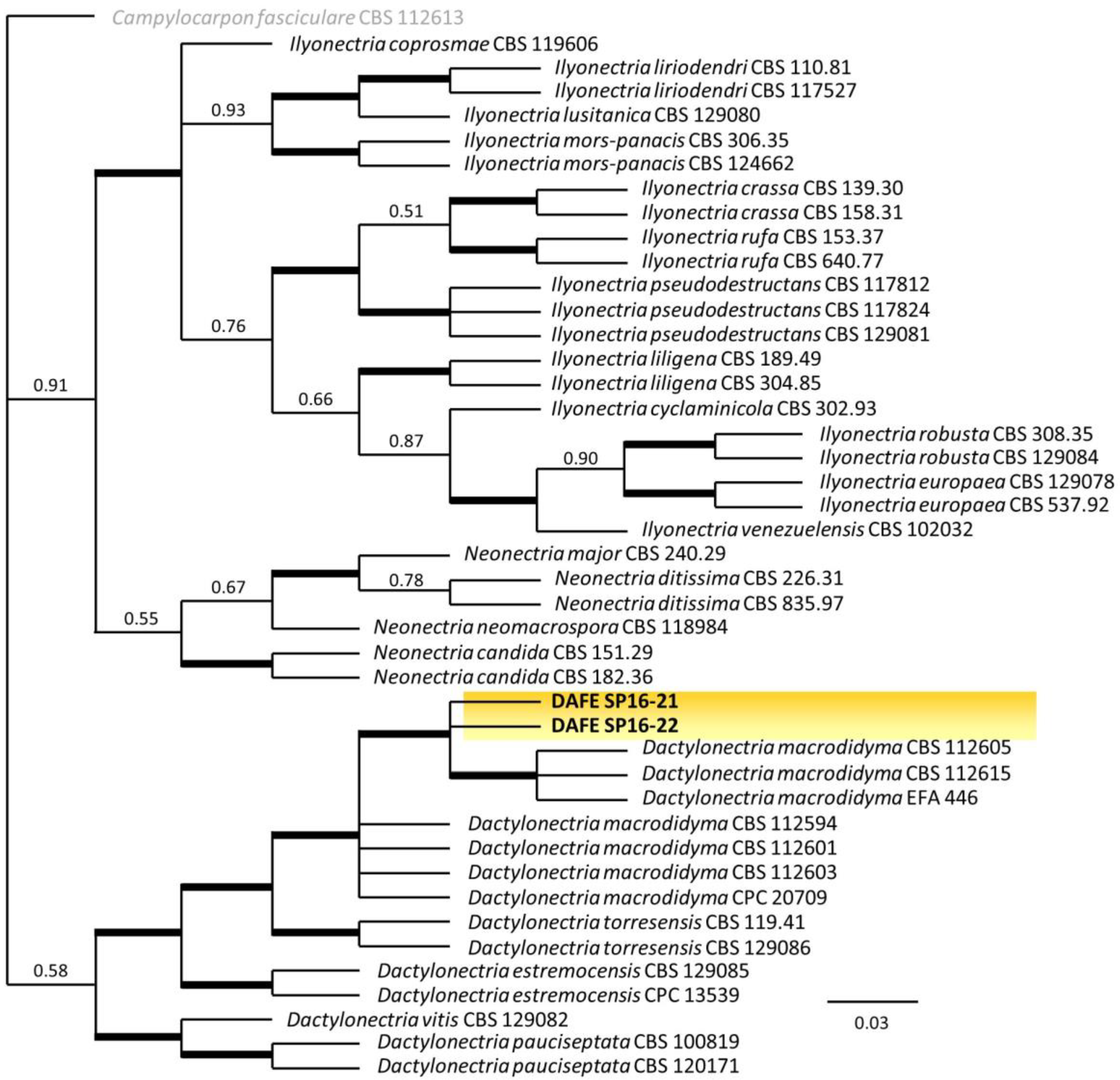
2.4. Phylogenetic Analysis

Sequences were assembled and edited using the BioEdit program (v.7.2.5) [30] to obtain consensus sequences from those generated from forward and reverse primers and have been deposited in GenBank (Table S1). The histone 3, β-tubulin and ITS consensus sequences were used as queries in BLASTn searches of the GenBank database hosted by NCBI.
Phylogenetic analysis was first performed using the histone 3 alignment as this locus was reported to be very informative [31,32,33]. In addition, a combined alignment of three loci (HIS3, TUB2 and ITS) was also analyzed. Both analyses were conducted on a set of subjects (42) selected from the BLASTn searches, along with sequences of type species retrieved from the literature [21,31]. All the reference sequences are listed in Table S1 and are available on GenBank.
The sequences obtained were aligned using MAFFT v. 7 online software [34], and the resulting multiple alignments were end-trimmed in order to compare the nucleotides of the sequence. Using MEGA11 [35], the best-fit substitution model for the multiple sequence alignments was calculated for each separate sequence dataset. The multilocus concatenated alignment (HIS3, TUB2 and ITS) was performed using Geneious 10.2.3. The Markov chain Monte Carlo (MCMC) algorithm was performed using MrBayes 3.2.6 [36] to generate phylogenetic trees with Bayesian posterior probabilities for a combined sequence dataset. For each locus, the nucleotide substitution models determined by MEGA11 were used in the analysis. Four MCMC chains were run simultaneously for random trees for 5,000,000 generations. Samples were taken every 1000 generations. The first 25% of trees were discarded as the burn-in phase of each analysis, and posterior probabilities were determined from the remaining trees.

2.5. Pathogenicity Test

Pathogenicity was confirmed by inoculating healthy 2-year-old P. lusitanica ‘Angustifolia’ plants with the representative isolates DAFE SP16-21 and DAFE SP16-22. Inoculations were carried out by removing a 9-mm bark disc with a cork borer and replacing it with a 9-mm plug from a 10-day-old PDA culture. The wounds were covered with sterile wet cotton wrapped with parafilm to prevent contamination and desiccation. Three inoculation points per plant were used, with a total of three replicates plus one control per plant inoculated with PDA plugs only. After inoculation, plants were incubated in a growth chamber at 24 ± 1 °C with a 12-h dark and light cycle and were visually inspected for disease for up to 6 months. To fulfill Koch’s postulates, re-isolation was carried out following the same procedure described above.

3. Results

3.1. Morphological Observations

Cylindrocarpon-like isolates were consistently recovered from the cross sections of diseased P. lusitanica stems with a frequency of 68% of the total isolations performed.
Colonies on PDA appeared with a highly diffuse aerial mycelium, which was initially grayish-white and then varied in color from brown to sepia. On the reverse of the substrate, the colony color was burnt umber (6E7) in the center of the colony, brownish yellow (6D7) towards the margin and amber-yellow (4B6–4B7) at the margin. The colony diameter (mm) after 7 days was 34–35 on PDA, 45–47 on OA and 43–45 on SNA.
On PDA, all cultures produced hyaline, ellipsoidal to ovoidal 0–1-septate microconidia measuring and hyaline, straight, cylindrical, somewhat clavate 1–3 septate macroconidia; 0-septate microconidia measuring (3.1-)12.5-13.3-14.1(-15.2) × (3.1-)4.1-4.3-4.6(-5.5) μm (n = 11) and 3-septate macroconidia measuring (36.6-)44.3-46.2-48.1(-53.6) × (4.9-)6.6-7.0-7.4(-9.4) μm (n = 30). Conidia were formed on simple or branched conidiophores. Chlamydospores were not abundant, generally intercalary, and single or in chains (Figure 3a–d). Perithecia did not develop on agar media.

3.2. Phylogenetic Analysis

Amplicons of 569 and 544 bp (ITS), 347 bp (TUB2) and 499 bp (HIS3) were obtained for the fungal isolates DAFE SP16-21 and DAFE SP16-22, respectively.
BLASTn search using ITS and TUB2 sequences were ineffective in distinguishing D. macrodidyma from other Dactylonectria species (e.g., D. torresensis and D. alcacerensis) even though they showed 99–100% identity.
In contrast, the HIS3 gene sequence exhibited 99–100% identity only with D. macrodidyma isolates and among them with D. macrodidyma ex-type isolate CBS 112615 (GenBank Accession No. JF735647) [31].
The phylogenetic analysis included the fungal isolates DAFE SP16-21 and DAFE SP16-22 and 41 ingroup taxa, with Campylocarpon fasciculare (CBS 112613) as an outgroup taxon. Two phylogenetic trees were obtained using the alignment of a combined dataset (HIS3, TUB2 and ITS) and the HIS3 single-locus alignment (Figure 4 and Figure 5).
The combined sequence dataset of 1300 characters containing alignment gaps consisted of 833 conserved, 318 parsimony-informative and 98 singleton characters. The gene boundaries were HIS3: 1–506, ITS: 507–1010 and TUB2: 1011–1300. Using MEGA11, the GTR + G + I evolution model was selected for the partitioned Bayesian inference for HIS3, while the K2 + G evolution model was selected for ITS and TUB2, and the consensus tree is presented in Figure 4.
The HIS3 single-locus sequence dataset of 506 characters containing alignment gaps consisted of 298 conserved, 172 parsimony-informative and 33 singleton characters. Using MEGA11, the GTR + G + I evolution model was selected for the partitioned Bayesian inference, and the consensus tree is presented in Figure 5.
In both trees, the isolates DAFE SP16-21 and DAFE SP16-22 clustered in the Dactylonectria (≡ Ilyonectria, Neonectria) macrodidyma (Cylindrocarpon macrodidymum) clade, which include the ex-type isolate CBS 112615, with high Bayesian Posterior Probability (0.99). The isolates used in this study were therefore identified as Dactylonectria macrodidyma (Halleen, Schroers & Crous) L. Lombard & Crous [21,24,37].

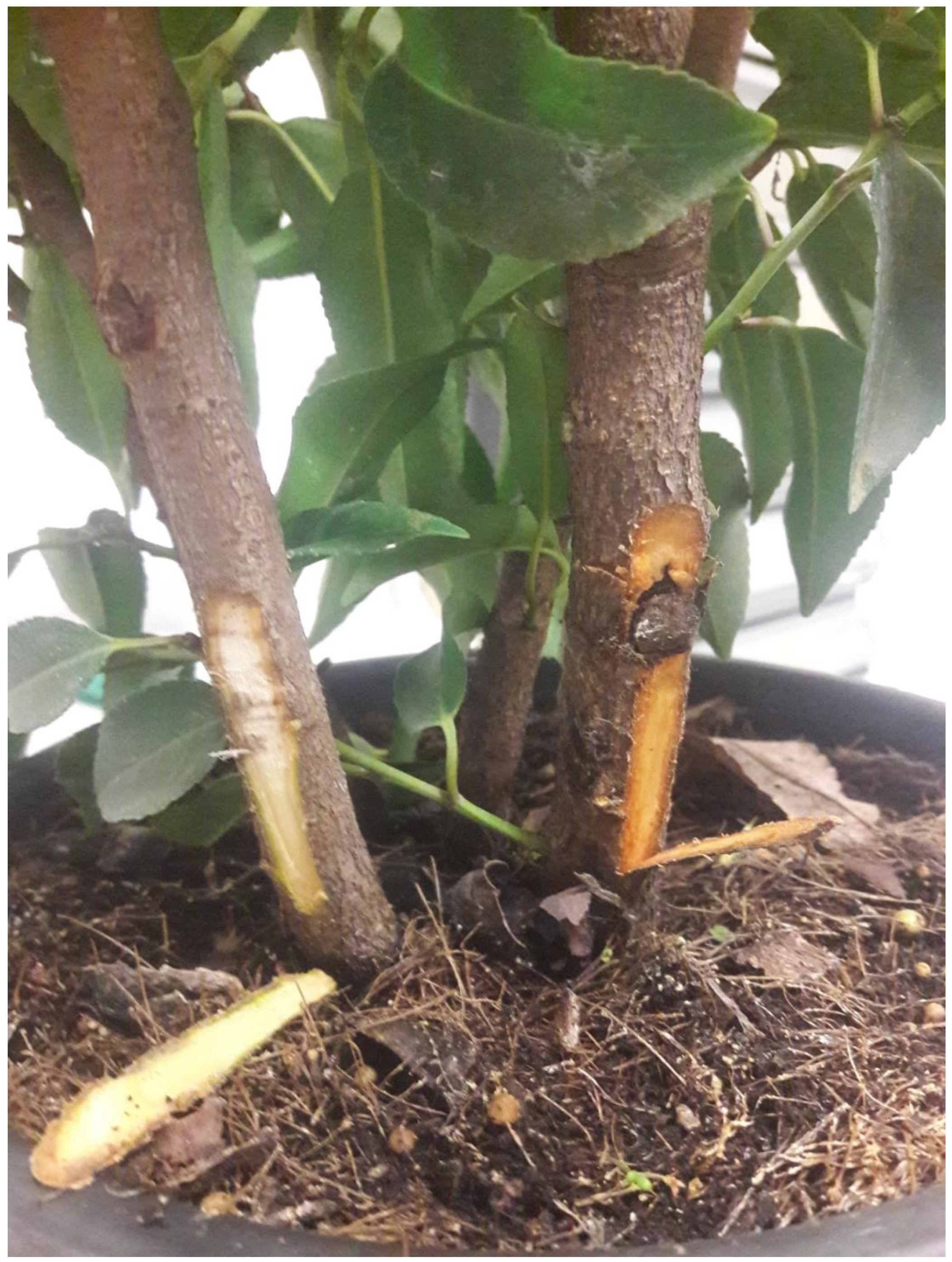
3.3. Pathogenicity Test

Six months after inoculation with isolates DAFE SP16-21 and DAFE SP16-22 of D. macrodidyma, most of the inoculated P. lusitanica plants showed chlorotic leaves and large reddish-brown areas along the stems similar to those observed in the commercial nursery (Figure 6). Both isolates of D. macrodidyma were recovered from symptomatic P. lusitanica plants and identified based on morphology, fulfilling Koch’s postulates. The re-isolation percentages from the lesions varied between 90% and 100%. No symptoms were observed in the controls.

4. Discussion

A polyphasic approach to identify fungal isolates associated with the dieback found on Prunus lusitanica in an Italian nursery (Tuscany, Pistoia) was used. The combination of morphological traits, pathogenicity assay, and molecular phylogeny using the HIS3 sequence and a combined alignment of three loci (HIS3, TUB2 and ITS) [24,38] revealed the identity of the fungal isolates to be Dactylonectria macrodidyma. Sequence analysis of the histone 3 gene has been recognized as the most appropriate to identify Dactylonectria species, especially those belonging to the “macrodidyma” species complex [31,32,33].
Fungal genera such as Dactylonectria, Ilyonectria and Neonectria are important pathogens of large varieties of plants worldwide. The species belonging to these genera showed common phenotypic traits such as orange to red ascomata, intercalary globose chlamydospores and Cylindrocarpon-like asexual forms with ellipsoidal to ovoid 1–4 septate macroconidia and 0–1 septate microconidia [21,37].
Recently the generic status of Ilyonectria and allied genera was re-evaluated using multi-gene DNA data and morphological comparisons, and the genus Dactylonectria was introduced with 10 new combinations, several of which were previously treated as Ilyonectria [21].
Dactylonectria macrodidyma has been mainly reported as the causal agent of the black foot disease of grapevine, but the pathogen has also been reported on many herbaceous and woody plants with high economic importance (Table 1).
Symptoms described included various forms of decline as well as typical black foot symptoms (root, collar, stem and crown rot), which cause a rapid collapse of branches during the summer, chlorosis of leaves, which suddenly wilted and died, as well as shoot dieback, abnormal development of roots and necrotic root crowns. Internal wood symptoms, visible in cross sections as wedge-shaped necroses, were frequently observed. Affected young nursery plants died within a few weeks of showing the first symptoms.
When inoculated on P. lusitanica, the isolates DAFE SP16-21 and DAFE SP16-22 of D. macrodidyma were pathogenic, and the plants developed similar disease symptoms observed in the nursery after six months. The disease symptoms caused by D. macrodidyma on our samples of P. lusitanica were similar to those described by Halleen et al. [24] as black foot disease.
Colony morphology and colony growth were in agreement with previous descriptions of D. macrodidyma. Macroconidia were predominantly three septate, and microconidia, scarcely produced on PDA, were aseptate. Both types of conidia were longer than those reported for the D. macrodidyma ex-type isolate CBS 112615 [24]. Our measurements were more similar to those reported by Úrbez-Torres et al. [13] for the isolate PARC398.
Cylindrocarpon/Dactylonectria spp. are soil-borne pathogens that can survive in the soil for a long time in the absence of the host crops and often have a wide range of hosts in different habitats [12,13]. These pathogens produce different types of propagules: mycelium within crop debris, conidia (macro and micro-conidia), which spread in soil water, and chlamydospores, which allow the pathogen to survive in soil for long periods [11,14]. Recently Probst et al. [53,54] reported that for D. macrodidyma, all three propagule types caused infections, and the pathogen is capable of infesting different soil types (clay loam, silt loam and sandy loam soils).
Generally, these fungi are considered opportunistic necrotic pathogens that usually infect their hosts through wounds producing toxins and cell wall degrading enzymes to invade host tissues. Under favorable conditions such as stress and/or reduction of plant vitality, they have the potential to be damaging pathogens and can be of great economic importance [15,16].
The potted nursery plants have a role in promoting infections because they are frequently stressed, remain containerized throughout production, and several wounds could be caused during transplanting [17,18,19].
The potential impact of increased drought on eight different pathosystems in New Zealand has been recently reported. For the disease caused by Ilyonectria/Dactylonectria spp., an increase in drought corresponds to an increase in disease expression [55]. The variability in seasonal rainfall patterns and the higher temperatures recorded in 2016 in the Pistoia province (compared to the averages from 1951 to 2016) [56] suggested, as elsewhere described [57] a warmer climate. Since temperatures were increasing and rainfall was decreasing, extreme climate events such as droughts could have influenced agriculture in the province. Moreover, 2016 was the hottest year on record since 1850 [58].
Different management strategies to prevent or reduce disease caused by Cylindrocarpon/Dactylonectria spp. with promising results have been exploited: host resistance, hot water treatments, fungicides, chitosan applications, biofumigation, biological control agents such as Trichoderma spp. and/or arbuscular-mycorrhizal fungi [11,59,60,61]. However, limited single effective measures are currently available for the control of the disease [50,51,59,62].
In the frame of nursery management, the phytosanitary status of plant material and the use of clean potting soil and containers (especially if reused) are considered essential measures to avoid the spread of the pathogen inoculum from nurseries to plantations [43,63,64].

5. Conclusions

To the best of our knowledge, this is the first report of the occurrence of dieback on P. lusitanica caused by D. macrodidyma. The fungal pathogen caused a crown rot disease on Portugal laurel, and many affected plants died within a few weeks of showing the first symptoms. This study showed the ability of D. macrodidyma to impact the ornamental nursery industry in Italy. The accurate identification of the pathogen will help to adopt correct management strategies to prevent or reduce the disease in potted nursery plants.

Supplementary Materials

The following supporting information can be downloaded at: https://www.mdpi.com/article/10.3390/horticulturae9020145/s1, Table S1: Campylocarpon, Dactylonectria, Ilyonectria and Neonectria species used for phylogenetic analysis.

Author Contributions

Conceptualization, S.P., B.C. and E.R.; methodology, S.P. and B.C.; validation, S.P. and B.C.; formal analysis, S.P. and D.D.L.; investigation, S.P., B.C., D.D.L. and E.R.; resources, S.P.; data curation, S.P., B.C. and D.D.L.; writing—original draft preparation, S.P. and D.D.L.; writing—review and editing, S.P., B.C., D.D.L. and E.R.; visualization, S.P., B.C., D.D.L. and E.R.; supervision, S.P. and E.R.; funding acquisition, S.P. All authors have read and agreed to the published version of the manuscript.

Funding

This research received no external funding.

Institutional Review Board Statement

Not applicable.

Informed Consent Statement

Not applicable.

Data Availability Statement

Not applicable.

Acknowledgments

Grateful thanks are expressed to Grazia Puntoni (University of Pisa, Italy) for the helpful technical support and Vannucci Piante Company (Pistoia) for kindly providing Prunus lusitanica plants for artificial inoculation experiments.

Conflicts of Interest

The authors declare no conflict of interest.

References

  1. Rieuf, P. Parasites and saprophytes des plantes au Maroc. Les Cah. De La Rech. Agron. 1969, 27, 1–178. [Google Scholar]
  2. Bean, W.J. Trees and Shrubs Hardy in British Isles. Murray, J., Ed.; John Murray Pubs Ltd.: London, UK, 1981; Volume 1–4. [Google Scholar]
  3. Huxley, A.J.; Griffiths, M.; Levy, M. The New Royal Horticultural Society Dictionary of Gardening; Macmillan: London, UK, 1992; Volume 4. [Google Scholar]
  4. Hiemstra, J.A. Some general features of Verticillium wilts in trees. In A Compendium of Verticillium Wilts in Tree Species; Hiemstra, J.A., Harris, D.C., Eds.; Ponsen & Looijen: Wageningen, The Netherlands, 1998; pp. 5–11. [Google Scholar]
  5. Halstead, A.J.; Scrace, J.M. Pests and diseases of outdoor ornamentals, including hardy nursery stock. In Pest and Disease Management Handbook; Alford, D.V., Ed.; Blackwell Science: Oxford, UK, 2000; pp. 429–541. [Google Scholar]
  6. Cunnington, J.H.; Lawrie, A.C.; Pascoe, I.G. Genetic variation within Podosphaera tridactyla reveals a paraphyletic species complex with biological specialization towards specific Prunus subgenera. Mycol. Res. 2005, 109, 357–362. [Google Scholar] [CrossRef]
  7. Lázaro, B.B. Identification of the Main Plagues and Diseases that Affect the Azorean Plum—Prunus lusitanica L. ssp. azorica (Mouillef.) Franco. 2006, pp. 1–22. Available online: www.life-priolo.spea.pt/en/priolo-and-its-habitat/documents/ (accessed on 17 November 2022).
  8. Cave, G.L.; Randall-Schadel, B.; Redlin, S.C. Risk Analysis for Phytophthora Ramorum Werres, de Cock & Man In’t Veld, Causal Agent of Sudden Oak Death, Ramorum Leaf Blight, and Ramorum Dieback; USDA, APHIS, PPQ, CPHST, PERAL: Raleigh, NC, USA, 2008; p. 88. [Google Scholar]
  9. Joshi, V.; Jeffries, M.; Jesperson, G. Diseases diagnosed on commercial crops submitted to the British Columbia Ministry of Agriculture Plant Health Laboratory in 2011. Can. Plant Dis. Surv. 2012, 92, 7–17. [Google Scholar]
  10. Schulze, J.A.; Contreras, R.N. In Vivo chromosome doubling of Prunus lusitanica and preliminary morphological observations. HortScience 2017, 52, 332–337. [Google Scholar] [CrossRef] [Green Version]
  11. Agustí-Brisach, C.; Armengol, J. Black-foot disease of grapevine: An update on taxonomy, epidemiology and management strategies. Phytopathol. Mediterr. 2013, 52, 245–261. [Google Scholar]
  12. Tewoldemedhin, Y.T.; Mazzola, M.; Mostert, L.; McLeod, A. Cylindrocarpon species associated with apple tree roots in South Africa and their quantification using real-time PCR. Eur. J. Plant Pathol. 2011, 129, 637–651. [Google Scholar] [CrossRef]
  13. Úrbez-Torres, J.R.; Haag, P.; Bowen, P.; O’Gorman, D.T. Grapevine trunk diseases in British Columbia: Incidence and characterization of the fungal pathogens associated with black foot disease of grapevine. Plant Dis. 2014, 98, 456–468. [Google Scholar] [CrossRef] [Green Version]
  14. Petit, E.; Barriault, E.; Baumgartner, K.; Wilcox, W.F.; Rolshausen, P.E. Cylindrocarpon species associated with black-foot of grapevine in Northeastern United States and Southeastern Canada. Am. J. Enol. Vitic. 2011, 62, 177–183. [Google Scholar] [CrossRef]
  15. Adesemoye, A.O.; Mayorquin, J.S.; Peacock, B.B.; Moreno, K.; Hajeri, S.; Yokomi, R.; Eskalen, A. Association of Neonectria macrodidyma with dry root rot of Citrus in California. J. Plant Pathol. Microbiol. 2016, 8, 391. [Google Scholar]
  16. Menkis, A.; Burokiene, D. Distribution and genetic diversity of the root-rot pathogen Neonectria macrodidyma in a forest nursery. For. Pathol. 2012, 42, 79–83. [Google Scholar] [CrossRef]
  17. Mora-Sala, B.; Cabral, A.; León, M.; Agustì-Brisach, C.; Armengol, J.; Abad-Campos, P. Survey, identification, and characterization of Cylindrocarpon-like asexual morphs in Spanish forest nurseries. Plant Dis. 2018, 102, 2083–2100. [Google Scholar] [CrossRef] [PubMed] [Green Version]
  18. Capote, N.; Del Río, M.Á.; Herencia, J.F.; Arroyo, F.T. Molecular and pathogenic characterization of Cylindrocarpon-like anamorphs causing root and basal rot of almonds. Plants 2022, 11, 984. [Google Scholar] [CrossRef]
  19. Aigoun-Mouhous, W.; Elena, G.; Cabral, A.; León, M.; Sabaou, N.; Armengol, J.; Chaouia, C.; Mahamedi, A.E.; Berraf-Tebbal, A. Characterization and pathogenicity of Cylindrocarpon-like asexual morphs associated with black foot disease in Algerian grapevine nurseries, with the description of Pleiocarpon algeriense sp. nov. Eur. J. Plant Pathol. 2019, 154, 887–901. [Google Scholar] [CrossRef]
  20. Wollenweber, H.W. Ramularia, Mycosphaerella, Nectria, Calonectria. Eine morphologisch pathologische studie zur abgrenzung von pilzgruppen mit cylindrischen und sichelförmigen konidienformen. Phytopathology 1913, 3, 198–242. [Google Scholar]
  21. Lombard, L.; Van der Merwe, N.A.; Groenewald, J.Z.; Crous, P.W. Lineages in Nectriaceae: Re-evaluating the generic status of Ilyonectria and allied genera. Phytopathol. Mediterr. 2014, 53, 515–532. [Google Scholar]
  22. Dhingra, O.D.; Sinclair, J.B. Basic Plant Pathology Methods, 2nd ed.; CRC Press: Boca Raton, FL, USA, 1995; p. 355. [Google Scholar]
  23. Nirenberg, H.I. Untersuchungen über die morphologische und biologische Differenzierung in der Fusarium-Sektion Liseola. In Mitteilungen Aus Der Biologischen Bundesanstalt Für Land- Und Forstwirtschaft (Berlin-Dahlem); Biologische Bundesanst. für Land- und Forstwirtschaft: Berlin, Germany, 1976; Volume 169, pp. 1–117. [Google Scholar]
  24. Halleen, F.; Schroers, H.F.; Groenewald, J.Z.; Crous, P.W. Novel species of Cylindrocarpon (Neonectria) and Campylocarpon gen. nov. associated with black foot disease of grapevines (Vitis spp.). Stud. Mycol. 2004, 50, 431–455. [Google Scholar]
  25. Kornerup, A.; Wanscher, J.H. Methuen Handbook of Colour; Eyre Methuen: London, UK, 1978; p. 252. [Google Scholar]
  26. Pecchia, S.; Da Lio, D. Development of a rapid PCR-Nucleic Acid Lateral Flow Immunoassay (PCR-NALFIA) based on rDNA IGS sequence analysis for the detection of Macrophomina phaseolina in soil. J. Microbiol. Methods 2018, 151, 118–128. [Google Scholar] [CrossRef] [PubMed]
  27. White, T.J.; Bruns, T.; Lee, S.; Taylor, J. Amplification and direct sequencing of fungal ribosomal RNA genes for phylogenetics. In PCR Protocols: A Guide to Methods and Applications; Innis, M.A., Gelfand, D.H., Sninsky, J.J., White, T.J., Eds.; Academic Press: San Diego, CA, USA, 1990; pp. 315–322. [Google Scholar]
  28. Glass, N.L.; Donaldson, G.C. Development of primer sets designed for use with the PCR to amplify conserved genes from filamentous ascomycetes. Appl. Environ. Microbiol. 1995, 61, 1323–1330. [Google Scholar] [CrossRef] [PubMed] [Green Version]
  29. Crous, P.W.; Groenewald, J.Z.; Risede, J.M.; Hywel-Jones, N.L. Calonectria species and their Cylindrocladium anamorphs: Species with sphaeropedunculate vesicles. Stud. Mycol. 2004, 50, 415–429. [Google Scholar]
  30. Hall, T.A. BioEdit: A user-friendly biological sequence alignment editor and analysis program for Windows 95/98/NT. Nucleic Acids Symp. Ser. 1999, 41, 95–98. [Google Scholar]
  31. Cabral, A.; Groenewald, J.Z.; Rego, C.; Oliveira, H.; Crous, P.W. Cylindrocarpon root rot: Multi-gene analysis reveals novel species within the Ilyonectria radicicola species complex. Mycol. Prog. 2012, 11, 655–688. [Google Scholar] [CrossRef] [Green Version]
  32. Cabral, A.; Rego, C.; Nascimento, T.; Holiveira, H.; Groenewald, J.Z.; Crous, P.W. Multi-gene analysis and morphology reveal novel Ilyonectria species associated with black foot disease of grapevines. Fungal Biol. 2012, 116, 62–80. [Google Scholar] [CrossRef] [PubMed]
  33. Lawrence, D.P.; Nouri, M.T.; Trouillas, F.P. Taxonomy and multi-locus phylogeny of cylindrocarpon-like species associated with diseased roots of grapevine and other fruit and nut crops in California. Fungal Syst. Evol. 2019, 4, 59–75. [Google Scholar] [CrossRef]
  34. Katoh, K.; Rozewicki, J.; Yamada, K.D. MAFFT online service: Multiple sequence alignment, interactive sequence choice and visualization. Brief. Bioinform. 2019, 20, 1160–1166. [Google Scholar] [CrossRef] [PubMed] [Green Version]
  35. Tamura, K.; Stecher, G.; Kumar, S. MEGA11: Molecular evolutionary genetics analysis version 11.0. Mol. Biol. Evol. 2021, 38, 3022–3027. [Google Scholar] [CrossRef]
  36. Ronquist, F.; Huelsenbeck, J.P. MrBayes 3: Bayesian phylogenetic inference under mixed models. Bioinformatics 2003, 19, 1572–1574. [Google Scholar] [CrossRef] [Green Version]
  37. Chaverri, P.; Salgado, C.; Hirooka, Y.; Rossman, A.Y.; Samuels, G.J. Delimitation of Neonectria and Cylindrocarpon (Nectriaceae, Hypocreales, Ascomycota) and related genera with Cylindrocarpon-like anamorphs. Stud. Mycol. 2011, 68, 57–78. [Google Scholar] [CrossRef]
  38. Schroers, H.-J.; Zerjav, M.; Munda, A.; Halleen, F.; Crous, P.W. Cylindrocarpon pauciseptatum sp. nov., with notes on Cylindrocarpon species with wide, predominantly 3-septate macroconidia. Mycol. Res. 2008, 112, 82–92. [Google Scholar] [CrossRef]
  39. Dart, N.L.; Weeda, S.M. First report of Cylindrocarpon macrodidymum on Acer palmatum in Virginia. Plant Dis. 2011, 95, 1592. [Google Scholar] [CrossRef]
  40. Auger, J.; Pérez, I.; Esterio, M. Occurrence of root rot disease of cherimoya (Annona cherimola Mill.) caused by Dactylonectria macrodidyma in Chile. Plant Dis. 2015, 99, 1282. [Google Scholar] [CrossRef]
  41. Machín Barreiro, J.A. Identificación de Los Organismos Asociados a la Muerte de Plantas de Frutilla (Fragaria Ananassa Duch.) En El Departamento de Salto, Uruguay. Degree Thesis, Universidad de la República, Facultad de Agronomía, Montevideo, Uruguay, 2017; p. 41. Available online: https://www.colibri.udelar.edu.uy/jspui/bitstream/20.500.12008/18658/1/TTS_Mach%C3%ADnBarreiroJorgeAlex.pdf (accessed on 20 November 2022).
  42. Habibi, A.; Ghaderi, F. First record of Dactylonectria macrodidyma causing black root rot on strawberry. Mycologia Iranica 2020, 72, 241–246. [Google Scholar]
  43. Lombard, L.; Bezuidenhout, C.M.; Crous, P.W. Ilyonectria black foot rot associated with Proteaceae. Australas. Plant Pathol. 2013, 42, 337–349. [Google Scholar] [CrossRef]
  44. Úrbez-Torres, J.R.; Peduto, F.; Gubler, W.D. First report of Ilyonectria macrodidyma causing root rot of olive trees (Olea europaea) in California. Plant Dis. 2012, 96, 1378. [Google Scholar] [CrossRef]
  45. El-ddin, S.N.; Haleem, R.A. First record of Dactylonectria macrodidyma: A cause of olive root rot in Iraq. For. Pathol. 2022, 52, e12729. [Google Scholar]
  46. Vitale, A.; Aiello, D.; Guarnaccia, V.; Perrone, G.; Stea, G.; Polizzi, G. First report of root rot caused by Ilyonectria (=Neonectria) macrodidyma on avocado (Persea americana) in Italy. J. Phytopathol. 2012, 160, 156–159. [Google Scholar] [CrossRef]
  47. Parkinson, L.E.; Shivas, R.G.; Dann, E.K. Pathogenicity of Nectriaceous fungi on avocado in Australia. Phytopathology 2017, 107, 1479–1485. [Google Scholar] [CrossRef] [PubMed] [Green Version]
  48. Menkis, A.; Vasiliauskas, R.; Taylor, A.F.S.; Stenström, E.; Stenlid, J.; Finlay, R. Fungi in decayed roots of conifer seedlings from forest nurseries, afforested clearcuts and abandoned farmland. Plant Pathol. 2006, 55, 117–129. [Google Scholar] [CrossRef]
  49. van Der Merwe, R. Occurrence of Canker and Wood Rot Pathogens on Stone Fruit Propagation Material and Nursery Stone Fruit Trees. Master’s Thesis, Faculty of AgriSciences at the University of Stellenbosch, Stellenbosch, South Africa, 2019. Available online: https://scholar.sun.ac.za (accessed on 20 November 2022).
  50. Cabral, A.; Rego, C.; Crous, P.W.; Oliveira, H. Virulence and cross-infection potential of Ilyonectria spp. to grapevine. Phytopathol. Mediterr. 2012, 51, 340–354. [Google Scholar]
  51. Halleen, F.; Fourie, P.H.; Crous, P.W. A review of black-foot disease of grapevine. Phytopathol. Mediterr. 2006, 45, S55–S67. [Google Scholar]
  52. Ye, Q.; Zhang, W.; Jia, J.; Li, X.; Zhou, Y.; Han, C.; Wu, X.; Yan, J. Fungal pathogens associated with black foot of grapevine in China. Phytopathol. Mediterr. 2021, 60, 303–319. [Google Scholar] [CrossRef]
  53. Probst, C.M.; Ridgway, H.J.; Jaspers, M.V.; Jones, E.E. Pathogenicity of Ilyonectria liriodendri and Dactylonectria macrodidyma propagules in grapevines. Eur. J. Plant Pathol. 2019, 154, 405–421. [Google Scholar] [CrossRef]
  54. Probst, C.M.; Ridgway, H.J.; Jaspers, M.V.; Jones, E.E. Propagule and soil type affects the pathogenicity of Ilyonectria and Dactylonectria spp., the causal agents of black foot disease of grapevines. Vitis 2022, 61, 11–19. [Google Scholar]
  55. Wakelin, S.A.; Gomez-Gallego, M.; Jones, E.; Smaill, S.; Lear, G.; Lambie, S. Climate change induced drought impacts on plant diseases in New Zealand. Australas. Plant Pathol. 2018, 47, 101–114. [Google Scholar] [CrossRef]
  56. Marzialetti, P. Banca Dati Agrometeorologici Di Pistoia, 2018. Centro Sperimentale per Il Vivaismo. Available online: www.cespevi.it (accessed on 17 November 2022).
  57. Pendergrass, A.G.; Knutti, R.; Lehner, F.; Deser, C.; Sanderson, B.M. Precipitation variability increases in a warmer climate. Sci. Rep. 2017, 7, 17966. [Google Scholar] [CrossRef]
  58. World Meteorological Organization WMO Confirms 2016 as Hottest Year on Record, about 1.1 °C above Pre-Industrial Era. 2017. Available online: https://public.wmo.int/en/media/press-release/ (accessed on 26 November 2022).
  59. Gramaje, D.; Úrbez-Torres, J.R.; Sosnowski, M.R. Managing grapevine trunk diseases with respect to etiology and epidemiology: Current strategies and future prospects. Plant Dis. 2018, 102, 12–39. [Google Scholar] [CrossRef] [Green Version]
  60. Martínez-Diz, M.P.; Díaz-Losada, E.; Andrés-Sodupe, M.; Bujanda, R.; Maldonado-González, M.M.; Ojeda, S.; Yacoub, A.; Rey, P.; Gramaje, D. Field evaluation of biocontrol agents against black-foot and Petri diseases of grapevine. Pest Manag. Sci. 2021, 77, 697–708. [Google Scholar] [CrossRef]
  61. van Jaarsveld, W.J.; Halleen, F.; Bester, M.C.; Pierron, R.J.; Stempien, E.; Mostert, L. Investigation of Trichoderma species colonization of nursery grapevines for improved management of black foot disease. Pest Manag. Sci. 2021, 77, 397–405. [Google Scholar] [CrossRef]
  62. Halleen, F.; Crous, P.W.; Petrini, O. Fungi associated with healthy grapevine cuttings in nurseries, with special reference to pathogens involved in the decline of young vines. Australas. Plant Pathol. 2003, 32, 47–52. [Google Scholar] [CrossRef]
  63. Alaniz, S.; Abad-Campos, P.; García-Jiménez, J.; Armengol, J. Evaluation of fungicides to control Cylindrocarpon liriodendri and Cylindrocarpon macrodidymum in vitro, and their effect during the rooting phase in the grapevine propagation process. Crop Prot. 2011, 30, 489–494. [Google Scholar] [CrossRef]
  64. Dumroese, R.K.; James, R.L.; Wenny, D.L. Hot water and copper coatings in reused containers decrease inoculum of Fusarium and Cylindrocarpon and increase Douglas Fir seedling growth. Hortic. Sci. 2002, 37, 943–947. [Google Scholar] [CrossRef]
Figure 1. Potted Prunus lusitanica plants growing in a nursery: (a) hedging plants and (b) topiary balls.
Horticulturae 09 00145 g001
Figure 2. Symptoms associated with Prunus lusitanica dieback. (a) Three-year-old plants showing, from left to right, severe decline symptoms, including weak vegetation, yellowing, summer wilting and crown rot symptoms. A healthy plant is shown on the far left. (b) Part of the stem with bark removed, showing large reddish-brown areas along the stem beginning below the crown and running up along the stem. (c) Cross-sections of a healthy P. lusitanica stem. (d) Cross sections of an infected P. lusitanica stem revealed dark brown sectorial necrosis extending from the bark to the pith.
Horticulturae 09 00145 g002
Figure 3. Morphology of Dactylonectria macrodidyma (asexual form Cylindrocarpon macrodidymum) isolate DAFE SP16-21: (a) Colony on PDA. (b) Chlamydospores. (c) Micro- and macroconidia formed on PDA. (d) Simple, unbranched septate conidiophores on PDA. Scale bars = 10 μm.
Horticulturae 09 00145 g003
Figure 4. Bayesian inference phylogenetic tree generated using concatenated sequences of HIS3, TUB2 and ITS. A GTR + G + I evolution model was used for HIS3 sequences, while a K2 + G evolution model was selected for ITS and TUB2 sequences. The representative isolates DAFE SP16-21, and DAFE SP16-22 are highlighted in yellow. The values above branches show Bayesian Posterior Probability (BPP). Thicker branches represent nodes with BPP  ≥  0.95. The scale bar indicates the number of expected substitutions per site. Campylocarpon fasciculare (CBS 112613) was used as an outgroup.
Horticulturae 09 00145 g004
Figure 5. Bayesian inference phylogenetic tree generated using sequences of HIS3. A GTR + G + I evolution model was used. The representative isolates DAFE SP16-21, and DAFE SP16-22 are highlighted in yellow. The values above branches show Bayesian Posterior Probability (BPP). Thicker branches represent nodes with BPP  ≥  0.95. The scale bar indicates the number of expected substitutions per site. Campylocarpon fasciculare (CBS 112613) was used as an outgroup.
Horticulturae 09 00145 g005
Figure 6. Pathogenicity test with the isolate DAFE SP16-21 of Dactylonectria macrodidyma. On the left: healthy control. On the right: reddish brown symptoms.
Horticulturae 09 00145 g006
Table 1. List of plants on which the presence of the fungal pathogen Dactylonectria macrodidyma has been reported.
PlantCommon NameDiseaseReferences
Acer palmatumJapanese mapleCrown cankers and root rot[39]
Annona cherimolaCherimoyaRoot-rot[40]
CitrusTrifoliate seedlings (Carrizo)Dry Root Rot[15]
Fragaria x ananassaStrawberryBlack root rot[41,42]
Ilex aquifoliumHollyRoot rot and dieback[17]
Juniperus phoeniceaPhoenician juniperRoot rot and dieback[17]
Leucospermum sp.PincushionBlack foot rot[43]
Lonicera sp.HoneysuckleRoot rot and dieback[17]
Malus domesticaAppleApple Replant Disease, Root rot[12,31]
Myrtus communisCommon myrtleRoot rot and dieback[17]
Olea europeaOliveRoot rot[44,45]
Persea americanaAvocadoRoot rot, Black root rot[46,47]
Picea abiesNorway spruceRoot dieback[48]
Pinus halepensisAleppo pineRoot rot and dieback[17]
Pinus sylvestrisScots pineRoot-rot[16,48]
Protea sp.SugarbushBlack foot rot[43]
Prunus lusitanicaPortugal laurelCrown rotThis study
Prunus dulcisAlmondRoot and basal rot[18]
Prunus salicinaPlumCrown infections[49]
Pyracantha sp.FirethornRoot rot and dieback[17]
Quercus fagineaPortuguese oakRoot rot and dieback[17]
Quercus ilexHolm oakRoot rot and dieback[17]
Rosmarinus officinalisRosemaryRoot rot and dieback[17]
Vitis viniferaGrapeBlack foot[13,50,51,52]
Disclaimer/Publisher’s Note: The statements, opinions and data contained in all publications are solely those of the individual author(s) and contributor(s) and not of MDPI and/or the editor(s). MDPI and/or the editor(s) disclaim responsibility for any injury to people or property resulting from any ideas, methods, instructions or products referred to in the content.

