Article

Quantitative Determination of the Main Phenolic Compounds, Antioxidant Activity, and Toxicity of Aqueous Extracts of Olive Leaves of Greek and Spanish Genotypes

by
María Esther Martínez-Navarro
1,
Eleftheria H. Kaparakou
2,
Charalabos D. Kanakis
2,
Cristina Cebrián-Tarancón
1,
Gonzalo L. Alonso
1,
María Rosario Salinas
1,* and
Petros A. Tarantilis
2
1
Cátedra de Química Agrícola, E.T.S.I. Agronómica y de Montes y Biotecnología, Universidad de Castilla-La Mancha, Avda. de España s/n, 02071 Albacete, Spain
2
Laboratory of Chemistry, Department of Food Science and Human Nutrition, Agricultural University of Athens, Iera Odos 75, 118 55 Athens, Greece
*
Author to whom correspondence should be addressed.
Horticulturae 2023, 9(1), 55; https://doi.org/10.3390/horticulturae9010055
Submission received: 29 November 2022 / Revised: 16 December 2022 / Accepted: 27 December 2022 / Published: 3 January 2023

Abstract

:
Olive leaves are rich in phenolic compounds, which give them antioxidant properties that are associated with a lower incidence of disease. Therefore, the aim of this work was to determine the phenolic content, antioxidant activity, and toxicity of the aqueous extracts of olive leaves of the main Spanish and Greek cultivated and wild genotypes. For these purposes, ‘Picual’ and ‘Arbequina’ leaves from Spain and ‘Koronoeiki’ and ‘Kalamon’ leaves from Greece were collected, as were wild olive leaves from both countries. The aqueous extracts of these genotypes were analyzed by HPLC-DAD, and the DPPH·, ABTS·+ Folin–Ciocalteu, and Microtox® methods were also used. ‘Picual’ had the highest oleuropein values, followed by wild olive leaves from both countries and ‘Arbequina’. The latter was reflected in the antioxidant activity measured by DPPH· and ABTS·+, which positioned the leaves of ‘Arbequina’, ‘Picual’, and the wild genotypes as having the most antioxidant activity. As expected, these leaves also had the highest total phenol content, as measured by Folin–Ciocalteu. Regarding the inhibition of the bioluminescence of Aliivibrio fischeri of the aqueous leaf extracts measured by Microtox®, the EC5015 ranged between 11.82 and 82.50 mg/mL, demonstrating similar behavior to other herbal infusions.

1. Introduction

In recent years, the role of diet in human health has been recognized as essential, with vegetable and infusion intake being associated with reduced incidence of various chronic diseases, atherosclerosis, inflammation, diabetes, and certain types of cancer [1]. These beneficial effects have been attributed in part to compounds with antioxidant capacities.
Free radical formation is associated with the normal natural metabolism of aerobic cells. The oxygen consumption inherent to cell growth leads to the generation of a series of oxygen free radicals. The interaction of these species with molecules of a lipidic nature produces new radicals, hydroperoxides, and different peroxides, which can interact with biological systems in a clearly cytotoxic way [2]. Many antioxidant compounds are known, but the ones that have the greatest antioxidant activity are gallic acid, followed by hydroxytyrosol [3].
Hydroxytyrosol is found in almost all parts of the olive tree (Olea europaea L.), but mainly in the leaves. It is usually found after the hydrolysis of oleuropein, which is formed by a molecule of elenolic acid linked to hydroxytyrosol by an ester bond, and this is linked to glucose by a glycosidic bond [4]. Oleuropein is characterized as the compound responsible for the bitter taste of the olive fruit [5].
Olive leaves are an agricultural by-product that accumulates during pruning and harvesting periods, with volumes estimated to be in excess of 18 million tonnes per annum [6]. The main olive-oil-producing countries, with more than 50% of the world’s harvest, are Spain (1.272.324.000 L) and Greece (251.900.000 L). It is estimated that producing one liter of olive oil generates 6.23 kg of pruning waste [7]. The leaves have a high antioxidant capacity because they are rich in secoiridoids (oleuropein, hydroxyoleuropein), alcohol phenols (hydroxytyrosol, tyrosol), hydroxycinnamic acid derivatives (verbascoside), and flavonoids (apigenin-7-O-glucoside, diosmetin-7-O-glucoside). Furthermore, flavonoids, secoiridoids, and verbascoside possess significant antioxidant activity against superoxide, hydroxyl, and peroxide radicals, which is mainly due to the redox properties of their phenolic hydroxyl groups [2,8].
Traditionally, olive leaf infusions have been used in folk medicine to treat fever or hypertension. Infusions are often consumed without any control. However, ‘natural’ and ‘safe’ are not synonymous, and some phenolic compounds are known to show toxicity in plants during extraction or in olive mill wastewater containing catechol, benzoic acids, and cinnamic acids, among others [9,10]. It is therefore essential to estimate and understand their potential [9]
Therefore, the aim of this work was to quantify the main phenolic compounds and to assess the antioxidant activity and toxicity in aqueous extracts of the olive leaves (O. europaea) from the main genotypes cultivated in Spain and Greece (‘Picual’, ‘Arbequina’, ‘Koroneiki’, and ‘Kalamon’) as well as in the wild olive leaves (Olea europaea spp. oleaster) from each country.

2. Materials and Methods

2.1. Chemicals and Reagents

Methanol (HPLC grade), 2,2-diphenyl-1-picrylhydrazyl (DPPH·), ethylhexadecyldimethylammonium bromide, and magnesium chloride were purchased from Sigma-Aldrich (St Louis, MO, USA). Oleuropein, hydroxytyrosol, verbascoside, apigenin-7-O-glucoside, and Trolox (6-hydroxy-2,5,7,8-tetramethylchroman-2-carboxylic acid) were purchased from Sigma-Aldrich (Steinheim, Germany). Diosmetin-7-O-glucoside was obtained from Extrasynthèse (Genay, France). Potassium persulfate (K2S2O8), sodium carbonate, and hydrogen peroxide (H2O2) were supplied by Merck KGaA (Darmstadt, Germany). Water was purified using a Millipore Direct-Q 3UV apparatus (18.2 mΩ s).

2.2. Raw Material

Olive leaves (O. europaea) were collected around harvest time [11] from typical Spanish and Greek olive genotypes. In Spain, ‘Picual’ and ‘Arbequina’ genotypes were selected in the Castilla-La Mancha region (southwest Spain, 881 m altitude, 38°58′40″ N latitude and 1°51′21″ W longitude). In Greece, ‘Koroneiki’ and ‘Kalamon’ genotypes were chosen from the Attica region (central Greece, 170 m altitude, 37°58′57″ N latitude and 23°42′19″ E longitude). In addition, wild olive trees (O.e. spp. oleaster) from both countries were collected from the Murcia region in Spain (WOLS) and from the Peloponnese region in Greece (WOLG).

2.3. Sample Preparation

Olive leaves were dried in the dark for seven days at room temperature (22 ± 2 °C) and at ambient relative humidity (45 ± 3%) and were then stored under the same conditions until use according to Martínez-Navarro et al. [3]. Dried leaves were ground in a knife mill, sieved (35 mesh), and subjected to microwave extraction at 800 W for 30 s using water as an extractant according to the methodology described by Martínez-Navarro et al. [12]. All extractions were performed in triplicate.

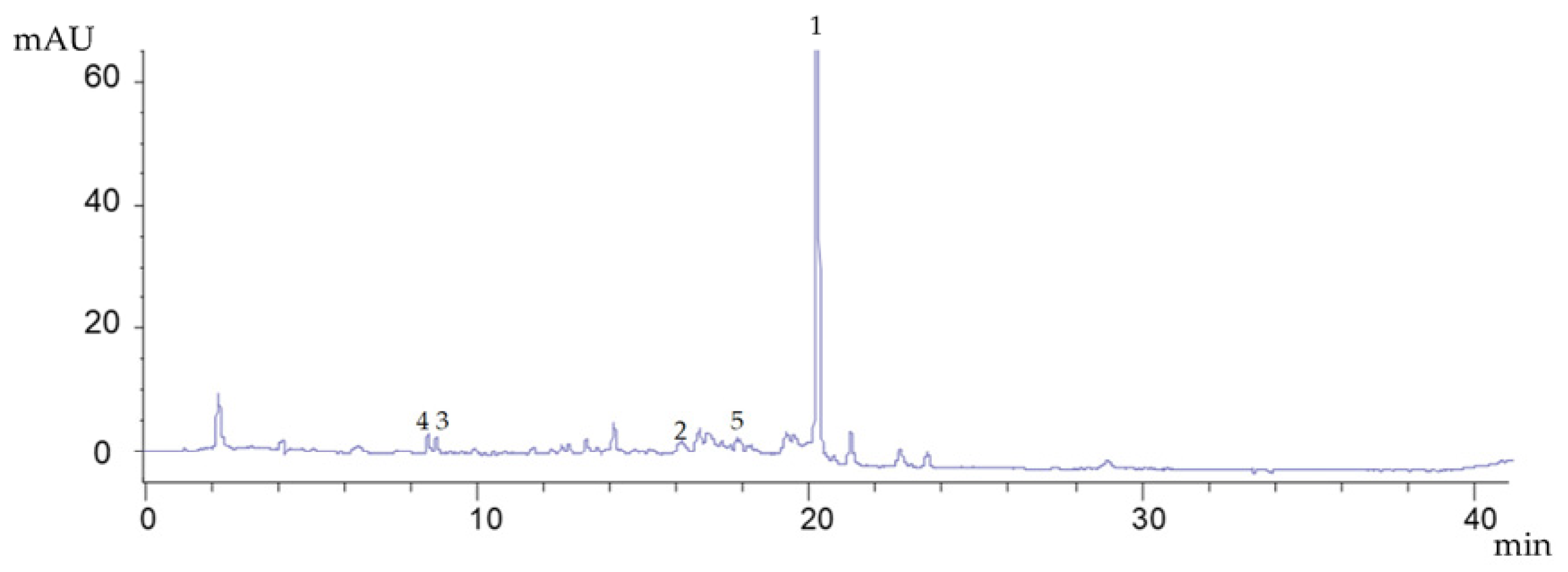
2.4. Determination of Oleuropein and Other Phenolic Compounds

Analyses were carried out according to Martínez-Navarro et al.’s method [12]. Aqueous extracts were injected into an Agilent 1100 high-performance liquid chromatograph (Palo Alto, CA, USA) equipped with a diode array detector (Agilent G1315D) coupled to a ChemStation, version B.03.01 (Agilent), data-processing station. Separation was performed on a reverse-phase C18 column, Brisa LC2 (250 mm × 4.6 mm, 5 μm particle size), purchased from Teknokroma (Barcelona, Spain) at 30 °C. The phenolic compounds studied were oleuropein, verbascoside, hydroxytyrosol (HT), and hydroxytyrosol hexoside and the flavonoids apigenin-7-O-glucoside and diosmetin-7-glucoside. All analyses were performed in triplicate and were expressed as milligrams of compound per gram of olive leaves.

2.5. Determination of Antioxidant Activity

The ABTS·+ (radical cation azino-bis[3-ethylbenzthiazoline-6-sulfonic acid]) and DPPH· (2, 2-diphenyl-1-picrylhydrazyl radical) methods were used to determine the antioxidant activity of different aqueous extracts. ABTS·+ and DPPH· solutions were prepared and measured according to Kaparakou et al. [13]. A calibration line was obtained using Trolox standard solution at concentrations in methanol from 0 to 1 mg/mL for ABTS·+. For DPPH·, the calibration line used was from 0 to 1.2 mg/mL. The results were expressed as micromole Trolox equivalents per milliliter. In addition, the concentration at which 50% of the DPPH· (EC50DPPH·) radicals in the olive leaf samples were scavenged was calculated. All determinations were conducted in triplicate.

2.6. Determination of Total Phenolic Content (TPC)

The total phenolic content (TPC) was determined using the Folin–Ciocalteu method described by Singleton et al. [14]. For quantification, 25 µL samples were added to 1500 µL of milli-Q water and 125 µL of Folin–Ciocalteu reagents. After 3 min, 375 µL of 20% sodium carbonate solution and 475 µL of milli-Q water were mixed, and after 120 min of incubation time at room temperature in dark conditions, the absorbance was recorded at 750 nm. Standard gallic acid was used to prepare the calibration line from 0 to 1.5 mg/mL, and the results were reported in milligrams of gallic acid equivalents (GAE) per milliliter. All samples were analyzed in triplicate.

2.7. Toxicity Test

The toxicity of the aqueous extracts of the different genotypes was tested at room temperature using the Microtox Analyzer 500 (Azur Environmental, Carlsbad, CA, USA). The procedure used was based on Kaparakou et al. [13], which consisted of performing the protocol known as Basic Test 81.9% [15] using the bio-luminescent bacterium Aliivibrio fischeri. Prior to this step, a preliminary test was completed to determine the adequate concentration of the aqueous extract. Once the appropriate dilution of the extracts was determined, Basic Test 81.9% was performed to estimate the effective concentration at which bioluminescence inhibition was 50% without data extrapolation after 15 min (EC5015).

2.8. Statistical Analysis

All experimental data obtained were analyzed in three replicated trials, and the data were expressed in the form of mean ± standard deviation using Excel (Microsoft Corporation. Redmond, WA, USA). One-way analysis of variance (ANOVA) along with Tukey’s post hoc HSD test (α < 0.05) were used to determine the significance of the data obtained using SPSS version 24 for Windows (SPSS INC., Chicago, IL, USA). In addition, Pearson correlation analysis was completed to assess the correlation between the phenolic compounds, DPPH·, ABTS·+, and TPC with SPSS software.

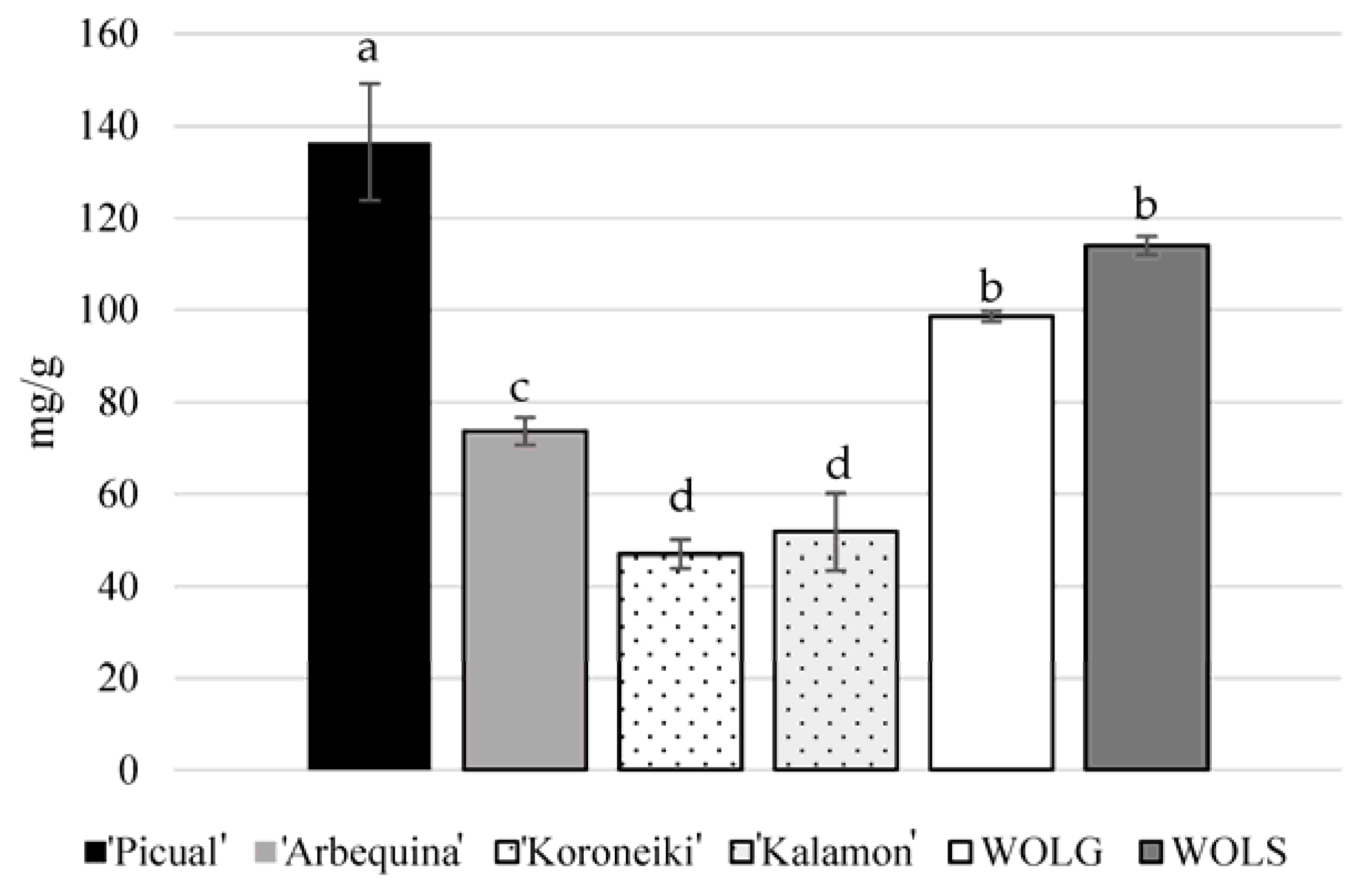
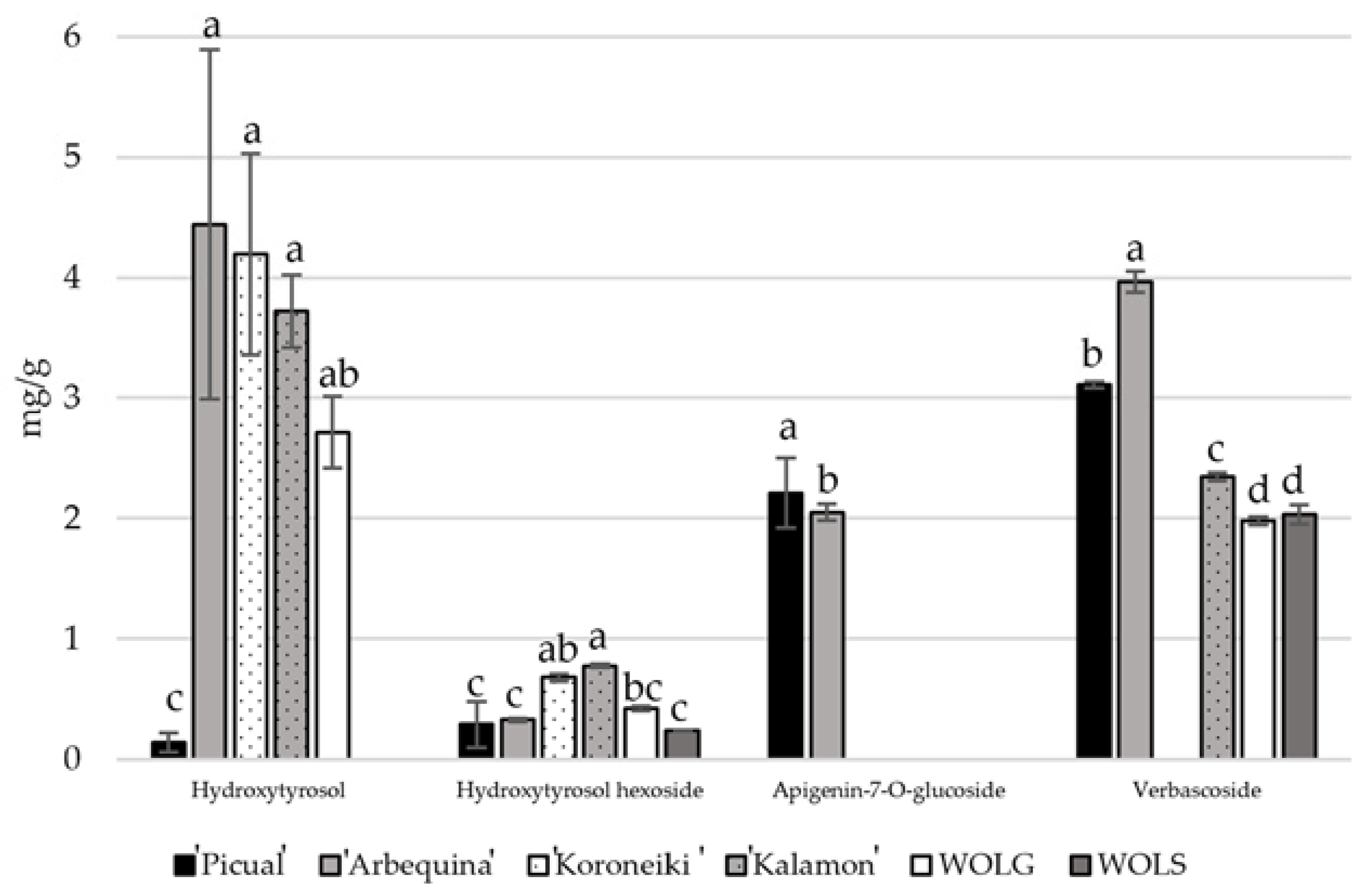
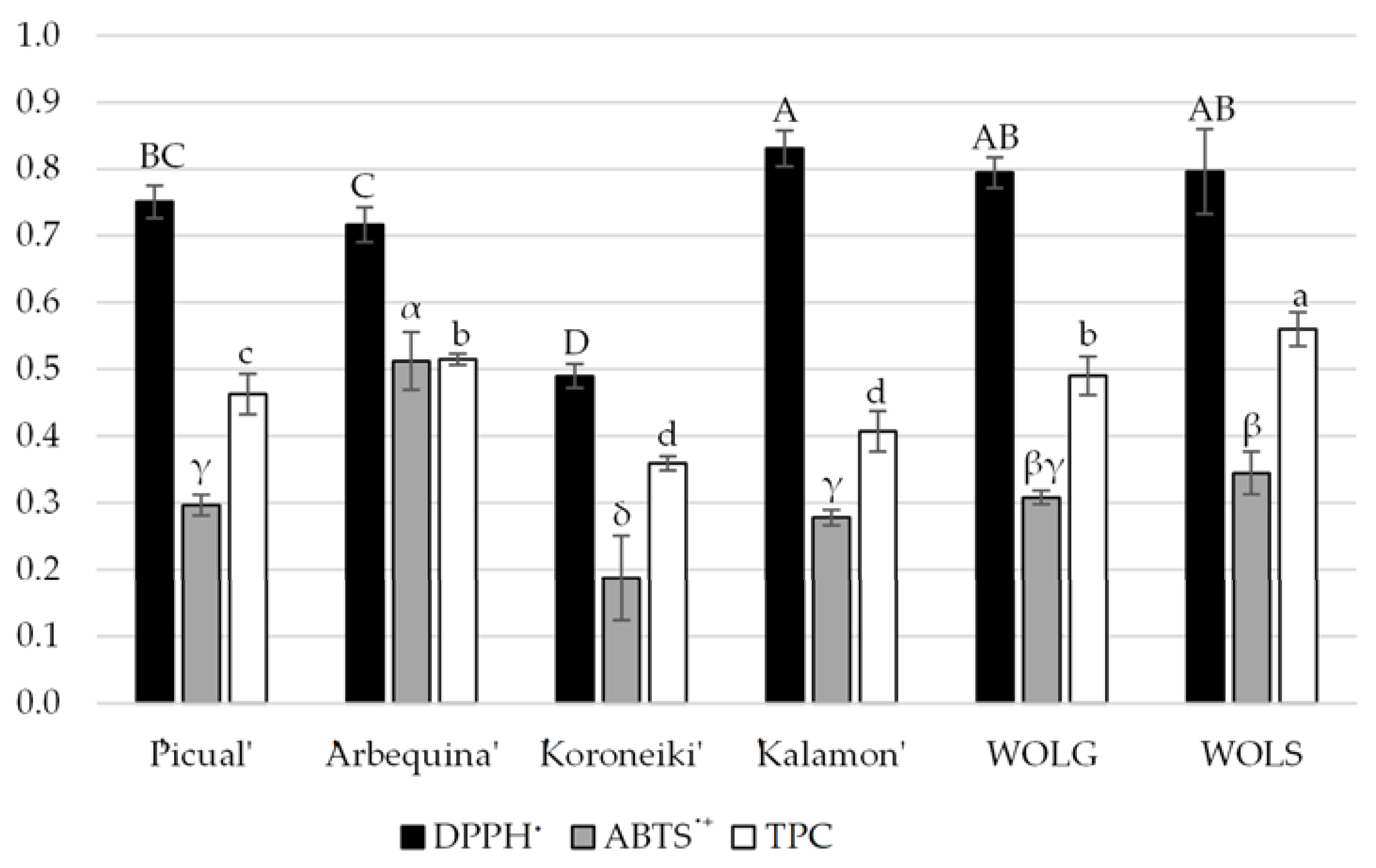
3. Results and Discussion

Figure 1 shows the chromatogram of the compounds obtained in the aqueous extracts. Figure 2 depicts the oleuropein content of all of the analyzed genotypes. The leaves with the highest oleuropein content were those of the ‘Picual’ genotype (136.48 mg/g), followed by wild olive leaves from Spain (WOLS, 114.04 mg/g) and Greece (WOLG, 98.59 mg/g), while the genotypes with the lowest amount of oleuropein were ‘Kalamon’ and ‘Koroneiki’ (51.83 and 47.02 mg/g, respectively). In contrast, Figure 3 indicates that ‘Kalamon’ and ‘Koroneiki’ had the highest hydroxytyrosol hexoside contents, with values of 0.78 and 0.68 mg/g, respectively.
The leaves with the lowest hydroxytyrosol hexoside content were ‘Picual’ (0.29 mg/g) and the WOLS (0.24 mg/g). As for hydroxytyrosol, the highest content was found in ‘Arbequina’ (4.44 mg/g) and ‘Koroneiki’ (4.19 mg/g), while the lowest content was found in ‘Picual’ (0.14 mg/g), and in the WOLS, it was not even detected. Verbascoside was found in concentrations ranging from 1.98 mg/g to 3.97 mg/g in ‘Arbequina’, followed by ‘Picual’ with 3.11 mg/g. Apigenin-7-O-glucoside was only found in the ‘Picual’ and ‘Arbequina’ at concentrations of 2.21 and 2.05 mg/g, respectively. Previous studies carried out by Martínez-Navarro et al. [11,16] on the same plots during the 2019/2020 season showed that, for the same month, ‘Arbequina’ leaves contained oleuropein values similar to those found in the present study (63.80 mg/g), while ‘Picual’ had a slightly lower value (103.41 mg/g). In a study carried out by Petridis et al. [17], comparing the phenolic compounds of olive leaves of different genotypes, including ‘Koroneiki’ and ‘Kalamon’, they also observed that ‘Kalamon’ had the highest phenolic content. In a study conducted by Talhaoui et al. [18] in mid-June on the phenolic content and antioxidant activity of ‘Picual’ and ‘Arbequina’ leaves, the oleuropein content was 17.08 mg/g in ‘Arbequina’ and 18.01 mg/g in ‘Picual’, which were below the values of 73.71 and 136.49 mg/g, respectively, found in the present work. As for other compounds studied by Talhaoui et al. [18], verbascoside was found to have values of 4.07 mg/g in ‘Arbequina’ and 1.12 mg/g in ‘Picual’, similar to the values obtained in this study (3.10 and 3.90 mg/g, respectively). The hydroxytyrosol hexoside content was also similar, at around 0.30–0.40 mg/g in both genotypes and in both studies.
The correlations between the different compounds contained in the olive leaves are included in Table 1. As expected, oleuropein was negatively correlated with the hydroxytyrosol (p < 0.01, r = −0.851) and hydroxytyrosol hexoside contents (p < 0.01, r = −0.683) and positively correlated with verbascoside (p < 0.01, r = 0.685). All of these correlations were observed by the authors of previous works [11,16]. The hydroxytyrosol content was negatively correlated with hydroxytyrosol hexoside (p < 0.01, r = −0.605) and was positively correlated with the verbascoside content (p < 0.01, r = −0.741), with the former suggesting that the hydroxytyrosol in the plant could be derived from glucosides or vice versa. Finally, apigenin-7-O-glucoside only demonstrated a positive correlation with the verbascoside content (p < 0.01, r = 0.662).
Figure 4 shows the antioxidant activity and total phenols of the studied leaf genotypes. Regarding the antioxidant activity measured by DPPH·, the genotypes with the highest activity were ’Kalamon’ (0.831 μM), WOLG (0.794 μM), and WOLS (0.796 μM), while ‘Koroneiki’ (0.490 μM) had the lowest values.
The measurements obtained by the ABTS·+ method had a similar order in the genotypes with the highest antioxidant activity measured by DPPH·, but although genotype is one of the most influential factors, other factors can influence the content of phenolic compounds, such as location, climatic factors, soil, cultivation practices, and pests and diseases, among others [19]. Regarding TPC, the highest amount was found in WOLS, followed by in ‘Arbequina’, WOLG, and ‘Picual’. This coincided with the varieties that showed the highest antioxidant activity potentials.
Similarly, the calculated EC50DPPH (Table 2) indicated that ‘Kalamon’, wild olive leaves (WOLS and WOLG), and ‘Arbequina’ had higher scavenging activity. An investigation carried out by Talhaoui et al. [18] studied the olive leaves of ‘Picual’ and ‘Arbequina’, which showed the highest scavenging activity via the EC50DPPH of ‘Arbequina’ compared to ‘Picual’, as is the case in this work.
The correlations of the antioxidant activity and Folin–Ciocalteu methods used with the phenolic compounds studied are shown in Table 3. As expected, the oleuropein content is positively correlated with ABTS·+ (p < 0.01, r = 0.669) and TPC (p < 0.01, r = 0.649), although the correlation with DPPH· is not statistically significant. Likewise, the verbascoside content was positively correlated with DPPH· (p < 0.01, r = 0.653) and ABTS·+ (p < 0.01, r = 0.702). On the contrary, hydroxytyrosol and its hexoside content were negatively correlated with ABTS·+ (p < 0.05, r = −0.600) and TPC (p < 0.01, r = −0.805), respectively. According to the study by Özcan et al. [20], hydroxytyrosol has a higher antioxidant activity than oleuropein and verbascoside. However, the results obtained in this work suggest that in olive leaf aqueous extracts, both the hydroxytyrosol and the hydroxytyrosol hexoside are more labile compounds and are therefore less stable than oleuropein and verbascoside. Furthermore, these last two compounds are precursors of hydroxytyrosol.
Concerning toxicity, the appropriate concentration in aqueous extracts to be used was eight milligrams of olive leaves per milliliter of water. Table 4 presents the results of the Microtox® assay in terms of EC5015.
Generally, EC5015 represents the effective lethal concentration that corresponds to the proportion of extract that causes the mortality or inhibition of 50% of the exposed bacteria, A. fischeri, in 15 min. The aqueous olive leaf extracts yielded significant differences. The ‘Picual’, ‘Arbequina’, and ‘Koroneiki’ genotypes stood out for their lower EC5015 values, while the highest values were found in ‘Kalamon’ and WOLS. Thus, the first three genotypes were the most active against A. fischeri, while WOLS and ‘Kalamon’ were the least active against this microorganism.
To our knowledge, no studies in the literature have focused on performing comparisons on the acute toxicity of aqueous olive leaf extracts. However, since the extractant used was water, some comparisons have been made with the results obtained for aqueous infusions of aromatic plants. Research by Skotti et al. [21] studied the toxicity of several aqueous extracts from different Greek medicinal and aromatic plants. Infusions of oregano (Origanum vulgare L.) and dittany (Origanum dictamnus L.) at a concentration of 10 mg/mL and water at 85 °C were found to have lower EC5015 values of 8.6 and 10.6, respectively, which are lower than those obtained for aqueous olive leaf extracts. For chamomile (Matricaria chamomilla L.) and sage (Salvia officinalis L.) infusions at 10 mg/mL and 25, 80, and 100 °C, Sotiropoulou et al. [9] reported EC5015 values between 0.032 and 1.264 mg/mL. Aqueous extracts of chamomile had a higher bacterial inhibition capacity compared to those of sage, and the effect increased with increasing temperatures.
In 2017, the Committee for Herbal Medicinal Products of the European Union completed an evaluation of the different products available on the European market and concluded that olive leaf products can be accepted as safe, and no toxicity was observed [22]. Therefore, olive leaf extracts behave in a similar way to other herbal infusions and, when consumed in moderation, should not cause toxicity.

4. Conclusions

The results of this study show that the contents of oleuropein and verbascoside in olive leaves are related to the antioxidant capacity of their aqueous infusions, with ‘Arbequina’ and wild olive leaves from Spain and Greece being the most active of the genotypes studied. In terms of toxicity, aqueous olive leaf extracts behave similarly to other herbal infusions against A. fischeri, so their consumption as an bitter antioxidant infusion should not be a problem with moderate consumption.

Author Contributions

Project administration, G.L.A. and M.R.S.; funding acquisition, G.L.A. and M.R.S.; writing—review and editing, all authors; visualization, all authors; supervision, P.A.T., G.L.A. and M.R.S.; conceptualization, all authors; methodology, all authors; software, M.E.M.-N. and C.C.-T.; validation, M.E.M.-N. and C.C.-T.; formal analysis, M.E.M.-N., E.H.K., C.D.K. and C.C.-T.; investigation, M.E.M.-N.; resources, all authors; data curation, M.E.M.-N., E.H.K., and C.C.-T.; writing—original draft preparation, all authors. All authors have read and agreed to the published version of the manuscript.

Funding

This study was supported by the Government of Castilla-La Mancha (Spain) in collaboration with FEDER for financing the project SBPLY/17/180501/000191.

Data Availability Statement

Not applicable.

Acknowledgments

M. E. Martínez-Navarro thanks the University of Castilla–La Mancha for the predoctoral contract 2019-PREDUCLM and the grant for predoctoral stays in foreign universities and research centers in collaboration with Banco Santander (590186 [2021/11379]). We are grateful to the Government of Castilla–La Mancha (Spain) in collaboration with FEDER for funding the project SBPLY/17/180501/000191 and to the Department of Food Science and Human Nutrition of the Agricultural University of Athens (Greece). Thank you to the owners of the olive plots and, especially, to the company Acebuche Campo Coy S.L. in Murcia (Spain).

Conflicts of Interest

The authors declare no conflict of interest.

References

  1. Xie, P.-J.; Huang, L.-X.; Zhang, C.-H.; Zhang, Y.-L. Phenolic compositions, and antioxidant performance of olive leaf and fruit (Olea europaea L.) extracts and their structure–activity relationships. J. Funct. Foods 2015, 16, 460–471. [Google Scholar] [CrossRef]
  2. Benavente-García, O.; Castillo, J.; Lorente, J.; Ortuño, A.; Del Rio, J.A. Antioxidant activity of phenolics extracted from Olea europaea L. leaves. Food Chem. 2000, 68, 457–462. [Google Scholar] [CrossRef]
  3. Martínez-Navarro, M.E.; Cebrián-Tarancón, C.; Oliva, J.; Salinas, M.R.; Alonso, G.L. Oleuropein Degradation Kinetics in Olive Leaf and Its Aqueous Extracts. Antioxidants 2021, 10, 9163. [Google Scholar] [CrossRef] [PubMed]
  4. Yuan, J.-J.; Wang, C.-Z.; Ye, J.-Z.; Tao, R.; Zhang, Y.-S. Enzymatic Hydrolysis of Oleuropein from Olea europea (Olive) Leaf Extract and Antioxidant Activities. Molecules 2015, 20, 2903–2921. [Google Scholar] [CrossRef] [PubMed] [Green Version]
  5. Cayan, H.; Erener, G. Effect of Olive Leaf (Olea europaea) Powder on Laying Hens Performance, Egg Quality and Egg Yolk Cholesterol Levels. Asian-Australas J. Anim. Sci. 2015, 28, 538–543. [Google Scholar] [CrossRef] [PubMed] [Green Version]
  6. Espeso, J.; Isaza, A.; Lee, J.Y.; Sörensen, P.M.; Jurado, P.; Avena-Bustillos, R.d.J.; Olaizola, M.; Arboleya, J.C. Olive Leaf Waste Management. Front. Sustain. Food Syst. 2021, 5, 660582. [Google Scholar] [CrossRef]
  7. COI (International Olive Council). The Wold of Olive Oil. 2022. Available online: https://www.internationaloliveoil.org/the-world-of-olive-oil (accessed on 15 July 2022).
  8. Topuz, S.; Bayram, M. Oleuropein extraction from leaves of three olive varieties (Olea europaea L.): Antioxidant and antimicrobial properties of purified oleuropein and oleuropein extracts. J. Food Process. Preserv. 2022, 46, e15697. [Google Scholar] [CrossRef]
  9. Sotiropoulou, N.S.; Megremi, S.F.; Tarantilis, P. Evaluation of Antioxidant Activity, Toxicity, and Phenolic Profile of Aqueous Extracts of Chamomile (Matricaria chamomilla L.) and Sage (Salvia officinalis L.) Prepared at Different Temperatures. Appl. Sci. 2020, 10, 2270. [Google Scholar] [CrossRef] [Green Version]
  10. Abbas, M.; Adil, M.; Ehtisham-ul-Haque, S.; Munir, B.; Yameen, M.; Ghaffar, A.; Shar, G.A.; Asif Tahir, M.; Iqbal, M. Vibrio fischeri bioluminescence inhibition assay for ecotoxicity assessment: A review. Sci. Total Environ. 2018, 626, 1295–1309. [Google Scholar] [CrossRef] [PubMed]
  11. Martínez-Navarro, M.E.; Cebrián-Tarancón, C.; Alonso, G.L.; Salinas, M.R. Determination of the Variability of Bioactive Compounds and Minerals in Olive Leaf along an Agronomic Cycle. Agronomy 2021, 11, 2447. [Google Scholar] [CrossRef]
  12. Martínez-Navarro, E.M.; Cebrián-Tarancón, C.; Moratalla-López, N.; Lorenzo, C.; Alonso, G.L.; Salinas, R.M. Development and validation of an HPLC-DAD method for determination of oleuropein and other bioactive compounds in olive leaf by-products. J. Sci. Food Agric. 2021, 101, 1447–1453. [Google Scholar] [CrossRef] [PubMed]
  13. Kaparakou, E.H.; Kanakis, C.D.; Gerogianni, M.; Maniati, M.; Vekrellis, K.; Skotti, E.; Tarantilis, P.A. Quantitative determination of aloin, antioxidant activity, and toxicity of Aloe vera leaf gel products from Greece. J. Sci. Food Agric. 2021, 101, 414–423. [Google Scholar] [CrossRef] [PubMed]
  14. Singleton, V.L.; Orthofer, R.; Lamuela-Raventós, R.M. [14] Analysis of total phenols and other oxidation substrates and antioxidants by means of folin-ciocalteu reagent. In Methods in Enzymology; Academic Press: Cambridge, MA, USA, 1999; Volume 299, pp. 152–178. [Google Scholar]
  15. Azur, M.M. Azur Environmental; Microbics Corporation: Carlsbad, CA, USA, 1997; pp. 1–4. [Google Scholar]
  16. Martínez-Navarro, M.E.; Cebrián-Tarancón, C.; Salinas, M.R.; Alonso, G.L. Evolution of Oleuropein and Other Bioactive Compounds in Arbequina Olive Leaves under Different Agronomic Conditions. Horticulturae 2022, 8, 530. [Google Scholar] [CrossRef]
  17. Petridis, A.; Therios, I.; Samouris, G.; Koundouras, S.; Giannakoula, A. Effect of water deficit on leaf phenolic composition, gas exchange, oxidative damage and antioxidant activity of four Greek olive (Olea europaea L.) cultivars. Plant Physiol. Biochem. 2012, 60, 1–11. [Google Scholar] [CrossRef] [PubMed]
  18. Talhaoui, N.; Gómez-Caravaca, A.M.; León, L.; De la Rosa, R.; Segura-Carretero, A.; Fernández-Gutiérrez, A. Determination of phenolic compounds of ‘Sikitita’ olive leaves by HPLC-DAD-TOF-MS. Comparison with its parents ‘Arbequina’ and ‘Picual’ olive leaves. LWT-Food Sci. Technol. 2014, 58, 28–34. [Google Scholar] [CrossRef]
  19. Talhaoui, N.; Taamalli, A.; Gómez-Caravaca, A.M.; Fernández-Gutiérrez, A.; Segura-Carretero, A. Phenolic compounds in olive leaves: Analytical determination, biotic and abiotic influence, and health benefits. Food Res. Int. 2015, 77, 92–108. [Google Scholar] [CrossRef]
  20. Özcan, M.M.; Matthäus, B. A review: Benefit and bioactive properties of olive (Olea europaea L.) leaves. Eur. Food Res. Technol. 2017, 243, 89–99. [Google Scholar] [CrossRef]
  21. Skotti, E.; Anastasaki, E.; Kanellou, G.; Polissiou, M.; Tarantilis, P.A. Total phenolic content, antioxidant activity and toxicity of aqueous extracts from selected Greek medicinal and aromatic plants. Ind. Crops Prod. 2014, 53, 46–54. [Google Scholar] [CrossRef]
  22. European Medicines Agency. Oleae Folium. Available online: https://www.ema.europa.eu/en/medicines/herbal/oleae-folium (accessed on 7 July 2022).
Figure 1. Chromatogram by HPLC-DAD at 280 nm of \ aqueous extracts of ‘Arbequina’ olive leaf, where 1: oleuropein; 2: verbascoside; 3: hydroxytyrosol; 4: hydroxytyrosol hexoside; and 5: apigenin-7-O-glucoside.
Horticulturae 09 00055 g001
Figure 2. Oleuropein concentration (mg/g) contained in dried olive leaves. WOLG: wild olive leaves (O.e. spp. oleaster) from Greece; WOLS: wild olive leaves (O.e. spp. oleaster) from Spain. Different lower case letters indicate significant differences among genotypes according to Tukey’s HSD test (α < 0.05).
Horticulturae 09 00055 g002
Figure 3. Concentrations (mg/g) of other phenolic compounds contained in dried olive leaves. WOLG: wild olive leaves (O.e. spp. oleaster) from Greece; WOLS: wild olive leaves (O.e. spp. oleaster) from Spain. For each compound, different lower case letters indicate significant differences among genotypes according to Tukey’s HSD test (α < 0.05).
Horticulturae 09 00055 g003
Figure 4. Antioxidant activity determined by DPPH· and ABTS·+ methods (μM Trolox) and total phenol content (TPC) determined by Folin–Ciocalteu (mg/mL GAE) in olive leaves. WOLG: wild olive leaves (O.e. spp. oleaster) from Greece; WOLS: wild olive leaves (O.e. spp. Oleaster) from Spain. Different capital, Greek, and lower case letters indicate significant differences among genotypes analyzed by the same method according to the Tukey’s HSD (α < 0.05).
Horticulturae 09 00055 g004
Table 1. Correlation between concentration of phenolic compounds studied in olive leaves.
OleuropeinHTHT
Hexoside
Apigenin-7-O-glucoside
HT−0.851 **
HT hexoside−0.683 **−0.605 **
Apinegin-7-O-glucoside0.428−0.274−0.447
Verbascoside0.685 **−0.741 **−0.4290.662 **
HT: Hydroxytyrosol; significant correlation values are typed in bold according to ** p value < 0.01.
Table 2. Half maximal effective concentration as EC50DPPH in olive leaves.
GenotypeLinear Regression Equation for DPPH· Radical ScavengingC.V.
(m)
C.V
(n)
R2EC50DPPH (mg/mL)
‘Picual’y = 0.192x + 1.0270.0040.5210.99510.21 ± 0.03
‘Arbequina’y = 7.250x + 0.2991.7199.1990.9996.86 ± 0.08
‘Koroneiki’y = 4.268x + 3.0601.1015.8890.99711.00 ± 0.09
‘Kalamon’y = 6.715x + 8.4891.8519.9060.9965.10 ± 0.13
WOLGy = 7.901x + 2.3461.8039.6450.9996.62 ± 0.00
WOLSy = 7.484x + 5.8151.94910.3270.9945.90 ± 0.02
C.V.: Coefficient of variation. WOLG: wild olive leaves (O.e. spp. oleaster) from Greece; WOLS: wild olive leaves (O.e. spp. oleaster) from Spain.
Table 3. Correlation between the concentration of phenolic compounds studied, antioxidant activity (DPPH· and ABTS·+), and total phenolic compounds (TPC) in olive leaves.
Oleuropein HTHT
Hexoside
Apigenin-7-O-glucosideVerbascoside
DPPH·0.418−0345−0.2440.250.653 **
ABTS·+0.669 **−0.600 *−0.399−0.0890.702 **
TPC0.649 **−0.453−0.805 **0.2940.101
HT: hydroxytyrosol; significant correlation values are in bold according to * p value < 0.05; ** p value < 0.01.
Table 4. Toxicity results from Microtox® assay of aqueous olive leaf extracts as EC5015.
GenotypesEC5015 (mg/mL)
‘Picual’20.9 ± 4.5 c
‘Arbequina’13.9 ± 3.4 c
‘Koroneiki’11.8 ± 1.1 c
‘Kalamon’69.1 ± 7.9 a
WOLG45.2 ± 12.5 b
WOLS82.5 ± 1.0 a
WOLG: wild olive leaves (O.e. spp. oleaster) from Greece; WOLS: wild olive leaves (O.e. spp. oleaster) from Spain. Lower case letters indicate significant differences among all different extracts from the genotypes according to Tukey’s test (α < 0.05).
Disclaimer/Publisher’s Note: The statements, opinions and data contained in all publications are solely those of the individual author(s) and contributor(s) and not of MDPI and/or the editor(s). MDPI and/or the editor(s) disclaim responsibility for any injury to people or property resulting from any ideas, methods, instructions or products referred to in the content.

