Article

Hormone–Flavonoid Patterns in Two Genotypes of Campanula portenschlagiana with Distinct Adventitious Rooting Competence

1
Department of Food Science, Aarhus University, 8200 Aarhus, Denmark
2
Department of Agroecology, Aarhus University, 4200 Slagelse, Denmark
3
Hortiadvice, 5250 Odense, Denmark
*
Authors to whom correspondence should be addressed.
Horticulturae 2023, 9(1), 121; https://doi.org/10.3390/horticulturae9010121
Submission received: 23 November 2022 / Revised: 13 January 2023 / Accepted: 14 January 2023 / Published: 16 January 2023
(This article belongs to the Section Propagation and Seeds)

Abstract

:
In horticulture and floriculture, plants are predominantly propagated vegetatively from stem cuttings. Consequently, the success of propagation depends on a plant’s capacity to form adventitious roots (AR) at the basal part of the cutting, and AR formation depends on the interaction between flavonoids and plant hormones in the basal part. In ornamental plants, flavonoid accumulation is important for flower color and can interact with plant hormone activity. In this study, we used an aeroponic system to culture two Campanula portenschlagiana genotypes contrasting in flower color to estimate cultivar differences in AR formation, flavonoid concentrations, and hormone composition. We found a greater accumulation of flavonols and flavones in petals in a blue-flowered genotype than in a white-flowered genotype. The blue-flowered genotype also accumulated more flavonols and flavones in the basal part of cuttings and showed better AR formation. The better AR formation in the blue-colored genotype was related to a greater accumulation of auxin and a lower accumulation of ABA in the basal part of the cuttings. In conclusion, higher flavonoid levels in the basal part of cuttings may interact with hormone action to affect excision-induced AR formation during the vegetative propagation of Campanula.

1. Introduction

Asexual reproduction is a highly effective and economically important technique in horticulture and floriculture [1]. This reproduction depends on the plant’s capacity to develop adventitious roots (ARs) at the basal part of stem cuttings, but this capacity varies greatly among different plant species and cultivars, making this plant trait an important consideration in plant breeding and marketing.
Flower color is another important trait in ornamental plants and is related to the accumulation of plant pigments as flavonoids, a class of low-molecular-weight phenolic compounds that are widely distributed in the plant kingdom. Flavonoids are accumulated in flower petals and all other parts of plants, where they play various roles, including defense against pathogens and herbivores, reduction of damage from reactive oxygen species (ROS), ultraviolet light protection, and regulation of development [2].
Differences in flavonoid concentrations in flowers might be related to differences in the biosynthesis of flavonoids in flower petals and/or differences in flavonoid biosynthesis in other plant tissues and subsequent flavonoid transport to flowers. Thus, the selection of a genotype with a specific flower color might also favor the selection of plants with additional industrially important traits related to the accumulation of flavonoids in other plant parts. One of these traits could be the plant’s capacity to form ARs because of flavonoid interactions with the action of auxin, a key player in AR formation. The effects of auxin on root development also depend on the interactions of auxin with other plant hormones, such as gibberellin (GA), cytokinin (CYT), abscisic acid (ABA), ethylene, and brassinosteroids [3]. Therefore, the modulation of concentrations of other hormones can also regulate AR formation through interactions with auxin.
Auxin is the most important plant hormone in root development. Natural auxin, indole-3-acetic acid (IAA), binds TIR1/AFB (TRANSPORT INHIBITOR RESPONSE1/AUXIN SIGNALING F-BOX)—Aux/IAA co-receptor complex that induces degradation of Aux/IAA proteins and subsequent initiation expression of auxin-responsive genes [4]. The key role in auxin action is defined by auxin gradients in plant tissue, where both biosynthesis and polar auxin transport play essential roles [4]. IAA is primarily synthesized through a two-step tryptophan-dependent auxin biosynthesis pathway catalyzed by families of enzymes related to the tryptophan aminotransferase of Arabidopsis (TAA) and YUCCA (a flavin monooxygenase enzyme) (reviewed in [5]). Transporters involved in polar IAA transport include both uptake and efflux carriers. IAA uptake is conducted by members of the amino acid permease-like AUXIN RESISTANT1 (AUX1) family. Two transporter families mediate cellular IAA efflux: the family of PIN-FORMED (PIN) proteins [6] and the MULTIDRUG RESISTANCE/P-GLYCOPROTEIN (MDR/PGP) class of ATP-binding cassette (ABC) transporters [7].
One type of interaction between flavonoids and auxin occurs through the regulation of auxin transport, as flavonoids can inhibit polar auxin transport [8]. Several targets have been proposed for flavonoid effects on auxin transport. Specifically, flavonoids can modulate the activity of MDR/PGP transporters, as shown by analysis of auxin transport in mdr/pgp and flavonoid biosynthesis mutants, MDP/PGP gene expression patterns, subcellular MDP/PGP protein localization, and of MDR/PGP in heterogeneous systems (reviewed in [9]). In contrast to the known effects on MDR/PGP, the effects of flavonoids on PIN activity are less obvious. Flavonoids do not modulate PIN1 and PIN2 trafficking [10,11]; however they regulate auxin transport by promoting the stabilization of PIN dimers [12]. Another possible interaction might be an inhibitory action of flavonoids against IAA oxidase activity, which would protect IAA from oxidation/decarboxylation [13]. The inhibitory action of flavonoids on IAA oxidase might be a result of competition for enzyme substrates or scavenging of reactive oxygen species (ROS), which are also involved in the IAA oxidation reaction [14]. Increased levels of flavonoids could therefore enhance IAA concentrations by reducing ROS levels in plant cells. In addition, ROS themselves are known regulators of numerous physiological processes, including root development. For example, ROS are involved in root hair elongation [15,16], primary root elongation [17], lateral root emergence [18,19], and AR formation [20].
The ability of flavonoids to stabilize auxin levels while decreasing ROS levels suggests that flavonoid effects on AR development might vary depending on the plant genotype and environmental conditions. Indeed, several investigations have shown different results for flavonoids (and other polyphenolic compounds with antioxidant activity) on AR formation. Specifically, flavonoids had positive effects on AR formation in chestnut hybrids [21]. Similarly, a comparative study on two olive cultivars with different rooting capacities revealed a strong positive correlation between total flavonoid content and rooting percentage [14]. Experiments with Eucaplyptus genotypes with distinct adventitious rooting competence also showed a higher flavonoid content in an easy-to-root genotype than in a hard-to-root genotype [22]. Several flavonoids (i.e., orientin, isoorientin, luteolin, and luteolin-7-glucoside), when combined with IAA, have improved the rooting of mung bean stem cuttings [23]. The accumulation of anthocyanins also appears to have positive effects on rooting, cuttings from a red-flowered Hibiscus variety showed better rooting than a white-flowered variety [24].
The aim of the present study was to address the question of whether distinct flavonoid patterns in flower petals are related to flavonoid composition in the basal parts of excised cuttings, as these are the sites of AR formation. We also sought to explore whether a relationship exists between AR development and hormonal composition in the basal parts of cuttings. We chose Campanula portenschlagiana, a widely cultivated ornamental plant, as an experimental material, as it has genotypes with strongly contrasting flower colors (i.e., flavonoid content) and propagation success (i.e., AR formation by stem cuttings).

2. Materials and Methods

2.1. Experimental Setup

Shoot cuttings from two genotypes of Campanula portenschlagiana, Deep Blue Ocean and White Ocean (Supplementary Figure S1), were collected from 4-months old stock plants in a nursery located in Odense, Denmark. The cuttings were placed in an aeroponic system, consisting of a closed irrigation system. For each cultivar, 20 cuttings per genotype per replication were used. Five independent replicates were conducted. The cuttings were cultivated in a nutrient solution that contained 1 mM CaSO4, 1 mM K2HPO4, 1 mM KH2PO4, 2 mM MgSO4, and micronutrients with the following concentrations: 30 µmol B, 15 µmol Fe, 10 µmol Mn, 5 µmol Zn, 0.75 µmol Cu, and 0.5 µmol Mo. The plants were grown in a photoperiod of 16/8 h (day/night), at 23 °C, relative humidity of 70–80%, and 150 μmol m−2 s−1 of light intensity.

2.2. Morphological and Physiological Analyses

After 21 days of treatments, cuttings from Deep Blue Ocean and White Ocean genotypes were evaluated for fresh and dry weight of the root, number of rooted cuttings, length of the root, number of adventitious roots per explant, and number of lateral roots. The numbers of rooted cuttings were counted every day, from day 0 to day 21. Dry weight, average leaf area, number of leaves, stem length, and diameter of the shoot were measured after 21 days. For the dry weight measurement, fresh root and shoot parts were dried at 65 °C in a constant flux oven for 48 h.

2.3. Plant Hormone Analyses

The 1-cm fragments at the basal cut site from the shoot explant were collected and immediately frozen in liquid nitrogen (Supplementary Figure S2). In short, 20 mg of homogenized sample material were extracted with 80% methanol containing 1% acetic acid. After 45 min of sonication, the samples were centrifugated at 1500 rpm for 10 min at 20 °C. The supernatant was transferred to another vial, and the pellet was re-extracted with the same process. The first supernatant was combined with the second supernatant obtained. The samples were diluted 4 times and filtered through a syringe filter (0.22 µm).
The standard solutions for the calibration curve were prepared from the mixed stock solution containing indole-3-acetic acid, indole-3-butyric acid, abscisic acid, gibberellic acid 3, and trans-zeatin 6-purine, in the range of 0.19–400 ng/mL. The content of all hormones was detected by QTRAP 4500 LC/LC-MS platform [25]. Analyst Software (version 1.6.1, Concord, ON, Canada) was used for instrument control, data acquisition, and quantification. Three independent biological replicates were analyzed.

2.4. Flavonoid Analyses

Flavonoid compounds were extracted from shoots and from petals. Briefly, 1 cm shoot fragments were excised at the basal cut site of the shoot cuttings (Supplementary Figure S2) and collected and frozen in liquid nitrogen. Whole petals were detached from the stock plants and collected and frozen in liquid nitrogen. Each shoot sample and each petal sample was processed separately for flavonoid extraction using the same extraction protocol. Ground-up shoot tissues (20 mg) or ground-up petals (20 mg) were transferred to 2 mL Eppendorf tubes, and 1 mL of 70% methanol was added. The samples were sonicated for 45 min and centrifuged at 1500 rpm for 10 min at 20 °C. The supernatant was transferred to another vial, and the pellet was re-extracted with the same process. The supernatant of the two extractions were combined in the same vial, diluted 4 times, and filtered through a syringe filter (0.22 µm).
The standard solutions for the calibration curve were prepared from the mixed stock solution containing epicatechin, kaempferol, nicotiflorin, quercentin-rha-xyl-gal, quercetin-xyl-gal, rutin, apigenin, orientin, isovitexin, luteolin, luteolin-4-O-glc, luteolin-di-glc, myricetin, naringin, naringenin, biochanin A, daidzein, daidzin, formononetin, genistein, genistin, sissotrin, coumestrol, and medicarpin, in the range of 0.095–400 ng/mL. The content of the flavonoid compounds was detected by QTRAP 4500 LC/LC-MS platform [25]. Analyst Software (version 1.6.1, Concord, ON, Canada) was used for instrument control, data acquisition, and quantification. Three independent biological replicates were analyzed.
LC-MS/MS analysis of the basal cut end of the stem and the petals identified 24 flavonoid compounds (Table 1). Flavonoid groups detected in this study included flavanol, flavonol, flavone, flavanone, isoflavone, coumestan, and pterocarpan, with one, five, seven, two, seven, one, and one metabolites, detected, respectively.

2.5. Statistical Analyses

Data analyses from three independent experiments were performed using R (version 3.3.1, R Core Development Team, 2017, Vienna, Austria). Pairwise comparisons were performed by t-test.

3. Results

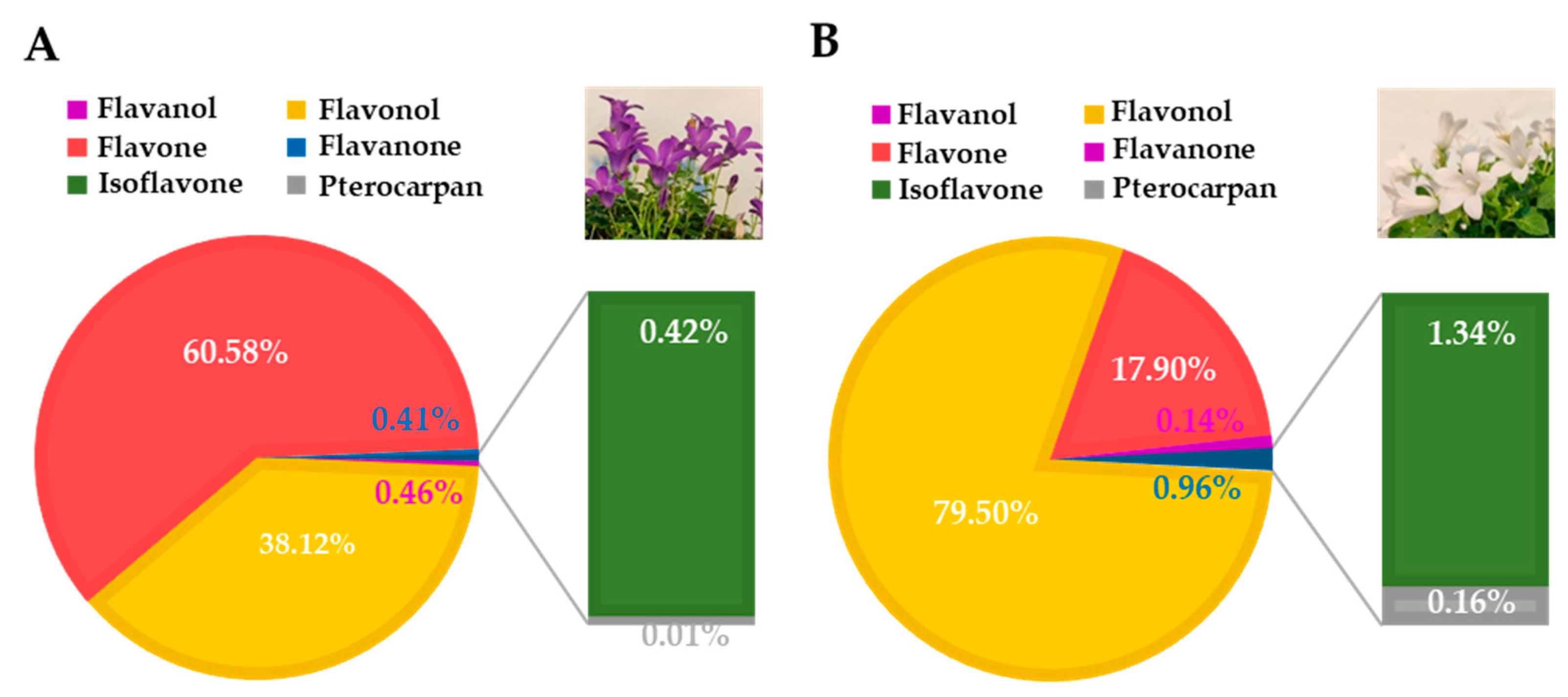
3.1. Deep Blue Ocean and White Ocean Genotypes Present Differences in the Flavonoid Composition of Petals

Flavonoids are a diverse group of phenolic compounds and play an important role as flower pigments [26]. The obtained metabolic profile revealed a clear difference in the accumulation of flavonoids between genotypes with different colored flowers. In Deep Blue Ocean petals, flavones were predominant, accounting for 60.58% of the total flavonoids analyzed (Figure 1A), whereas in White Ocean, flavonols were predominant, accounting for 79.50% of the total of the flavonoids analyzed (Figure 1B). Flavonols accounted for 38.12% of the total flavonoids investigated in Deep Blue Ocean petals, followed by flavanols (0.46%), flavanones (0.41%), isoflavones (0.42%), and pterocarpans (0.01%). In White Ocean petals, flavones accounted for 17.90% of the total flavonoids investigated, followed by isoflavones (1.34%), flavanones (0.96%), pterocarpans (0.16%), and flavanols (0.14%).
Luteolin-di-glc was one of four major flavonoids in Deep Blue Ocean petals (16,500 ng/g DW), the other three major components were nicotiflorin (7160 ng/g DW), quercetin-xyl-gal (2004 ng/g DW), and rutin (1726 ng/g DW) (Table 2). All of these were components of the flavonol group (Figure 1). Together, these four compounds accounted for 94.4% of the total flavonoids found in Deep Blue Ocean petals. In White Ocean petals, nicotiflorin was the main compound found (4400 ng/g DW), followed by myricetin (602 ng/g DW), rutin (240 ng/g DW), and kaempferol (228 ng/g DW), which together accounted for 88.6% of the total flavonoids found in the petals of the White Ocean genotype. The Deep Blue Ocean and White Ocean genotypes showed differences in flavonoid accumulations. Kaempferol, apigenin, and genistein were detected only in White Ocean petals, and myricetin was significantly higher in the White Ocean petals (2.2-fold higher) than in the Deep Blue Ocean petals (Table 2). Eleven compounds accumulated to greater levels in Deep Blue Ocean petals than in White Ocean petals. For example, rutin and luteolin were 7.1 and 2.7-fold higher, respectively, in Deep Blue Ocean than in White Ocean petals.

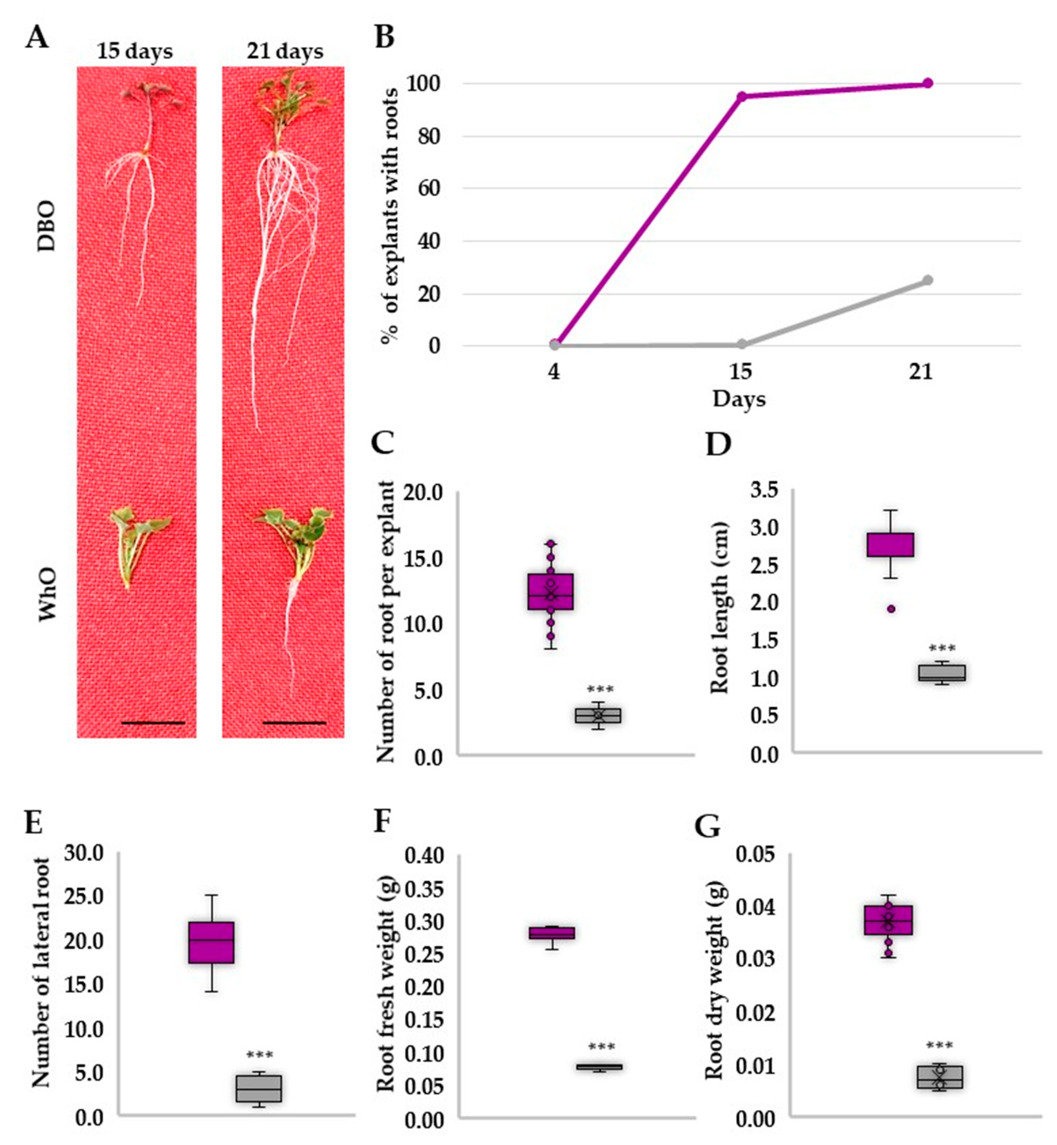
3.2. Deep Blue Ocean and White Ocean Genotypes Have Contrasting Efficiency to Adventitious Root Formation

To explore the ability of two genotypes to form AR, explants were set up in an aeroponic system and root formation was observed (Figure 2A). After 4 days of growth in the aeroponic system, Deep Blue Ocean explants started to form ARs (Figure 2B). AR formation in White Ocean explants started after 15 days of growth in the aeroponic system (Figure 2B). After 21 days, 100% of the Deep Blue Ocean explants and 25% of the White Ocean explants showed ARs. Deep Blue Ocean showed an average of 4 times more AR per explant compared to the White Ocean genotype (Figure 2C). The average root length in the Deep Blue Ocean genotype was 155% longer than the root length in the White Ocean (Figure 2D). Deep Blue Ocean showed an average of 6.5 times more lateral roots compared to the White Ocean genotype (Figure 2E).
Analysis of the root biomass of the two genotypes revealed that Deep Blue Ocean showed 260% and 400% higher fresh and dry weight, respectively, when compared with White Ocean (Figure 2F,G). The analysis of shoot biomass showed no significant differences between the two genotypes (Supplementary Figure S3). Average leaf area, number of leaves, stem length, and diameter also did not present statistically significant differences between genotypes (Supplementary Figure S3). Taken together, the results showed that the ability to form ARs was greater for the Deep Blue Ocean than for the White Ocean genotype of C. portenschlagiana, and both genotypes had the same vegetative growth rate. Deep Blue Ocean can be considered an easy-to-root C. portenschlagiana genotype, whereas White Ocean is a hard-to-root genotype.

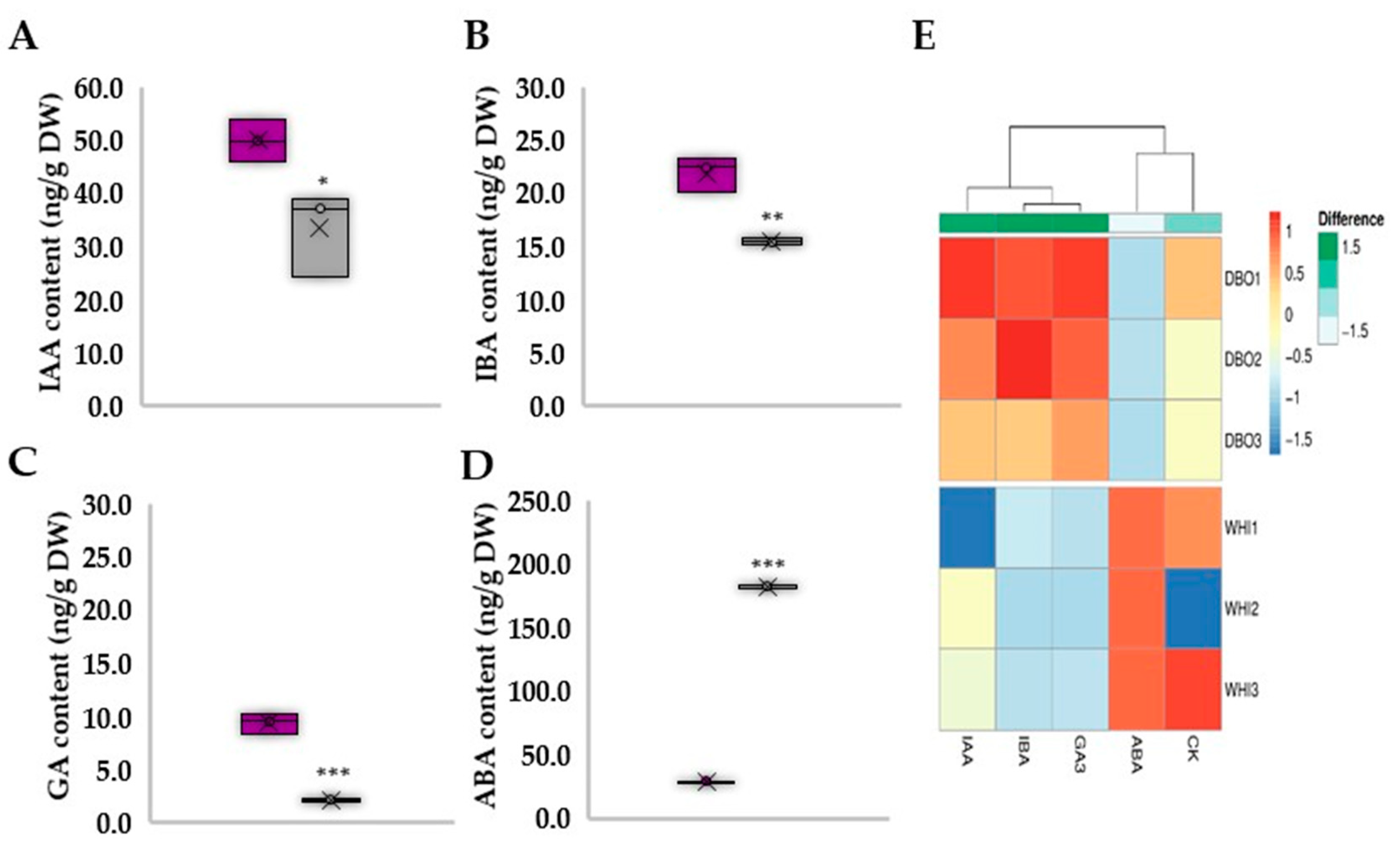
3.3. Deep Blue Ocean and White Ocean Genotypes Show Discrepant Concentrations of Endogenous Plant Hormones

Plant hormone analyses were performed at the basal cut site of both the Deep Blue Ocean and White Ocean genotypes for a better understanding of the differences in AR formation. The analyses showed that two forms of auxin, IAA and indole-3-butyric acid (IBA), had higher concentrations in the Deep Blue Ocean than in the White Ocean genotype (Figure 3A,B). High levels of free IAA at the basal cut site in explants are essential for AR formation [28], and the conversion of IBA to IAA is also important in AR formation in many species [29]. The IAA and IBA content differences between Deep Blue Ocean and White Ocean could partially explain the differences in the ability to form ARs.
Our results showed that GA content is higher in the Deep Blue Ocean than in the White Ocean genotype (Figure 3C). The content of ABA was higher in the White Ocean than in the Deep Blue Ocean genotype (Figure 3D), suggesting that a lower level of ABA may collaborate to AR formation in C. portenschlagiana. In Vaccinium corymbosum, treatment with exogenous auxin increased the AR formation and decreased ABA endogenous content [30]. Although CYT were shown to negatively regulate AR formation in some species [31], no significant difference in trans-zeatin 6-purine content was observed between the Deep Blue Ocean and White Ocean genotypes (Figure 3E and Figure S4).

3.4. Deep Blue Ocean and White Ocean Genotypes Present Differences in the Flavonoid Composition in the Basal Part of the Stems

Flavonoids have been demonstrated to play significant roles in regulating root growth and lateral root formation [32]. The stems of both Deep Blue Ocean and White Ocean genotypes contained flavones as the predominant compounds, accounting for 97.67% and 86.02%, respectively, of the total flavonoids analyzed. The Deep Blue Ocean stem also contained 2.13% flavonols, 0.15% flavanols, and 0.02% flavanones. The amount of isoflavones and coumestan, pooled together, corresponded to 0.004% of the total compounds analyzed in Deep Blue Ocean (Figure 4A). In the White Ocean stem, flavanols accounted for 9.63% of the total flavonoids analyzed, followed by flavanones (4.11%), isoflavones (0.16%), and coumestan (0.049%). Further, in Deep Blue Ocean flavonols accounted for 2.13% of the total flavonoids analyzed, whereas in White Ocean genotypes they are not observed.
The diversity of flavonoid compounds was smaller in the cut stems than in the petals (Figure 4B–G). The following nine flavonoid components were identified in petals, but they were absent from the stems: medicarpin, daidzin, genistein, myricetin, biochanin A, quercetin-rha-xyl-gal, quercetin-xyl-gal, kaempferol, and apigenin (Table 2 and Table 3). Analogous to the petal findings, luteolin-di-glc was also the main compound found in the Deep Blue Ocean stem (185,200 ng/g DW), accounting for 97.4% of the total flavonoid (Table 3). The other three major components were rutin (2586 ng/g DW), nicotiflorin (1476 ng/g DW), and epicatechin (295 ng/g DW). Luteolin-di-glc was also the main flavonoid detected in White Ocean stem (3670 ng/g DW), accounting for 75.8% of the total flavonoids in the stem of that genotype, although its level was 50-fold lower than in the Deep Blue Ocean genotype. Epicatechin (466 ng/g DW), isovitexin (280 ng/g DW), and luteolin (190 ng/g DW) were the three other major components found in White Ocean stems. Two compounds were not found in petals but were exclusive components in the stem: coumestrol (the only coumestan analyzed in this study) and isovitexin (a flavone).
The PCA (Principal Component Analysis) biplot illustrated the relationship of the 24 flavonoids analyzed in the Deep Blue Ocean and White Ocean genotypes in the stem end and the flowers (Supplementary Figure S5). Two principal components, Dim1 and Dim2, were extracted at 58.3% and 29.5%, respectively. The cumulative contribution rate reached 87.8%. The Deep Blue Ocean and White Ocean genotypes have contrasting petal colors purple and white, respectively (Supplementary Figure S5). The PCA score plot showed clear separation of the petals of the Deep Blue Ocean and White Ocean genotypes. Deep Blue Ocean petals were associated with quercetin-xyl-gal, quercetin-rha-xyl-gal, sissotrin, luteolin, formononetin, biochanin A, naringin, daidzin, nicotiflorin, and medicarpin; whereas White Ocean petals were associated with myricetin, daidzein, genistein, apigenin, and kaempferol. Analysis of the score plot revealed that Deep Blue Ocean and White Ocean stems were allocated in the same quadrant, with both associated with orientin, epicatechin, naringenin, genistin, isovitexin, coumestrol, and luteolin-di-glc.

4. Discussion

Flavonoids are secondary metabolites that accumulate in most plant species, where they play various roles. As antioxidants, flavonoids protect plants against abiotic and abiotic stresses; as color pigments, they help to attract pollinators to flowers. By interacting with plant hormones, flavonoids also affect many growth and developmental processes in plants. In this study, we found a greater accumulation of flavones and flavonols in petals of a blue-flowered Campanula genotype than in a white-flowered genotype (Table 2). The blue-flowered genotype also accumulated more flavonoids in the basal part of the cutting (Table 3) and had a greater capacity for AR formation (Figure 2). The enhanced capacity to form AR in the blue-flowered genotype was related to higher auxin and lower ABA concentrations in the basal part of the cuttings than were found in the cuttings of the white-flowered genotype (Figure 3). The higher concentrations of auxins and lower concentrations of ABA, a stress hormone, in the genotype with better rooting capacity are in agreement with the known hormonal effects of auxin and ABA on AR development [33]. Flavonoids might contribute to AR formation through a modulation of plant hormone activity in the basal part of the stem.

4.1. Crosstalk of Flavonoids, Auxin, and ROS

Higher accumulations of flavonols and flavones in the petals and in the basal part of the stem of the Deep Blue Ocean cultivar indicate a greater synthesis of these flavonoids in the easy-to-root genotype, independent of plant tissue. In addition, greater biosynthesis of flavonoids in one plant part can contribute to enhanced concentrations in another part due to flavonoid transport, as shown in experiments with Arabidopsis [34]. The higher concentrations of both flavonoids and auxins in the basal parts of cuttings might indicate a causal relationship between these compounds, where a higher flavonoid concentration can contribute to higher auxin levels. The effects of flavonoids on auxin levels might occur through modulation of auxin stability and/or auxin transport.
The effects of flavonoids on auxin stability arise through their interference with the first step of IAA oxidation [35]. The oxidative degradation of auxin appears to be developmentally important, especially under conditions of oxidative stress [36]. Interestingly, in our study, the concentrations of both the major natural auxin IAA [4] and IBA were higher in the easy-to-root than in the hard-to-root genotype. IBA was initially considered a “synthetic auxin” that elicits auxin-like effects, such as root initiation, and is widely used as the active ingredient in plant propagation media to induce AR initiation in stem cuttings [37]. Subsequent studies have shown that IBA is an endogenous compound in a variety of plant species [38]. However, because of its lengthened side chain, IBA cannot bind to the auxin receptor. Therefore, IBA can contribute to auxin responses only after its carbon side chain is reduced and it is converted to IAA through a fatty acid β-oxidation process [39].
A higher endogenous pool of IBA can positively contribute to IAA levels; however, IBA itself is synthesized from IAA [40]. Therefore, the higher level of IAA in the easy-to-root genotype cannot be explained by a higher level of IBA accumulation. Thus, de novo auxin biosynthesis and/or reduced oxidative degradation might contribute to the observed increases in the levels of both IAA and IBA.
The importance of auxin biosynthesis in AR formation is also indirectly supported by our previous experiments, where the application of amino acid tryptophan, the precursor for auxin biosynthesis, enhances AR formation, especially for the hard-to-root genotype of Campanula plants [27]. Interestingly, the same study shows a similar stimulating effect on AR formation by applying another amino acid, glutamate. However, glutamate seems to decrease the responses to auxin [41], indicating that in addition to auxin, other signaling pathways are involved in the regulation of AR formation.
The higher auxin concentration in the basal part of shoots after cutting can be due to a reduction in oxidative IAA degradation and/or enhanced polar auxin transport (PAT). Indeed, previous studies with petunia (Petunia hybrid) showed that PAT is essential for AR formation [42], as the basipetal PAT is conserved after cutting as an important component explaining early IAA accumulation in the rooting zone.
Flavonols, a major class of flavonoids, inhibit PAT, as shown in studies with Arabidopsis [8]. If flavonols inhibit PAT, we would expect a smaller auxin accumulation in the basal parts of cuttings in the easy-to-root Deep Blue Ocean cultivar, which had a higher flavonol level. However, we found the opposite result, as this cultivar accumulated more auxin and more flavonols in basal parts of the cuttings. The higher auxin concentration might reflect the contribution of higher concentrations of flavonoids to IAA stability due to a reduction in oxidative IAA degradation. Moreover, the inhibitory effect of flavonols on PAT might be related to the molecular structure of specific flavonols, such as derivatives of quercetin and kaempferol, as shown in Arabidopsis studies [43,44]. However, these flavonols were not found in stems of both genotypes. By contrast, in our study, other flavonols (nicotiflorin and rutin) showed differential accumulation in the basal parts of the cuttings from the two contrasting Campanula genotypes (Table 3).
One possible mechanism of action of flavonols on PAT occurs through their binding to the ABCB1/19 protein [45,46], which is an auxin transporter. However, flavonols can have less specific actions on auxin transport, because flavonoids have antioxidant properties and reduce ROS levels in cells. The ROS level itself is important in the regulation of auxin transport. For example, treatments that increase ROS levels in the cells, such as pharmacological inhibition of glutathione (GSH) biosynthesis, as well as mutations in GSH biosynthesis (cad2 and rml1) and THIOREDOXIN REDUCTASE (ntra ntrb), all reduce the expression of the auxin efflux-mediating PIN proteins, which are the main facilitators of PAT [47]. The higher levels of several flavonoids in the Deep Blue Ocean cultivar might contribute to reduced ROS levels, resulting in an enhancement of auxin transport through the upregulation of PIN proteins. Thus, we can hypothesize that the antioxidant properties of flavonoids can contribute to higher auxin concentrations at the basal parts of cuttings due to decreased IAA oxidative degradation and stimulation of PIN-related PAT.
In our study, different classes of flavonoids were regulated differently in the easy-to-root genotype than in the hard-to-root genotype. In the basal parts of cuttings, the easy-to-root genotype has a higher concentration of flavonols and flavones and a lower concentration of flavonones (Table 3). High accumulation of the flavone luteolin-di-glc in the easy-to-root genotype raises the question of a potential role for this compound in AR formation. This requires further investigation. Different classes of flavonoids have different efficiencies in scavenging different ROS. Flavonols are the most effective at reducing ROS levels. By contrast, flavones are the least effective, having no antioxidant capacity for H2O2 and a very slight capacity for 1O2, while flavonones show very little action on H2O2 and no antioxidant action on 1O2 [48].
Thus, the modulation of flavonoid concentrations can change the total ROS level as well as the ratio between different ROS, such as H2O2 and 1O2, and different ROS play different roles in root development. For example, in the root tip, the H2O2/superoxide ratio regulates the transition of cells from a zone of cell division to a zone of cell elongation and differentiation [17]. In lateral roots, both H2O2 and superoxide are localized in all developmental stages of lateral root primordia [19].
The participation of ROS in adventitious rooting is still poorly understood. Some lines of evidence indicate that H2O2 might act as a signaling molecule that induces AR formation [20,49]. For example, in mung bean hypocotyl, H2O2 concentrations increase with time after excision, and ascorbate, a reducing substrate for H2O2 removal, prevents AR induction [50]. In poplar, the optimal concentration of H2O2 accelerated AR formation, whereas higher concentrations slowed down this process and reduced root growth [51].
Other important relationships between the major players guiding root development have also been identified. Specifically, auxin itself can enhance ROS formation. For example, application of the auxin IBA promoted H2O2 production in mung bean seedling [49]. Wounding induced ROS-modulated auxin biosynthesis-related and transport-related genes, leading to the accumulation of this phytohormone at the base of Arabidopsis cuttings [20]. In addition, ROS enhance flavonoid biosynthesis [52]. All these functional relationships indicate the operation of a complex network of interactions between flavonoids, ROS, and auxin that guide AR formation in Campanula.

4.2. ABA Is a Potential Additional Regulator of Ar Development

The other plant hormone that regulates AR development is ABA [53]. Our funding that the easy-to-root genotype contained lower ABA concentrations in the basal part of cuttings indicates that ABA can be a second regulator responsible for inhibition of AR formation in the hard-to-root genotype. ABA blocks both AR [53] and lateral root development [54]. However, experiments with Arabidopsis mutants of ABA receptors showed that the mechanisms that block root development differ for adventitious and lateral root development [53].
Antagonistic interactions between flavonoids and ABA have been found in “downstream” components of ABA signaling, such as H2O2 and a range of protein kinases (MAPKs) [55,56]. Flavonols may regulate the ABA signaling pathway primarily by countering the oxidative burst and the consequent generation of H2O2 driven by enhanced ABA biosynthesis [57,58]. ABA, in turn, can enhance flavonoid biosynthesis, possibly, through the generation of ROS. Thus, an additional feedback loop, where some flavonoids inhibit ABA signaling and ABA stimulates flavonoid biosynthesis might be involved in the regulation of AR formation. In our study, the stimulatory effect of ABA on flavonoid biosynthesis did not appear to play a dominant role in the regulation of flavonoid accumulation, because the easy-to-root genotype accumulated a higher amount of both ABA and flavonoids. The regulation of ABA biosynthesis can also affect AR development independent of flavonoids. Specifically, a disruption in ABA biosynthesis leads to the accumulation of carotenoid precursors, which have been shown to stimulate anchor root formation [59].
Another player that can contribute to AR development is GA. In previous research, gibberellins inhibited AR formation in hybrid aspen and Arabidopsis by affecting auxin transport [60]. In rice, GA stimulates AR formation via a mechanism that requires ethylene [1]. In our investigation, the easy-to-root genotype accumulated a higher GA content. Further investigations are required to understand the role of GA in AR formation in C. portenschlagiana.

5. Outlook

Several players, such as flavonoids, auxins, and ROS, are involved in a network that guides AR formation. Flavonoids inhibit ROS, but ROS stimulate flavonoid biosynthesis. Conversely, ROS inhibit auxin activity, but auxins stimulate ROS production. Among other hormones, ABA can also contribute to the regulation of AR formation, as indicated by the lower level of ABA in the easy-to-root genotype and the well-known inhibitory effect of ABA on AR formation. Further investigations with higher temporal and special resolution are required regarding the crosstalk between flavonoids and plant hormones to understand the different strategies by which AR development is regulated in ornamental plants.

Supplementary Materials

The following supporting information can be downloaded at: https://www.mdpi.com/article/10.3390/horticulturae9010121/s1, Figure S1: Deep Blue Ocean and White Ocean genotypes pictures showing the colour of the petals; Figure S2: Illustration of how the 1 cm segment of the basal cut site was collected; Figure S3: Stem biomass accumulation and morphological analysis at Deep Blue Ocean (DBO) and White Ocean (WhO) Campanula portenschlagiana genotypes; Figure S4: Trans-zeatin quantification in Deep Blue Ocean (dark purple box) and White Ocean (light gray box); Figure S5: Principal component analysis (PCA) of flavonoids in the cut stems and petals of Deep Blue Ocean (DBO) and White Ocean (WhO) Campanula portenschlagiana genotypes.

Author Contributions

Conceptualization, T.B. and I.A.P.; methodology, T.B., I.A.P. and I.S.F.; software, T.B.; validation, T.B.; formal analysis, T.B.; investigation, T.B.; resources, I.A.P.; data curation, T.B.; writing—original draft preparation, T.B. and I.A.P.; writing—review and editing, I.A.P. and T.B.; visualization, T.B.; supervision, I.A.P.; project administration, T.B., I.A.P. and K.H.K.; funding acquisition, K.H.K. and I.A.P. All authors have read and agreed to the published version of the manuscript.

Funding

This research was funded by the Green Development and Demonstration Program (GUDP) grant FUTFLOR (Project number: 34009-20-1752).

Data Availability Statement

Not applicable.

Acknowledgments

The authors wish to thank Thomas Bak-Østerby and Bente Birgitte Laursen for providing technical support; and Jan Uglebjerg Storm and Jacob Morten Andersen for providing the Campanula portenschlagiana genotypes.

Conflicts of Interest

The authors declare no conflict of interest.

References

  1. Steffens, B.; Rasmussen, A. The physiology of adventitious roots. Plant Physiol. 2016, 170, 603–617. [Google Scholar] [CrossRef] [PubMed] [Green Version]
  2. Buer, C.S.; Imin, N.; Djordjevic, M.A. Flavonoids: New roles for old molecules. J. Integr. Plant Biol. 2010, 52, 98–111. [Google Scholar] [CrossRef] [PubMed]
  3. Teale, W.D.; Ditengou, F.A.; Dovzhenko, A.D.; Li, X.; Molendijk, A.M.; Ruperti, B.; Paponov, I.; Palme, K. Auxin as a model for the integration of hormonal signal processing and transduction. Mol. Plant 2008, 1, 229–237. [Google Scholar] [CrossRef] [PubMed] [Green Version]
  4. Teale, W.D.; Paponov, I.A.; Palme, K. Auxin in action: Signalling, transport and the control of plant growth and development. Nat. Rev. Mol. Cell Biol. 2006, 7, 847–859. [Google Scholar] [CrossRef] [PubMed]
  5. Zhao, Y. Auxin biosynthesis: A simple two-step pathway converts tryptophan to indole-3-acetic acid in plants. Mol. Plant 2012, 5, 334–338. [Google Scholar] [CrossRef] [Green Version]
  6. Paponov, I.A.; Teale, W.D.; Trebar, M.; Blilou, K.; Palme, K. The PIN auxin efflux facilitators: Evolutionary and functional perspectives. Trends Plant Sci. 2005, 10, 170–177. [Google Scholar] [CrossRef]
  7. Cho, M.; Cho, H.T. The function of ABCB transporters in auxin transport. Plant Signal. Behav. 2013, 8, e22990. [Google Scholar] [CrossRef] [Green Version]
  8. Brown, D.E.; Rashotte, A.M.; Murphy, A.S.; Normanly, J.; Tague, B.W.; Peer, W.A.; Taiz, L.; Muday, G.K. Flavonoids act as negative regulators of auxin transport in vivo in Arabidopsis. Plant. Physiol. 2001, 126, 524–535. [Google Scholar] [CrossRef] [Green Version]
  9. Peer, W.A.; Murphy, A.S. Flavonoids and auxin transport: Modulators or regulators? Trends Plant Sci. 2007, 12, 556–563. [Google Scholar] [CrossRef]
  10. Peer, W.A.; Bandyopadhyay, A.; Blakeslee, J.J.; Makam, S.I.; Chen, R.J.; Masson, P.H.; Murphy, A.S. Variation in expression and protein localization of the PIN family of auxin efflux facilitator proteins in flavonoid mutants with altered auxin transport in Arabidopsis thaliana. Plant Cell 2004, 16, 1898–1911. [Google Scholar] [CrossRef]
  11. Paponov, I.A.; Budnyk, V.; Paponov, M.; Teale, W.; Palme, K. Butylated Hydroxytoluene (BHT) Inhibits PIN1 Exocytosis From BFA Compartments in Arabidopsis Roots. Front. Plant Sci. 2020, 11, 393. [Google Scholar] [CrossRef]
  12. Teale, W.D.; Pasternak, T.; Dal Bosco, C.; Dovzhenko, A.; Kratzat, K.; Bildl, W.; Schwörer, M.; Falk, T.; Ruperti, B.; Schaefer, J.V.; et al. Flavonol-mediated stabilization of PIN efflux complexes regulates polar auxin transport. EMBO J. 2021, 40, e104416. [Google Scholar] [CrossRef] [PubMed]
  13. De Klerk, G.-J.; Guan, H.; Huisman, P.; Marinova, S. Effects of phenolic compounds on adventitious root formation and oxidative decarboxylation of applied indoleacetic acid in Malus ‘Jork 9’. Plant Growth Regul. 2011, 63, 175–185. [Google Scholar] [CrossRef] [Green Version]
  14. Denaxa, N.-K.; Roussos, P.A.; Vemmos, S.N. Assigning a role to the endogenous phenolic compounds on adventitious root formation of olive stem cuttings. J. Plant Growth Regul. 2020, 39, 411–421. [Google Scholar] [CrossRef]
  15. Gayomba, S.R.; Muday, G.K. Flavonols regulate root hair development by modulating accumulation of reactive oxygen species in the root epidermis. Development 2020, 147, 185819. [Google Scholar] [CrossRef]
  16. Foreman, J.; Demidchik, V.; Bothwell, J.H.F.; Mylona, P.; Miedema, H.; Torres, M.A.; Linstead, P.; Costa, S.; Brownlee, C.; Jones, J.D.G.; et al. Reactive oxygen species produced by NADPH oxidase regulate plant cell growth. Nature 2003, 422, 442–446. [Google Scholar] [CrossRef]
  17. Tsukagoshi, H.; Busch, W.; Benfey, P.N. Transcriptional regulation of ROS controls transition from proliferation to differentiation in the root. Cell 2010, 143, 606–616. [Google Scholar] [CrossRef] [Green Version]
  18. Orman-Ligeza, B.; Parizot, B.; de Rycke, R.; Fernandez, A.; Himschoot, E.; Van Breusegem, F.; Bennett, M.J.; Périlleux, C.; Beeckman, T.; Draye, X. RBOH-mediated ROS production facilitates lateral root emergence in Arabidopsis. Development 2016, 143, 3328–3339. [Google Scholar] [CrossRef] [Green Version]
  19. Manzano, C.; Pallero-Baena, M.; Casimiro, I.; De Rybel, B.; Orman-Ligeza, B.; Van Isterdael, G.; Beeckman, T.; Draye, X.; Casero, P.; Del Pozo, J.C. The emerging role of reactive oxygen species signaling during lateral root development. Plant Physiol. 2014, 165, 1105–1119. [Google Scholar] [CrossRef] [Green Version]
  20. Huang, A.; Wang, Y.; Liu, Y.; Wang, G.; She, X. Reactive oxygen species regulate auxin levels to mediate adventitious root induction in Arabidopsis hypocotyl cuttings. J. Integr. Plant Biol. 2020, 62, 912–926. [Google Scholar] [CrossRef]
  21. Osterc, G.; Štefančič, M.; Solar, A.; Stampar, F. Potential involvement of flavonoids in the rooting response of chestnut hybrid (Castanea crenata × Castanea sativa) clones. Aust. J. Exp. Agr. 2007, 47, EA05149. [Google Scholar] [CrossRef]
  22. Vilasboa, J.; da Costa, C.T.; Ransan, L.G.; Mariath, J.E.D.A.; Fett-Neto, A.G. Microcutting redox profile and anatomy in Eucalyptus spp. with distinct adventitious rooting competence. Front Plant Sci 2021, 11, 620832. [Google Scholar] [CrossRef] [PubMed]
  23. Curir, P.; Sulis, S.; Mariani, F.; van Sumere, C.F.; Marchesini, A.; Dolci, M. Influence of endogenous phenols on rootability of Chamaelaucium uncinatum Schauer stem cuttings. Sci. Hortic. 1993, 55, 303–314. [Google Scholar] [CrossRef]
  24. Van Overbeek, J.; Gordon, S.; Gregory, L. An analysis of the function of the leaf in the process of root formation in cuttings. Am. J. Bot. 1946, 33, 100–107. [Google Scholar] [CrossRef]
  25. Barba de la Rosa, A.; León-Rodríguez, A.; Laursen, B.; Fomsgaard, I. Influence of the growing conditions on the flavonoids and phenolic acids accumulation in amaranth (Amaranthus hypochondriacus L.) leaves. Rev. Terra Latinoam. 2019, 37, 449. [Google Scholar] [CrossRef]
  26. Grotewold, E. The genetics and biochemistry of floral pigments. Annu. Rev. Plant Biol. 2006, 57, 761–780. [Google Scholar] [CrossRef]
  27. Bergonci, T.; Paponov, I.A. Amino acid treatments induce adventitious root formation in two different genotypes of Campanula portenschlagiana. Biol. Life Sci. Forum 2022, 16, 8. [Google Scholar]
  28. Fattorini, L.; Falasca, G.; Kevers, C.; Rocca, L.M.; Zadra, C.; Altamura, M.M. Adventitious rooting is enhanced by methyl jasmonate in tobacco thin cell layers. Planta 2009, 231, 155–168. [Google Scholar] [CrossRef]
  29. Chen, L.; Tong, J.; Xiao, L.; Ruan, Y.; Liu, J.; Zeng, M.; Huang, H.; Wang, J.W.; Xu, L. YUCCA-mediated auxin biogenesis is required for cell fate transition occurring during de novo root organogenesis in Arabidopsis. J. Exp. Bot. 2016, 67, 4273–4284. [Google Scholar] [CrossRef] [Green Version]
  30. An, H.; Zhang, J.; Xu, F.; Jiang, S.; Zhang, X. Transcriptomic profiling and discovery of key genes involved in adventitious root formation from green cuttings of highbush blueberry (Vaccinium corymbosum L.). BMC Plant Biol. 2020, 20, 182. [Google Scholar] [CrossRef] [Green Version]
  31. Gonin, M.; Bergougnoux, V.; Nguyen, T.D.; Gantet, P.; Champion, A. What Makes Adventitious Roots? Plants 2019, 8, 240. [Google Scholar] [CrossRef] [PubMed]
  32. Franco, D.M.; Silva, E.M.; Saldanha, L.L.; Adachi, S.A.; Schley, T.R.; Rodrigues, T.M.; Dokkedal, A.L.; Nogueira, F.T.; Rolim de Almeida, L.F. Flavonoids modify root growth and modulate expression of SHORT-ROOT and HD-ZIP III. J. Plant Physiol. 2015, 188, 89–95. [Google Scholar] [CrossRef] [PubMed]
  33. Lakehal, A.; Bellini, C. Control of adventitious root formation: Insights into synergistic and antagonistic hormonal interactions. Physiol. Plantarum 2019, 165, 90–100. [Google Scholar] [CrossRef] [Green Version]
  34. Buer, C.S.; Muday, G.K.; Djordjevic, M.A. Implications of long-distance flavonoid movement in Arabidopsis thaliana. Plant Signal. Behav. 2008, 3, 415–417. [Google Scholar] [CrossRef] [PubMed] [Green Version]
  35. Lee, T.T. Role of phenolic inhibitors in peroxidase-mediated degradation of indole-3-acetic acid. Plant Physiol. 1977, 59, 372–375. [Google Scholar] [CrossRef] [PubMed] [Green Version]
  36. Tognetti, V.B.; Van Aken, O.; Morreel, K.; Vandenbroucke, K.; van de Cotte, B.; De Clercq, I.; Chiwocha, S.; Fenske, R.; Prinsen, E.; Boerjan, W.; et al. Perturbation of indole-3-butyric acid homeostasis by the UDP-glucosyltransferase UGT74E2 modulates Arabidopsis architecture and water stress tolerance. Plant Cell 2010, 22, 2660–2679. [Google Scholar] [CrossRef] [Green Version]
  37. Frick, E.M.; Strader, L.C. Roles for IBA-derived auxin in plant development. J. Exp. Bot. 2017, 69, 169–177. [Google Scholar] [CrossRef] [Green Version]
  38. Korasick, D.A.; Enders, T.A.; Strader, L.C. Auxin biosynthesis and storage forms. J. Exp. Bot. 2013, 64, 2541–2555. [Google Scholar] [CrossRef] [Green Version]
  39. Strader, L.C.; Bartel, B. Transport and metabolism of the endogenous auxin precursor indole-3-butyric acid. Mol. Plant 2011, 4, 477–486. [Google Scholar] [CrossRef] [Green Version]
  40. Strader, L.C.; Culler, A.H.; Cohen, J.D.; Bartel, B. Conversion of endogenous indole-3-butyric acid to indole-3-acetic acid drives cell expansion in Arabidopsis seedlings. Plant Physiol. 2010, 153, 1577–1586. [Google Scholar] [CrossRef] [Green Version]
  41. Ravelo-Ortega, G.; López-Bucio, J.S.; Ruiz-Herrera, L.F.; Pelagio-Flores, R.; Ayala-Rodríguez, J.Á.; de la Cruz, H.R.; Guevara-García, Á.A.; López-Bucio, J. The growth of Arabidopsis primary root is repressed by several and diverse amino acids through auxin-dependent and independent mechanisms and MPK6 kinase activity. Plant Sci. 2021, 302, 110717. [Google Scholar] [CrossRef] [PubMed]
  42. Ahkami, A.H.; Melzer, M.; Ghaffari, M.R.; Pollmann, S.; Ghorbani Javid, M.; Shahinnia, F.; Hajirezaei, M.R.; Druege, U. Distribution of indole-3-acetic acid in Petunia hybrida shoot tip cuttings and relationship between auxin transport, carbohydrate metabolism and adventitious root formation. Planta 2013, 238, 499–517. [Google Scholar] [CrossRef] [Green Version]
  43. Yin, R.; Han, K.; Heller, W.; Albert, A.; Dobrev, P.I.; Zažímalová, E.; Schäffner, A.R. Kaempferol 3-O-rhamnoside-7-O-rhamnoside is an endogenous flavonol inhibitor of polar auxin transport in Arabidopsis shoots. New Phytol. 2014, 201, 466–475. [Google Scholar] [CrossRef] [PubMed] [Green Version]
  44. Geisler, M.; Blakeslee, J.J.; Bouchard, R.; Lee, O.R.; Vincenzetti, V.; Bandyopadhyay, A.; Titapiwatanakun, B.; Peer, W.A.; Bailly, A.; Richards, E.L.; et al. Cellular efflux of auxin catalyzed by the Arabidopsis MDR/PGP transporter AtPGP1. Plant J. 2005, 44, 179–194. [Google Scholar] [CrossRef] [PubMed]
  45. Noh, B.; Murphy, A.S.; Spalding, E.P. Multidrug resistance-like genes of Arabidopsis required for auxin transport and auxin-mediated development. Plant Cell 2001, 13, 2441–2454. [Google Scholar]
  46. Murphy, A.S.; Hoogner, K.R.; Peer, W.A.; Taiz, L. Identification, purification, and molecular cloning of N-1- naphthylphthalmic acid-binding plasma membrane-associated aminopeptidases from Arabidopsis. Plant Physiol. 2002, 128, 935–950. [Google Scholar] [CrossRef] [Green Version]
  47. Bashandy, T.; Meyer, Y.; Reichheld, J.-P. Redox regulation of auxin signaling and plant development in Arabidopsis. Plant Signal. Behav. 2011, 6, 117–119. [Google Scholar] [CrossRef] [Green Version]
  48. Csepregi, K.; Hideg, É. Phenolic compound diversity explored in the context of photo-oxidative stress protection. Phytochem. Anal. 2018, 29, 129–136. [Google Scholar] [CrossRef]
  49. Li, S.-W.; Xue, L.; Xu, S.; Feng, H.; An, L. Hydrogen peroxide acts as a signal molecule in the adventitious root formation of mung bean seedlings. Environ. Exp. Bot. 2009, 65, 63–71. [Google Scholar] [CrossRef]
  50. Huang, A.-X.; She, X.-P.; Cao, B.-H.; Ren, Y. Distribution of hydrogen peroxide during adventitious roots initiation and development in mung bean hypocotyls cuttings. Plant Growth Regul. 2011, 64, 109–118. [Google Scholar] [CrossRef]
  51. Zhang, Y.; Xiao, Z.A.; Zhan, C.; Liu, M.; Xia, W.; Wang, N. Comprehensive analysis of dynamic gene expression and investigation of the roles of hydrogen peroxide during adventitious rooting in poplar. BMC Plant Biology. 2019, 19, 99. [Google Scholar] [CrossRef] [PubMed] [Green Version]
  52. Shi, H.; Liu, G.; Wei, Y.; Chan, Z. The zinc-finger transcription factor ZAT6 is essential for hydrogen peroxide induction of anthocyanin synthesis in Arabidopsis. Plant Mol. Biol. 2018, 97, 165–176. [Google Scholar] [CrossRef] [PubMed]
  53. Zeng, Y.; Verstraeten, I.; Trinh, H.K.; Heugebaert, T.; Stevens, C.V.; Garcia-Maquilon, I.; Rodriguez, P.L.; Vanneste, S.; Geelen, D. Arabidopsis hypocotyl adventitious root formation is suppressed by ABA signaling. Genes 2021, 12, 1141. [Google Scholar] [CrossRef] [PubMed]
  54. De Smet, I.; Signora, L.; Beeckman, T.; Inzé, D.; Foyer, C.H.; Zhang, H. An abscisic acid-sensitive checkpoint in lateral root development of Arabidopsis. Plant J. 2003, 33, 543–555. [Google Scholar] [CrossRef] [Green Version]
  55. Brunetti, C.; Sebastiani, F.; Tattini, M. Review: ABA, flavonols, and the evolvability of land plants. Plant Sci. 2019, 280, 448–454. [Google Scholar] [CrossRef]
  56. Brunetti, C.; Di Ferdinando, M.; Fini, A.; Pollastri, S.; Tattini, M. Flavonoids as antioxidants and developmental regulators: Relative significance in plants and humans. Int. J. Mol. Sci. 2013, 14, 3540–3555. [Google Scholar] [CrossRef] [Green Version]
  57. Tossi, V.; Lamattina, L.; Cassia, R. An increase in the concentration of abscisic acid is critical for nitric oxide-mediated plant adaptive responses to UV-B irradiation. New Phytol. 2009, 181, 871–879. [Google Scholar] [CrossRef]
  58. Choudhury, F.K.; Rivero, R.M.; Blumwald, E.; Mittler, R. Reactive oxygen species, abiotic stress and stress combination. Plant J. 2017, 90, 856–867. [Google Scholar] [CrossRef]
  59. Jia, K.P.; Dickinson, A.J.; Mi, J.; Cui, G.; Xiao, T.T.; Kharbatia, N.M.; Guo, X.; Sugiono, E.; Aranda, M.; Blilou, I.; et al. Anchorene is a carotenoid-derived regulatory metabolite required for anchor root formation in Arabidopsis. Sci. Adv. 2019, 5, eaaw6787. [Google Scholar] [CrossRef] [Green Version]
  60. Mauriat, M.; Petterle, A.; Bellini, C.; Moritz, T. Gibberellins inhibit adventitious rooting in hybrid aspen and Arabidopsis by affecting auxin transport. Plant J. 2014, 78, 372–384. [Google Scholar] [CrossRef]
Figure 1. Flavonoid compounds accumulated in Deep Blue Ocean (DBO) and White Ocean (WhO) Campanula portenschlagiana genotypes. (A) Percentage of flavonoid groups in DBO petals. (B) Percentage of flavonoid groups in WhO petals.
Horticulturae 09 00121 g001
Figure 2. Adventitious root formation by Deep Blue Ocean (DBO) and White Ocean (WhO) genotypes Campanula portenschlagiana. (A) DBO and WhO genotypes at the fifteenth and twenty-first day after cutting. (B) Percentage of explants of DBO (dark purple line) and WhO (light gray line) with adventitious root at the fourth, fifteenth, and twenty-first day after cutting. (C) Average number of adventitious roots of DBO (dark purple box) and WhO (light gray box) at the twenty-first day after cutting. (D) Average root length (cm) of the adventitious root of DBO (dark purple box) and WhO (light gray box) at the twenty-first day after cutting. (E) Average number of lateral roots of DBO and WhO at the twenty-first day after cutting. (F) Average fresh root weight of DBO (dark purple box) and WhO (light gray box) genotypes at the twenty-first day after cutting. (G) Average dry root weight of DBO (dark purple box) and WhO (light gray box) at the twenty-first day after cutting. Scale bars: 0.5 cm. Error bars represent standard deviation. t-test significance: *** p < 0.001; n= 20. The data for genotypic differences in AR formation were previously presented at the 1st International Electronic Conference on Horticulture [27].
Horticulturae 09 00121 g002
Figure 3. Plant hormone quantification in Deep Blue Ocean (DBO) and White Ocean (WhO) Campanula portenschlagiana genotypes. (A) Indole-3-acetic acid (IAA) content DBO (dark purple box) and WhO (light gray box) in ng/g dry weight (DW). (B) Indole-3-butyric acid (IBA) content. (C) Gibberellic acid 3 (GA) content. (D) Abscisic acid (ABA) content. (E) Clustering heat map of all plant hormone analyzed. Each sample is represented by a column, and each metabolite is represented by a row. The up-regulated and down-regulated metabolites are shown with different color shades. WHI: White Ocean. GA3: Gibberellic acid. CK: Trans-zeatin. t-test significance: * p < 0.05, ** p < 0.01, *** p < 0.001, n= 3. The data for hormone composition in the basal parts of cuttings were presented at the 1st International Electronic Conference on Horticulture [27].
Horticulturae 09 00121 g003
Figure 4. Flavonoid groups accumulated in stems of Deep Blue Ocean (DBO) and White Ocean (WhO) Campanula portenschlagiana genotypes. (A) Percentage of flavonoid groups in DBO and WhO. Concentrations (ng per g dry weight) of (B) flavanols, (C) flavonols, (D) flavone, (E) flavanones, (F) isoflavones, and (G) coumestan in stems of DBO (dark purple column) and WhO (light gray column) genotypes. t-test significance: * p < 0.05, ** p < 0.01, *** p < 0.001, ns = no significance; n= 5.
Horticulturae 09 00121 g004
Table 1. Names and classification of flavonoid compounds quantified in Campanula portenschlagiana.
Common NameSystematic NameChemical Group
Epicatechin2,3-trans-catechinFlavanol
Kaempferol3,4‘,5,7-tetrahydroxyflavoneFlavonol
NicotiflorinKaempferol-3-O-rutinoside
Quercetin-rha-xyl-galQuercetin-3-O-D-rhamnosyl-(1-6)D-[xylosyl-(1-2])—D-galactoside
Quercetin-xyl-galQuercetin-3-O-[xyl(1-2)-gal]
RutinQuercetin-3-O-rutinoside
Apigenin4‘,5,7-trihydroxyflavoneFlavone
OrientinLuteolin-8-C-glucoside
Isovitexin6-(β-D-glucopyranosyl)-4’,5,7-trihydroxyflavone
Luteolin3‘,4‘,5,7-tetrahydroxyflavone
Luteolin-4-O-glcLuteolin-4-O-glucoside
Luteolin-di-glcLuteolin-3‘,7-di-O-glucoside
Myricetin3,5,7-trihydroxy-2-(3,4,5-trihydroxyphenyl)chromen-4-one
Naringenin4‘,5,7-trihydroxyflavanoneFlavanone
NaringinNaringenin-7-O-neohesperidoside
Biochanin A5,7-dihydroxy-4‘-methoxyisoflavoneIsoflavone
Daidzein4‘,7-dihydroxyisoflavone
DaidzinDaidzein-7-O-glucoside
Formononetin7-hydroxy-4‘-methoxyisoflavone
Genistein4‘,5,7-trihydroxyisoflavone
GenistinGenistein-7-O-D-glucoside
SissotrinBiochanin A 7-O-β-D-glucopyranoside
Coumestrol3,9-dihydroxy-[1]benzofuro[3,2-c]chromen—6-oneCoumestan
Medicarpin3-hydroxy-9-methoxypterocarpanPterocarpan
Table 2. Predominant flavonoids compounds in Deep Blue Ocean and White Ocean Campanula portenschlagiana petals. Flavonoid concentrations are expressed in ng/g of dry weight (DW) of petals.
Flavonoid CompoundChemical GroupDeep Blue OceanSDWhite
Ocean
SDt-Test
EpicatechinFlavanol1321292***
KaempferolFlavonol0---228------
Nicotiflorin 71601254400110**
Quercetin-rha-xyl-gal 1581481***
Quercetin-xyl-gal 2004210254***
Rutin 172612324021***
ApigeninFlavone0---228------
Luteolin 5803621315**
Luteolin-4-O-glc 1842115816ns
Luteolin-di-glc 16,50025571***
Myricetin 2702560230**
NaringeninFlavanone381072**
Naringin 8012538*
GenisteinIsoflavone0---74------
Sissotrin 1061810.6***
The ng/g DW numbers are mean. SD: standard deviation. t-test significance: * p < 0.05, ** p < 0.01, *** p < 0.001, ns: no significance; n= 5.
Table 3. Predominant flavonoids compounds in Deep Blue Ocean and White Ocean Campanula portenschlagiana stems. Flavonoid concentrations are expressed in ng/g of dry weight (DW) of the basal parts of cuttings.
Flavonoid CompoundChemical GroupDeep Blue
Ocean
SDWhite
Ocean
SDt-Test
EpicatechinFlavanol2955846656*
NicotiflorinFlavonol14761450------
Rutin 25862360------
OrientinFlavone2612183ns
Isovitexin 1845628025**
Luteolin 1302619024*
Luteolin-4-O-glc 2230------
Luteolin-di-glc 185,20025413670265***
NaringeninFlavanone21314625**
Naringin 2655312*
DaidzeinIsoflavone0.60.10------
Formononetin 0---0.040.01---
Genistin 4282*
Sissotrin 0.80.10------
CoumestrolCoumestan20.821ns
The ng/g DW numbers are mean. SD: standard deviation. t-test significance: * p < 0.05, ** p < 0.01, *** p < 0.001, ns: no significance; n = 5.
Disclaimer/Publisher’s Note: The statements, opinions and data contained in all publications are solely those of the individual author(s) and contributor(s) and not of MDPI and/or the editor(s). MDPI and/or the editor(s) disclaim responsibility for any injury to people or property resulting from any ideas, methods, instructions or products referred to in the content.

