Article

Effects of Fusarium proliferatum on Aboveground Physiological Indicators of Superior Apple Rootstock Line 12-2 (Malus spectabilis) with Improved Apple-Replant-Disease Resistance

National Key Laboratory of Crop Biology, College of Horticulture Science and Engineering, Shandong Agricultural University, Tai’an 271000, China
*
Authors to whom correspondence should be addressed.
Horticulturae 2022, 8(8), 723; https://doi.org/10.3390/horticulturae8080723
Submission received: 18 July 2022 / Revised: 1 August 2022 / Accepted: 9 August 2022 / Published: 11 August 2022
(This article belongs to the Special Issue Research Advances in Plant Stress Biology)

Abstract

:
(1) Background: Cultivating resistant rootstocks is an effective way to mitigate apple replant disease (ARD), and we developed superior apple rootstock line 12-2 (self-named), which shows improved ARD resistance. (2) Methods: We used ARD-associated pathogen Fusarium proliferatum MR5 (MR5) to test the fungal infection in the 12-2 line. Seedlings of the 12-2, T337, and M26 rootstock lines were planted in a substrate with potato dextrose broth and MR5 spore solution, and aboveground physiological indicators were measured. (3) Results: MR5 had the greatest effect on the leaf growth of T337 and M26. The incidence rates of infectious symptoms in the T337 and M26 lines were 68 and 100%, respectively. MR5 significantly affected the leaf chlorophyll content, ETR, and NPQ of T337 and M26, as well as Pn and Tr of M26. MR5 tended to reduce the leaf photosynthetic parameters of T337, but the decreases were not significant. The leaf reactive-oxygen-species levels of T337 and M26, the leaf antioxidant-enzyme activities of M26, and the superoxide-dismutase activity of T337 were significantly affected by MR5. MR5 also had a significant effect on the leaf malondialdehyde, proline, and soluble-sugar contents of T337 and M26. None of these aboveground physiological indicators were affected by MR5 in the 12-2 rootstock. (4) Conclusions: The 12-2 rootstock was more resistant to ARD-associated MR5 and could serve as an important test material for resistant-apple-rootstock breeding in China.

1. Introduction

Apple is a popular fruit worldwide, and many countries list it as a major consumer product because of its ecological adaptability, high nutritional value, good storage properties, and long supply cycle [1]. Apple replant disease (ARD) is a compound disease that commonly occurs after replanting an apple orchard. ARD slows the growth of young apple trees, inhibits root development, reduces yield, and can cause death, ultimately shortening the life of a new orchard [2]. ARD is estimated to cost growers USD 70,000–150,000 per acre during the first 4 years of orchard production [3]. ARD is a worldwide problem that seriously hinders the sustainable development of the apple industry [4]. As a result, it is increasingly important to study the etiology, prevention, and control of ARD.
Studies have shown that the main cause of ARD is an imbalance in the microbial community structure [5]. The identified causative agents of ARD include Pratylenchus penetrans, oomycetes (Pythium and Phytopthora spp.), and species from fungal genera Cylindrocarpon/Ilyonectria and Rhizoctonia [6]. However, the specific harmful fungi differ among replanted orchard soils from different regions [4]. Fusarium is an important pathogen that has been confirmed to cause ARD in some regions [4,7], including China [8] and South Africa [7]. A study of 57 perennial trees planted in ARD soil in the Bohai Bay region of China showed that the abundances of Fusarium and Mortierella species were significantly correlated with ARD severity [9]. Fusarium proliferatum and other suspected pathogenic Fusarium spp. were also identified in ARD soils in Hebei Province, China [10], and Duan et al. [11] recently identified 89 genera and 219 species, most of which were Fusarium. Using pathogenicity assays, Fusarium was confirmed to be the most abundant pathogen causing ARD in China. Among Fusarium spp., specialized, ARD-associated F. proliferatum strain MR5 (MW600437.1) was screened and identified, and it shows strong pathogenicity in apple rootstocks [11,12]. Diseased seedlings, particularly, exhibit leaf chlorosis and the browning of leaf edges, followed by leaf rolling and yellowing, withering, and death [11].
Many methods have been proposed to control ARD, such as appropriate crop rotation, intercropping, soil sterilization, and the use of beneficial bacterial fertilizer [13]. Planting resistant rootstocks is one of the more effective methods to alleviate ARD [14]. This method effectively controls the diseases and pests in the reseeded soil, alleviates the row of the same crop in the same plot of land continuously finding obstacles caused by some pathogenic bacteria [15], strengthens plant stress resistance, and increases fruit yield and quality [16]. Many studies have been carried out on resistant rootstocks in Europe, the United States, and other countries. The G11, G16, and G41 rootstocks developed by the Geneva rootstock-breeding program are tolerant to some ARD-associated pathogens, although this assessment has not been consistently confirmed in all studies [17,18]. However, these rootstocks have not been popularized in China for various reasons. Rootstock breeding for fruit trees has long been neglected in China, as hundreds of apple varieties have been reported since the founding of the People’s Republic of China, but few apple rootstock varieties have been developed. In contrast, the T337 and M26 dwarf rootstocks remain the main apple rootstocks used for production in China. T337 is characterized by a yellowish-brown trunk, from red to silvery-white branches, oval dark-green leaves with sharply serrated margins, and a sharp apex [19]. M26 seedlings are characterized by large, smooth, and leathery leaves; wavy serrated leaf margins; thick and upright shoots; and obvious lenticels [20]. T337 has the advantages of early fruiting and high yield [21], and M26 has higher graft compatibility and stronger healing ability [3]. However, these two rootstocks have shallow roots and short lifespans. T337 and M26 are generally considered to be ARD-“susceptible” rootstocks [3,13]. Through the patented technology of in situ breeding [22], our research group selected a new apple rootstock superior line, named 12-2, which is tolerant to ARD. 12-2 is a new line of Malus spectabilis that has not been previously identified. This new line has red stems and new purple-red leaves in 30-day- and 3-year-old plants [14]. We initially selected more than 30 high-quality ARD-resistant lines and planted them in replanted soil with 20-year-old Fuji/Malus × robusta (CarriŠre) Rehder apples in 2010. By November 2014, only 12-2 and other fine strains survived, and the trees continue to thrive to this day [23,24].
The roots of 12-2 show good resistance to ARD-associated F. proliferatum MR5 [12]. The Fusarium infectious process can be divided into root identification, root-surface adhesion and colonization, penetration and colonization of the root cortex, and mycelium proliferation in the xylem vasculum [25]. Fusarium that reaches the woody tissue vessels spreads within the plant through the proliferation of hyphae and the production of microspores [26]. Infected plants must adopt a series of stress responses to reduce infection and pathogenic bacteria [27]. Fungi secrete phytotoxins during plant colonization that can reduce plant-cell viability or cause cell death at low concentrations [28]. Fusaric acid (5-n-butyl-pyridine-2-carboxylic acid, C10H13NO2) is a nonspecific toxin produced by Fusarium bacteria that interferes with the metabolism of infected plants, alters membrane permeability, and inhibits oxygen absorption [25]. Studies have shown that enniatins, equisetin, fusarium acid, and several other mycotoxins are directly involved in the pathological systems of many Fusarium host plants [29]. Secondary fungal metabolites have also been widely studied in host-specific pathogenesis mediated by specific toxins. Secondary metabolites released by filamentous fungi into the rhizosphere, such as fusaric acid, enniatins, and equisetin, significantly affect plant growth [30]. Pathogenic fungi of the Fusarium complex produce various hormones, such as auxins, cytokinins, and gibberellins [31]. The accumulation of active cytokinins may lead to organ malformation after Fusarium infection [32]. Plants infected with Fusarium develop symptoms, such as yellowing, curling, leaf senescence, decreased photosynthesis, cell damage caused by oxidative stress, decreased antioxidant-enzyme activities, and increased malondialdehyde (MDA) and proline contents [33]. Severely infected plants die [34]. Studies have shown that seedlings are more likely to be infected with ARD, and the growth of rootstocks infected with ARD during the seedling period is affected [35]. The higher the resistance of the apple rootstock, the less it is affected by stress [36]. Because 12-2, T337, and M26 are different varieties within the Malus genus, it is impossible to determine Fusarium resistance by directly comparing the growth of the three varieties. Therefore, we used the intraspecies comparative method to detect Fusarium resistance and tested the aboveground physiological indicators. Our study provides useful test materials for the breeding of resistant apple rootstocks in China, which are important for fundamentally solving the ARD problem.

2. Materials and Methods

2.1. Experimental Materials and Treatments

The experiment was carried out from June to September 2021. The test materials were 12-2 (self-named), which is a tolerant rootstock produced by our group using patented breeding technology, and the T337 and M26 tissue-culture rootstocks purchased from Shandong Horticultural Techniques & Services Co., Ltd. (Tai’an, Shandong, China). In mid-June, tissue-culture rootstocks that had been subcultured multiple times were inoculated into a rooting medium (modified 1/2 MS medium with 20 g·L−1 sucrose, 7.5 g·L−1 agar, 0.2 mg·L−1 6-BA, and 1.0 mg·L−1 IBA (pH 5.8)). Five explants were placed in each bottle of induction medium and grown in a tissue-culture room at 25 ± 2 °C with a 16 h light photoperiod and a light intensity of 1,000 lx. In mid-August 2021, rooted seedlings of similar size were selected from each superior line and transplanted into a sterile substrate in early September after acclimating the seedlings. Specialized, ARD-associated F. proliferatum strain MR5 (MW600437.1) [11] was characterized by the research group of Professor Mao Zhiquan of Shandong Agricultural University. In early September 2021, a layer of pathogenic fungal strains was inoculated in a potato glucose liquid broth medium (PDB; Qingdao, Shandong, China). After 7 days of culture, a spore suspension was obtained through 8 layers of sterile gauze filtration. A hemocytometer (Thermo Fisher Scientific, Waltham, MA, USA) was used to measure the spore concentration under a microscope (Nikon ni-u, Tokyo, Japan), and the final concentration was adjusted to 106 spores ml− 1 with sterile water. On September 8, the spore suspension (20 mL) was inoculated into plugs containing sterile substrate (substrate: Perlite = 1:1), and a control group received the same volume of PDB medium. One hundred rooted seedlings of each cultivar (12-2, T337, and M26) were randomly divided into two groups and planted in the two media. Inoculated mycelia were grown in a tissue-culture chamber at 24 ± 2 °C under a 16 h photoperiod and 1,000 lx light intensity. All indicators were measured on September 14, 2021 [12]. Five biological replicates were measured in each treatment.

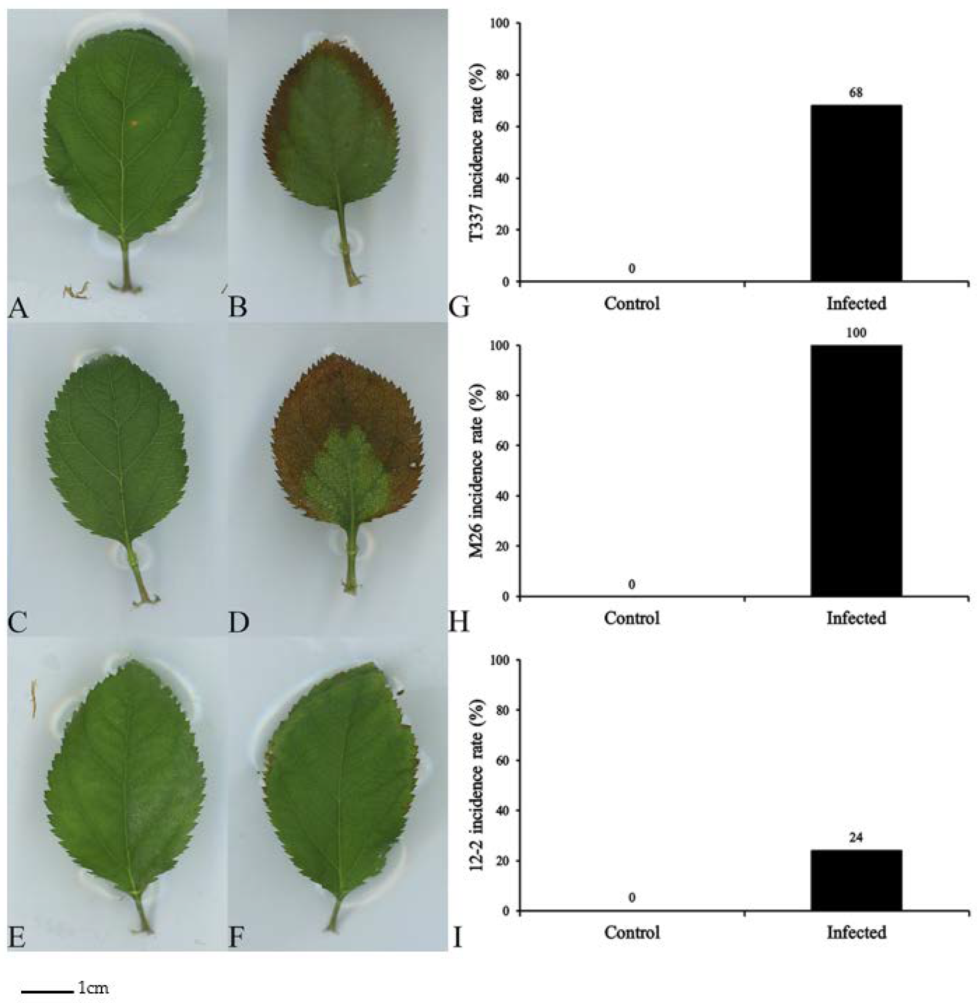
2.2. Assessment of Infection Symptoms

Disease resistance was assessed based on the plant survival rate, disease incidence rate, and the disease index [37]. The disease incidence rate was based on the appearance of brown lesions on the leaves, and the disease index grades were defined as shown below [37].
Grade 0: No symptoms.
Grade 1: Yellowed or wilted true leaves and cotyledons did not exceed 50% of the total area.
Grade 2: Yellowed or wilted true leaves and cotyledons exceeded 50% of the total area;
Grade 3: Leaves wilted or dead, with only the growing points surviving.
Grade 4: The entire plant was severely wilted or dead.
Disease index = ∑ (Plant number in the grade × Grade number)/ (Total plant number × Highest grade number) × 100.

2.3. Measurement of Leaf Chlorophyll Content

The relative chlorophyll contents of fully expanded leaves (leaves 3–5 from the bottom) were measured with an SPAD-502 portable chlorophyll meter (Beijing Harvesting Science and Technology Co., Ltd., Beijing, China). The same leaves were later removed, and the contents of chlorophyll a, chlorophyll b, and carotenes were measured using the acetone extraction method [38].

2.4. Measurement of Leaf Photosynthetic Parameters

The same fully expanded leaves used for the SPAD-meter measurements were placed in a CIRAS-2 portable photosynthesis measurement system (PP-Systems, Hansha Scientific Instruments, Beijing, China) to measure the leaf net photosynthetic rate (Pn), intercellular CO2 concentration (Ci), stomatal conductance (Gs), and the transpiration rate (Tr). Five replicates of each treatment were measured.

2.5. Measurement of Leaf Fluorescence Parameters

The chlorophyll fluorescence parameters were measured on fully expanded leaves as described above using a German WALZ Junior-PAM portable fluorometer (Zealquest Scientific and Technology Co., Ltd., Shanghai, China). The measured fluorescence parameters were the quantum efficiency of PSII (Fv/Fm), the actual photochemical efficiency of PSII (ΦPSII), the non-photochemical quenching coefficient (NPQ), the photochemical quenching coefficient (qP), and the electron transfer rate (ETR). Five replicates of each treatment were measured.

2.6. Measurement of Leaf Reactive-Oxygen-Species Levels

Fully expanded leaves were harvested from each treatment as described above to measure H2O2 content and the O2 production rate. The method by Bai et al. [39] was used to determine H2O2 content and the O2 production rate in five replicate samples from each treatment.

2.7. Measurement of Leaf Antioxidant-Enzyme Activities and Malondialdehyde Content

Fully expanded leaves were harvested as described above and used to measure enzyme activities. Superoxide dismutase (SOD) activity was measured using the method by Sun et al. [40]. Peroxidase (POD) activity was measured with the guaiacol method as described by Omran [41]. Catalase (CAT) activity was measured according to the method by Singh et al. [42]. MDA content was determined with the thiobarbituric acid method [43].

2.8. Measurement of Leaf Proline and Soluble-Sugar Contents

Fully expanded leaves from each treatment were harvested as described above to measure proline and soluble-sugar contents. Proline content was determined with the ninhydrin color method [3], and soluble-sugar content was determined with the phenol method [44]. Five replicate samples from each treatment were measured.

2.9. Data Analysis

The analysis of variance was performed using SPSS software (version 17; IBM SPSS, Chicago, IL, USA). Differences were detected among treatment means using Student’s t-test or Duncan’s multiple range test. A p-value < 0.05 was considered significant.

3. Results

3.1. Effects on Apple Rootstocks

Seven days after applying the MR5 spore solution, T337, M26, and 12-2 all showed different degrees of disease (Figure 1). On day 3 after MR5 infection, yellowish-brown spots appeared on the tips of the M26 leaf edges, and the leaves were dry and necrotic, but there were no obvious symptoms on T337 or 12-2. On day 5, yellowish-brown spots were observed on the tips of the T337 leaf edges. The dry and necrotic areas of the M26 leaves had expanded from the outside and inside to cover about one-half of the entire leaf area, whereas the 12-2 leaves had not developed any obvious symptoms. On day 7, the leaf edges of 12-2 also began to exhibit yellowish-brown spots (Figure 1F). At this time, the dry and necrotic areas of T337 leaves had expanded from the inside to the outside, and a circle of yellowish-brown lesions appeared on the leaf edges (Figure 1B). The dry and necrotic areas of M26 had expanded to cover > 50% of the total area (Figure 1D). The plant survival rates of the three rootstocks were all 100% 7 days after MR5 was inoculated (Table 1). The disease incidence rates on T337, M26, and 12-2 were 68, 100, and 24%, respectively (Figure 1G–I). The disease indices for T337, M26, and 12-2 were 68.00, 74.00, and 24.00, respectively (Table 1). These results show that M26 leaves had the most significant disease symptoms 7 days after MR5 infection, followed by T337 and 12-2.

3.2. Effects on Leaf Chlorophyll Contents

The chlorophyll a, chlorophyll b, carotene, and relative chlorophyll contents of T337 decreased by 82.91%, 77.30%, 83.54%, and 25.46% in treated plants compared with controls 7 days after MR5 infection (Figure 2). The chlorophyll a, chlorophyll b, carotene, and relative chlorophyll contents of M26 decreased by 78.34%, 60.75%, 42.65%, and 36.43%, respectively. These results show that MR5 significantly reduced the leaf chlorophyll contents of T337 and M26 but had no significant effect on 12-2.

3.3. Effects on Leaf Photosynthetic Parameters

MR5 significantly reduced Pn and Tr of M26 by 48.26% and 29.50% in treated plants compared with controls 7 days after infection (Figure 3). Although Gs and Ci of M26 decreased, the differences were not significant. MR5 produced nonsignificant reductions in the T337 photosynthetic parameters but did not affect the leaf photosynthetic parameters of 12-2. These results show that MR5 reduced some leaf photosynthetic parameters of T337 and M26 but had no significant effect on those of 12-2.

3.4. Effects on Leaf Fluorescence Parameters

MR5 significantly reduced the ETR of T337 and M26 and significantly increased the NPQ of T337 and M26 but had no significant effect on the other leaf fluorescence parameters of T337 or M26 7 days after infection (Figure 4). MR5 infection had no significant effect on the leaf fluorescence parameters of 12-2. These results show that MR5 had some effect on the leaf fluorescence parameters of T337 and M26 but had no significant effect on those of 12-2.

3.5. Effects on Leaf Reactive-Oxygen-Species Levels

MR5 significantly increased the H2O2 content of treated versus control T337 and M26 plants by 14.95% and 75.22%, respectively, and significantly increased the O2 production rate of T337 and M26 by 30.23% and 77.96%, respectively, 7 days after infection (Figure 5). These results show that MR5 significantly increased the leaf reactive oxygen species (ROS) levels of T337 and M26 but had no significant effect on those of 12-2.

3.6. Effects on Leaf Antioxidant-Enzyme Activities and MDA Content

MR5 significantly reduced SOD activity of treated vs. control T337 plants by 55.60% and increased MDA content by 28.55% 7 days after the infection (Figure 6). Although no significant differences in POD nor CAT activity were observed in T337, POD and CAT activities decreased by 18.96% and 29.43%, respectively. MR5 significantly increased SOD, POD, and CAT activities in M26 by 77.59%, 43.20%, and 18.15%, respectively, and increased MDA content by 141.20%. MR5 infection increased the antioxidant-enzyme activities and MDA content of 12-2 leaves, but these effects were not significant. These results show that MR5 decreased leaf antioxidant-enzyme activities and significantly increased leaf MDA contents of T337 and M26 but had no significant effect on these parameters in 12-2.

3.7. Effects on Leaf Proline and Soluble-Sugar Contents

MR5 significantly increased the proline contents of treated vs. control T337 and M26 plants by 71.82% and 102.35%, respectively, and significantly increased the soluble-sugar contents of T337 and M26 by 57.08% and 117.98%, respectively, 7 days after infection (Figure 7). These results show that MR5 significantly increased proline and soluble-sugar contents in T337 and M26 leaves but had no significant effect on these parameters in 12-2.

4. Discussion

The morphological structure of and physiological changes in leaves directly reflect the adaptability and stress resistance of plants, as leaves are the most sensitive plant part to disease [45]. Studies have shown that 5–7 days after Fusarium infection, the pathogen invades the vascular cylinder and pith of the host, causing leaves to fall and lodging to occur [46]. The wilting symptoms and physiological responses of plants after Fusarium infection are similar to those observed in plants under water stress [47]. In the present experiment, the leaves of the three rootstocks displayed different degrees of dryness and necrosis. The onset of disease symptoms occurred earlier in M26, followed by T337 and 12-2. These findings were similar to the results obtained by Choi et al. [48] in an apple-rootstock tolerance test. The greater the sensitivity of the rootstock was, the earlier the onset of disease was.
Chlorophyll absorbs the optical energy required for photosynthesis. A decrease in chlorophyll content inevitably leads to a decline in the photosynthetic rate and the accumulation of photosynthetic products, which eventually affect the healthy development of plants [49]. Botyanszka et al. [50] reported that chlorophyll content can be used as a technical index to recognize rootstock disease resistance. The worse the resistance of the rootstock is, the more significant the effect of infection is on chlorophyll content. In the present experiment, the chlorophyll contents of infected T337 and M26 were significantly lower than those of the controls. This may have occurred because the pathogen destroyed the structure of the chloroplasts, causing the chlorophyll synthetic rate to be much slower than the degradation rate [51]. In contrast, the chlorophyll content of 12-2 was not significantly affected by MR5. This may be because 12-2 is an ARD-resistant variety with mechanisms that alleviate chloroplast damage, thereby maintaining high chlorophyll levels [52].
Photosynthesis is the principal physiological process of plant growth, and its susceptibility to Fusarium infection has been confirmed in bananas, tomatoes, and wheat [53,54]. Fusarium disrupts the photosynthetic metabolism [55] and blocks the transport of water through the xylem by clogging the vessels, causing leaf dehydration, stomatal closure, and reduced Pn [56]. Studies have shown that Ci typically increases when Gs decreases, as carboxylation efficiency is reduced [57]. The leaf photosynthetic limits (Pn, Gs, Ci, and Tr) tend to fall under a stressed state [58], and this phenomenon was observed here for T337 and M26. The water loss per unit leaf area of mildly infected plants was significantly lower than that of healthy plants. Reduced Gs and Tr decrease the water loss of infected plants [25]. We speculate that MR5 infection induced photoprotective mechanisms in T337 and M26 and that their reduced Pn was caused by stomatal factors [54,59]. The photosynthetic parameters of 12-2 were less affected by MR5, as 12-2 may have maintained relatively high Gs, which increased Tr and heat dissipation, prevented leaf oxidative damage, and enhanced disease resistance [60].
Stress can also impair normal photochemical reactions. PSII is more susceptible to environmental stress damage than PSI, so chlorophyll fluorescence parameters can be used to assess plant stress tolerance [61]. Previous studies have shown that the carboxylation efficiency and photochemical ability of PSII decrease with Fusarium infection, indicating that the infection negatively affected Rubisco and the PSII reaction center [53]. Fusarium infection depresses PSII performance and impairs the coordination between PSI and PSII by inducing pathological wilting in apple seedlings [62]. Fv/Fm is commonly used as an indicator of photoinhibition or PSII damage [63]. After MR5 infection, Fv/Fm declined in all three varieties, but this decrease was less severe in 12-2 than in T337 and M26. PSII of 12-2 may have experienced less damage under MR5 stress than T337 and M26, resulting in a lesser slowing of photosynthetic electron transport and preserving the redox ability of initial electron acceptor QA, thereby maintaining photosynthetic carbon assimilation at a higher level [64]. The ETR values of the three varieties were consistent with this scenario. At the same time, qP decreased in T337 and M26, indicating that the actual efficiency of light capture, carbon assimilation capacity, and the ratio of the open PSII reaction center decreased, limiting electron transfer from the PSII reaction center, and inhibiting photosynthesis [65]. NPQ significantly increased in T337 and M26 in response to infection, showing that the leaf heat dissipation mechanism was destroyed and light energy dissipation increased in the form of heat [66]. Therefore, a large amount of excitation energy likely accumulated, damaging the thylakoid membranes and photosynthetic complexes, leading to a loss of photoprotective ability [67] and the appearance of lesions on diseased leaves (Figure 1). No significant changes in the NPQ of 12-2 were observed, indicating that the light energy conversion rate and photosynthesis remained unchanged under stress [68].
Active disease resistance is a host defense response induced by pathogen infection, and ROS are the earliest defense response of host plants to invasion by a causative agent [69]. Bacterial infection affects the balance of ROS metabolism in plants, destroying or reducing ROS scavengers, increasing ROS production, and damaging membrane structures [70]. Here, MR5 infection caused a significant increase in H2O2 content and O2 production rate in T337 and M26, suggesting that the ROS produced by T337 and M26 actively participated in the plant disease-resistance response, as ROS are toxic to pathogenic bacteria and may directly kill them [70]. The ROS content did not significantly increase in 12-2, because disease-resistant varieties are more capable of eliminating ROS than susceptible varieties [71], and ROS induce the immune-mediated necrosis of host cells, thereby inhibiting the spread of a pathogen [72].
However, overproduced ROS react with proteins, lipids, and nucleic acids in the host cell, resulting in cell-membrane peroxidation and damage, enzyme inactivation, cell metabolic disorders, and the eventual death of the host cell [73]. The host plant’s ROS scavenging system regulates the ROS balance, thereby maintaining a normal cellular redox state and reducing ROS damage to the host cell [74]. Excessive ROS in plant leaves are removed by SOD, POD, CAT, and other defense-related enzymes [47]. Here, the activities of SOD, POD, and CAT decreased to varying degrees in T337 and M26 7 days after MR5 infection. It may be that when the stress exceeded a certain threshold, T337 and M26 could not remove the oxygen free radicals rapidly enough, eventually leading to reduced enzyme activities and oxidative damage [75]. The POD activity of 12-2 increased 7 days after MR5 infection. One possible reason is that the O2 generation rate of 12-2 increased, resulting in increased SOD activity and rapid disproportionation of O2 to H2O2 and O2 [76]. At this time, POD plays a protective role in decomposing O2 into H2O2 and H2O [68]. CAT activity acts as a reserve force for POD, removing H2O2 and converting it to O2 and H2O [77].
MDA is the final product of membrane lipid peroxidation. MDA content has been used as a representative of the degree of membrane damage [78]. The more serious the lipid peroxidation is, the higher the MDA content is, indicating a greater plant-damaging effect [79]. Here, the MDA content of T337 and M26 significantly increased 7 days after MR5 infection, whereas that of 12-2 did not significantly change. Therefore, we speculated that the cell membrane functions of T337 and M26 were severely damaged, whereas 12-2 responded to stress in time, and its cell membranes experienced less injury.
Soil-borne pathogens infect the xylem of plant roots and hinder water transport in the xylem [62]; this leads to severe water stress and the pathological wilting of apple seedlings [47]. To restrict water coerced loss, plants synthesize specific consistent solutes to abate the cell osmotic potential and promote water absorption [80]. Soluble-sugars and proline act as plant protective substances under stress. Their production is one of the basic mechanisms for plants to react to stress, and it is probably a manifestation of cell structural damage [81]. Here, the proline and soluble-sugar contents of T337 and M26 increased significantly 7 days after MR5 infection. MR5 infection may have led to a rapid burst of ROS production and an excessive oxidation of membrane lipids, causing cellular damage and inactivating antioxidant enzymes, leading to an increase in proline and soluble-sugars [70]. No significant changes in proline or soluble-sugar contents were detected in 12-2. We speculated that 12-2 was more tolerant to MR5, experienced less stress, and thus maintained relatively normal cellular function [82].
Although we provided some evidence that 12-2 is resistant to ARD-associated F. proliferatum strain MR5, the molecular mechanisms by which 12-2 inhibits MR5 are unknown. We only tested MR5. Whether 12-2 is resistant to other pathogens is an important question for future research. We do not have the Geneva apple rootstock, which would also be a good test material. Although the performance of the three varieties was significantly different within 7 days of infection, 12-2 exhibited mild symptoms. Because there were no other disease-resistant varieties with which to compare 12-2 and the experimental soil contained much higher concentrations of pathogenic fungi than actual soil, it was not necessary to continue this particular experiment. However, the behavior of 12-2 under continued inoculation with a fungal solution would be worth studying. The risk of the damaging effect of low-molecular-weight compounds liberated by Fusarium into the plant rhizosphere is another research topic. The interaction between 12-2 and secondary metabolites released by fungi should also be the focus of future research. Many experiments should be performed on new rootstocks, including evaluations of their graft consistency, gain outputs, and survival and growth in other areas.

5. Conclusions

The 12-2 rootstock had good resistance to ARD-associated F. proliferatum MR5. Leaf growth, the disease incidence rate, chlorophyll content, photosynthetic and fluorescence parameters, ROS levels, antioxidant-enzyme activity, MDA content, and proline and soluble-sugar contents did not significantly differ between 12-2 plants treated with PDB and those treated with the MR5 spore solution. The MR5 spore solution had the greatest effect on most of the aboveground physiological indicators of T337 and M26. An infection test showed that 12-2 was more resistant to ARD-associated F. proliferatum MR5 and may serve as an important test material for apple-rootstock resistance breeding in China.

Author Contributions

Conceptualization, X.C. (Xueli Cui), Y.M. and X.S.; methodology, X.C. (Xueli Cui), Y.M. and X.S.; software, X.C. (Xueli Cui) and Y.M.; validation, X.C. (Xueli Cui); formal analysis, X.C. (Xueli Cui) and Y.M.; investigation, X.C. (Xueli Cui), X.Q. and Y.L.; resources, Y.H., Z.M. and X.C. (Xuesen Chen); data curation, X.C. (Xueli Cui); writing—original draft preparation, X.C. (Xueli Cui); writing—review and editing, X.C. (Xueli Cui), Z.M., X.C. (Xuesen Chen), Y.M. and X.S.; visualization, X.C. (Xueli Cui); supervision, X.S.; project administration, X.S.; funding acquisition, X.S. All authors have read and agreed to the published version of the manuscript.

Funding

This research study was funded by a project supported by National Natural Science Foundation of China, grant number 32072520; Fruit innovation team project of Shandong Province (CN), grant number SDAIT-06-07; and Natural Science Foundation of Shandong Province (CN), grant number ZR2020MC132.

Institutional Review Board Statement

Not applicable.

Informed Consent Statement

Not applicable.

Data Availability Statement

Data are contained within the article. All data and materials presented in the manuscript are available.

Conflicts of Interest

The authors declare no conflict of interest.

Abbreviations

ARD, apple replant disease; 12-2, apple rootstock superior line named 12-2; MR5, ARD-associated F. proliferatum MR5; Pn, net photosynthetic rate; Ci, intercellular CO2 concentration; Gs, stomatal conductance; Tr, transpiration rate; Fv/Fm, PSII original light energy conversion efficiency; ΦPSII, PSII actual photochemical efficiency; NPQ, non-photochemical quenching coefficient; qP, photochemical quenching coefficient; ETR, electron transfer rate; SOD, superoxide dismutase; POD, peroxidase; CAT, catalase; MDA, malondialdehyde.

References

  1. Chen, P.; Wang, Y.Z.; Liu, Q.Z.; Zhang, Y.T.; Li, X.Y.; Li, H.Q.; Li, W.H. Phase changes of continuous cropping obstacles in strawberry (Fragaria × ananassa Duch.) production. Appl. Soil Ecol. 2020, 155, 103626. [Google Scholar] [CrossRef]
  2. Mao, Y.F.; Zhang, L.L.; Wang, Y.Y.; Yang, L.; Yin, Y.J.; Su, X.F.; Liu, Y.P.; Pang, H.L.; Xu, J.; Hu, Y.L.; et al. Effects of polycyclic aromatic hydrocarbons (PAHs) from different sources on soil enzymes and microorganisms of Malus prunifolia var. Ringo. Arch. Agron. Soil Sci. 2021, 14, 2048–2062. [Google Scholar] [CrossRef]
  3. Wang, Y.F.; Ma, Z.T.; Wang, X.W.; Sun, Q.R.; Dong, H.Q.; Wang, G.S.; Chen, X.S.; Yin, C.M.; Han, Z.H.; Mao, Z.Q. Effects of biochar on the growth of apple seedlings, soil enzyme activities and fungal communities in replant disease soil. Sci. Hortic-Amst. 2019, 256, 108641. [Google Scholar] [CrossRef]
  4. Manici, L.M.; Kelderer, M.; Franke-Whittle, I.H.; Rühmer, T.; Baab, G.; Nicoletti, F.; Caputo, F.; Topp, A.; Insam, H.; Naef, A. Relationship between root-endophytic microbial communities and replant disease in specialized apple growing areas in Europe. Appl. Soil Ecol. 2013, 72, 207–214. [Google Scholar] [CrossRef]
  5. Wang, H.Y.; Zhao, L.; Jiang, W.T.; Zhang, R.; Chen, R.; Mao, Y.F.; Chen, X.S.; Shen, X.; Yin, C.M.; Mao, Z.Q. Effects of Allium fistulosum-Brassica juncea-Triticum aestivum rotation a year on the soil microbial environment and the subsequent growth of young apple trees. Sci. Hortic-Amst. 2021, 290, 110549. [Google Scholar] [CrossRef]
  6. Zhu, Y.M.; Shin, S.; Mazzola, M. Genotype responses of two apple rootstocks to infection by Pythium ultimum causing apple replant disease. Can. J. Plant Pathol. 2016, 38, 483–491. [Google Scholar] [CrossRef]
  7. van Schoor, L.; Denman, S.; Cook, N.C. Characterisation of apple replant disease under South African conditions and potential biological management strategies. Sci. Hortic.-Amst. 2009, 119, 153–162. [Google Scholar] [CrossRef]
  8. Wang, G.S.; Yin, C.M.; Pan, F.B.; Wang, X.B.; Xiang, L.; Wang, Y.F.; Wang, J.Z.; Tian, C.P.; Chen, J.; Mao, Z.Q. Analysis of the Fungal Community in Apple Replanted Soil Around Bohai Gulf. Hortic. Plant J. 2018, 4, 175–181. [Google Scholar] [CrossRef]
  9. Xiang, L.; Wang, M.; Jiang, W.T.; Wang, Y.F.; Chen, X.S.; Yin, C.M.; Mao, Z.Q. Key indicators for renewal and reconstruction of perennial trees soil: Microorganisms and phloridzin. Ecotoxicol. Environ. Saf. 2021, 225, 112723. [Google Scholar] [CrossRef]
  10. Zou, Q.J.; Wang, S.T.; Liang, K.J.; Wang, Y.N.; Hu, T.L.; Han, Z.Q.; Cao, K.Q. Suspected pathogenic Fusarium spp. isolated from apple orchard soils in Hebei Province. Mycosystema 2014, 33, 976–983. [Google Scholar] [CrossRef]
  11. Duan, Y.; Jiang, W.; Zhang, R.; Chen, R.; Chen, X.; Yin, C.; Mao, Z. Discovery of Fusarium proliferatum f. sp. malus domestica causing Apple Replant Disease in China. Plant Dis. 2022. [Google Scholar] [CrossRef] [PubMed]
  12. Mao, Y.F.; Yin, Y.J.; Cui, X.L.; Wang, H.Y.; Su, X.F.; Qin, X.; Liu, Y.B.; Hu, Y.L.; Shen, X. Detection of Root Physiological Parameters and Potassium and Calcium Currents in the Rhizoplane of the Apple Rootstock Superior Line 12-2 With Improved Apple Replant Disease Resistance. Front. Plant Sci. 2021, 12, 734430. [Google Scholar] [CrossRef] [PubMed]
  13. Pan, F.B.; Xiang, L.; Wang, S.; Li, J.J.; Shen, X.; Chen, X.S.; Yin, C.M.; Mao, Z.Q. Effects of short-term rotation and Trichoderma application on the soil environment and physiological characteristics of Malus hupehensis Rehd. seedlings under replant conditions. Acta Ecol. Sin. 2017, 37, 315–321. [Google Scholar] [CrossRef]
  14. Mao, Y.F.; Yin, Y.J.; Cui, X.L.; Wang, H.Y.; Su, X.F.; Qin, X.; Liu, Y.B.; Hu, Y.L.; Shen, X. Detection of Above-Ground Physiological Indices of an Apple Rootstock Superior Line 12-2 with Improved Apple Replant Disease (ARD) Resistance. Horticulturae 2021, 7, 337. [Google Scholar] [CrossRef]
  15. Rivard, C.L.; O’Connell, S.; Peet, M.M.; Welker, R.M. Grafting Tomato to Manage Bacterial Wilt Caused by Ralstonia solanacearum in the Southeastern United States. Plant Dis. 2012, 96, 973–978. [Google Scholar] [CrossRef] [Green Version]
  16. Zhu, Y.M.; Saltzgiver, M. A systematic analysis of apple root resistance traits to Pythium ultimum infection and the underpinned molecular regulations of defense activation. Hortic. Res. 2020, 7, 62. [Google Scholar] [CrossRef]
  17. Auvil, T.D.; Schmidt, T.R.; Hanrahan, I.; Castillo, F.; McFerson, J.R.; Fazio, G. Evaluation of dwarfing rootstocks in Washington apple replant sites. Acta Hortic. 2011, 903, 265–271. [Google Scholar] [CrossRef]
  18. Norelli, J.L.; Holleran, H.T.; Johnson, W.C.; Robinson, T.L.; Aldwinckle, H.S. Resistance of Geneva and other apple rootstocks to Erwinia amylovora. Plant Dis. 2003, 87, 26–32. [Google Scholar] [CrossRef] [Green Version]
  19. Ma, S.S. Apple dwarfing from root stock—M9T337. Northwest Hortic. (Fruit Tree) 2017, 4, 6–8. (In Chinese) [Google Scholar]
  20. Li, M. Application of M26 Rootstock in Potted Apple Tree. Ph.D. Dissertation, Northwest A&F University, Xi’an, China, 2015. [Google Scholar]
  21. Fallahi, E.; Colt, W.M.; Fallahi, B.; Chun, I.J. The Importance of Apple Rootstocks on Tree Growth, Yield, Fruit Quality, Leaf Nutrition, and Photosynthesis with an Emphasis on ‘Fuji’. HortTechnology 2001, 12, 38–44. [Google Scholar] [CrossRef] [Green Version]
  22. Shen, X.; Mao, Z.Q.; Ni, W.R.; Wang, R.; Hu, Y.L.; Chen, X.S.; Wu, S.J.; Han, T.T.; Zhang, W.H. In Situ Breeding Method for Three-Stage Selection of Apple Rootstock Tolerant to ARD. China Patent No. CN104488645A, 8 April 2015. Available online: https://patents.google.com/patent/CN104488645A (accessed on 4 January 2017). (In Chinese).
  23. Gao, F.F. The Continuous Cropping Testing and Rapid Propagation of Primary Apple Rootstocks Superior Lines. Bachelor’s Thesis, Shandong Agricultural University, Tai’an, China, 2018. [Google Scholar]
  24. Su, X.F.; Chai, S.S.; Mao, Y.F.; Zhang, L.L.; Yin, Y.J.; Liu, Y.P.; Pang, H.L.; Hu, Y.L. Polyploid Induction and Identification of Apple Rootstocks with Tolerance to Continuous Cropping. Mol. Plant Breed. 2021, 2021, 1–12. Available online: https://kns.cnki.net/kcms/detail/46.1068.S.20210521.1458.010.html (accessed on 16 September 2021). (In Chinese).
  25. Dong, X.; Ling, N.; Wang, M.; Shen, Q.R.; Guo, S.W. Fusaric acid is a crucial factor in the disturbance of leaf water imbalance in Fusarium-infected banana plants. Plant Physiol. Bioch. 2012, 60, 171–179. [Google Scholar] [CrossRef] [PubMed]
  26. Czymmek, K.J.; Fogg, M.; Powell, D.H.; Sweigard, J.; Park, S.-Y.; Kang, S. In vivo time-lapse documentation using confocal and multiphoton microscopy reveals the mechanisms of invasion into the Arabidopsis root vascular system by Fusarium Oxysporum. Fungal Genet. Biol. 2007, 44, 1011–1023. [Google Scholar] [CrossRef] [PubMed]
  27. Krivanek, A.F.; Stevenson, J.F.; Walker, M.A. Development and comparison of symptom indices for quantifying grapevine resistance to Pierce’s disease. Phytopathology 2005, 95, 36–43. [Google Scholar] [CrossRef] [PubMed] [Green Version]
  28. Berestetskiy, A.O. A review of fungal phytotoxins: From basic studies to practical use. Appl. Biochem. Microbiol. 2008, 44, 453–465. [Google Scholar] [CrossRef]
  29. Shimizu, B.; Saito, F.; Miyagawa, H.; Watanabe, K.; Ueno, T.; Sakata, K.; Ogawa, K. Phytotoxic components produced by pathogenic Fusarium against morning glory. Z. Naturforsch C J. Biosci. 2005, 60, 862–866. [Google Scholar] [CrossRef]
  30. Manici, L.M.; Caputo, F.; Saccà, M.L. Secondary metabolites released into the rhizosphere by Fusarium oxysporum and Fusarium spp. as underestimated component of nonspecific replant disease. Plant Soil 2017, 415, 85–98. [Google Scholar] [CrossRef]
  31. Vrabka, J.; Niehaus, E.-M.; Münsterkötter, M.; Proctor, R.H.; Brown, D.W.; Novák, O.; Pěnčik, A.; Tarkowská, D.; Hromadová, K.; Hradilová, M.; et al. Production and Role of Hormones During Interaction of Fusarium Species With Maize (Zea mays L.) Seedlings. Front. Plant Sci. 2019, 9, 1936. [Google Scholar] [CrossRef]
  32. Blum, A.; Benfield, A.H.; Sørensen, J.L.; Nielsen, M.R.; Bachleitner, S.; Studt, L.; Beccari, G.; Covarelli, L.; Batley, J.; Gardiner, D.M. Regulation of a novel Fusarium cytokinin in Fusarium pseudograminearum. Fungal Biol-UK 2019, 123, 255–266. [Google Scholar] [CrossRef] [Green Version]
  33. Islam, M.M.; Hoque, M.A.; Okuma, E.; Banu, M.N.; Shimoishi, Y.; Nakamura, Y.; Murata, Y. Exogenous proline and glycinebetaine increase antioxidant enzyme activities and confer tolerance to cadmium stress in cultured tobacco cells. J. Plant Physiol. 2009, 166, 1587–1597. [Google Scholar] [CrossRef]
  34. Balbín-Suárez, A.; Lucas, M.; Vetterlein, D.; Sørensen, S.J.; Winkelmann, T.; Smalla, K.; Jacquiod, S. Exploring microbial determinants of apple replant disease (ARD): A microhabitat approach under split-root design. FEMS Microbiol. Ecol. 2020, 96, fiaa211. [Google Scholar] [CrossRef]
  35. Atucha, A.; Emmett, B.; Bauerle, T.L. Growth rate of fine root systems influences rootstock tolerance to replant disease. Plant Soil 2014, 376, 337–346. [Google Scholar] [CrossRef]
  36. Xu, H.; Ediger, D. Rootstocks with Different Vigor Influenced Scion–Water Relations and Stress Responses in AmbrosiaTM Apple Trees (Malus Domestica var. Ambrosia). Plants 2021, 10, 614. [Google Scholar] [CrossRef] [PubMed]
  37. Li, M.; Ma, G.; Lian, H.; Su, X.; Tian, Y.; Huang, W.; Mei, J.; Jiang, X. The effects of Trichoderma on preventing cucumber fusarium wilt and regulating cucumber physiology. J. Integr. Agric. 2019, 18, 607–617. [Google Scholar] [CrossRef] [Green Version]
  38. Su, S.Q.; Zhou, Y.M.; Qin, J.G.; Yao, W.Z.; Ma, Z.H. Optimization of the Method for Chlorophyll Extraction in Aquatic Plants. J. Freshw. Ecol. 2010, 25, 531–538. [Google Scholar] [CrossRef] [Green Version]
  39. Bai, R.; Ma, F.; Liang, D.; Zhao, X. Phthalic Acid Induces Oxidative Stress and Alters the Activity of Some Antioxidant Enzymes in Roots of Malus prunifolia. J. Chem. Ecol. 2009, 35, 488–494. [Google Scholar] [CrossRef] [PubMed]
  40. Sun, D.Q.; Sun, X.M.; Xu, Y.Y.; Wu, T.J.; Tao, L.X. Superoxide dismutase activity and risk of cognitive decline in older adults: Findings from the Chinese Longitudinal Healthy Longevity Survey. Exp. Gerontol. 2019, 118, 72–77. [Google Scholar] [CrossRef]
  41. Omran, R.G. Peroxide levels and the activities of catalase, peroxidase, and indoleacetic acid oxidase during and after chilling cucumber seedlings. Plant Physiol. 1980, 65, 407–408. [Google Scholar] [CrossRef] [Green Version]
  42. Singh, B.K.; Sharma, S.R.; Singh, B. Antioxidant enzymes in cabbage: Variability and inheritance of superoxide dismutase, peroxidase and catalase. Sci. Hortic.-Amst. 2010, 124, 9–13. [Google Scholar] [CrossRef]
  43. Lykkesfeldt, J. Determination of Malondialdehyde as Dithiobarbituric Acid Adduct in Biological Samples by HPLC with Fluorescence Detection: Comparison with Ultraviolet-Visible Spectrophotometry. Clin. Chem. 2001, 47, 1725–1727. [Google Scholar] [CrossRef]
  44. Masuko, T.; Minami, A.; Iwasaki, N.; Majima, T.; Nishimura, S.; Lee, Y.C. Carbohydrate analysis by a phenol-sulfuric acid method in microplate format. Anal. Biochem. 2005, 339, 69–72. [Google Scholar] [CrossRef] [PubMed]
  45. Li, X.P.; Li, Y.P.; Zhang, Z.; Li, X.G. Influences of Environmental Factors on Leaf Morphology of Chinese Jujubes. PLoS ONE 2015, 10, e0127825. [Google Scholar] [CrossRef]
  46. Yao, J.; Huang, P.; Chen, H.; Yu, D.Y. Fusarium oxysporum is the pathogen responsible for stem rot of the succulent plant Echeveria ‘Perle von Nürnberg’ and observation of the infection process. Eur. J. Plant Pathol. 2021, 159, 555–568. [Google Scholar] [CrossRef]
  47. Wang, S.; Zhang, K.K.; Huang, X.; Fan, Y.J.; Yang, L.T.; Li, Y.R. Cloning and Functional Analysis of Thylakoidal Ascorbate Peroxidase (TAPX) Gene in Sugarcane. Sugar Tech. 2015, 17, 356–366. [Google Scholar] [CrossRef]
  48. Choi, B.H.; Bhusal, N.; Jeong, W.T.; Park, I.H.; Han, S.G.; Yoon, T.M. Waterlogging tolerance in apple trees grafted on rootstocks from G., CG, and M series. Hortic. Environ. Biote. 2020, 61, 685–692. [Google Scholar] [CrossRef]
  49. Wang, S.; Wang, X.D.; Shi, X.B.; Wang, B.L.; Zheng, X.C.; Wang, H.B.; Liu, F.Z. Red and Blue Lights Significantly Affect Photosynthetic Properties and Ultrastructure of Mesophyll Cells in Senescing Grape Leaves. Hortic. Plant J. 2016, 2, 82–90. [Google Scholar] [CrossRef] [Green Version]
  50. Botyanszka, L.; Zivcak, M.; Chovancek, E.; Sytar, O.; Barek, V.; Hauptvogel, P.; Halabuk, A.; Brestic, M. Chlorophyll Fluorescence Kinetics May Be Useful to Identify Early Drought and Irrigation Effects on Photosynthetic Apparatus in Field-Grown Wheat. Agronomy 2020, 10, 1275. [Google Scholar] [CrossRef]
  51. Lu, Y.; Yao, J. Chloroplasts at the crossroad of photosynthesis, pathogen infection and plant defense. Int. J. Mol. Sci. 2018, 19, 3900. [Google Scholar] [CrossRef] [Green Version]
  52. Han, Y.Q.; Wang, H.; Wang, H.N.; Cheng, F.Y.; Tian, N.N.; Yan, X.; Han, Y.H. Physiological, biochemical and gene expression analysis of foxtail millet against infection of Sclerospora graminicola. Acta Phytopathol. Sin. 2020, 50, 657–665. (In Chinese) [Google Scholar] [CrossRef]
  53. Dong, X.; Wang, M.; Ling, N.; Shen, Q.R.; Guo, S.W. Potential role of photosynthesis-related factors in banana metabolism and defense against Fusarium oxysporum f. sp cubense. Environ. Exp. Bot. 2016, 129, 4–12. [Google Scholar] [CrossRef]
  54. Yang, S.M.; Li, X.; Chen, W.Q.; Liu, T.G.; Zhong, S.F.; Ma, L.X.; Zhang, M.; Zhang, H.Y.; Yu, D.L.; Luo, P.G. Wheat Resistance to Fusarium Head Blight is Associated With Changes in Photosynthetic Parameters. Plant Dis. 2016, 100, 847–852. [Google Scholar] [CrossRef] [PubMed] [Green Version]
  55. Saeed, I.; MacGuidwin, A.E.; Rouse, D.I.; Sharkey, T.D. Limitation to Photosynthesis in Pratylenchus penetrans- and Verticillium dahliae-Infected Potato. Crop Sci. 1999, 39, 1340–1346. [Google Scholar] [CrossRef]
  56. Pinto, L.S.R.C.; Azevedo, J.L.; Pereira, J.O.; Vieira, M.L.C.; Labate, C.A. Symptomless infection of banana and maize by endophytic fungi impairs photosynthetic efficiency. New Phytol. 2000, 147, 609–615. [Google Scholar] [CrossRef] [PubMed]
  57. Wang, H.L.; Lin, K.D.; Hou, Z.A.; Richardson, B.; Gan, J. Sorption of the herbicide terbuthylazine in two New Zealand forest soils amended with biosolids and biochars. J. Soil Sediment 2010, 10, 283–289. [Google Scholar] [CrossRef]
  58. Zhang, A.Q.; Liu, M.X.; Gu, W.; Chen, Z.Y.; Gu, Y.C.; Pei, L.F.; Tian, R. Effect of drought on photosynthesis, total antioxidant capacity, bioactive component accumulation, and the transcriptome of Atractylodes lancea. BMC Plant Biol. 2021, 21, 293. [Google Scholar] [CrossRef]
  59. Wright, H.; Delong, J.; Lada, R.; Prange, R. The relationship between water status and chlorophyll a fluorescence in grapes (Vitis spp.). Postharvest Biol. Technol. 2009, 51, 193–199. [Google Scholar] [CrossRef]
  60. Farber, M.; Attia, Z.; Weiss, D. Cytokinin activity increases stomatal density and transpiration rate in tomato. J. Exp. Bot. 2016, 67, 6351–6362. [Google Scholar] [CrossRef]
  61. Yang, L.L.; Zhao, Y.; Zhang, Q.; Cheng, L.; Han, M.; Ren, Y.Y.; Yang, L.M. Effects of drought–re-watering–drought on the photosynthesis physiology and secondary metabolite production of Bupleurum chinense DC. Plant Cell Rep. 2019, 38, 1181–1197. [Google Scholar] [CrossRef]
  62. Yan, K.; Han, G.X.; Ren, C.G.; Zhao, S.J.; Wu, X.Q.; Bian, T.T. Fusarium solani Infection Depressed Photosystem Performance by Inducing Foliage Wilting in Apple Seedlings. Front. Plant Sci. 2018, 9, 479. [Google Scholar] [CrossRef] [Green Version]
  63. Gao, W.Q.; Lv, L.H.; Srivastava, A.K.; Wu, Q.S.; Kuča, K. Effects of Mycorrhizae on Physiological Responses and Relevant Gene Expression of Peach Affected by Replant Disease. Agron.-Basel 2020, 10, 186. [Google Scholar] [CrossRef] [Green Version]
  64. Gill, S.S.; Tuteja, N. Reactive oxygen species and antioxidant machinery in abiotic stress tolerance in crop plants. Plant Physiol. Biochem. 2010, 48, 909–930. [Google Scholar] [CrossRef] [PubMed]
  65. Li, Q.; Zhao, C.Z.; Wang, J.W.; Wen, J.; Li, Z.Q.; Ma, J.Y. Morphological and photosynthetic physiological characteristics of Saussurea salsa in response to flooding in salt marshes of Xiao Sugan Lake, Gansu, China. Chin. J. Plant Ecol. 2019, 43, 685–696. [Google Scholar] [CrossRef]
  66. Ruban, A.V. Nonphotochemical Chlorophyll Fluorescence Quenching: Mechanism and Effectiveness in Protecting Plants from Photodamage. Plant Physiol. 2016, 170, 1903–1916. [Google Scholar] [CrossRef] [Green Version]
  67. Chen, Y.E.; Mao, H.T.; Wu, N.; Mohi Ud Din, A.; Khan, A.; Zhang, H.Y.; Yuan, S. Salicylic Acid Protects Photosystem II by Alleviating Photoinhibition in Arabidopsis thaliana under High Light. Int. J. Mol. Sci. 2020, 21, 1229. [Google Scholar] [CrossRef] [PubMed] [Green Version]
  68. Tung, J.; Goodwin, P.H.; Hsiang, T. Chlorophyll fluorescence for quantification of fungal foliar infection and assessment of the effectiveness of an induced systemic resistance activator. Eur. J. Plant Pathol. 2013, 136, 301–315. [Google Scholar] [CrossRef]
  69. Qi, F.S.; Song, C.P.; Wang, B.S.; Zhou, J.M.; Kangasjärvi, J.; Zhu, J.K.; Gong, Z.Z. Reactive oxygen species signaling and stomatal movement in plant responses to drought stress and pathogen attack. J. Integr. Plant Biol. 2018, 60, 805–826. [Google Scholar] [CrossRef] [Green Version]
  70. Sharma, P.; Jha, A.B.; Dubey, R.S.; Pessarakli, M.; Polle, A. Reactive Oxygen Species, Oxidative Damage, and Antioxidative Defense Mechanism in Plants under Stressful Conditions. J. Bot. 2012, 2012, 217037. [Google Scholar] [CrossRef] [Green Version]
  71. Chen, Y.E.; Cui, J.M.; Su, Y.Q.; Yuan, M.; Zhang, H.Y. Influence of stripe rust infection on the photosynthetic characteristics and antioxidant system of susceptible and resistant wheat cultivars at the adult plant stage. Front. Plant Sci. 2015, 6, 779. [Google Scholar] [CrossRef] [Green Version]
  72. Wang, C.F.; Huang, L.L.; Zhang, H.C.; Han, Q.M.; Buchenauer, H.; Kang, Z.S. Cytochemical localization of reactive oxygen species (O2 and H2O2) and peroxidase in the incompatible and compatible interaction of wheat- Puccinia striiformis f. sp. triitici. Physiol. Mol. Plant Pathol. 2010, 74, 221–229. [Google Scholar] [CrossRef]
  73. Juan, C.A.; de la Lastra, J.M.P.; Plou, F.J.; Pérez-Lebeña, E. The Chemistry of Reactive Oxygen Species (ROS) Revisited: Outlining Their Role in Biological Macromolecules (DNA, Lipids and Proteins) and Induced Pathologies. Int. J. Mol. Sci. 2021, 22, 4642. [Google Scholar] [CrossRef]
  74. Tanveer, M.; Shah, A. An insight into salt stress tolerance mechanisms of Chenopodium album. Environ. Sci. Pollut. Res. Int. 2017, 24, 16531–16535. [Google Scholar] [CrossRef] [PubMed]
  75. Ghosh, N.; Das, A.; Chaffee, S.; Roy, S.; Sen, C.K. Reactive Oxygen Species, Oxidative Damage and Cell Death. In Immunity and Inflammation in Health and Disease; Academic Press: Cambridge, UK, 2018; pp. 45–55. [Google Scholar] [CrossRef]
  76. Guo, D.L.; Ma, J.; Su, W.Y.; Xie, B.M.; Guo, C.H. Contribution of reactive oxygen species (ROS) to genotoxicity of Nitrobenzene on V. faba. Ecotoxicology 2014, 23, 657–664. [Google Scholar] [CrossRef] [PubMed]
  77. Hadwan, M.H. Simple spectrophotometric assay for measuring catalase activity in biological tissues. BMC Biochem. 2018, 19, 7. [Google Scholar] [CrossRef] [PubMed]
  78. Liu, J.J.; Wei, Z.; Li, J.H. Effects of copper on leaf membrane structure and root activity of maize seedling. Bot. Stud. 2014, 55, 47. [Google Scholar] [CrossRef] [Green Version]
  79. Song, X.L.; Wang, Y.H.; Lv, X.M. Responses of plant biomass, photosynthesis and lipid peroxidation to warming and precipitation change in two dominant species (Stipa grandis and Leymus chinensis) from North China Grasslands. Ecol. Evol. 2016, 6, 1871–1882. [Google Scholar] [CrossRef]
  80. Delauney, A.J.; Verma, D.P. Proline biosynthesis and osmoregulation in plants. Plant J. 1993, 4, 215–223. [Google Scholar] [CrossRef]
  81. Li, H.Y.; Tang, X.Q.; Yang, X.Y.; Zhang, H.X. Comprehensive transcriptome and metabolome profiling reveal metabolic mechanisms of Nitraria sibirica Pall. to salt stress. Sci. Rep. 2021, 11, 12878. [Google Scholar] [CrossRef]
  82. Amoah, J.N.; Seo, Y.W. Effect of progressive drought stress on physio-biochemical responses and gene expression patterns in wheat. 3 Biotech. 2021, 11, 440. [Google Scholar] [CrossRef]
Figure 1. Effects of F. proliferatum MR5 on the three rootstocks after 7 days. (A) Control T337 leaves. (B) Infected T337 leaves. (C) Control M26 leaves. (D) Infected M26 leaves. (E) Control 12-2 leaves. (F) Infected 12-2 leaves. (G) T337 incidence rate. (H) M26 incidence rate. (I) 12-2 incidence rate. Note: Scale bars in (AF) are 1 cm.
Horticulturae 08 00723 g001
Figure 2. Effects of F. proliferatum MR5 on leaf chlorophyll contents: (A) chlorophyll a; (B) chlorophyll b; (C) carotene; (D) SPAD. The Student’s t-test was used to detect the differences between the control and infected treatments within a rootstock. * p < 0.05; ** p < 0.01.
Horticulturae 08 00723 g002
Figure 3. Effects of F. proliferatum MR5 on leaf photosynthetic parameters: (A) Pn; (B) Ci; (C) Gs; (D) Tr. The Student’s t-test was used to detect the differences between the control and infected treatments within a rootstock. * p < 0.05; ** p < 0.01.
Horticulturae 08 00723 g003
Figure 4. Effects of F. proliferatum MR5 on leaf fluorescence parameters: (A) ETR; (B) NPQ; (C) qP; (D) Fv/Fm; (E) ΦPSII. The Student’s t-test was used to detect differences between the control and infected treatments within a rootstock. * p < 0.05; ** p < 0.01.
Horticulturae 08 00723 g004
Figure 5. Effects of F. proliferatum MR5 on leaf reactive-oxygen-species levels: (A) H2O2 content; (B) O2 production rate. The Student’s t-test was used to detect differences between the control and infected treatments within a rootstock. * p < 0.05; ** p < 0.01.
Horticulturae 08 00723 g005
Figure 6. Effects of F. proliferatum MR5 on leaf antioxidant-enzyme activities and malondialdehyde (MDA) content: (A) SOD activity; (B) POD activity; (C) CAT activity; (D) MDA content. The Student’s t-test was used to detect differences between the control and infected treatments within a rootstock. * p < 0.05; ** p < 0.01.
Horticulturae 08 00723 g006
Figure 7. Effects of F. proliferatum MR5 on leaf proline and soluble-sugar contents: (A) proline content; (B) soluble-sugar content. The Student’s t-test was used to detect differences between the control and infected treatments within a rootstock. * p < 0.05; ** p < 0.01.
Horticulturae 08 00723 g007
Table 1. The number of plants of the three rootstocks in each grade 7 days after MR5 infection.
Grade NumberT337M2612-2
016038
134012
20390
30110
4000
Publisher’s Note: MDPI stays neutral with regard to jurisdictional claims in published maps and institutional affiliations.

