Article

Copper Stress Enhances the Lignification of Axial Organs in Zinnia elegans

1
Department of Experimental Biology and Biotechnology, Institute of Natural Sciences and Mathematics, Ural Federal University, 620000 Ekaterinburg, Russia
2
Inner Mongolia Academy of Agriculture and Animal Husbandry Science, Hohhot 232813, China
3
Key Laboratory of Forage and Endemic Crop Biology, Ministry of Education, School of Life Sciences, Inner Mongolia University, Hohhot 010070, China
*
Authors to whom correspondence should be addressed.
Horticulturae 2022, 8(6), 558; https://doi.org/10.3390/horticulturae8060558
Submission received: 30 May 2022 / Revised: 15 June 2022 / Accepted: 18 June 2022 / Published: 20 June 2022

Abstract

:
Zinnia elegans Jacq. is an ornamental plant, widely used in landscaping. Heavy-metal pollution in urban and rural areas is still increasing, which determines the actuality of studying plants’ reactions to pollutants. Zinnia was not sufficiently studied in this regard, so the aim of our research was to identify morphophysiological changes in this species under excess copper concentration in the soil. For this, we treated a growth substrate with 200 µM CuSO4 solution for 20 days. At the end of the treatment, several morphological, biochemical, and molecular genetic traits were evaluated: the root and the shoot size; the concentration of H2O2 and malondialdehyde (MDA), as indicators of stress; the amount of the phenolic compounds and lignin; and the level of the expression of genes, which encoded their biosynthesis. The Cu amount in the substrate and zinnia organs was quantified using atomic-absorption spectroscopy; hydrogen peroxide, MDA, and phenolic compounds were determined spectrophotometrically, while the amount of lignin was determined according to Klason. Real-time PCR was used for estimation of the gene-transcription level. Lignin in tissues was visualized by fluorescent microscopy. In experimental plants, Cu accumulation was higher in the root than in the stem. This caused an increase in stress markers and a decrease in the root and stem lengths. For the first time for zinnia, it was shown that for several genes—4-coumarate-CoA ligase (4CL), cinnamoyl alcohol dehydrogenase (CAD), and class III peroxidase (PRX)—the level of expression increased under copper treatment. The rise of the transcripts’ amount of these genes was accompanied by a thickening and lignification of the cell walls in the metaxylem vessels. Thus, the adaptation of zinnia to the excess Cu in the growth medium was associated with the metabolic changes in the phenylpropanoid pathway. As a result, the lignification increased in the root, which led to the accumulation of Cu in this organ and limited its translocation through the xylem to the stem, which provided plant growth.

1. Introduction

Pollution of garden and agricultural lands with heavy metals (HMs), in particular copper, is a common problem. The widely used fungicides and phosphate fertilizers can be sources of excess copper in garden farms [1,2]. The high amount of this element (up to 2000 mg kg−1) in the soil of certain regions can also be a consequence of the mining and processing of copper ore, as well as natural soil-forming processes [1,2,3].
Copper is one of the trace elements essential for plant life and plant growth; 4–15 mg kg−1 of copper in dry matter is considered sufficient for the synthesis of chlorophylls, cytochromes, nitrogen, carbon metabolism, respiration, and photosynthesis [1,3,4,5]. In its ion form, copper is a part of the electron-transport-chain proteins in chloroplasts and mitochondria as well as copper-containing enzymes (Cu/Zn-superoxide dismutase, laccase, etc.) [4,5]. Copper is a redox active metal; its excess in plant tissues leads to the development of oxidative stress, which manifests itself in the growth of the reactive oxygen species and products of lipid peroxidation [4,5,6]. Copper toxicity is also associated with the ability to bind the SH-groups of proteins, disrupting their conformation and loss of functions [6]. Phenotypically, copper stress limits the growth of the root and aboveground plant organs, causing browning of the root and chlorosis [3,4,5,6].
Plants have several protective mechanisms against the impact of HMs and copper in particular, for example, chelation, sequestration in cell walls and vacuoles, deposition in root tissues, limitation of translocation to the shoot, and activation of the non-enzymatic and enzymatic systems of antioxidant defense [3,4,5,6]. These reactions are well-studied; however, the data on plant tolerance to copper excess in specific taxa and horticultural crops are limited. Plants that accumulate heavy-metal ions in aboveground organs are promising for phytoremediation [7]. Those that keep heavy-metal ions in their roots could be used for reclamation or re-cultivation of the disturbed territories.
For plants, growing in conditions contaminated by HMs, the root cell wall is the first barrier preventing the penetration of ions into cells. It plays a key role in the absorption, immobilization, and translocation of heavy-metal ions [8,9]. The cell wall consists of polysaccharides (cellulose, pectin, hemicellulose), phenolic compounds (lignin, suberin), and proteins, the ratio of which may vary depending on the tissue type, ontogenetic state, intensity, and duration of the stress factor [10,11]. Lignin defines the rigidity and hydrophobicity of cell walls, and it is also involved in the binding of copper ions by the carboxyl and hydroxyl groups [9,10]. Increased lignification is suggested as a nonspecific reaction of plants to the excess of HMs in the medium [8,9]. Lignin precursors, monolignols (coniferyl, synapyl, p-coumaryl alcohols), are synthesized in the cytosol via the phenylpropanoid pathway, then transported to the apoplast, where they are oxidized by class III peroxidases (PRX, EC 1.11.1.7) and laccases (LAC, EC 1.10.3.2) with the formation of radicals. The polymerization of lignin occurs according to the free-radical mechanism [12,13].
The first three reactions of the phenylpropanoid pathway are the sequential conversion of phenylalanine to p-Coumaroyl-CoA [14,15]. Phenylalanine ammonium lyase (PAL, EC 4.3.1.24) deaminates phenylalanine to cinnamic acid. Then, cinnamate-4-hydroxylase (C4H, EC 1.14.13.11) catalyzes the hydroxylation of cinnamic acid to p-coumaric acid. Moreover, 4-coumarate-CoA ligase (4CL, EC 6.2.1.12) catalyzes the formation of p-Coumaroyl-CoA, which is a precursor of oxycinnamic alcohols, flavonoids, lignins, and isoflavonoids [14,15]. Further, cinnamoyl-CoA reductase (CCR, EC 1.2.1.44) synthesizes hydroxycinnamaldehydes from hydroxycinnamoyl-CoA, and then they are converted to cinnamyl alcohols by cinnamoyl-alcohol dehydrogenase (CAD, EC 1.1.1.195), which are the precursors of p-hydroxyphenyl (H)-, guaiacyl (G)-, and syringyl (S)-lignin monomers [14,15].
Zinnia elegans Jacq. is a model object for studying lignification as well as Arabidopsis sp. and Populus sp. [14,15,16]. Zinnia is an annual, fast-growing plant with a long flowering period (about 90 days). It is an ornamental, widely cultivated flower culture, used in landscape design [17].
We suggest that under excess copper in the soil, zinnia like other plants will enhance the lignification of the axial organs and, thereby, prevent the translocation of this element from the root to the shoot, and allow the plant to form aboveground organs. The excess deposition of lignin under copper stress could be associated with a modified expression of the genes that are involved in phenylpropanoid metabolism and lignin synthesis. The aim of our study was to identify the morphophysiological and biochemical changes in zinnia plants, grown under conditions of excess copper in the substrate. For this purpose, the anatomical and morphological characteristics of zinnia axial organs, the deposition of lignin in cell walls, the level of stress markers, and some traits of the phenolics metabolism in the control and experimental plants were evaluated.

2. Materials and Methods

2.1. Plant-Growth Conditions

Zinnia (Zinnia elegans Jacq.) is an annual plant from the Asteraceae family, which is often used for decoration of parks and gardens, but rarely studied in biochemical and molecular genetic experiments.
Zinnia plants (cv. Rotkappchen) were cultivated on a pre-autoclaved substrate—a mixture of soil (neutralized peat, pH 6.5, containing total nitrogen 1500 mg kg−1 per dry weight (DW), phosphorus 2500 mg kg−1 per DW, and potassium 3000 mg kg−1 per DW) and coco substrate (3:1, v/v) in 0.2 L vegetative vessels. An aqueous 200 µM CuSO4 solution (30 mL) was added to the experimental plants every 5 days. Control plants were poured by water. Plants were grown for 20 days under a 16 h (day):8 h (night) photoperiod; 23 ± 2 °C temperature; and 65 ± 5% humidity.
The concentration of copper and the duration of the treatment were selected to avoid acute toxicity and to assess the long-term response of plants to the stressor, according to previous study—200 µM CuSO4 worsened seed germination and inhibited seedling growth [18].

2.2. Quantification of Copper

The substrate, dried to a constant weight, was ground thoroughly and sieved to remove large fragments (more than 2 mm); the total amount of copper was determined by digesting 0.25 g soil with HNO3:HClO4:HF (5:1:1, v/v/v) on a hot plate, followed by filtration through a Whatman filter No. 42. The extraction of mobile forms of copper ions was carried out by treating the soil sample with 4 mM Na2EDTA in a ratio of 1:25 (w/v) (shaken at 150 rpm for 24 h, pH 4.5); then the extract was acidified with 1% HNO3 [19]. The amount of copper ions in the substrate was expressed in mg copper kg−1. The analysis was performed in 5 independent replicates.
To determine the copper amount in the zinnia organs (μg g−1 DW), 50 mg of dried to constant-weight biomass (separately root and stem) was ashed in HNO3. All measurements were done using atomic-emission spectroscopy (ICP-AES, iCAP 6500 Duo, Thermo Fisher, Waltham, MA, USA). The analysis was performed in 5 independent replicates, and each replicate was formed from 3 plants.
The bioconcentration factor (BCF) was calculated as the ratio of the Cu concentration in the organ (μg g−1 DW) to the amount of available Cu in the substrate (μg g−1), performed in relative units. The translocation factor (TF) was determined as the ratio of Cu concentration in the stem (μg g−1 DW) to its concentration in the root (μg g−1 DW).

2.3. Biochemical Characteristics

The H2O2 concentration was assessed in a crude extract of root and stem tissues (0.1 M Tris-HCl buffer, pH 7.8) using a method based on the oxidation of xylenol orange chelates with iron (III) by peroxide, according to Bellincampi et al. [20], and expressed in µmol of hydrogen peroxide g−1 fresh weight (FW). The intensity of lipid peroxidation was estimated spectrophotometrically as the production of malondialdehyde (MDA) in the reaction with thiobarbituric acid and expressed in µmol MDA g−1 FW [21].
Phenolic compounds were extracted by 70% ethanol, and their concentration was determined using the Folin–Ciocalteu reagent and performed in µg g−1 FW in terms of gallic acid [22]. The content of Klason lignin (KL) and acid-soluble lignin (ASL) were determined in the dry ground roots or stems by the sulfuric acid method [23] and expressed in percentages (%). The optical density of the samples was measured on a Tecan Infinite M200 Pro spectrophotometer (Tecan Austria GmbH, Grödig, Austria). The analysis was performed in 3 biological and 15 analytical replicates.

2.4. Quantitative Real-Time PCR (qRT-PCR) Analysis

Total RNA was isolated using Trizol (TransGen Biotech, Beijing, China) [24]. The concentration and the quality of the isolated RNA was assessed spectrophotometrically using a NanoDrop ND-1000 instrument (ThermoScientific, Waltham, MA, USA). In total, 100 ng of total RNA was used for each sample with Oligo(dT)23VN and Random Hexamer primers to obtain the first strand of c-DNA, in accordance with the instructions of the manufacturer (HiScriptII 1st standard cDNA synthesis kit, Vasyme, Nanjing, China). Gene expression was assessed by qRT-PCR in a qTOWER 2.0 96-well optical amplifier (Analytikjena, Jena, Germany) using TransStrat® Tip Green qPCR SuperMix (TransGenBiotech, Beijing, China, Cat#AQ141).
The forward and reverse primers for the reaction were selected using the Blast Primer designee online program (www.ncbi.nlm.nih.gov/tools/primer-blast, accessed on 1 May 2022). Gene-specific primers are performed in Table 1. Amplification was carried out under standard conditions (1 cycle: 30 s at 94 °C; 40 cycles: 5 s at 94 °C, 15 s at 60 °C, and 10 s at 72 °C; 5 s at 60 °C). The relative expression level was calculated using the 2−ΔΔCt method [25]. The data were normalized to the gene encoding the 18S rRNA. The analysis was performed in 3 biological and 3 analytical replicates.

2.5. Biometric and Anatomical Analysis

At the end of experiment, 30 plants from each variant were used for the determination of biometric characteristics. The length of the shoot and root were measured for each plant. Then, the samples were oven-dried at 85 °C for 48 h, and the dry weight (DW) was determined.
Plant axial organs (the root in the mature zone, the hypocotyl, and the first internode above the cotyledons) were fixed in a mixture of ethyl alcohol and acetic acid (3:1, v/v) [26] for the investigation of their anatomy. The fixation time was 48 h at 4 °C. Then, the plant material was washed and stored in 96% ethanol. Cross sections of the axial organs were made by hand using a razor and placed in glycerin. The transverse sections were visualized on a wide-field microscope Leica DM5500 (“Leica Microsystem”, Wetzlar, Germany). Autofluorescence of lignin was detected with a standard GFP filter. The diameter of the root and stem, the thickness of the cortex and stele, and the cross-sectional area and cell-wall thickness of the metaxylem vessels were measured in cross-sectional photographs using SIMAGIS® Meso-Plant™ software for Windows XP. The number of measurements was 50 for each characteristic.

2.6. Statistical Analysis

The experiment was repeated three times. The data are presented as the arithmetic mean and the standard error. Statistical data processing was carried out in the STATISTICA 13 program for Windows 10 using Student’s t-test (p < 0.05), Mann–Whitney U-test (p < 0.05), and Spearman’s r-test to calculate correlations (p < 0.01).

3. Results

3.1. Copper Amount

The amount of the available forms of copper in the substrate was estimated as 0.56% of its total amount, in the case of treatment with 200 µM CuSO4 (Table 2). The total copper amount increased by 21.6 times compared to the untreated substrate.
The strong accumulation of copper in the root and its weak redistribution into the shoot were detected under the treatment with 200 µM CuSO4 (Table 2). In the roots of the experimental plants, the copper amount increased by 168% in comparison with the control, but in the stems it did not change. The BCF for copper was higher in the root than in the stem (Table 2).
In untreated plants, the TF for copper was greater than one, i.e., the plants absorbed it from the substrate and translocated it from the root to the shoot as an essential element. In the case of the copper excess in the substrate, the TF was less than one, which proves the barrier role of the root system in the long-distance transport and allows for classifying zinnia as a copper-excluder plant (Table 2).

3.2. Concentration of Hydrogen Peroxide and MDA Products

The levels of lipid peroxidation (LPO) and hydrogen-peroxide concentration were determined as the stress markers. In the case of substrate treatment by the copper solution, the development of oxidative stress reached 120% compared to the control in the root and 150% in the stem (Table 3). There was a 5.4-fold increase in the H2O2 concentration in the roots of treated plants and a 2.1-fold decrease in the stems, compared to untreated plants (Table 3).

3.3. Concentration of Phenolics and Lignin

A strong decrease (37.3%) in the phenolics amount was observed in the roots and in the stems (19.6%), in the case of the soil treatment with 200 μM CuSO4, compared to the control (Table 4).
A statistically significant increase in the KL quantity (Table 4) was found in the zinnia organs, which contributed to the increase in the total lignin amount. Both in the root and the stem, the KL content under the copper treatment increased, respectively, by 32.1% and 31.8% (Table 4).
The amount of ASL in the root of treated plants was the same as in the control group of plants (Table 4). In the stem it significantly decreased by 30.5% compared to the control (Table 4).

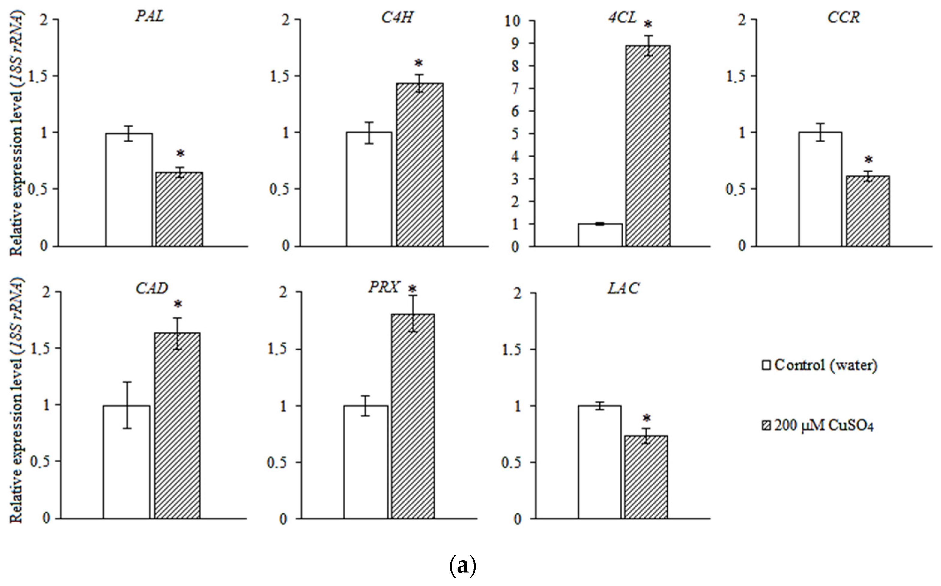
3.4. Expression of the Genes of Phenylpropanoid Metabolic Pathway and Lignin Biosynthesis

Under CuSO4 treatment, the relative number of transcripts of PAL and CCR genes decreased in the root, and the expression of the C4H, 4CL, and CAD genes increased (Figure 1a).
The relative quantity of transcripts of the C4H and CCR gene in the stem did not differ significantly both in the control and experimental plants, but transcripts of the PAL, 4CL, and CAD increased in number compared to the untreated plants (Figure 1b).
The expression level of the genes involved in lignin biosynthesis has changed under stress conditions. The relative number of transcripts of PRX gene increased by 1.8 times in the root, and by 2.5 times in the stem, compared to the control. The expression level of the LAC gene tended to decrease in both organs.

3.5. Anatomical and Morphological Characteristics of Zinnia Plants

The shoot growth of zinnia plants delayed under the excess copper in the medium (Figure 2a). The length of the main root decreased by 17.2% (Figure 2c), and the root transection area increased by 31.0% (Table 5) compared to the control. The stem of treated plants was shorter by 12.7% compared to the control (Figure 2c), and the hypocotyl and stem diameter decreased by 8.6% and 22.9%, respectively (Table 5). The proportion of the cortex in the transection area increased in the root, while in the hypocotyl and stem it changed insignificantly compared to the control (Figure 2b). In response to copper stress, the thickness of the metaxylem vessels cell walls increased by 7.2% in the mature zone of the root compared to the control (Table 5). In the hypocotyl and internodes, this characteristic did not change. The cross-sectional area of the metaxylem vessels decreased by 12.2% in the root, 18.6% in the hypocotyl, and 28.9% in the stem compared to the control (Table 5, Figure 3).

4. Discussion

Copper as both an essential element and a heavy metal revealed a complicated action on plants. In our study, the treatment of the growth substrate with 200 µM CuSO4 led to an increase in the available copper forms and its total amount. Since copper is a redox active metal, it induced oxidative stress through Fenton and Haber–Weiss reactions [2,3], which manifested in an increase in the levels of LPO and hydrogen peroxide (Table S1), more significant in the roots of zinnia than in the shoots, which was also shown in other species, for example, Helianthus annuus L. [5], Salvinia auriculata Aubl. [27], Oryza sativa L. [28], etc. The development of stress is also evidenced by the suppression of zinnia growth, which is a typical response to copper stress [5,27,29,30]. At the same time, leaf chlorosis did not appear in our experiment, as was shown in [2,5,6], which was probably due to a slight increase in the copper concentration in the shoots. The roots contacted directly with copper ions in the substrate; therefore, the toxic effects of HMs were more pronounced in them than in the above-ground organs. The root accumulated a greater amount of copper compared to the stem, thereby performing a barrier function. The same results were obtained on Arabidopsis thaliana L. and Oreganum vulgare L., treated with copper [29,30]. The translocation factor for copper was less than one, which makes it possible to attribute zinnia to copper excluders. The reason for the limitation of copper translocation from the root to the shoot could be increased root lignification. The deposition of lignin into the cell walls could be considered as a protective mechanism, which was enhanced by the production of hydrogen peroxide under excess copper in plant tissues [8,9]. Increased lignification of cell walls is a nonspecific plant response to HMs. Cu stimulated lignin biosynthesis in the roots of many plants: A. thaliana [29], Raphanus sativus L. [31], Glycine max L. [32], Panax ginseng C.A. Meyer [4], etc. Some authors also noted the higher level of lignification in the roots of plant varieties sensitive to copper. It is known that lignin amount and composition can change under different kinds of stress. In our experiment, both in the root and in the stem the amount of KL increased, while ASL did not change in the root, it decreased in the stem in experimental plants compared to the control. KL is based on H-, S-, and G-units of lignin, while S-units predominate in ASL [33], so our results demonstrate the qualitative changes in lignin under copper stress.
Phenolic compounds are known as the precursors of lignin biosynthesis and as antioxidants, which may be involved in the quenching of reactive oxygen species (ROS) [27,34]. In our study, the increase in the copper amount in the zinnia root was accompanied by a decrease in the amount of phenolics (Table S1), that could be explained by their use for lignin synthesis and ROS deactivation [27,35], which led to a decrease in their detectable amount. The drop of phenolics was shown in response to both short-term treatment with 0.01–10 mM copper ions [27] in S. auriculata and two-week treatment in Lycopersicon esculentum L., in the case of with 10 ppm, 20 ppm, and 50 ppm of Cu [34]. The changes in lignin were the result of a complex and time-coordinated regulation of the phenylpropanoid-pathway enzymes. We have found changes in 4CL and CAD transcription in the root as well as in 4CL, CAD, and PAL in the stem, in treated plants compared to the control. Therefore, the 4CL gene is responsible for the synthesis of G-units [36] and KL (Table S1). Our data on the increase in KL content in zinnia root in response to copper stress are likely due to this fact. An increase in the number of transcripts of the 4CL, CAD, and PAL genes was also shown in response to Al stress in Oryza sativa L. [37] and in Gossypium hirsutum L. under Cd stress [38].
At the final stages of lignin formation, its composition and amount depend on the activity of the III class peroxidases and laccases. It was shown that under both normal and stress conditions, lignin biosynthesis was stimulated by an increase in the activity of class III peroxidases, which use hydrogen peroxide as an electron acceptor and phenolics as a substrate [11,12,13]. In A. thaliana, the level of AtPRX62 gene transcripts in roots increased under the treatment with Cd2+ [39]. The induction of PRX7 and PRX8 gene expression was observed in the roots of Hordeum vulgare L., in response to 1 mM Cu2+. The excess transcription of these genes led to the inhibition of growth [40]. The POD gene, encoding anionic peroxidase, which is involved in lignification, increased in Paeonia ostii T. Hong and J.X. Zhang plants, cultivated with high Cu2+ concentrations [41]. In our study, the enhanced expression of the zinnia PRX gene under copper stress was also shown. It was shown that promoters of many genes contain cis-regulatory elements, which are associated with transcription factors, the work of which is modified by the H2O2 generated during the stress caused by HMs [42,43].
When plants are exposed to metal stress, the biosynthesis of phenylpropanoids and lignin is activated, resulting in thickening of the cell walls [9,44]. In zinnia root, we also found an increase in the cell walls’ thickness in the metaxylem vessels under copper stress; in the stem, it did not change. The thickening of cell walls and their lignification could provide the deposition of copper ions in the apoplast, limiting its translocation to the shoot [29,40].
Cell walls and root diameter both increased, and the root length decreased in the experimental zinnia plants treated with copper. The same effects were shown in A. thaliana [29] and O. vulgare [30]. The root cortex also thickened under stress. In maize treated with Cd, the same changes were shown [45]. The increase in cortical cell sizes led to the deposition of heavy-metal ions in vacuoles, limiting their entry into the stele [44,45]. Stress from HMs affected water uptake from the soil and, in turn, reduced the root water content [46]. Increased lignification can also limit the apoplast transport of water and minerals and cause their loading into the symplast [10,11,46]. Probably, the violation of mineral nutrition [47], the water regime [46], and the development of oxidative stress led to disorders in the development of vascular tissues in the zinnia stem, therefore, the number of xylem vessels in the vascular bundle decreased compared to the untreated plants. As a result, the radial growth of the hypocotyl and zinnia stem was limited.

5. Conclusions

Like most cultivated plants, zinnia could survive under excess of heavy metals, copper in particular, in the soil. One of the mechanisms that provide its tolerance to the moderate CuSO4 concentration is the increased lignification of axial organs. For the first time for zinnia, it was shown that this process is associated with the enhanced expression of several phenylpropanoid metabolic-pathway genes (4CL, CAD) and the PRX gene that participates in lignin biosynthesis. Another mechanism of zinnia adaptation to excess copper is the increase in the transcript amount of PAL, 4CL, and CAD genes that provide biosynthesis of phenolics, involved in the quenching of the reactive oxygen species in stress conditions. These changes allow zinnia to tolerate moderate copper stress.
Zinnia is an ornamental plant, characterized by rapid growth and abundant flowering. This species could be recommended for the reclamation of contamination by copper urban and industrial areas. The use of flowering plants will also increase the aesthetic value of the disturbed territories.

Supplementary Materials

The following supporting information is available online at https://www.mdpi.com/article/10.3390/horticulturae8060558/s1. Table S1: Spearman’s rank correlation coefficients between copper amount, biochemical traits, and relative gene expression.

Author Contributions

Conceptualization, A.T. and I.K.; methodology, A.T., A.E., H.W. and I.K.; software, A.T. and A.E.; validation, A.T. and A.E.; formal analysis, A.T. and H.W.; investigation, A.T.; resources, A.E., H.W. and I.K.; data curation, A.E. and I.K.; writing—original draft preparation, A.T.; writing—review and editing, A.T., A.E., M.M., G.B. and I.K.; visualization, A.T.; supervision, I.K.; project administration, A.E.; funding acquisition, A.T. and A.E. All authors have read and agreed to the published version of the manuscript.

Funding

This research was supported by the Russian Science Foundation, project no. 22-24-00817, https://rscf.ru/project/22-24-00817/, accessed date: 17 July 2022.

Institutional Review Board Statement

Not applicable.

Informed Consent Statement

Not applicable.

Data Availability Statement

Not applicable.

Conflicts of Interest

The authors declare no conflict of interest.

References

  1. Ballabio, C.; Panagos, P.; Lugato, E.; Huang, J.-H.; Orgiazzi, A.; Jones, A.; Fernández-Ugalde, O.; Borrelli, P.; Montanarella, L. Copper distribution in European topsoils: An assessment based on LUCAS soil survey. Sci. Total Environ. 2018, 636, 282–298. [Google Scholar] [CrossRef] [PubMed]
  2. Shabbir, Z.; Sardar, A.; Shabbir, A.; Abbas, G.; Shamshad, S.; Khalid, S.; Ghulam Murtaza, N.; Dumat, C.; Shahid, M. Copper uptake, essentiality, toxicity, detoxification and risk assessment in soil-plant environment. Chemosphere 2020, 259, 127436. [Google Scholar] [CrossRef]
  3. Adriano, D.C. Trace Elements in Terrestrial Environments Biogeochemistry, Bioavailability, and Risks of Metals; Springer: New York, NY, USA, 2001; 867p. [Google Scholar] [CrossRef]
  4. Ali, M.B.; Singh, N.; Shohael, A.M.; Hahn, E.J.; Paek, K.Y. Phenolics metabolism and lignin synthesis in root suspension cultures of Panax ginseng in response to copper stress. Plant Sci. 2006, 171, 147–154. [Google Scholar] [CrossRef]
  5. Jouili, H.; Ferjani, E. Changes in antioxidant and lignifying enzyme activities in sunflower roots (Helianthus annuus L.) stressed with copper excess. Comp. Rend. Biolog. 2003, 326, 639–644. [Google Scholar] [CrossRef]
  6. Elleuch, A.; Chaâbene, Z.; Grubb, D.C.; Drira, N.; Mejdoub, H.; Khemakhem, B. Morphological and biochemical behavior of fenugreek (Trigonella foenum-graecum) under copper stress. Ecotoxicol. Environ. Saf. 2013, 98, 46–53. [Google Scholar] [CrossRef]
  7. Aioub, A.A.A.; Zuo, Y.; Aioub, A.A.A.; Hu, Z. Biochemical and phytoremediation of Plantago major L. to protect tomato plants from the contamination of cypermethrin pesticide. Environ. Sci. Pollut. Res. 2021, 28, 43992–44001. [Google Scholar] [CrossRef] [PubMed]
  8. Loix, C.; Huybrechts, M.; Vangronsveld, J.; Gielen, M.; Keunen, E.; Cuypers, A. Reciprocal interactions between cadmium-induced cell wall responses and oxidative stress in plants. Front. Plant Sci. 2017, 8, 1867. [Google Scholar] [CrossRef] [PubMed] [Green Version]
  9. Bouazizi, H.; Jouili, H.; Geitmann, A.; Ferjani, E. Cell wall accumulation of Cu ions and modulation of lignifying enzymes in primary leaves of bean seedlings exposed to excess copper. Biol. Trace Elem. Res. 2010, 139, 97–107. [Google Scholar] [CrossRef]
  10. Tenhaken, R. Cell wall remodeling under abiotic stress. Front. Plant Sci. 2015, 5, 771. [Google Scholar] [CrossRef] [Green Version]
  11. Vaahtera, L.; Schulz, J.; Hamann, T. Cell wall integrity maintenance during plant development and interaction with the environment. Nat. Plants 2019, 5, 924–932. [Google Scholar] [CrossRef]
  12. Berthet, S.; Thevenin, J.; Baratiny, D.; Demont-Caulet, N.; Debeaujon, I.; Bidzinski, P.; Leple, J.C.; Huis, R.; Hawkins, S.; Gomez, L.D.; et al. Role of plant laccases in lignin polymerization. Adv. Bot. Res. 2012, 61, 145–172. [Google Scholar] [CrossRef]
  13. Jovanovic, S.V.; Kukavica, B.; Vidovic, M.; Morina, F.; Menckhoff, L. Class III peroxidases: Functions, localization and redox regulation of isoenzymes. In Antioxidants and Antioxidant Enzymes in Higher Plants; Gupta, D., Palma, J., Corpas, F., Eds.; Springer: Cham, Switzerland, 2018; pp. 269–300. [Google Scholar] [CrossRef]
  14. Fraser, C.M.; Chapple, C. The phenylpropanoid pathway in Arabidopsis. Arab. Book. 2011, 9, e0152. [Google Scholar] [CrossRef] [Green Version]
  15. Barros, J.; Serk, H.; Granlund, I.; Pesquet, E. The cell biology of lignification in higher plants. Ann. Bot. 2015, 115, 1053–1074. [Google Scholar] [CrossRef] [Green Version]
  16. Pesquet, E.; Zhang, B.; Andras Gorzsas, A.; Puhakainen, T.; Serk, H.; Escamez, S.; Barbier, O.; Gerber, L.; Courtois-Moreau, C.; Alatalo, E.; et al. Non-cell-autonomous postmortem lignification of tracheary elements in Zinnia elegans. Plant Cell 2013, 25, 1314–1328. [Google Scholar] [CrossRef] [Green Version]
  17. Ehsan, N.; Nawaz, R.; Ahmad, S.; Arshad, M.; Umair, M.; Sarmad, M. Remediation of heavy metal-contaminated soil by ornamental plant Zinnia (Zinnia elegance L.). Asian J. Chem. 2016, 28, 1338–1342. [Google Scholar] [CrossRef]
  18. Plotnikov, D.S.; Tugbaeva, A.S.; Ermoshin, A.A.; Kiseleva, I.S. Response reactions of Zinnia elegans seedlings to the impact of different copper ions concentrations. AIP Conf. Proc. 2021, 2388, 030034. [Google Scholar] [CrossRef]
  19. Roccotiello, E.; Marescotti, P.; Di Piazza, S.; Cecchi, G.; Mariotti, M.G.; Zotti, M. Biodiversity in Metal-Contaminated Sites—Problem and Perspective. In A Case Study, Biodiversity in Ecosystems—Linking Structure and Function; Blanco, J., Ed.; IntechOpen: London, UK, 2015; 1865p. [Google Scholar] [CrossRef] [Green Version]
  20. Bellincampi, D.; Dipierro, N.; Salvi, G.; Cervone, F.; De Lorenzo, G. Extracellular H2O2 induced by oligogalacturonics is not involved in the inhibition of the auxin-regulated rolB gene expression in tobacco leaf explants. Plant Physiol. 2000, 122, 1379–1385. [Google Scholar] [CrossRef] [Green Version]
  21. Uchiyama, M.; Mihara, M. Determination of malonaldehyde precursor in tissues by thiobarbituric acid test. Anal. Bioch. 1978, 86, 287–297. [Google Scholar] [CrossRef]
  22. Ermoshin, A.A.; Kiseleva, I.S.; Nsengiyumva, I.V.; Nsengiumva, D.S.; Duan, S.; Ma, C.; Kurchenko, V.P. Antioxidant activity and chemical composition of extracts from fruiting bodies of xylotrophic fungi growing on birch. J. Sib. Fed. Univ. Biol. 2021, 14, 339–353. [Google Scholar] [CrossRef]
  23. Bajpai, P. Pulp Bleaching. In Biermann’s Handbook of Pulp and Paper; Bajpai, P., Ed.; Elsevier: New York, NY, USA, 2018; pp. 465–491. [Google Scholar] [CrossRef]
  24. Kuluev, B.; Mikhaylova, E.; Ermoshin, A.; Veselova, S.; Tugbaeva, A.; Gumerova, G.; Gainullina, K.; Zaikina, E. The ARGOS-LIKE genes of Arabidopsis and tobacco as targets for improving plant productivity and stress tolerance. J. Plant Physiol. 2019, 242, 153033. [Google Scholar] [CrossRef]
  25. Livak, K.J.; Schmittgen, T.D. Analysis of relative gene expression data using real-time quantitative PCR and the 2−ΔΔCt method. Methods 2001, 25, 402–408. [Google Scholar] [CrossRef] [PubMed]
  26. Berlin, G.P.; Miksche, J.P. Botanical Microtechnique and Cytochemistry; Iowa State University Press: Ames, IA, USA, 1976; 321p. [Google Scholar]
  27. Bizzo, A.L.; Intorne, A.C.; Gomes, P.H.; Susuki, M.S.; Esteves, B.D. Short-term physiological responses to copper stress in Salvinia auriculata Aubl. Acta Limnol. Bras. 2014, 26, 268–277. [Google Scholar] [CrossRef] [Green Version]
  28. Thounaojam, T.C.; Panda, P.; Choudhury, S.; Patra, H.K.; Panda, S.K. Zinc ameliorates copper-induced oxidative stress in developing rice (Oryza sativa L.) seedlings. Protoplasma 2014, 251, 61–69. [Google Scholar] [CrossRef]
  29. Lequeux, H.; Hermans, C.; Lutts, S.; Verbruggen, N. Response to copper excess in Arabidopsis thaliana: Impact on the root system architecture, hormone distribution, lignin accumulation and mineral profile. Plant Physiol. Biochem. 2010, 48, 673–682. [Google Scholar] [CrossRef]
  30. Panou-Filotheou, H.; Bosabalidis, A.M. Root structural aspects associated with copper toxicity in oregano (Origanum vulgare subsp. hirtum). Plant Sci. 2004, 166, 1497–1504. [Google Scholar] [CrossRef]
  31. Chen, E.L.; Chen, Y.A.; Chen, L.M.; Liu, Z.H. Effect of copper on peroxidase activity and lignin content in Raphanus sativus. Plant Physiol. Biochem. 2002, 40, 439–444. [Google Scholar] [CrossRef]
  32. Lin, C.-C.; Chen, L.-M.; Liu, Z.-H. Rapid effect of copper on lignin biosynthesis in soybean roots. Plant Sci. 2005, 168, 855–861. [Google Scholar] [CrossRef]
  33. Yasuda, S.; Fukushima, K.; Kakehi, A. Formation and chemical structures of acid-soluble lignin I: Sulfuric acid treatment time and acid-soluble lignin content of hardwood. J. Wood Sci. 2001, 47, 69–72. [Google Scholar] [CrossRef]
  34. Michalak, M. Phenolic compounds and their antioxidant activity in plants growing under heavy metal stress. Pol. J. Environ. Stud. 2006, 15, 523–530. [Google Scholar]
  35. Kısa, D.; Öztürk, L.; Doker, S.; Gökçe, İ. Expression analysis of metallothioneins and mineral contents in tomato (Lycopersicon esculentum) under heavy metal stress. J. Sci. Food Agric. 2017, 97, 1916–1923. [Google Scholar] [CrossRef]
  36. Lee, D.; Meyer, K.; Chapple, C.; Douglas, C.J. Antisense suppression of 4-Coumarate: Coenzyme A Ligase activity in Arabidopsis leads to altered lignin subunit composition. Plant Cell 1997, 9, 1985–1998. [Google Scholar] [CrossRef] [Green Version]
  37. Mao, C.; Yi, K.; Yang, L.; Zheng, B.; Wu, Y.; Liu, F.; Wu, P. Identification of aluminium-regulated genes by cDNA-AFLP in rice (Oryza sativa L.): Aluminium-regulated genes for the metabolism of cell wall components. J. Exp. Bot. 2004, 55, 137–143. [Google Scholar] [CrossRef] [Green Version]
  38. Chen, H.; Li, Y.; Ma, X.; Guo, L.; He, Y.; Ren, Z.; Kuang, Z.; Zhang, X.; Zhang, Z. Analysis of potential strategies for cadmium stress tolerance revealed by transcriptome analysis of upland cotton. Sci. Rep. 2019, 9, 86. [Google Scholar] [CrossRef] [Green Version]
  39. Weber, M.; Trampczynska, A.; Clemens, S. Comparative transcriptome analysis of toxic metal responses in Arabidopsis thaliana and the Cd2+-hypertolerant facultative metallophyte Arabidopsis halleri. Plant Cell Environ. 2006, 29, 950–963. [Google Scholar] [CrossRef]
  40. Tamas, L.; Durcekova, K.; Huttova, J.; Mistrik, I. Expression of defence-related peroxidases Prx7 and Prx8 during abiotic stresses in barley roots. Acta Physiol. Plant. 2009, 31, 139. [Google Scholar] [CrossRef]
  41. Wang, Y.; Dong, C.; Xue, Z.; Jin, Q.; Xu, Y. De novo transcriptome sequencing and discovery of genes related to copper tolerance in Paeonia ostii. Gene 2016, 576, 126–135. [Google Scholar] [CrossRef]
  42. Li, G.-Z.; Zheng, Y.-X.; Chen, S.-J.; Liu, J.; Wang, P.-F.; Wang, Y.-H.; Guo, T.-C.; Kang, G.-Z. TaWRKY74 participates copper tolerance through regulation of TaGST1 expression and GSH content in wheat. Ecotoxicol. Environ. Saf. 2021, 221, 112469. [Google Scholar] [CrossRef]
  43. Gómez-Ros, L.V.; Gabaldón, C.; Núñez-Flores, M.J.L.; Gutiérrez, J.; Herrero, J.; Zapata, J.M.; Sottomayor, M.; Juan Cuello, J.; Barceló, A.R. The promoter region of the Zinnia elegans basic peroxidase isoenzyme gene contains cis-elements responsive to nitric oxide and hydrogen peroxide. Planta 2012, 236, 327–342. [Google Scholar] [CrossRef]
  44. Maksimovic, I.; Kastori, R.; Krstic, L.; Lukovic, J. Steady presence of cadmium and nickel affects root anatomy, accumulation and distribution of essential ions in maize seedlings. Biol. Plant. 2007, 51, 589–592. [Google Scholar] [CrossRef]
  45. Yadav, V.; Arif, N.; Kováč, J.; Singh, V.P.; Tripathi, D.K.; Chauhan, D.K.; Vaculík, M. Structural modifications of plant organs and tissues by metals and metalloids in the environment: A review. Plant Physiol. Biochem. 2021, 159, 100–112. [Google Scholar] [CrossRef]
  46. Tewari, R.K.; Yadav, N.; Gupta, R.; Kumar, R. Oxidative stress under macronutrient deficiency. J. Soil Sci. Plant Nutr. 2021, 21, 832–859. [Google Scholar] [CrossRef]
  47. Rucińska-Sobkowiak, R. Water relations in plants subjected to heavy metal stresses. Acta Physiol. Plant. 2016, 38, 257. [Google Scholar] [CrossRef] [Green Version]
Figure 1. Relative expression levels of phenylpropanoid biosynthesis pathway genes in: (a) the root and (b) the stem in zinnia on the 20th day of growth; 18S rRNA was used as the reference gene. Result is presented as mean ± standard error (n = 9). Asterisks represent significant differences (p < 0.05, U-test) from control.
Horticulturae 08 00558 g001aHorticulturae 08 00558 g001b
Figure 2. Morphological characteristics of zinnia plants: (a) zinnia plants on the 20th day of growth; (b) the ratio of cortex and stele in transverse sections of zinnia organs; (c) the length of main root and stem height (n = 30). Asterisks represent significant differences (p < 0.05, t-test) from control.
Horticulturae 08 00558 g002
Figure 3. Transverse sections of roots and stems in zinnia plants (Z. elegans) visualized by wide-field fluorescence (green autofluorescence of lignin). Bars = 100 µm; mxy, metaxylem; pxy, protoxylem.
Horticulturae 08 00558 g003
Table 1. Forward and reverse primers for qRT-PCR.
Gene,
GenBank
Access No.
Forward Primer Sequence (5′ → 3′)Reverse Primer Sequence
(5′ → 3′)
PAL
FM879196
GTCACCAGGCGAAGAGTTTGCGGAACACCATCCCATCCTT
C4H
FM880082
GAACTTTGAGCTGTTGCCGCTGAAAAACCCACAAACAACAATCC
4CL
AU294519
ACGTCACCTTCCGTTACACCCGTCAGCGATTATCGACGGT
CCR
FM881365
CCTCGGCTTCTGGTCGATACTGTATGGCTTTGCTCGTGGT
CAD
FM881026
CCGTAAACCATCCTCTTGCGCAAGCTTCCTCCCCACAATC
PRX
AB023959
TCGCAGCTTCAATGGTCAAACTCTCTTCTTCTTTCATACTTCCCTT
LAC
AU286008
AATAAGGACGGGTTGGGCTGAGGGTAAGGGATACCACGCT
18S rRNA
AB089282
ATGTGGTAGCCGTTTCTCAGGTGCCCGTTGCTGCGAT
Table 2. Copper amount in substrate and zinnia organs; BCF and TF on the 20th day of growth.
TreatmentCopper Amount,
µg g−1 Dry Substrate
Copper Amount,
µg g−1 DW
BCFTF
AvailableTotalRootStemRootStem
Control (water)n.d.7.75 ± 0.659.80 ± 0.5211.36 ± 0.70n.d.n.d.1.16 ± 0.07
200 µM CuSO41.12 ± 0.17 * 1167.63 ± 1.87 *26.32 ± 1.32 *12.04 ± 0.6923.5 ± 1.2 *10.75 ± 0.65 *0.46 ± 0.02 *
1 Result is presented as mean ± standard error (n = 5); n.d.—the copper amount below detection limit. Asterisks represent significant differences (p < 0.05, U-test).
Table 3. The concentration of hydrogen peroxide and MDA in zinnia organs on the 20th day of growth.
TreatmentH2O2, µmol g−1 FWMDA, µmol g−1 FW
RootStemRootStem
Control (water)29.5 ± 3.6 1120.5 ± 9.10.56 ± 0.030.31 ± 0.02
200 µM CuSO4159.61 ± 5.88 *61.50 ± 3.8 *0.67 ± 0.02 *0.48 ± 0.02 *
1 Result is presented as mean ± standard error (n = 15). Asterisks represent significant differences (p < 0.05, U-test) from control.
Table 4. The concentration of phenolic compounds and lignin (KL, ASL, and total lignin) in zinnia organs on the 20th day of growth.
TreatmentPhenolics, mg g−1 FWKL, %ASL, %Total Lignin, %
RootStemRootStemRootStemRootStem
Control (water)0.43 ± 0.01 10.61 ± 0.039.29 ± 0.088.02 ± 0.444.90 ± 0.236.43 ± 0.1114.19 ± 0.3114.19 ± 0.31
200 µM CuSO40.27 ± 0.01 *0.50 ± 0.01 *12.27 ± 0.20 *10.58 ± 0.56 *4.35 ± 0.424.47 ± 0.42 *16.62 ± 0.76 *15.05 ± 0.98
1 Result is presented as mean ± standard error (n = 15). Asterisks represent significant differences (p < 0.05, U-test) from control.
Table 5. Anatomy characteristics of zinnia organs on the 20th day of growth.
TreatmentCross-Sectional Diameter of
Organ, mm
Metaxylem Cell-Wall Thickness, µmCross-Sectional Area of Metaxylem Vessels, µm2
RootHypocotylStem
(1st Internode)
RootHypocotylStem
(1st Internode)
RootHypocotylStem
(1st Internode)
Control (water)1.74 ± 0.06 12.44 ± 0.052.57 ± 0.082.90 ± 0.062.62 ± 0.052.94 ± 0.0851.34 ± 1.4342.19 ± 2.1742.97 ± 1.59
200 µM CuSO42.24 ± 0.06 *2.23 ± 0.05 *1.98 ± 0.13 *3.11 ± 0.06 *2.60 ± 0.062.90 ± 0.0745.06 ± 1.88 *34.35 ± 1.41 *30.55 ± 0.59 *
1 Result is presented as mean ± standard error (n = 50). Asterisks represent significant differences (p < 0.05, t-test) from control.
Publisher’s Note: MDPI stays neutral with regard to jurisdictional claims in published maps and institutional affiliations.

