Article

Thidiazuron Induced In Vitro Clonal Propagation of Lagerstroemia speciosa (L.) Pers.—An Important Avenue Tree

1
Plant Biotechnology Laboratory, Department of Botany, Aligarh Muslim University, Aligarh 202 002, India
2
Department of Botany & Microbiology, College of Science, King Saud University, P.O. Box 2455, Riyadh 11451, Saudi Arabia
3
Department of Plant Pathology, University of Minnesota, Saint Paul, MN 55108, USA
*
Author to whom correspondence should be addressed.
Horticulturae 2022, 8(5), 359; https://doi.org/10.3390/horticulturae8050359
Submission received: 28 February 2022 / Revised: 11 April 2022 / Accepted: 18 April 2022 / Published: 20 April 2022
(This article belongs to the Special Issue Seed Germination and Micropropagation of Ornamental Plants)

Abstract

:
A high throughput regeneration protocol has been developed for Lagerstroemia speciosa through node explants under the regime of various plant growth regulators (PGRs). This protocol can provide an alternative mode to seed-grown plants and minimize the cost–time of regeneration, significantly. Murashige and Skoog (MS) medium containing various combinations of PGRs exhibited a marked stimulatory effect on morphogenesis. Of the various combinations tried, node explant pretreated with thidiazuron (TDZ; 5.0 µM) for 4 weeks and followed with transfer into MS medium containing 1.0 μM 6-benzyladenine (BA) and 0.25 μM α-naphthalene acetic acid (NAA) was reported to be the best treatment as it resulted in a maximum number of 24.5 shoots with an average shoot length of 7.1 cm per explant in 90% of cultures after 12 weeks of incubation. The in vitro-generated shoots rooted satisfactorily in the adopted ex vitro method of rooting, which saves time and cost. Among the different treatments, the greatest rooting percentage (85%) was observed in the 200 μM IBA-treated shoots, with the highest root number (8.7) and length (3.4 cm) occurring after 4 weeks. Four months after being transferred to ex vitro, some of the physiological attributes of the in vitro-propagated plants were examined and compared to the ex vitro plants. Further, analysis of the genetic integrity in tissue culture-raised plantlets along with the parental tree was accomplished through DNA-based RAPD technique. The monomorphic banding pattern obtained by the RAPD primers resulted in a high level of genetic uniformity in regenerated plants.

1. Introduction

Lagerstroemia speciosa (L.) Pers., commonly known as ‘Pride of India’ or ‘Banaba’, belongs to the family Lythraceae and is a tropical deciduous tree widely distributed in the Philippines, Malaysia, India, Vietnam, and China [1,2]. It is commonly planted in gardens for its aesthetic and ornamental value, with long-lasting gorgeous blossoms of various colors, and has been explored in current research for vital medicinal properties such as anti-diabetic [1,3], hyperuricemia [4], anti-obesity [5,6], anti-septic [7], and anti-cancer [8,9].
Due to its numerous applications in the pharmaceutical, paper, pulp, small-scale building, and wood industries [10,11], L. speciosa is in great demand and is continually overexploited. Although the multiplication of this species is accomplished through seeds, they have a short lifespan of viability (approximately 1–2 months); after this period germination is too difficult [12]. Due to its significant ethnomedicinal applications and unsustainable exploitation, the tree is on the verge of becoming extinct at both the national and international levels if appropriate conservation measures are not implemented immediately. Thus, it is important to provide a standardized micropropagation methodology for this multipurpose tree. This technique is currently used for multiplication of the planting stock of such types of tree species by several conversationalists [13,14]. In vitro clonal multiplication via axillary bud differentiation offers immense potential for the regeneration of healthy plantlets in a short period of time and space.
TDZ is an artificially modified phenyl urea (N-phenyl-N′-1, 2, 3-thidiazol-5-yl-urea) or non-purine cytokinin that has been commonly utilized in the in vitro regeneration of many tree species [13,15,16]. In general, the morphogenic response to TDZ is varied and depends on the concentration, exposure period, explant type, and genotype of the cells. It possesses the unique property of high efficacy at low concentrations with short exposure times, and it is less susceptible to enzymatic degradation in vivo than other naturally occurring aromatic cytokinins, and has been found to be most responsive at very low concentrations for the micropropagation of woody plants [17]. In plant tissues, it may exist in a variety of forms, including TDZ-free molecules, sequestered TDZ molecules, conjugated forms coupled to proteins or cell wall components, and TDZ-free molecules connected to cell wall components. Shoot regeneration was noted in other tree species on culture media with TDZ [18,19]. However, in many cases, a major problem is allied with micropropagated plantlets is the occurrence of somaclonal variation among the regenerants [20,21]. According to Fatima and Anis [22], somaclonal variation is a main disadvantage during the in vitro cultivation practice of any elite parental line. A number of scientific studies have also shown that it inhibited shoot elongation, created fasciated shoots (causing hyperhydricity), and induced other physiological abnormalities [23,24,25,26]. Therefore, it is important to evaluate the genetic fidelity of tissue culture-raised plantlets. Polymerase chain reaction (PCR)-based techniques, such as random amplified polymorphic DNA (RAPD), have been successfully used to investigate genetic diversity and clonal fidelity in several woody or perennial species such as Morus alba [21], Arbutus spp. [27], Canna indica [28], Calycophyllum spruceanum [29], and Paulownia [30]. Such study will provide a guarantee towards the in vitro propagation of normal, healthy, and true-to-type plantlets. Up to now, a few reports on in vitro regeneration in L. speciosa have been described using different explants [12,15] with limited success in terms of number of shoots per explant, and further efforts are needed to establish a practicable approach. To date there is no report available on in vitro regeneration using the highly active plant growth regulator (PGR) thidiazuron (TDZ). The major aim of the present study was to develop an efficient and reproducible in vitro regeneration and shoot multiplication protocol from nodal explant using thidiazuron with ex vitro rooting, acclimatization, assessment of physiological attributes like photosynthetic pigments, net photosynthetic rate, stomatal conductance, water use efficiency, transpiration rate, and genetic fidelity of regenerated plants using DNA-based RAPD markers.

2. Materials and Methods

2.1. Explants Source, Media and Culture Conditions

Newly sprouting shoots of L. speciosa were collected from an approximately 20-year-old mature tree growing in the Department of Botany, AMU, Aligarh, India (27°54′52.0″ N 78°04′21.8″ E). The harvested sprouting shoots were washed under running tap water for up to 10 min and then treated with fungicide (1% w/v Bavistin™, Mumbai, India) and again rinsed with running tap water for 15 min. Thereafter, they were treated with laboratory detergent (5% v/v Labolene™, Qualigens, Mumbai, India) for 8 min followed by being rinsed 5 to 6 times with autoclaved distilled water. Surface disinfection took place using 0.1% HgCl2 (w/v) for 5 min in aseptic conditions followed by thorough washing with sterile distilled water to remove the disinfectant. Sterile node explants (0.5–1.0 cm) excised under laminar flow hood were used as the starting materials in all the experiments. Murashige and Skoog [31] (MS) medium (Himedia®, Mumbai, India) was used as a nutrient–substrate in all the culture experimental setups with exogenously applied sucrose (3%, w/v) (Qualigens, Mumbai, India) and agar (0.8%, w/v) (Qualigens, Mumbai, India). The pH was adjusted (5.75 to 5.85) before autoclaving at 121 °C for 20 min. The cultures were maintained at 24 ± 2 °C during a 16 h light photoperiod with a light intensity of 50–150 µmol m−2 s−1 photon flux density provided by cool white fluorescent lamps. Relative humidity was adjusted up to 55 ± 5% and regularly monitored by thermo-hygrometer (Testo, India Pvt. Ltd., Pune, India). The sub-culturing was carried out by transfer of the cultures onto fresh medium after every 3–4 week of incubation. Data were collected on the percent response, shoot numbers, and shoot lengths per explant, after 4, 8, and 12 weeks of culture incubation.

2.2. Shoot Induction

The induction of multiple shoots from node explants was carried out in two different sets of experiments. In the first phase, node explants were grown for four weeks in a broad range of TDZ concentrations (1.0, 5.0, 10.0, 15.0, 20.0 µM) supplemented MS medium to determine the optimal concentration at which maximal bud induction could be achieved. In order to determine the optimal TDZ dosage, the shoot formation was carefully monitored in each tube. In the second step, based on the results of the experiment, the best TDZ dose among those listed in Table 1 was refined at a narrow level (3.5, 4.0, 4.5, 5.0, 5.5, 6.0, and 6.5 µM) for 4 weeks in a separate experiment to determine the optimal concentration of TDZ for further shoot bud induction from node explants.

2.3. Shoot Multiplication

After four weeks of bud induction period on culture medium with TDZ, responsive explants were transferred to MS culture medium (in glass tubes of 25 × 150 mm) supplemented with various cytokinins, such as 6-benzyladenine (BA), kinetin (Kin), and 2-isopentenyl adenine (2iP), each at a concentration of 0.25, 0.5, 1.0, 2.0, or 4.0 µM either singly or in combination with auxins, which included indole-3-acetic acid (IAA), indole-3-butyric acid (IBA), and α-naphthalene acetic acid (NAA) at three different concentrations (0.10, 0.25, and 0.50 µM) for shoot multiplication.

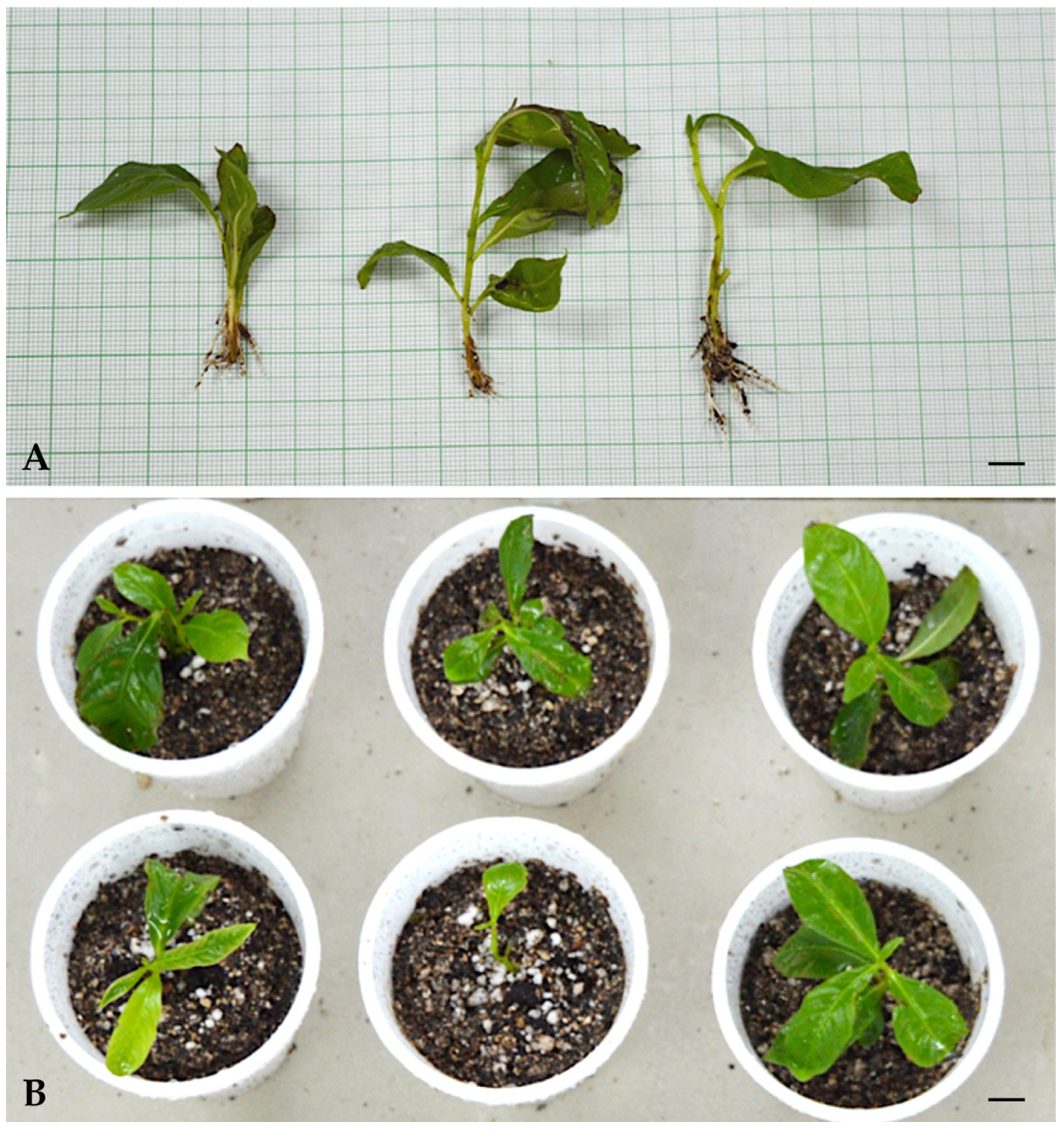
2.4. Ex Vitro Rooting and Acclimatization

Healthy in vitro-regenerated shoots of about 4–5 cm in length with 3–5 fully expended leaves were selected for the rooting experiment. The basal cut end of the shoots was dipped in different concentrations (200, 400, 800, or 1000 µM) of various auxins (IBA, IAA, or NAA) for about 30 min. The treated shoots were washed thrice with sterile distilled water and transplanted into plastic/thermocol cups full of a sterile potting mixture called Soilrite™ (Keltech Pvt. Ltd., Bangalore, India), and these cups were covered with transparent polybags to ensure high humidity up to a couple of weeks. The plantlets were irrigated with quarter-strength MS liquid medium without organic supplements at every alternate day for 2 weeks. The polybags were removed after each second day with increasing time period for the purpose of hardening. The percentage of rooting response, root numbers per shoot, and root length was noted after 4 weeks of rooting culture. Furthermore, these semi-acclimatized plantlets were again transplanted into a mixture of vermiculated garden soil with Soilrite (1:1) for the next 2 weeks. Thereafter, the healthy, acclimatized plants were transplanted on garden soil for the next 2 weeks under the same conditions. The acclimatized plants were transferred to greenhouse conditions for another couple of weeks, and, finally, they were successfully established in natural conditions.

2.5. Growth and Photosynthetic Traits

Plant height (in centimeters) and the number of branches on each plant were measured for five randomly selected micropropagated plants and ex vitro plants that were maintained in a greenhouse. At the same time, leaves from each plant were taken and analyzed spectrophotometrically to estimate the chlorophyll a and b concentrations, as well as the carotenoids content, using 80% acetone following the procedure described by Lichtenthaler and Wellburn [32]. The absorbance of the samples was measured at 645, 663, and 440 nm using a UV spectrophotometer for the determination of chlorophyll a, b, and carotenoid contents. Photosynthetic traits, i.e., net photosynthetic rate (Pn), stomatal conductance (gs), internal CO2 (Ci), and transpiration rate (E), were measured on the fully expanded leaves of the in vitro propagated plants and ex vitro plants with a portable photosynthetic system (LI-COR 6200, Lincoln, NE, USA) on a sunny day between 10 a.m. and 12 p.m. The measurement was repeated thrice. Water use efficiency (WUE) was calculated using the data of photosynthetic rate and stomatal conductance [33]. Five samples were used for each measurement, which was repeated twice.

2.6. Genomic DNA Isolation and Molecular Screening

Screening of the genetic fidelity in micropropagated plants was achieved using a PCR-based RAPD technique. Genomic DNA was extracted from young leaf tissues of the mother plant and 10 randomly selected 3-month-old tissue culture-raised plants using a slightly modified cetyl-trimethyl ammonium bromide (CTAB) technique established by Doyle and Doyle [34]. The quantification of genomic DNA was accomplished by Nanodrop Spectrophotometer (Implen, München, Germany). The preliminary screenings of the DNA samples were done by 20 RAPD primers (Operon Kit B primer). Of these, 11 primers were chosen based on their unambiguous, reproducible, and clear banding pattern and were employed for final screening. Polymerase chain reactions (PCR) for RAPD primers were completed in a thermocycler machine (Biometra, Göttingen, Germany). About 25 µL of DNA amplification reaction mixture consisted of various major components including 10× (NH4)2SO4 buffer, MgCl2 (25 mM), dNTPs (10 mM), primers (10 µM), Taq pol (2 Unit), and 50 ng/µL DNA template. The program of DNA amplification initially started with a denaturation step at 94 °C for 5 min followed by 38 repeated cycles of denaturation at 94 °C (1 min), annealing at 45–60 °C (1 min), and elongation at 72 °C (2 min), and final extension of DNA was carried out at 72 °C (8 min). The PCR assays were performed twice in order to eliminate the possibility of false positive results and to confirm the reproducibility of the RAPD markers. These amplified DNA fragments were separated by electrophoresis technique in agarose (1.2%, v/w) gel containing 4 µL ethidium bromide in TAE buffer run at 65 V (110 min) and imaged on a UV transilluminator (Bio Rad, Hercules, CA, USA). Thereafter, clear and well-distinct and reproducible bands were scored as present (1) or absent (0) for RAPD primers in each DNA sample. The relative size of each amplified DNA fragment was compared with 1 kb DNA ladder (Gene Ruler™ 1 kb DNA ladder, Thermo Scientific, Waltham, MA, USA). The amplification of each RAPD primer was repeated thrice to confirm the reproducibility pattern.

2.7. Statistical Analysis

All research experiments were followed on a completely randomized block design. Each treatment was repeated three times with 10 replicates. The data on different parameters were subjected one-way analysis of variance (ANOVA) using SPSS ver. 16 (SPSS Inc., Chicago, IL, USA). The significance of variances among mean values was calculated using Duncan’s multiple range tests at p = 0.05. The results of each treatment are represented as the mean ± SE (standard error) in 3 repeated experiments.

3. Results and Discussion

3.1. Shoot Induction

A node explant procured from a mature tree produced a single shoot when cultured on a PGR-free medium even after 4 weeks of incubation. However, a response in bud induction was recorded within 4 weeks when the node explants were grown in TDZ-fortified culture medium. The TDZ treatment on node explants significantly improved the rate of shoot bud induction. Among different concentrations, the optimal medium for the highest shoot bud induction was recorded as MS medium containing 5.0 μM TDZ (Figure 1A). At this level, a maximum of 75% shoot induction, a highest number of 14.5 shoots per node explant and a mean shoot length of 2.1 cm were recorded within 4 weeks of incubation (Table 1 and Table 2). However, it was noted that an increased dose of TDZ inhibited the elongation of shoots when compared to a lower dose of TDZ individually after a period of 4 weeks of culture. The MS medium enriched with 3.5 μM TDZ exhibited 6.2 shoots per explant with a mean shoot length of 2.8 cm after 4 weeks of culture (Table 2). The bud induction response declined with increased concentrations of TDZ beyond the optimal dose resulted in small, condensed shoots and most of them showed bunching of shoots with no further shoot growth even after 4 weeks of culture. According to Liu et al. [35], shoot multiplication and growth are varying in response to TDZ exposure because TDZ is a more biologically active PGR than others. However, Matand and Prakash [36] suggested that TDZ-pretreated explants subsequently transferred to secondary medium or TDZ-free medium exhibited better response in stimulations of shoot multiplication across a large number of plants species. Murch and Saxena [37] suggested that TDZ may exist in different forms such as free molecules of TDZ, sequestrated molecules of TDZ, conjugated molecules with proteins, or cell wall in TDZ-pretreated cultures. Overall, these studies indicated that TDZ molecules promotes a high rate of shoot bud induction by stimulating cell division and multiplication in apical meristem while also reprograming cells to the appropriate developmental stage for shoot bud differentiation. In many plant species, long duration exposure and high dose TDZ pretreatment exhibited morphological abnormalities such as hyper-hydricity and suppressed shoot growth [26,38,39]. Similarly, in the present study, the shoot numbers per node explant were quite high in TDZ-pretreated samples, but their shoot elongation was not obvious even after 4 weeks of incubation (Table 1). Hence, it was clear that TDZ exhibited inhibitory results on the elongation and growth of shoots. However, it shows a promoting effect on shoot bud induction from node explants. Similar kinds of TDZ effects have also already been documented in a large number of trees [13,40,41]. Although their studies suggested such problems could be overcome via a two-fold culture approach. Similarly, in the present study, when TDZ-pretreated node explants were subsequently transferred to a secondary medium (or one without TDZ) containing another cytokinin alone or in combination with auxins, it successfully improved L. speciosa cultures’ protocol. This type of strategy using primary medium (for shoot bud induction) and secondary medium (for shoot elongation) was successfully applied in several tree species [26,42,43].

3.2. Shoot Multiplication

The TDZ-pretreated node explants were sub-cultured onto secondary MS medium containing different concentrations of cytokinins (Table 3) for 8 weeks. A maximum of 18.6 shoots per node explant and a subsequent shoot length of 5.8 cm was recorded in 80% of cultures containing 1.0 µM BA, whereas, pretreated node explants sub-cultured on hormone-free MS medium (or control medium) exhibited a remarkable declination in shoot multiplication (12.0) and elongation (2.4 cm) per node explant. Similarly, the positive response of BA over other cytokinins (Kin and 2iP) on shoot elongation and multiplication has been already documented in various plants [22,44]. Meanwhile, the subsequent transfer of TDZ-pretreated node explants onto a secondary medium having an optimal dose of BA exhibited a better effect on the rate of shoot multiplication and their subsequent elongation. This means that TDZ exposure at appropriate concentration is sufficient to induce better shoot multiplication rates in pretreated node explants. The possible reason for better shoot growth in TDZ-pretreated node cultures on secondary medium augmented with an optimum dose of BA may be its capability of changing to an active form of cytokinin from non-active storage forms at a later stage of culture through the modulation of the endogenous PGRs level, either directly or as a result of induced stress [15]. Similar results have been reported by different researchers in several woody plant species [16,45]. Furthermore, we examined the combined effect of an optimum dose of BA (1.0 µM) with auxins (IAA, IBA and NAA) at different combinations for improving the rate of shoot multiplication as well as proliferation. Among different cytokinin–auxin combinations, the highest shoot numbers (24.5) with an average shoot length of 7.1 cm per node explant were noted on 1.0 µM BA and 0.25 µM NAA supplemented MS medium in 90% node cultures after 12 weeks (Table 4; Figure 1B). Whereas 1.0 µM BA and 0.50 µM IBA yielded lesser shoot numbers (16.9) per node explant with a mean shoot length of 4.7 cm in 75% cultures after 12 weeks. Interestingly, in the present study, the rates of shoot multiplication and proliferation were positively correlated with a combination of cytokinin–auxin concentrations.
However, the growth of shoots was inhibited when the levels of auxin were increased beyond the 0.25 µM in the culture medium. Besides this, after 2 weeks of culture, the new shoots emerged directly from the axillary node of node explants and were further sub-cultured onto the same fresh medium for further shoot growth. In the present study, these positive results (in vitro shoot proliferation from nodal explants of L. speciosa may be due to a suitable combination of cytokinin–auxin in a synergistic, additive, or antagonistic manner to get an effective proliferation response on post-TDZ pretreated cultures) are in agreement with several earlier reports such as Husain and Anis [41] in Melia azedarach, Siddique and Anis [46] in Balanites aegyptiaca, Javed, et al. [47] in Acacia ehrenbergiana, Ahmad et al. (2018) in Pterocarpus marsupium, and in Tecoma stans Hussain, Ahmad, Anis, and Hakeem [45].

3.3. Ex Vitro Rooting and Acclimatization

The efficacy of the established micropropagation protocols is determined by the proportion of rooting and acclimation success with the transfer of regenerants under natural environmental conditions. The ex vitro rooting approach has been proven to be the most efficient among the numerous methods of rooting in isolated shootlets from culture and may be utilized as an alternative method [25,48]. Among the several treatments used, 200 µM (IBA) was shown to be the most successful in promoting rooting, with 85% of shoots rooted effectively with a mean number of 8.7 and length of 3.4 cm per shootlet after 4 weeks (Table 5, Figure 2A). The applications of exogenous auxins, namely IBA, IAA, and NAA, in culture medium are commonly utilized for in vitro rooting in micropropagated shootlets.
It has also been reported that IBA showed superior effect over other auxins on adventitious roots formation in a large number of plants. According to Elmongy et al. [49], IBA is an important plant hormone for in vitro rhizogenesis, because it shows more stability towards photo-degradation, adherence to shootlets, and, most importantly, activates the meristematic region of pericycle. Furthermore, IBA shows an active role in the formation of adventitious roots and acts as a signaling molecule that controls gene expression and the root development process. Similar effects of IBA treatment on in vitro root formation have been established in many scientific reports [41,47,50]. By combining rooting and hardening, the ex vitro approach not only aids in the creation of appropriate root systems without basal callusing, but also aids in the removal of one extra stage of in vitro rooting. The efficiency of IBA in comparison to all other auxins has previously been documented in Vitex negundo [51]. After 4 weeks, well-developed rooted juvenile plants were transplanted to sterilized Soilrite for a couple of weeks and artificial light irradiated with 50 µmol m−2 s−1 PPFD. Thereafter, these juvenile plants were transplanted into a mixture of Soilrite–vermiculated garden soil (1:1) for the next couple of weeks under 100 µmol m−2 s−1 PPFD light irradiance in order to harden. Subsequently, the acclimatized plants were shifted to normal garden soil for the next couple of weeks under 150 µmol m−2 s−1 PPFD light irradiance (Figure 2B). The constructive effect of Soilrite on the acclimatization process during the early stages is because it is a spongy material, meaning it holds more nutrients along with water content, and thus it supports the development of adventitious tender roots. Also, the gradual increment of light intensity helps with the hardening process of juvenile plants during acclimatization. Later, these juvenile plantlets were further nourished under greenhouse conditions for the next couple of weeks. Finally, the 8-week-old acclimatized plants were transplanted under field conditions where they grew normally with a 93% of survival rate.

3.4. Growth and Photosynthetic Traits

Four months after being transferred to ex vitro growth, the physiological traits of the in vitro-propagated plants were examined and compared to ex vitro plants maintained in the greenhouse. Table 6 shows the comparative data on plant height, branch number, chlorophylls, carotenoids, photosynthetic rate, stomatal conductance, water use efficiency, and transpiration rate. When comparing ex vitro plants to micropropagated plants, the height of the plants and the number of branches on each plant were both slightly higher in ex vitro plants. In contrast, the net photosynthetic rate, chlorophyll a, and chlorophyll b concentration were found to be very similar in both plants, while the carotenoids content was found to be higher in in vitro plants. The results are in agreement with the findings in Tylophora indica [52] and Clitoria ternatea [53]. The stomatal conductance of in vitro-regenerated plants was found to be lower than that of ex vitro plants. In vitro plants, on the other hand, showed a substantial improvement in water use efficiency compared to ex vitro plants. Meanwhile, ex vitro plants transpired at a higher rate than micropropagated plants.

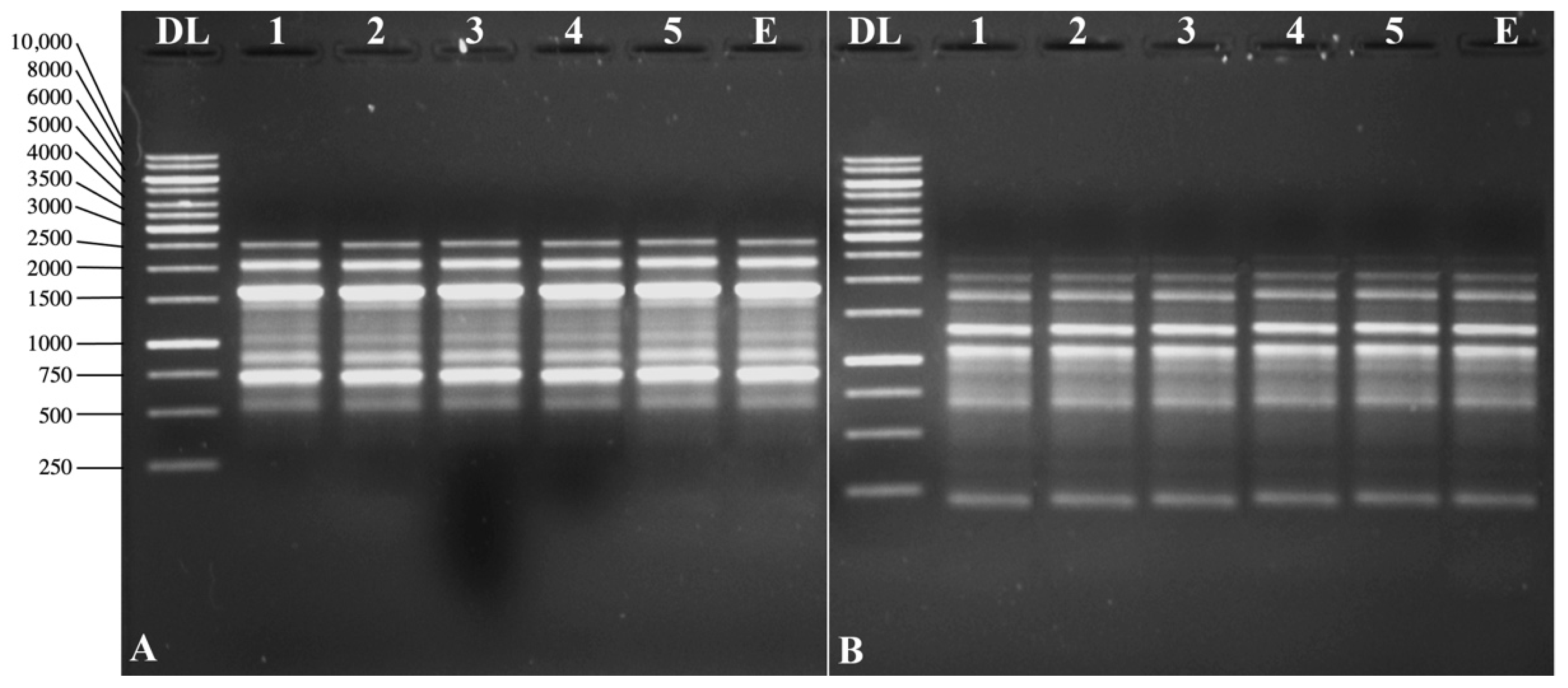
3.5. Molecular Screening

The plants raised from node explants of L. speciosa through the micropropagation technique were assessed for their clonal fidelity by DNA-based RAPD primers. Out of 20 IRAPD primers of Kit-B, 11 were selected because of their unambiguous, reproducible, and clear banding pattern results to evaluate the genetic fidelity in nine randomly selected micropropagated plantlets along with the mother plant. Monomorphic RAPD banding pattern exhibited a total of 70 DNA bands with an average of 6.3 DNA fragments per primer, indicating homogeneity among regenerants with the donor tree (Table 7). Meanwhile, no polymorphism was detected, revealing the complete genetic fidelity among the regenerated clones. The monomorphic banding patterns among regenerated plants were amplified by OPB1 and OPB10 and are depicted in Figure 3A,B.
Larkin and Scowcroft [54] reported that culture conditions during in vitro propagation practice are stressful and may sometimes exhibit genetic variations in the tissue cultured plants, known as somaclonal variations. The occurrence of somaclonal variations is more frequent when the in vitro cultures are exposed to stress. Thus, it is important to assess genetic integrity among micropropagated plantlets through DNA-based molecular markers. Such kinds of study provide a guarantee towards the regeneration of true-to-type plantlets. Generally, DNA-based molecular markers are not affected by any kind of environmental factors [55]. Although RAPD markers have become a more popular DNA-based molecular technique as they do not require any prior information on DNA sequences. Therefore, in the present study, RAPD primers exhibited a monomorphic DNA-banding pattern suggesting high-level genetic integrity among micropropagated plants. Consequently, the mode of clonal propagation using TDZ was appropriate for gaining true-to-type plantlets. There are a number of studies confirming the genetic fidelity of regenerated plants using DNA-based RAPD makers when cytokinin and auxin is optimized for micropropagation via axillary shoot proliferation in several plant species, such as Terminalia bellerica [20], Morus alba [21], Withania somnifera [25], Rauvolfia tetraphylla [56], Ruta chalepensis [57], Vitex negundo [58], Zanthoxylum armatum [59] Carnation [60], and Humulus lupulus [61].

4. Conclusions

The present work describes a reproducible and efficient in vitro protocol useful for scaling up the propagation of L. speciosa using mature nodal explants. Different concentrations of TDZ were optimized, and it was found that 5.0 µM TDZ is the most effective concentration for the induction of shoot buds from node explants. However, these buds showed stunted shoots and did not grow further. To overcome this problem, 4 weeks of TDZ exposure is optimum followed by their transfer to a secondary medium for maximum shoot elongation and multiplication. It is concluded that a two-step procedure is necessary for clonal propagation. Ex vitro rooting helps in the reduction of time period as it combines both the additional steps of in vitro rooting and acclimatization. From the physiological and molecular data, it can be postulated that the regenerated progeny is true to type. Thus, the developed clonal propagation system through node explant of L. speciosa will not only help in low-cost multiplication but also their conservation and commercial propagation.

Author Contributions

Conceptualization, N.A. and M.F.; methodology, N.A., M.F., A.A. (Anees Ahmad), A.A.A., A.A.Q. and A.A. (Anshu Alok); validation, N.A., M.F. and A.A. (Anees Ahmad); formal analysis, N.A., M.F., A.A. (Anees Ahmad) and A.A.Q.; investigation, N.A., M.F., A.A. (Anees Ahmad), A.A.A., A.A.Q. and A.A. (Anshu Alok); resources, N.A. and M.F.; writing—original draft preparation N.A. and M.F.; writing—review and editing, N.A., M.F., A.A. (Anees Ahmad), A.A.A., A.A.Q. and A.A. (Anshu Alok); supervision, M.F. and A.A.A.; project administration, A.A.A.; funding acquisition, M.F. and A.A.A. All authors have read and agreed to the published version of the manuscript.

Funding

This study was supported by the Researchers Supporting Project (RSP-2021/86), King Saud University, Riyadh, Saudi Arabia.

Institutional Review Board Statement

Not applicable.

Informed Consent Statement

Not applicable.

Data Availability Statement

Not applicable.

Acknowledgments

The authors are thankful to the Researchers Supporting Project (RSP-2021/86), King Saud University, Riyadh, Saudi Arabia for financial support.

Conflicts of Interest

There are no conflict of interest.

References

  1. Klein, G.; Kim, J.; Himmeldirk, K.; Cao, Y.; Chen, X. Antidiabetes and Anti-obesity Activity of Lagerstroemia speciosa. Evid.-Based Complement. Altern. Med. 2007, 4, 401–407. [Google Scholar] [CrossRef] [PubMed] [Green Version]
  2. Rahman, F.B.; Ahmed, S.; Noor, P.; Rahman, M.M.M.; Huq, S.M.A.; Akib, M.T.E.; Shohael, A.M. A comprehensive multi-directional exploration of phytochemicals and bioactivities of flower extracts from Delonix regia (Bojer ex Hook.) Raf., Cassia fistula L. and Lagerstroemia speciosa L. Biochem. Biophys. Rep. 2020, 24, 100805. [Google Scholar] [CrossRef] [PubMed]
  3. Kim, K.-H.; Roh, S.-G.; Li, C.-R.; Jin, C.-F.; Kim, A.; Choi, W.-C. Anti-diabetic effects of banaba leaf extracts (Lagerstroemia speciosa Pers.) through solvents. J. Life Sci. 2008, 18, 1305–1311. [Google Scholar] [CrossRef]
  4. Unno, T.; Sugimoto, A.; Kakuda, T. Xanthine oxidase inhibitors from the leaves of Lagerstroemia speciosa (L.) Pers. J. Ethnopharmacol. 2004, 93, 391–395. [Google Scholar] [CrossRef] [PubMed]
  5. Suzuki, Y.; Unno, T.; Ushitani, M.; Hayashi, K.; Kakuda, T. Antiobesity Activity of Extracts from Lagerstroemia speciosa L. Leaves on Female KK-Ay Mice. J. Nutrf. Sci. Vitaminol. 1999, 45, 791–795. [Google Scholar] [CrossRef]
  6. Karsono, A.H.; Tandrasasmita, O.M.; Tjandrawinata, R.R. Bioactive fraction from Lagerstroemia speciosa leaves (DLBS3733) reduces fat droplet by inhibiting adipogenesis and lipogenesis. J. Exp. Pharm. 2019, 11, 39–51. [Google Scholar] [CrossRef] [Green Version]
  7. Garo, E.; Eldridge, G.R.; Goering, M.G.; DeLancey Pulcini, E.; Hamilton, M.A.; Costerton, J.W.; James, G.A. Asiatic acid and corosolic acid enhance the susceptibility of Pseudomonas aeruginosa biofilms to tobramycin. Antimicrob. Agents Chemother. 2007, 51, 1813–1817. [Google Scholar] [CrossRef] [Green Version]
  8. Thakur, R.S.; Devaraj, E. Lagerstroemia speciosa (L.) Pers. triggers oxidative stress mediated apoptosis via intrinsic mitochondrial pathway in HepG2 cells. Environ. Toxicol. 2020, 35, 1225–1233. [Google Scholar] [CrossRef]
  9. Rohit Singh, T.; Ezhilarasan, D. Ethanolic Extract of Lagerstroemia Speciosa (L.) Pers., Induces Apoptosis and Cell Cycle Arrest in HepG2 Cells. Nutr. Cancer 2020, 72, 146–156. [Google Scholar] [CrossRef]
  10. Zobayed, S.M.A. In vitro propagation of Lagerstroemia spp. from nodal explants and gaseous composition in the culture headspace. Environ. Control Biol. 2000, 38, 1–11. [Google Scholar] [CrossRef]
  11. Jayakumar, K.S.; Sajan, J.S.; Aswati Nair, R.; Padmesh Pillai, P.; Deepu, S.; Padmaja, R.; Agarwal, A.; Pandurangan, A.G. Corosolic acid content and SSR markers in Lagerstroemia speciosa (L.) Pers.: A comparative analysis among populations across the Southern Western Ghats of India. Phytochemistry 2014, 106, 94–103. [Google Scholar] [CrossRef] [PubMed]
  12. Hadiuzzaman, S.; Zobayed, S.; Roy, S.; Miah, M. In vitro cloning from seedling explants of Lagerstroemia speciosa Pers. and L. thorellii Gagnep. Bangladesh J. Bot. 1992, 21, 59–64. (In Bengali) [Google Scholar]
  13. Ahmad, A.; Ahmad, N.; Anis, M.; Alatar, A.A.; Abdel-Salam, E.M.; Qahtan, A.A.; Faisal, M. Gibberellic acid and thidiazuron promote micropropagation of an endangered woody tree (Pterocarpus marsupium Roxb.) using in vitro seedlings. Plant Cell Tissue Organ Cult. (PCTOC) 2021, 144, 449–462. [Google Scholar] [CrossRef]
  14. Faisal, M.; Alatar, A.A.; Abdel-Salam, E.M.; Qahtan, A.A. Effects of 4-CPPU on in vitro multiplication and sustainable generation of Hibiscus rosa-sinensis L. ‘White Butterfly’. Saudi J. Biol. Sci. 2020, 27, 412–416. [Google Scholar] [CrossRef]
  15. Huetteman, C.A.; Preece, J.E. Thidiazuron: A potent cytokinin for woody plant tissue culture. Plant Cell Tissue Organ Cult. 1993, 33, 105–119. [Google Scholar] [CrossRef]
  16. Javed, S.B.; Alatar, A.A.; Anis, M.; El-Sheikh, M.A. In vitro regeneration of coral tree from three different explants using thidiazuron. HortTechnology 2019, 29, 946–951. [Google Scholar] [CrossRef] [Green Version]
  17. Lu, C.-Y. The use of thidiazuron in tissue culture. Vitr. Cell. Dev. Biol.-Plant 1993, 29, 92–96. [Google Scholar] [CrossRef]
  18. Corredoira, E.; Ballester, A.; Vieitez, A.M. Thidiazuron-induced high-frequency plant regeneration from leaf explants of Paulownia tomentosa mature trees. Plant Cell Tissue Organ Cult. 2008, 95, 197–208. [Google Scholar] [CrossRef] [Green Version]
  19. Feng, J.-C.; Yu, X.M.; Shang, X.L.; Li, J.D.; Wu, Y.X. Factors influencing efficiency of shoot regeneration in Ziziphus jujuba Mill. ‘Huizao’. Plant Cell Tissue Organ Cult. (PCTOC) 2010, 101, 111–117. [Google Scholar] [CrossRef]
  20. Dangi, B.; Khurana-Kaul, V.; Kothari, S.L.; Kachhwaha, S. Micropropagtion of Terminalia bellerica from nodal explants of mature tree and assessment of genetic fidelity using ISSR and RAPD markers. Physiol. Mol. Biol. Plants 2014, 20, 509–516. [Google Scholar] [CrossRef] [Green Version]
  21. Saha, S.; Adhikari, S.; Dey, T.; Ghosh, P. RAPD and ISSR based evaluation of genetic stability of micropropagated plantlets of Morus alba L. variety S-1. Meta Gene 2015, 7, 7–15. [Google Scholar] [CrossRef] [PubMed]
  22. Fatima, N.; Anis, M. Role of growth regulators on in vitro regeneration and histological analysis in Indian ginseng (Withania somnifera L.) Dunal. Physiol. Mol. Biol. Plants 2012, 18, 59–67. [Google Scholar] [CrossRef] [PubMed] [Green Version]
  23. Papafotiou, M.; Bertsouklis, K.F.; Trigka, M. Micropropagation of Arbutus unedo, A. andrachne, and their natural hybrid, A. x andrachnoides from seedling explants. J. Hortic. Sci. Biotechnol. 2013, 88, 768–775. [Google Scholar] [CrossRef]
  24. Husain, M.K.; Anis, M.; Shahzad, A. In Vitro Propagation of Indian Kino (Pterocarpus marsupium Roxb.) Using Thidiazuron. Vitr. Cell. Dev. Biol.-Plant 2007, 43, 59–64. [Google Scholar] [CrossRef]
  25. Fatima, N.; Ahmad, N.; Ahmad, I.; Anis, M. Interactive effects of growth regulators, carbon sources, pH on plant regeneration and assessment of genetic fidelity using single primer amplification reaction (SPARS) techniques in Withania somnifera L. Appl. Biochem. Biotechnol. 2015, 177, 118–136. [Google Scholar] [CrossRef]
  26. Ahmad, A.; Ahmad, N.; Anis, M. Preconditioning of Nodal Explants in Thidiazuron-Supplemented Liquid Media Improves Shoot Multiplication in Pterocarpus marsupium (Roxb.). In Thidiazuron: From Urea Derivative to Plant Growth Regulator; Ahmad, N., Faisal, M., Eds.; Springer: Singapore, 2018; pp. 175–187. [Google Scholar]
  27. Bertsouklis, K.F.; Papafotiou, M. Morphometric and Molecular Analysis of the Three Arbutus Species of Greece. Not. Bot. Horti Agrobot. 2016, 44, 423–430. [Google Scholar] [CrossRef] [Green Version]
  28. Mishra, T.; Goyal, A.K.; Sen, A. Somatic Embryogenesis and Genetic Fidelity Study of the Micropropagated Medicinal Species, Canna indica. Horticulturae 2015, 1, 3–13. [Google Scholar] [CrossRef] [Green Version]
  29. Saldaña, C.L.; Cancan, J.D.; Cruz, W.; Correa, M.Y.; Ramos, M.; Cuellar, E.; Arbizu, C.I. Genetic Diversity and Population Structure of Capirona (Calycophyllum spruceanum Benth.) from the Peruvian Amazon Revealed by RAPD Markers. Forests 2021, 12, 1125. [Google Scholar] [CrossRef]
  30. Salem, J.; Hassanein, A.; El-Wakil, D.A.; Loutfy, N. Interaction between Growth Regulators Controls In Vitro Shoot Multiplication in Paulownia and Selection of NaCl-Tolerant Variants. Plants 2022, 11, 498. [Google Scholar] [CrossRef]
  31. Murashige, T.; Skoog, F. A Revised Medium for Rapid Growth and Bio Assays with Tobacco Tissue Cultures. Physiol. Plant. 1962, 15, 473–497. [Google Scholar] [CrossRef]
  32. Lichtenthaler, h.k.; Wellburn, A.R. Determinations of total carotenoids and chlorophylls a and b of leaf extracts in different solvents. Biochem. Soc. Trans. 1983, 11, 591–592. [Google Scholar] [CrossRef] [Green Version]
  33. Dudley, S.A. Differing selection on plant physiological traits in response to environmental water availability: A test of adaptive hypotheses. Evolution 1996, 50, 92–102. [Google Scholar] [CrossRef] [PubMed]
  34. Doyle, J.J.; Doyle, J.L. A rapid DNA isolation procedure for small quantities of fresh leaf tissue. Phytochem. Bull. 1987, 19, 11–15. [Google Scholar]
  35. Liu, Q.; Salih, S.; Hammerschlag, F. Etiolation of ‘Royal Gala’ apple (Malus × domestica Borkh.) shoots promotes high-frequency shoot organogenesis and enhanced, -glucuronidase expression from stem internodes. Plant Cell Rep. 1998, 18, 32–36. [Google Scholar] [CrossRef]
  36. Matand, K.; Prakash, C.S. Evaluation of peanut genotypes for in vitro plant regeneration using thidiazuron. J. Biotechnol. 2007, 130, 202–207. [Google Scholar] [CrossRef] [PubMed]
  37. Murch, S.J.; Saxena, P.K. Molecular fate of thidiazuron and its effects on auxin transport in hypocotyls tissues of Pelargonium × hortorum Bailey. Plant Growth Regul. 2001, 35, 269–275. [Google Scholar] [CrossRef]
  38. Murthy, B.N.S.; Murch, S.J.; Saxena, P.K. Thidiazuron: A potent regulator of in vitro plant morphogenesis. Vitr. Cell. Dev. Biol.-Plant 1998, 34, 267. [Google Scholar] [CrossRef]
  39. Hussain, S.A.; Ahmad, N.; Anis, M. Synergetic effect of TDZ and BA on minimizing the post-exposure effects on axillary shoot proliferation and assessment of genetic fidelity in Rauvolfia tetraphylla (L.). Rend. Lincei Sci. Fis. Nat. 2018, 29, 109–115. [Google Scholar] [CrossRef]
  40. Faisal, M.; Ahmad, N.; Anis, M. Shoot multiplication in Rauvolfia tetraphylla L. using thidiazuron. Plant Cell Tissue Organ Cult. 2005, 80, 187–190. [Google Scholar] [CrossRef]
  41. Husain, M.K.; Anis, M. Rapid in vitro multiplication of Melia azedarach L. (a multipurpose woody tree). Acta Physiol. Plant. 2009, 31, 765–772. [Google Scholar] [CrossRef]
  42. Vengadesan, G.; Ganapathi, A.; Anand, R.P.; Selvaraj, N. In vitro propagation of Acacia sinuata (Lour.) Merr. from nodal segments of a 10-year-old tree. Vitr. Cell. Dev. Biol.-Plant 2003, 39, 409–414. [Google Scholar] [CrossRef]
  43. Thomas, T.D. Thidiazuron induced multiple shoot induction and plant regeneration from cotyledonary explants of mulberry. Biol. Plant. 2003, 46, 529–533. [Google Scholar] [CrossRef]
  44. Perveen, S.; Javed, S.B.; Anis, M.; Aref, I.M. Rapid in vitro multiplication and ex vitro establishment of Caribbean copper plant (Euphorbia cotinifolia L.): An important medicinal shrub. Acta Physiol. Plant. 2013, 35, 3391–3400. [Google Scholar] [CrossRef]
  45. Hussain, S.A.; Ahmad, N.; Anis, M.; Hakeem, K.R. Development of an efficient micropropagation system for Tecoma stans (L.) Juss. ex Kunth using thidiazuron and effects on phytochemical constitution. Vitr. Cell. Dev. Biol.-Plant 2019, 55, 442–453. [Google Scholar] [CrossRef]
  46. Siddique, I.; Anis, M. Direct plant regeneration from nodal explants of Balanites aegyptiaca L. (Del.): A valuable medicinal tree. New For. 2009, 37, 53–62. [Google Scholar] [CrossRef]
  47. Javed, S.; Anis, M.; Khan, P.; Aref, I. In vitro regeneration and multiplication for mass propagation of Acacia ehrenbergiana Hayne: A potential reclaiment of denude arid lands. Agrofor. Syst. 2013, 87, 621–629. [Google Scholar] [CrossRef]
  48. Ahmad, N.; Shahid, A.; Javed, S.B.; Khan, M.I.; Anis, M. Micropropagation of Vitex spp. through in vitro manipulation: Current status and future prospectives. J. Appl. Res. Med. Aromat. Plants 2015, 2, 114–123. [Google Scholar] [CrossRef]
  49. Elmongy, M.S.; Cao, Y.; Zhou, H.; Xia, Y. Root development enhanced by using indole-3-butyric acid and naphthalene acetic acid and associated biochemical changes of in vitro azalea microshoots. J. Plant Growth Regul. 2018, 37, 813–825. [Google Scholar] [CrossRef]
  50. Ahmad, A.; Anis, M. Meta-topolin improves in vitro morphogenesis, rhizogenesis and biochemical analysis in Pterocarpus marsupium Roxb.: A potential drug-yielding tree. J. Plant Growth Regul. 2019, 38, 1007–1016. [Google Scholar] [CrossRef]
  51. Ahmad, N.; Anis, M. Rapid clonal multiplication of a woody tree, Vitex negundo L. through axillary shoots proliferation. Agrofor. Syst. 2007, 71, 195–200. [Google Scholar] [CrossRef]
  52. Faisal, M.; Singh, S.; Anis, M. In vitro regeneration and plant establishment of Tylophora indica (Burm. F.) Merrill: Petiole callus culture. Vitr. Cell. Dev. Biol.-Plant 2005, 41, 511–515. [Google Scholar] [CrossRef]
  53. Shahzad, A.; Faisal, M.; Anis, M. Micropropagation through excised root culture of Clitoria ternatea and comparison between in vitro—Regenerated plants and seedlings. Ann. Appl. Biol. 2007, 150, 341–349. [Google Scholar] [CrossRef]
  54. Larkin, P.J.; Scowcroft, W.R. Somaclonal variation—A novel source of variability from cell cultures for plant improvement. Theor. Appl. Genet. 1981, 60, 197–214. [Google Scholar] [CrossRef] [PubMed]
  55. Winter, P.; Kahl, G. Molecular marker technologies for plant improvement. World J. Microbiol. Biotechnol. 1995, 11, 438–448. [Google Scholar] [CrossRef]
  56. Faisal, M.; Alatar, A.A.; Ahmad, N.; Anis, M.; Hegazy, A.K. An Efficient and Reproducible Method for in vitro Clonal Multiplication of Rauvolfia tetraphylla L. and Evaluation of Genetic Stability using DNA-Based Markers. Appl. Biochem. Biotechnol. 2012, 168, 1739–1752. [Google Scholar] [CrossRef]
  57. Qahtan, A.A.; Faisal, M.; Alatar, A.A.; Abdel-Salam, E.M. High-Frequency Plant Regeneration, Genetic Uniformity, and Flow Cytometric Analysis of Regenerants in Ruta chalepensis L. Plants 2021, 10, 2820. [Google Scholar] [CrossRef]
  58. Yusuf, A.; Nikhilesh, S.; Rao, P.S. Micropropagation of Vitex negundo L. and Random Amplified Polymorphic DNA Analysis of Micropropagated Plants. J. Sustain. For. 2012, 31, 267–282. [Google Scholar] [CrossRef]
  59. Purohit, S.; Jugran, A.K.; Bhatt, I.D.; Palni, L.M.S.; Bhatt, A.; Nandi, S.K. In vitro approaches for conservation and reducing juvenility of Zanthoxylum armatum DC: An endangered medicinal plant of Himalayan region. Trees 2017, 31, 1101–1108. [Google Scholar] [CrossRef]
  60. Thu, H.T.M.; Naing, A.H.; Jeong, H.Y.; Kim, C.K. Regeneration of Genetically Stable Plants from In Vitro Vitrified Leaves of Different Carnation Cultivars. Plants 2020, 9, 950. [Google Scholar] [CrossRef]
  61. Clapa, D.; Hârța, M. Establishment of an Efficient Micropropagation System for Humulus lupulus L. cv. Cascade and Confirmation of Genetic Uniformity of the Regenerated Plants through DNA Markers. Agronomy 2021, 11, 2268. [Google Scholar] [CrossRef]
Figure 1. (A) Multiple shoot bud induction from node explants of L. speciosa on MS medium supplemented with 5.0 µM TDZ after 4 weeks of culture (Bar = 0.8 cm); (B) proliferation of shoots from TDZ-pretreated node explants on MS medium supplemented with 1.0 µM BA + 0.25 µM NAA after 12 weeks of culture (Bar = 1.3 cm).
Horticulturae 08 00359 g001
Figure 2. (A) Ex vitro rooted shoots of L. speciosa after pulse treatment with 200 µM IBA (Bar = 0.6 cm); (B) 1-month-old, acclimatized plantlets of L. speciosa (Bar = 0.6 cm).
Horticulturae 08 00359 g002
Figure 3. Showing genetic stability between randomly selected in vitro plants and donor plants using RAPD markers in L. speciosa. (A) Amplified profile from primer OPB-01; (B) amplified profile from primer OPB10. DL—DNA ladder; lanes 1–5: randomly selected in vitro plants; lane E: donor plant.
Horticulturae 08 00359 g003
Table 1. Primary (or wide) screening of TDZ on multiple shoot induction from node explants of L. speciosa after 4 weeks of incubation.
TDZ
(µM)
Percent (%)
Response
No. of Shoots per
Explant
Mean ± SE
Shoot Length (cm)
Mean ± SE
0.0 (C)251.00 ± 0.00 e1.58 ± 0.05 c
1.0453.48 ± 0.08 c2.63 ± 0.06 a
5.07514.52 ± 0.38 a2.17 ± 0.13 b
10.0556.05 ± 0.14 b0.86 ± 0.08 d
15.0302.08 ± 0.12 d0.40 ± 0.06 e
20.0000.00 ± 0.00 f0.00 ± 0.00 f
Mean values (n = 30) followed by the same letter within a column are not significantly different (p = 0.05) using Duncan’s multiple range test. (C, control; TDZ, thidiazuron).
Table 2. Secondary (or narrow) screening of TDZ for multiple shoot induction from node explants of L. speciosa after 4 weeks of incubation.
TDZ
(µM)
Percent (%)
Response
No. of Shoots per
Explant
Mean ± SE
Shoot Length (cm)
Mean ± SE
0.0 (C)251.00 ± 0.00 g1.58 ± 0.06 d
3.5506.25 ± 0.18 f2.86 ± 0.09 a
4.0609.10 ± 0.33 e2.51 ± 0.09 b
4.57012.83 ± 0.15 b2.24 ± 0.05 bc
5.07514.52 ± 0.38 a2.17 ± 0.13 c
5.56511.58 ± 0.32 c2.05 ± 0.08 c
6.06010.14 ± 0.11 d1.49 ± 0.06 d
6.5558.69 ± 0.21 e1.06 ± 0.17 e
Mean values (n = 30) followed by the same letter within a column are not significantly different (p = 0.05) using Duncan’s multiple range test. (C, control; TDZ, thidiazuron).
Table 3. Effect of different concentrations of cytokinins on 5.0 µM TDZ-exposed node explants of L. speciosa for shoot multiplication and elongation after 8 weeks of culture.
Cytokinins (µM)Percent (%)
Response
No. of Shoots per
Explant
Mean ± SE
Shoot Length (cm)
Mean ± SE
BAKin2iP
0.0 (C)0.00.06012.06 ± 0.47 i2.43 ± 0.18 h
0.25 7015.16 ± 0.28 fg4.32 ± 0.24 cd
0.5 7015.85 ± 0.24 cde4.94 ± 0.31 b
1.0 8018.62 ± 0.75 a5.87 ± 0.23 a
2.0 7516.26 ± 0.31 bc5.12 ± 0.28 b
4.0 7015.06 ± 0.37 fgh4.56 ± 0.19 c
0.25 5514.68 ± 0.16 gh3.85 ± 0.21 e
0.5 6015.82 ± 0.41 cde4.08 ± 0.21 de
1.0 7016.56 ± 0.44 b5.26 ± 0.20 b
2.0 6516.04 ± 0.26 bcd4.52 ± 0.22 c
4.0 6015.48 ± 0.21 def4.36 ± 0.21 cd
0.255014.62 ± 0.11 gh3.04 ± 0.11 g
0.55515.22 ± 0.32 efg3.43 ± 0.17 f
1.06015.95 ± 0.38 bcd3.74 ± 0.20 ef
2.05514.98 ± 0.41 fgh3.02 ± 0.23 g
4.05014.43 ± 0.12 h2.90 ± 0.21 g
Mean values (n = 30) followed by the same letter within a column are not significantly different (p = 0.05) using Duncan’s multiple range test. (C, control; TDZ, thidiazuron).
Table 4. Combined effect of different concentrations of auxins in combination with optimal cytokinin (1.0 µM BA) on multiple shoot proliferation from node explants of L. speciosa after 12 weeks of culture.
Auxin (µM)Percent (%)
Response
No. of Shoots per
Explant
Mean ± SE
Shoot Length (cm)
Mean ± SE
IAAIBANAA
0.10 7519.45 ± 0.40 de5.68 ± 0.09 c
0.25 8020.38 ± 0.48 cd6.04 ± 0.08 b
0.50 8518.62 ± 0.42 ef5.27 ± 0.15 de
0.10 8017.36 ± 0.28 g5.05 ± 0.06 ef
0.25 8017.55 ± 0.22 fg5.34 ± 0.07 de
0.50 7516.93 ± 0.34 g4.76 ± 0.15 f
0.108522.08 ± 0.22 b6.25 ± 0.10 b
0.259024.53 ± 0.61 a7.16 ± 0.12 a
0.508020.85 ± 0.32 c5.50 ± 0.09 cd
Mean values (n = 30) followed by the same letter within a column are not significantly different (p = 0.05) using Duncan’s multiple range test. (C, Control).
Table 5. Effect of different auxins on ex vitro root induction in tissue culture-raised microshoots of L. speciosa after 4 weeks of culture.
PGRs (µM)Percent (%)
Rooting
No. of Roots per
Microshoot
Mean ± SE
Root Length (cm)
Mean ± SE
IAAIBANAA
0.0 (C) 00.00 ± 0.00 i0.00 ± 0.00 h
200 603.62 ± 0.07 f2.10 ± 0.10 de
400 755.84 ± 0.12 c2.65 ± 0.09 b
600 654.78 ± 0.13 e2.37 ± 0.07 cd
800 552.93 ± 0.09 g1.76 ± 0.09 f
200 705.35 ± 0.07 d2.50 ± 0.08 bc
400 858.72 ± 0.20 a3.48 ± 0.10 a
600 756.45 ± 0.11 b2.63 ± 0.11 b
800 604.84 ± 0.12 e2.35 ± 0.06 cd
200352.92 ± 0.14 g1.85 ± 0.11 ef
400504.65 ± 0.05 e2.32 ± 0.12 cd
600403.47 ± 0.08 f2.05 ± 0.05 e
800302.25 ± 0.4 h1.50 ± 0.06 g
Mean values (n = 30) followed by the same letter within a column are not significantly different (p = 0.05) using Duncan’s multiple range test. (C, control).
Table 6. Comparison of plant height, number of branches per plant, chlorophylls, carotenoids, net photosynthetic rate, stomatal conductance, water use efficiency, and transpiration rate between in vitro plants and ex vitro grown plants of L. speciosa.
PlantsPlant HeightBranches per PlantChl a
(mg g−1 Fresh Mass)
Chl b
(mg g−1 Fresh Mass)
Car
(mg g−1 Fresh Mass)
Pn
(µmol m−2 s−1)
gs
(mol m−2 s−1)
WUE
(mol m−2 s−1)
E
(mmol m−2 s−1)
In vitro8.5 ± 0.524.10 ± 0.200.83 ± 0.050.64 ± 0.070.49 ± 0.035.3 ± 0.210.43 ± 0.0330.1 ± 1.150.43 ± 0.03
Ex vitro9.5 ± 0.654.52 ± 0.170.90 ± 0.030.57 ± 0.020.41 ± 0.025.7 ± 0.170.47 ± 0.0728.3 ± 0.880.57 ± 0.05
Chl a = chlorophyll a; Chl b = chlorophyll b; Car = carotenoids; Pn = photosynthetic rate; gs = stomatal conductance; WUE = water use efficiency; E = transpiration rate.
Table 7. Randomly amplified polymorphic DNA primers (Kit B) used to assessed in vitro-propagated L. speciosa plantlets.
S. No.PrimersSequence (5′-3′)No. of Bands
1OPB01GTTTCGCTCG10
2OPB02TGATCCCTGG0
3OPB03CATCCCCCTG6
4OPB04GGACTGGAGT0
5OPB05TGCGCCCTTC6
6OPB06TGCTCTGCCC9
7OPB07GGTGACGCAG3
8OPB08GTCCACACGG0
9OPB09TGGGGGACTC0
10OPB10CTGCTGGGAC10
11OPB11GTAGACCCGT0
12OPB12CCTTGACGCA7
13OPB13TTCCCCCGCT2
14OPB14TCCGCTCTGG5
15OPB15GGAGGGTGTT0
16OPB16TTTGCCCGGA10
17OPB17AGGGAACGAG0
18OPB18CCACAGCAGT0
19OPB19ACCCCCGAAG2
20OPB20GGACCCTTAC0
Publisher’s Note: MDPI stays neutral with regard to jurisdictional claims in published maps and institutional affiliations.

